skeletal system - mrs. canale's science site · skeletal system unit 4 anatomy and physiology...
TRANSCRIPT
Skeletal System Unit 4 Anatomy and Physiology I
Mrs. Canale
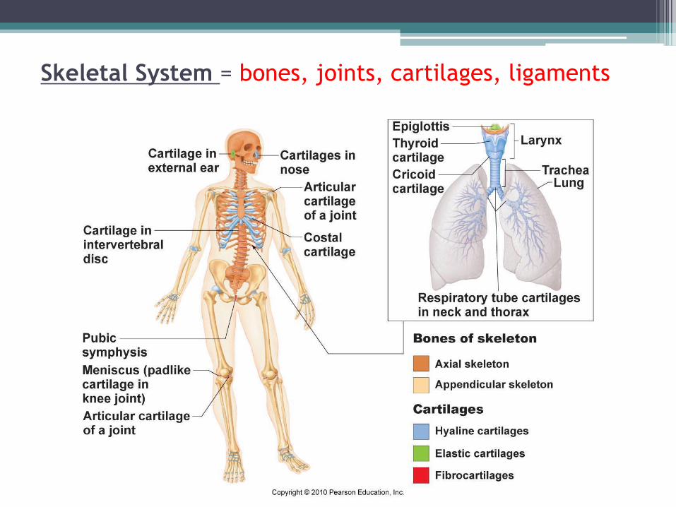
Skeletal System = bones, joints, cartilages, ligaments
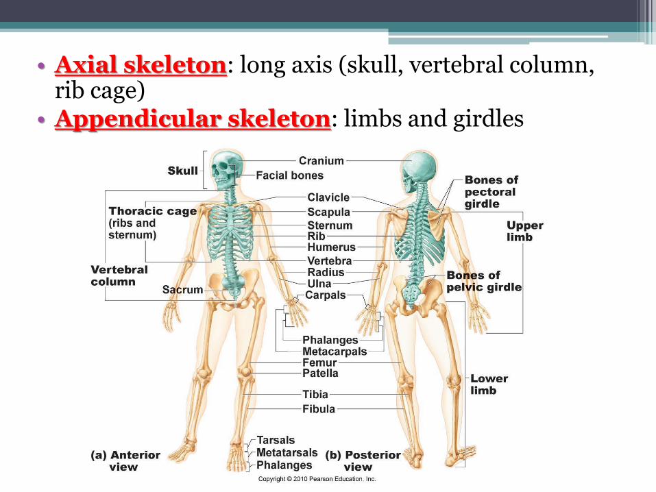
• Axial skeleton: long axis (skull, vertebral column, rib cage)
• Appendicular skeleton: limbs and girdles
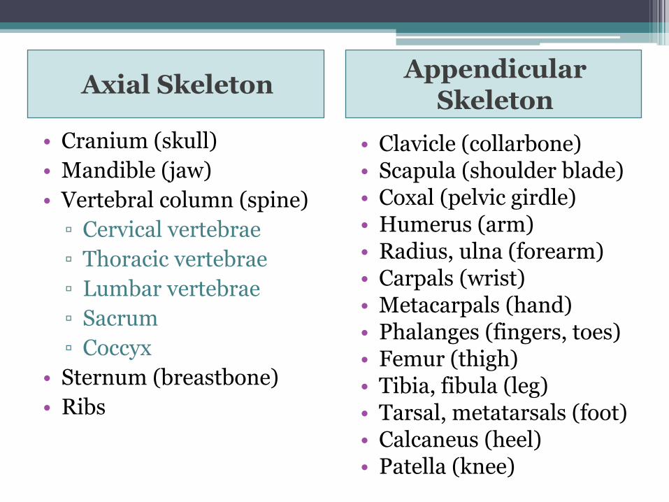
Axial Skeleton Appendicular
Skeleton
• Cranium (skull)
• Mandible (jaw)
• Vertebral column (spine)
▫ Cervical vertebrae
▫ Thoracic vertebrae
▫ Lumbar vertebrae
▫ Sacrum
▫ Coccyx
• Sternum (breastbone)
• Ribs
• Clavicle (collarbone) • Scapula (shoulder blade) • Coxal (pelvic girdle) • Humerus (arm) • Radius, ulna (forearm) • Carpals (wrist) • Metacarpals (hand) • Phalanges (fingers, toes) • Femur (thigh) • Tibia, fibula (leg) • Tarsal, metatarsals (foot) • Calcaneus (heel) • Patella (knee)
Functions of the Bones
• Support body and cradle soft organs
• Protect vital organs
• Movement: muscles move bones
• Storage of minerals (calcium, phosphorus) & growth factors
• Blood cell formation in bone marrow
• Triglyceride (fat) storage
Classification of Bones
1. Long bones
▫ Longer than they are wide (eg. femur, metacarpels)
2. Short bones
▫ Cube-shaped bones (eg. wrist and ankle)
▫ Sesamoid bones (within tendons – eg. patella)
3. Flat bones
▫ Thin, flat, slightly curved (eg. sternum, skull)
4. Irregular bones
▫ Complicated shapes (eg. vertebrae, hips)
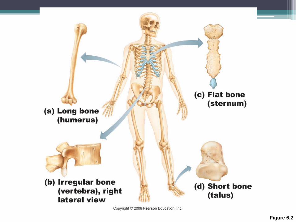
Figure 6.2
• Adult = 206 bones
• Types of bone tissue:
▫ Compact bone: outer layer – dense & solid
▫ Spongy bone: inner layer - open spaces, marrow
• Features:
▫ Very hard (calcium salts)
▫ Light weight
▫ Ability to resist tension and forces (collagen fibers)
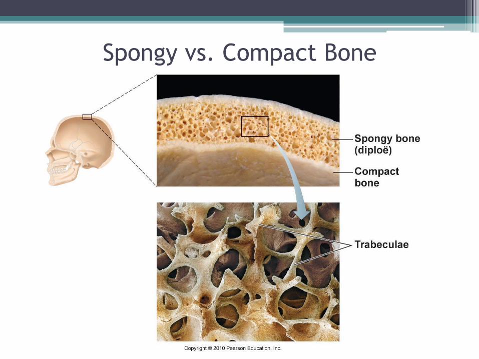
Spongy vs. Compact Bone
Bone Development
• Osteogenesis (ossification): bone tissue formation Stages: • Begins at 8 weeks gestation
• Start as cartilage replaced by bone • Post-natal bone growth early adulthood
• Epiphyseal plates: (growth plates) regions where long bones lengthen
• Appositional growth: bones increase in thickness
• Bone modeling and repair – lifelong
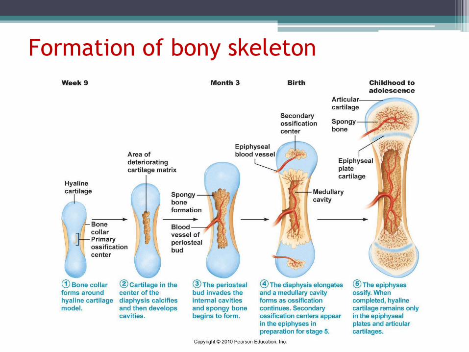
Formation of bony skeleton
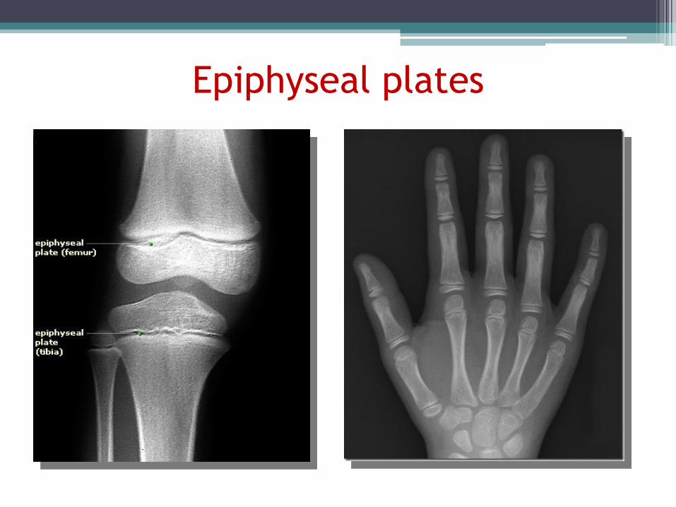
Epiphyseal plates
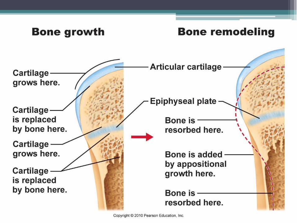
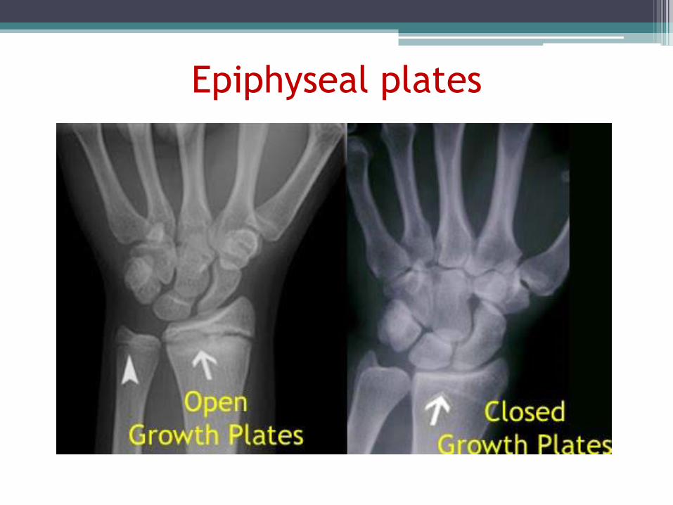
Epiphyseal plates
Hormonal Control
• Growth hormones: stimulate longitudinal bone growth
• Thyroid hormone: control activity of growth hormone
• Testosterone & estrogens (at puberty):
• Adolescent growth spurt
• Close epiphyseal plates end growth
Testosterone & estrogens
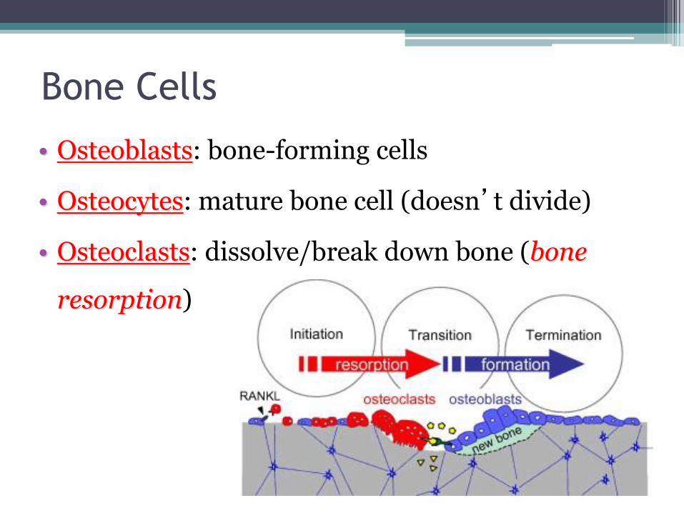
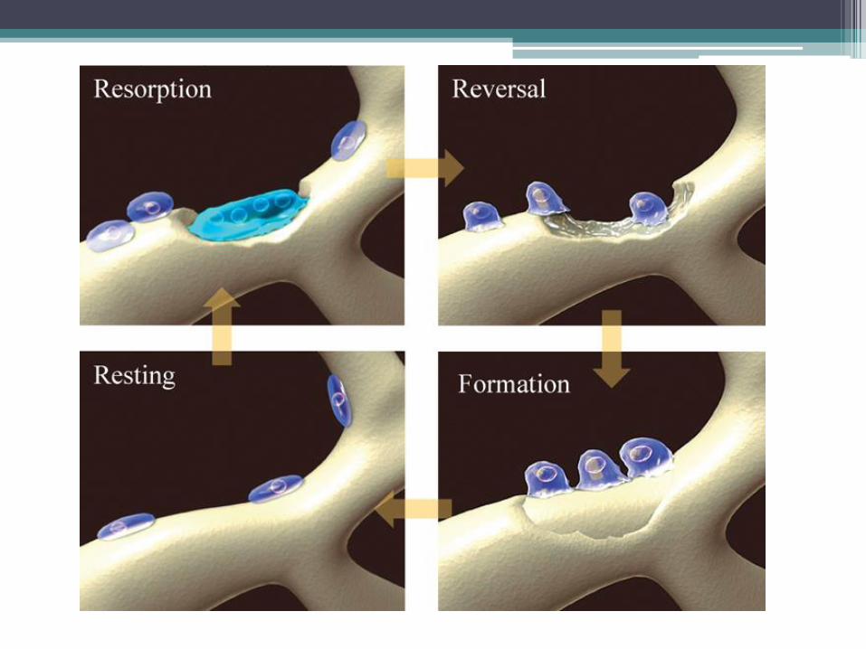
Bone Cells
• Osteoblasts: bone-forming cells
• Osteocytes: mature bone cell (doesn’t divide)
• Osteoclasts: dissolve/break down bone (bone
resorption)
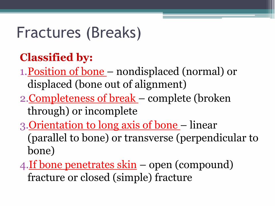
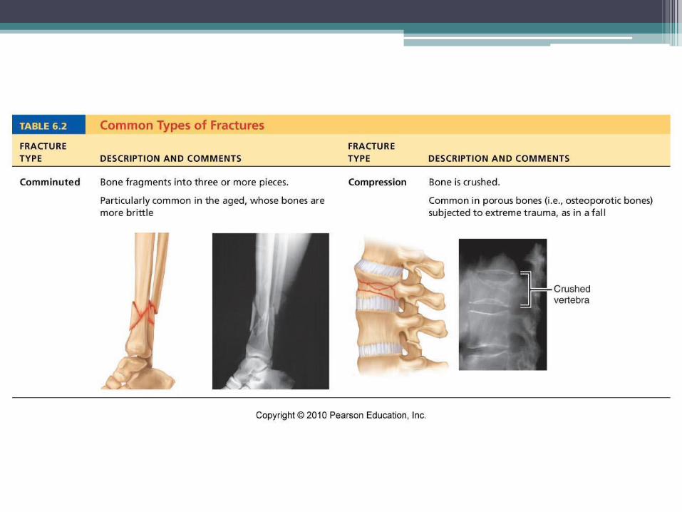
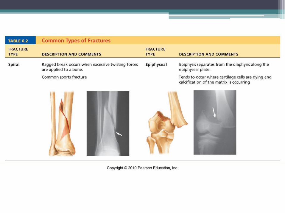
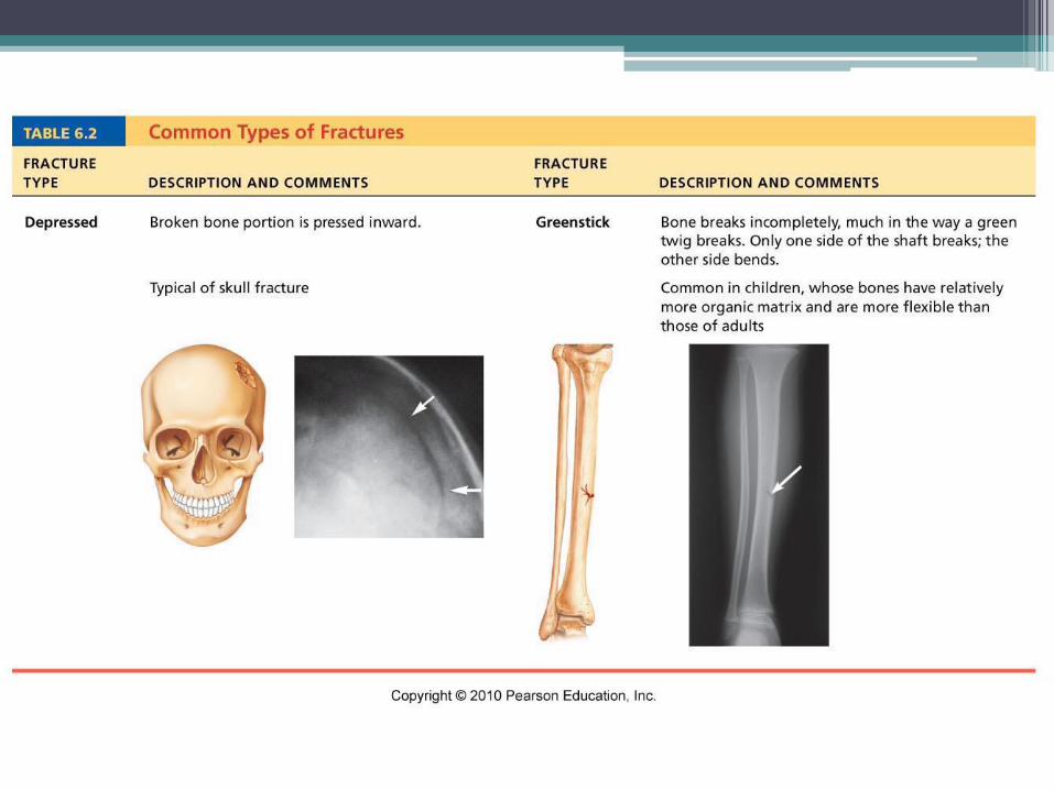
Fractures (Breaks)
Classified by:
1.Position of bone – nondisplaced (normal) or displaced (bone out of alignment)
2.Completeness of break – complete (broken through) or incomplete
3.Orientation to long axis of bone – linear (parallel to bone) or transverse (perpendicular to bone)
4.If bone penetrates skin – open (compound) fracture or closed (simple) fracture
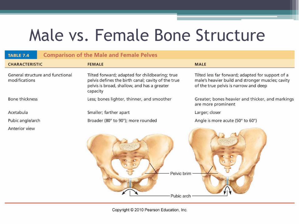
Male vs. Female Bone Structure
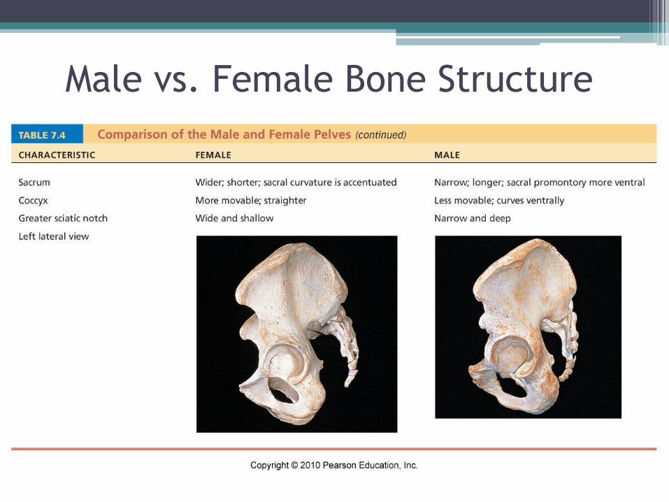
Male vs. Female Bone Structure
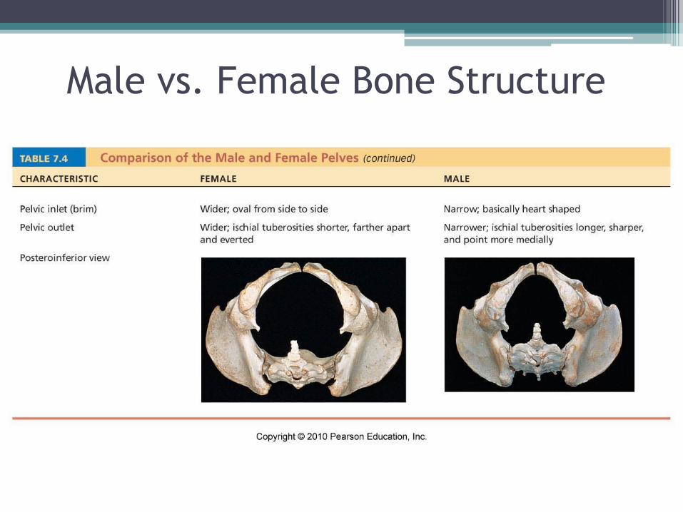
Male vs. Female Bone Structure
Bone Structure: Gender Differences
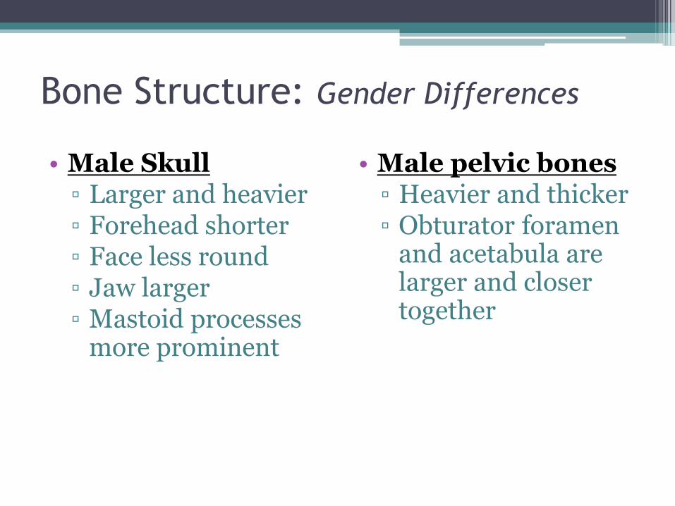
• Male Skull ▫ Larger and heavier ▫ Forehead shorter ▫ Face less round ▫ Jaw larger ▫ Mastoid processes
more prominent
• Male pelvic bones ▫ Heavier and thicker ▫ Obturator foramen
and acetabula are larger and closer together
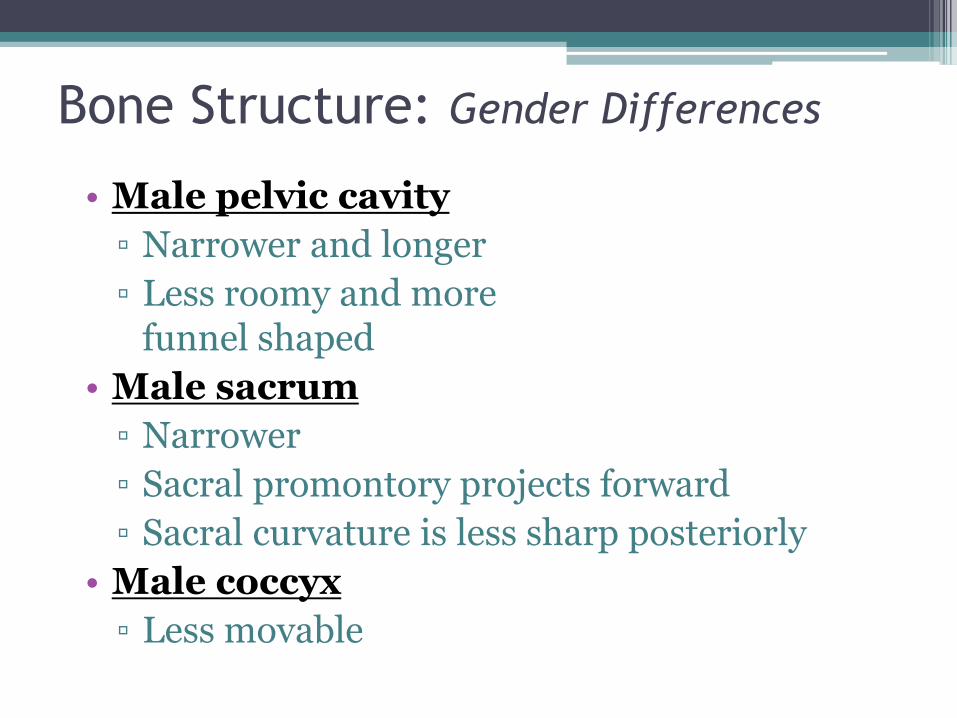
• Male pelvic cavity
▫ Narrower and longer
▫ Less roomy and more funnel shaped
• Male sacrum
▫ Narrower
▫ Sacral promontory projects forward
▫ Sacral curvature is less sharp posteriorly
• Male coccyx
▫ Less movable
Bone Structure: Gender Differences
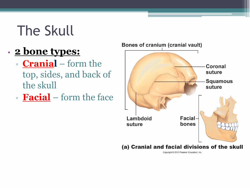
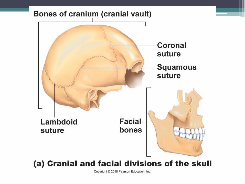
The Skull
• 2 bone types:
▫ Cranial – form the top, sides, and back of the skull
▫ Facial – form the face
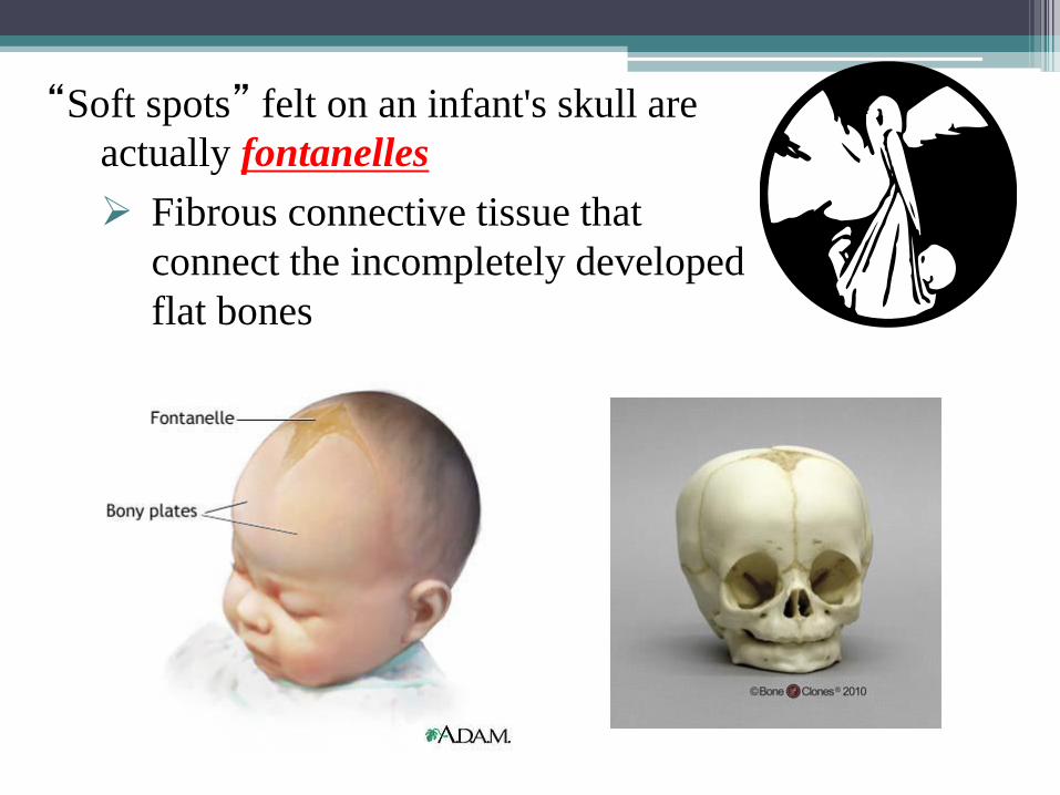
“Soft spots” felt on an infant's skull are
actually fontanelles
Fibrous connective tissue that
connect the incompletely developed
flat bones
The Skull: Cranial Bones
• Frontal – anterior
• Parietal – top and most of the sides
• Occipital – back
• Temporal – form the lower sides of the skull
• Sphenoid and ethmoid bones – floor
• Ear ossicles are the smallest bones of the body
▫ Malleus
▫ Incus
▫ Stapes
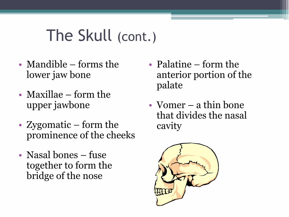
The Skull (cont.)
• Mandible – forms the lower jaw bone
• Maxillae – form the upper jawbone
• Zygomatic – form the prominence of the cheeks
• Nasal bones – fuse together to form the bridge of the nose
• Palatine – form the anterior portion of the palate
• Vomer – a thin bone that divides the nasal cavity
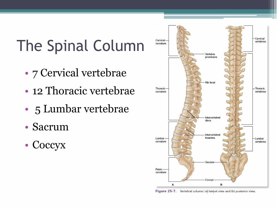
The Spinal Column
• 7 Cervical vertebrae
• 12 Thoracic vertebrae
• 5 Lumbar vertebrae
• Sacrum
• Coccyx
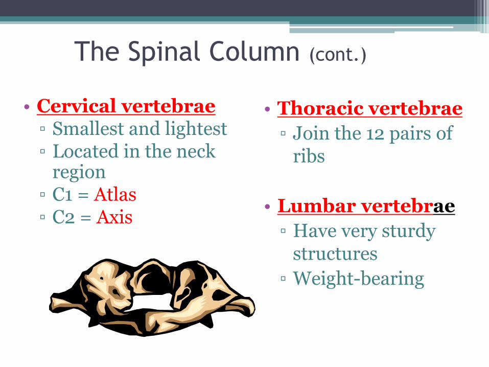
The Spinal Column (cont.)
• Cervical vertebrae ▫ Smallest and lightest ▫ Located in the neck
region ▫ C1 = Atlas ▫ C2 = Axis
• Thoracic vertebrae
▫ Join the 12 pairs of ribs
• Lumbar vertebrae
▫ Have very sturdy structures
▫ Weight-bearing
The Spinal Column (cont.)
• Sacrum
▫ Triangular-shaped bone 5 fused vertebrae
• Coccyx
▫ Small, triangular bone 3-5 fused vertebrae
▫ Considered unnecessary
▫ Also called the tailbone

Apply Your Knowledge
Identify the sections
of the spinal column
and give the number
of vertebrae for each. Thoracic – 12
Lumbar – 5 Sacrum –
5 fused
Coccyx –
3 to 5 fused
Cervical – 7
ANSWER:
Right!
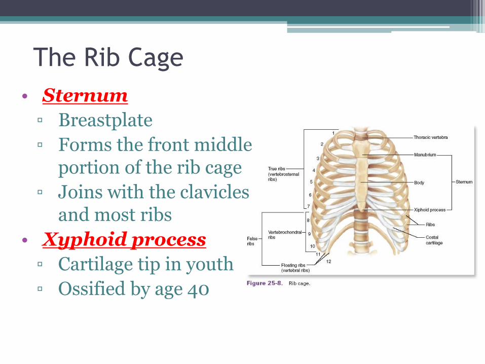
The Rib Cage
• Sternum
▫ Breastplate
▫ Forms the front middle portion of the rib cage
▫ Joins with the clavicles and most ribs
• Xyphoid process
▫ Cartilage tip in youth
▫ Ossified by age 40
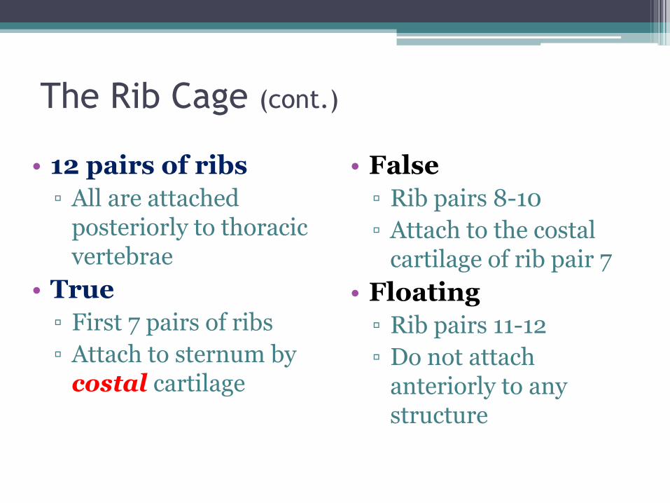
The Rib Cage (cont.)
• 12 pairs of ribs
▫ All are attached posteriorly to thoracic vertebrae
• True
▫ First 7 pairs of ribs
▫ Attach to sternum by costal cartilage
• False
▫ Rib pairs 8-10
▫ Attach to the costal cartilage of rib pair 7
• Floating
▫ Rib pairs 11-12
▫ Do not attach anteriorly to any structure
Apply Your Knowledge
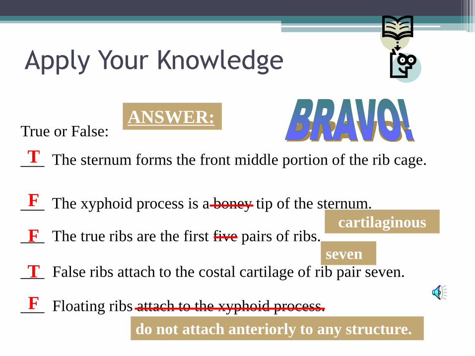
True or False:
___ The sternum forms the front middle portion of the rib cage.
___ The xyphoid process is a boney tip of the sternum.
___ The true ribs are the first five pairs of ribs.
___ False ribs attach to the costal cartilage of rib pair seven.
___ Floating ribs attach to the xyphoid process.
T
T
ANSWER:
F
cartilaginous F
seven
F
do not attach anteriorly to any structure.
Common Diseases and Disorders
• Arthritis – general term meaning joint inflammation
• Osteoarthritis – degenerative joint disease, primarily of weight-bearing joints
• Rheumatoid Arthritis – chronic systemic inflammatory disease of smaller joints and surrounding tissues
• Bursitis – inflammation of a bursa (fluid-filled sac that cushions tendons)
• Carpal Tunnel Syndrome – overuse of wrist; the median nerve in the wrist becomes compressed
• Ewing’s Family of Tumors (EFT) – a group of tumors that affect different tissue types; primarily bone
• Gout – a type of arthritis; deposits of uric acid crystals in the joints
Common Diseases and Disorders
• Kyphosis – abnormal curvature of the spine (humpback)
• Lordosis – exaggerated inward curvature of the lumbar spine (swayback)
• Osteogenesis imperfecta – brittle-bone disease
• Osteoporosis – a condition in which bones thin (become porous) over time
Common Diseases and Disorders
• Osteosarcoma – a type of bone cancer that originates from osteoblasts, the cells that make bony tissue
• Paget’s disease – causes bones to enlarge and become deformed and weak
• Scoliosis – an abnormal S-shaped curvature of the spine
Common Diseases and Disorders