understanding anatomy and physiology - part4b
DESCRIPTION
Understanding Anatomy and PhysiologyTRANSCRIPT

17
chapter RESPIRATORYSYSTEMDuring a 24-hour period, the average person will breathe 23,040 times.
FAST FACTYou can live about 40 dayswithout food, about 7 dayswithout water, but onlyminutes without oxygen.
Oxygen plays a powerful role in overall health and well-being. Most metabolic processes of the body—including thedigestion of food, the fighting of infection and disease, and the production of energy—depend on oxygen. Your ability tothink, feel, and act depend upon the availability of oxygen.
The respiratory and cardiovascular systems work closely together to provide the body with oxygen and to remove carbondioxide. The respiratory system assumes other roles as well: it influences sound production and speech; it makes the sense ofsmell (and therefore taste) possible; and, as will be discussed in Chapter 19, it helps the body maintain homeostasis throughthe regulation of acid-base balance. The respiratory system is divided into two tracts:
Nasal cavity
Nasopharynx
Oropharynx
Laryngopharynx
Larynx
Trachea
Left lung lower lobe
Left and rightprimary bronchi
Bronchioles
Diaphragm
Pharynx
The upper respiratory
tract consists of structures
located outside the thoracic
cavity.
The lower respiratory
tract consists of structures
located inside the thoracic
cavity.

Functionally, the respiratory system also includes the:
l Oral cavityl Rib cagel Respiratory muscles (including the diaphragm)
The structures of the upper respiratory tract—consisting of the nose, nasopharynx,oropharynx, laryngopharynx, and larynx—warm and humidify inspired air. They’re alsoresponsible for the senses of smell and taste as well as chewing and swallowing food.
Nose and Nasal Cavities
Air enters and leaves the respiratory system through the nose. Just inside the nostrils are smallhairs called cilia that filter out dust and large foreign particles.
The nasal cavity lies just over the mouth, separated from that orifice by a bony structurecalled the palate. A vertical plate of bone and cartilage—called the septum—separates the cavityinto two halves. The cavity is lined with epithelium rich in goblet cells that produce mucus.
FAST FACTSpontaneousnosebleeds (that resultwithout trauma orirritation) can be anearly sign ofhypertension.
Frontal sinus
Hard palate
Soft palate
Projecting from the lateral wall of
each cavity are three bones called
conchae. These bones create
narrow passages, ensuring that
most air contacts the mucous
membrane on the way through.
As it does, the air picks up
moisture and heat from the
mucosa. At the same time, dust
sticks to the mucus, which is then
swallowed.
Branches of the olfactory nerve (responsible
for the sense of smell) penetrate the upper
nasal cavity and lead to the brain.
The sphenoid sinus (shown here), as well
as the other paranasal sinuses (including
the frontal, maxillary, and ethmoidal
sinuses), drain mucus into the nasal
cavity. (For further discussion of the
paranasal sinuses, see Chapter 7, Skeletal
System).Pharynx
Just behind the nasal and oral cavities is a muscular tube called the pharynx. Commonly calledthe throat, the pharynx can be divided into three regions:
1 The nasopharynx extends from the
posterior nares to the soft palate. It
contains openings for the right and left
auditory (eustachian) tubes.
2 The oropharynx is a space between the
soft palate and the base of the tongue. It
contains the palatine tonsils (the ones most
commonly removed by tonsillectomy) as well
as the lingual tonsils, found at the base of the
tongue.
3 The laryngopharynx passes dorsal to
the larynx and connects to the esophagus.
The Body AT WORKOnly air passes through the
nasopharynx, while both
food and air pass through
the oropharynx and
laryngopharynx.
PA
RT
IV M
ain
ten
an
ce o
f the
Bo
dy
338
Upper Respiratory Tract

Larynx
Lying between the root of the tongue and the upper end of the trachea, the larynx is a chamberformed by walls of cartilage and muscle. Because it contains the vocal cords, it’s often called thevoice box; however, it actually has three functions:
1. It prevents food and liquids from entering the trachea.2. It acts as an air passageway between the pharynx and trachea.3. It produces sound.
Nasal cavity
Pharyngeal tonsil
Auditory tube
Uvula
Palatine tonsil
Lingual tonsil
Epiglottis
Trachea
Esophagus
Tongue
Hyoid bone
Base of tongue
Epiglottis
Vestibular fold
Vocal cord
Glottis
Vocal cords in the
closed position
Vocal cords in the
open position
Larynx
Trachea
Epiglottis
Hyoid bone
Thyroidcartilage
The larynx is formed by nine
pieces of cartilage that keep it
from collapsing; a group of
ligaments bind the pieces of
cartilage together and to
adjacent structures in the
neck.
• The epiglottis—which
closes over the top of the
larynx during swallowing to
direct food and liquids into
the esophagus—is the
uppermost cartilage.
• The largest piece of cartilage
is the thyroid cartilage,
which is also known as the
Adam’s apple.
The Body AT WORKAir passing between the vocal cords during exhalation produces sound. Loudness depends upon the force of the air: the
more forceful the air, the louder the sound. Only the vocal cords produce sound; however, the pharynx, oral cavity,
tongue, and lips shape the sounds to form words.
High-pitched sounds result when the cords are relatively taut; more relaxed cords produce lower-pitched sounds. The
vocal cords in adult males are usually longer and thicker and vibrate more slowly, producing lower-pitched sounds than
in females.
339
CH
AP
TE
R 1
7 R
esp
irato
ry S
yste
m
• The mucous membrane
lining the larynx forms two
pairs of folds. The superior
pair—called vestibular
folds, or, occasionally, false
vocal cords—play no role in
speech. They close the glottis
(the opening between the
vocal cords) during swallow-
ing to keep food and liquids
out of the airway.
• The inferior pair, the vocal
cords, produces sound when
air passes over them during
exhalation.
• The opening between the
cords is called the glottis.

The lower respiratory tract consists of the trachea, bronchi, and lungs. The tracheaand the bronchi distribute air to the interior of the lungs; deep within the lungs iswhere gas exchange occurs.
Larynx
Left primarybronchus
Left secondarybronchus
Left tertiarybronchus
Bronchioles
Lying just in front of the esophagus, the
trachea is a rigid tube about 4.5 inches
(11 cm) long and 1 inch (2.5 cm) wide.
C-shaped rings of cartilage encircle the
trachea to reinforce it and keep it from
collapsing during inhalation. The open part
of the “C” faces posteriorly, giving the
esophagus room to expand during
swallowing.
The trachea extends from the larynx to a
cartilaginous ridge called the carina.
FAST FACTThe trachea and the two bronchiwith their many branchesresemble an inverted tree: that’swhy it’s often called thebronchial tree.
Bronchial Tree
Trachea
At the carina, the trachea branches into
two primary bronchi. Like the trachea,
the primary bronchi are supported by
C-shaped rings of cartilage. (All of the
divisions of the bronchial tree also
consist of elastic connective tissue.)
The right bronchus is slightly wider
and more vertical than the left,
making this the most likely location for
aspirated (inhaled) food particles and
small objects to lodge.
Immediately after entering the lungs,
the primary bronchi branch into
secondary bronchi: one for each of
the lung’s lobes. Since the left lung
consists of two lobes, it has two
secondary bronchi; the right lung has
three lobes, so it has three bronchi.
Tertiary bronchi continue to branch,
resulting in very small airways called
bronchioles. Less than 1 mm wide and
lacking any supportive cartilage,
bronchioles divide further to form thin-
walled passages called alveolar ducts.
Secondary bronchi branch into smaller
tertiary bronchi. The cartilaginous
rings around the bronchi become
irregular and disappear entirely in the
smaller bronchioles. Alveolar ducts throughout the
lungs terminate in clusters of
alveoli called alveolar sacs, the
primary structures for gas
exchange.
PA
RT
IV M
ain
ten
an
ce o
f the
Bo
dy
340
Lower Respiratory Tract

Alveoli
The lung passages all exist to serve the alveoli because it’s within the alveoli that gasexchange occurs.
FAST FACTIt’s estimated that thelungs contain 300 million alveoli.
Pulmonary venule
Terminal bronchiole
Pulmonary arteriole
Alveolarsac
Alveoli
Alveolar duct
Alveolus
Capillary
O2
CO2
The alveoli are wrapped in a fine mesh of
capillaries. The extremely thin walls of the
alveoli, and the closeness of the capillaries,
allow for efficient gas exchange.
The exchange of air occurs through what’s called
the respiratory membrane, which consists of
the alveolar epithelium, the capillary
endothelium, and their joined basement
membranes.
For gas to enter or leave a cell, it must be dissolved in aliquid. Therefore, the inside of each alveolus is coated with athin layer of fluid. This fluid contains surfactant, a substancethat helps reduce surface tension (the force of attraction betweenwater molecules) to keep the alveolus from collapsing as airmoves in and out during respiration. (For more information, see“Alveolar Surface Tension” in this chapter.)
The Body AT WORKA large portion of the membrane lining the bronchial tree is
covered with a layer of protective mucus, which serves to purify
the air entering the respiratory tract. This cleansing mucus
moves up from the lower portions of the bronchial tree toward
the pharynx, propelled along by millions of hair-like cilia that
line the respiratory mucosa. The cilia beat in one direction—
upward—so that the mucus will move toward the pharynx.
Cigarette smoke paralyzes these cilia. As a result, mucus
accumulates in the lower portions of the bronchial tree, causing
the typical “smoker’s cough” as the lungs attempt to rid
themselves of the excess mucus.
341
CH
AP
TE
R 1
7 R
esp
irato
ry S
yste
m
FAST FACTInfants born before 28 weeks’gestation commonly lack surfactant.Without surfactant, surface tensionrestricts alveolar expansion duringinspiration and causes alveolar collapseduring expiration. That’s whyneonates often develop respiratorydistress syndrome and require theadministration of artificial surfactant.

PA
RT
IV M
ain
ten
an
ce o
f the
Bo
dy
342
Lungs
The lungs fill the pleural cavity: they extend from just above the clavicles to the diaphragm and lieagainst the anterior and posterior ribs. The medial portion of each lung is concave to allow room forthe heart and great vessels. The primary bronchi and pulmonary blood vessels enter each lungthrough an opening on the lung’s medial surface called the hilum.
Middlelobe
Inferiorvena cava
Descendingaorta
Superiorlobe
Superiorlobe
Inferiorlobe
Diaphragm
Inferiorlobe
The top, or apex, of each lung extends
about 1/2” (1.3 cm) above the first rib.
The base of each lung
rests on the diaphragm.
Right Lung
Left Lung
The right lung is shorter,
broader, and larger than
the left. It has three
lobes—the superior,
middle, and inferior—and
handles 55% of the gas
exchange. The right lung
contains two fissures:
• Horizontal fissure
• Oblique fissure
Because the heart extends
toward the left, the left
lung has only two lobes:
the superior and inferior. It
contains one fissure:
• Oblique fissure
Life lesson: AsthmaAsthma is the most common chronic illness in children; what’s more, the numberof people afflicted with asthma is on the rise. In the United States, about 5% of alladults and 10% of all children have asthma.
Typically, when someone with asthma is exposed to an allergen or otherrespiratory irritant (such as dust or fumes), the bronchioles constrict and spasm.These narrowed airways trigger coughing and wheezing—sometimes severe—as the person struggles to breathe. Inflammation in the airways also causesexcessive production of thick, sticky mucus that further clogs the airways. Airwayobstruction can become severe enough to cause suffocation and death.
Treatment involves dilating the airways through the administration ofepinephrine and beta-adrenergic stimulants. Also, anti-inflammatorymedications and inhaled corticosteroids are often prescribed to treatinflammation and minimize scarring.

343
CH
AP
TE
R 1
7 R
esp
irato
ry S
yste
m
Pleurae
Posterior
Aorta
Left lung
Sternum
Vertebra
Heart
Anterior
Right lung
Esophagus
Ribs
TracheaBronchi
The parietal pleura lines
the entire thoracic cavity.
A serous membrane—the
visceral pleura—covers the
surface of the lungs,
extending into the fissures.
The space between the visceral and parietal pleurae is called the pleural
cavity. The pleural cavity is only a potential space; the two membranes
are normally separated only by a film of slippery pleural fluid.
The fluid in the pleural cavity serves two purposes:
• It lubricates the pleural surfaces, allowing the two surfaces to glide
painlessly against each other as the lungs expand and contract.
• Because the pressure in the pleural cavity is lower than atmospheric
pressure, it creates a pressure gradient that assists in lung inflation.
Life lesson: Changes with agingAs the body ages, numerous changes occur in the respiratory system—allleading to a general reduction in respiratory efficiency. Here are a few of thosechanges:
• Decreased mobility of the chest wall• Increased lung rigidity• Decreased number and dilation of alveoli• Weakened respiratory muscles• Reduced volume of protective respiratory fluids
These changes reduce an older person’s ability to perform vigorous exercise.They also lead to increased risk for developing pneumonia following a bacterialor viral illness. In addition, these changes can compound the effects of heart andlung diseases.

• The diaphragm relaxes,
bulging upward and
pressing against the base
of the lungs, reducing the
size of the thoracic cavity.
• Air is pushed out of the
lungs.
• The internal intercostal
muscles pull the ribs
downward as the external
intercostals relax.
Expiration
Sternocleidomastoid
Scalenes
Pectoralisminor
Rectusabdominis
External abdominaloblique
• The diaphragm
contracts, flattens, and
drops, pressing the
abdominal organs
downward and enlarging
the thoracic cavity.
• Air rushes in to equalize
pressure.
• The internal
intercostals help elevate
the ribs.
• The external intercostal
muscles pull the ribs
upward and outward,
widening the thoracic
cavity.
Inspiration
Pulmonary ventilation is simply breathing: the repetitive process of inhaling (calledinspiration) and exhaling (called expiration). Both actions depend on the function ofrespiratory muscles and a difference between the air pressure within the lungs and the airpressure outside the body. One inspiration and one expiration comprise one respiratorycycle.
Respiratory Muscles
The lungs depend on the skeletal muscles of the trunk (especially the diaphragm and theintercostal muscles) to expand and contract to create airflow. The main muscleresponsible for pulmonary ventilation is the diaphragm: the dome-shaped muscleseparating the thoracic and pelvic cavities.
FAST FACTIn adults, normal respiratoryrates range from 12 to 20 breathsper minute.
The Body AT WORKPeople having difficulty breathing may depend heavily on accessory muscles
to breathe. For example, in emphysema, lungs lose their elasticity and
exhaling is no longer a passive process. Patients must use their accessory
muscles to exhale, making exhaling an active, exhausting process. In other
patients, the use of accessory muscles can indicate acute respiratory distress,
signaling a medical emergency.
PA
RT
IV M
ain
ten
an
ce o
f the
Bo
dy
344
Pulmonary Ventilation
Accessory Muscles of Respiration
During times of forced or labored breathing, additional muscles, called accessory musclesof respiration, join in to assist with breathing. For example:
l During deep inspiration, muscles of the neck (the sternocleidomastoids and scalenes)and the chest (the pectoralis minor) contract to help elevate the chest (see red arrows infigure).
l During forced expiration—such as when singing or shouting—the rectus abdominisand external abdominal obliques contract to pull down the ribs and sternum, furtherreducing chest size and expelling air more rapidly (see blue arrows in figure).

Neural Control of Breathing
Unlike cardiac muscle, which contains intrinsic pacemakers, the muscles used for breathing areskeletal muscles—and skeletal muscles require nervous stimulation to contract. Although a varietyof factors affect the rate and rhythm of breathing, the respiratory centers responsible for automatic,unconscious breathing reside in the medulla and pons—parts of the brainstem.
The medulla contains two interconnected centers that control breathing: the inspiratory centerand the expiratory center.
Medulla
Pons
Intercostal
nerve
Phrenic nerve
The inspiratory center is the primary respiratory center. It controls inspiration and, indirectly,
expiration as well. Here’s how it works:
1. The inspiratory center sends impulses to the intercostal muscles (via the intercostal
nerves) and to the diaphragm (via the phrenic nerves).
2. The inspiratory muscles contract, causing inhalation.
3. Nerve output then ceases abruptly, causing the inspiratory muscles to relax. The elastic
recoil of the thoracic cage produces exhalation.
• The apneustic center
stimulates the inspiratory
center to increase the
length and depth of
inspiration.
• The pneumotaxic center
inhibits both the apneustic
center and the inspiratory
center; this contributes to
a normal breathing
rhythm and prevents
overinflation of the lungs.
When more forceful exhalations are needed, such as during
exercise, the expiratory center sends impulses to the abdominal
and other accessory muscles.
The Body AT WORKThe cerebral cortex allows you to voluntarily change
your breathing rate or rhythm, such as to sing or
blow out a candle, or even to hold your breath.
However, when you hold your breath, CO2 isn’t
expelled through breathing and the CO2 level in the
blood rises. CO2 is a powerful respiratory stimulant.
When CO2 rises to a certain level, the respiratory
centers override your voluntary action and
breathing resumes.
Although the medulla is the main breathing center, the ponscontains two centers that can influence basic breathing rhythm:
345
CH
AP
TE
R 1
7 R
esp
irato
ry S
yste
m

PA
RT
IV M
ain
ten
an
ce o
f the
Bo
dy
346
FAST FACTCarbon dioxide—NOToxygen—is the primaryregulator of respiration. That’sbecause carbon dioxide easilycrosses the blood-brainbarrier.
Factor Sensory Receptor Action
O2Peripheral chemoreceptors
(located in the carotid
and aortic bodies)
Low blood levels of oxygen cause peripheral chemoreceptors to
send impulses to the medulla to increase the rate and depth of
respirations. This brings more air, and therefore oxygen, into the
lungs.
pHCentral chemoreceptors
(located in the brainstem)
Central chemoreceptors monitor the pH of cerebrospinal fluid
(CSF), which mirrors the level of carbon dioxide in the blood.
Falling pH levels indicate an excess of carbon dioxide.
When this occurs, central chemoreceptors signal the respiratory
centers to increase the rate and depth of breathing. This helps
the body “blow off“ excess carbon dioxide, raising the pH.
Receptors in the lungs
and chest wall
As the lungs inflate during inspiration, receptors detect the
stretching and signal the respiratory centers to exhale and inhibit
inspiration. Called the Hering-Breuer reflex, this mechanism
prevents lung damage from overinflation.
Hypothalamus and limbic
system
These areas of the brain send signals that affect breathing in
response to pain and emotions (such as fear, anger, and anxiety).
Nerve cells in the airway Nerve cells respond to irritants by signaling the respiratory
muscles to contract, resulting in a cough or a sneeze. Coughing
or sneezing propels air rapidly from the lungs, helping to
remove the offending substance.
Oxygen
Hydrogen ions
(pH)
Stretch
Irritants(such as smoke, dust, pollen, noxious chemicals, and mucus)
Pain and
emotion
Variations in BreathingDespite neural input, breathing patterns don’t remain constant. Respiratory rate andrhythm vary: with pain, emotion (such as fear, anger, or anxiety), exercise, andchanges in the body’s physical state. These variations occur because the respiratorycenters receive input from a number of sensory receptors throughout the body,alerting it to the body’s changing needs.
Factors Influencing Breathing

347
CH
AP
TE
R 1
7 R
esp
irato
ry S
yste
m
Pressure and Airflow
Air moves into and out of the lungs for the same reason that blood flows: because of a pressure gradient. The pressure thatdrives respiration is atmospheric pressure: the weight of the air around us.
When the pressure within the lungs drops lower than atmospheric pressure, air flows from the area of higher pressure—the air outside the body—to an area of lower pressure—the lungs; this is inspiration. When the pressure within the lungsrises above atmospheric pressure, air flows out of the lungs (expiration) until the two pressures equalize. Whereas inspirationis an active process, requiring the use of muscles, normal expiration is a passive process, resulting from the recoil of healthylungs.
Air inhaled
Lowpressure
Lowpressure
Air exhaled
Highpressure
Highpressure
Inspiration
l The intercostal muscles contract, pulling the ribs up and out; the
diaphragm contracts and moves downward. This enlarges the
chest cavity in all directions.
l The lungs expand along with the chest because of the two layers
of the pleurae.
l The parietal pleura is firmly attached to the ribs; the visceral
pleura covers the lungs.
l Although not attached to each other, the thin film of fluid
between the two pleurae causes them to cling together like
two pieces of wet paper.
l Furthermore, the potential space between the two pleurae
maintains a pressure slightly less than atmospheric pressure
(negative pressure). This is the intrapleural pressure. When
the ribs expand and the parietal pleura pulls away, intrapleural
pressure becomes even more negative. This has a suction-like
effect, causing the visceral pleura to cling even tighter to the
parietal pleura.
l The visceral pleura follows the parietal pleura, pulling the lung
along with it.
l When the lungs expand, the volume of air in the lungs spreads
throughout the enlarging space. This causes the pressure within
the bronchi and alveoli (the intrapulmonic pressure) to drop.
(See “That Makes Sense!”)
l When the intrapulmonic pressure drops lower than the
atmospheric pressure, air flows down the pressure gradient
into the lungs.
Expiration
l The diaphragm and external intercostal muscles relax, and the
thoracic cage springs back to its original size.
l The lungs are compressed by the thoracic cage.
l Intrapulmonary pressure rises.
l Air flows down the pressure gradient and out of the lungs.
That Makes SenseA law of physics called Boyle’s Law explains why
intrapulmonic pressure drops as the lungs expand. Boyle’s
Law states, “At a constant temperature, the pressure of a
given quantity of gas is inversely proportional to its volume.”
In other words, a given volume of gas will exert more
pressure in a smaller space than it will in a larger space.
To illustrate, imagine tying off the end of a large plastic
bag. The air in the bag would cause the bag to balloon
slightly but the bag would still be soft to touch. The air
inside the bag would have a low pressure. Now imagine
twisting the end of the bag, making the volume of the bag
smaller. This would force the air in the bag into a smaller
space and the bag would become firm to touch—
exhibiting a higher pressure. If you continued to twist,
making the bag even smaller, the pressure could become
great enough to pop the bag.
!
ANIMATION

PA
RT
IV M
ain
ten
an
ce o
f the
Bo
dy
348
Factors Affecting Airflow
Effective pulmonary ventilation depends on several factors. Two particularly important factors arepulmonary compliance and alveolar surface tension.
Pulmonary CompliancePulmonary compliance refers to the elasticity of lung tissue. Ventilation can’t occur unless the lungsand thorax can stretch and, just as important, recoil. Some diseases (such as tuberculosis or blacklung disease) cause scarring, which makes the lungs stiffer, or less compliant. The lungs havedifficulty expanding, and ventilation is impaired.
Alveolar Surface TensionThe inner surface of each alveoli is covered with a thin film of water. The water is necessary for gasexchange. However, water molecules are also electrically attracted to each other, just like weakmagnets. Left alone, the water molecules inside the alveolus will move toward each other, creating aforce that will collapse the alveoli. If the alveoli collapse, gas exchange can’t occur. To avoid thisproblem, alveolar cells secrete surfactant, a lipoprotein that disrupts the electrical attractionbetween the water molecules. This lowers surface tension and prevents alveolar collapse.
Life lesson: Pneumothorax
Life lesson: EmphysemaEmphysema is a progressive lung disease in which lung tissuesurrounding alveoli is destroyed. This leaves alveoli unsupported,allowing them to enlarge and eventually rupture. Alveolar walls fuse intolarge irregular spaces that offer less area available for gas exchange. Thealveoli also lose elasticity and the ability to recoil. Consequently, airbecomes trapped in the lungs. Over time, the trapped air causes thediameter of the chest to enlarge, and the chest assumes the shape of abarrel. (A person with emphysema is often said to be “barrel-chested.”)Persons with emphysema must work to exhale; they can expend three tofour times the normal amount of energy just to breathe.
Normal lungChest wall
Diaphragm
Mediastinum
Plural space
Outside air rushes in following a puncture of chest wall and parietal pleura.
Lung air rushes outfollowing disruptionof the visceral pleura
Normal chest Barrel chest
If the thoracic wall is punctured, air from the atmospherewill rush in to the pleural cavity, transforming what isnormally a potential space into a space filled with air. As aresult, the negative pressure that characterizes the pleuralcavity is lost, and the lung recoils and collapses. This iscalled a pneumothorax. Air can also enter the pleural spacewhen a weakened or diseased alveoli ruptures. This causesa disruption in the visceral pleura, and air from the lungenters the pleural cavity, again resulting in apneumothorax.

349
CH
AP
TE
R 1
7 R
esp
irato
ry S
yste
m
Measurements of Ventilation
Measuring lung capacity can provide information about the health of a person’s lungs. Thisinformation is usually obtained by having the person breathe through a device called a spirometer.(The values shown in the following spirographic record are typical for a healthy young adult.)
With every breath, about 150 ml of air remains in the conducting airways. Since this air doesn’treach the alveoli, it can’t participate in gas exchange. The air is said to be in the anatomical deadspace: in the passageways instead of the alveoli. Anatomical dead space occurs normally as airmakes its way to the alveoli.
Physiological dead space includes all the air in conducting airways (the anatomical dead space)plus the air in any alveoli that are poorly perfused and, therefore, less efficient in gas exchange.Certain diseases cause portions of the lung to be poorly perfused, leading to increased physiologicaldead space.
Inspiratory reserve volume
3000 ml
Expiratory reserve volume
1200 ml
Residual volume
1300 ml
Tidal volume 500 ml
Vital
capacity
4700 ml
Total lung
capacity
6000 ml
Lung v
olu
me (
ml)
6000
5000
4000
3000
2000
1000
0
Even after a forced expiration, about 1300 ml of air remains in the lungs. Called the residual volume, this air ensures that
gas exchange continues even between breaths.
Likewise, after a normal expiration, it’s possible to exhale even more air. The amount of air that can be exhaled after a
normal expiration by using maximum effort is the expiratory reserve volume.
The amount of air inhaled and exhaled during quiet breathing is known as tidal volume.
Total lung capacity is the maximum amount of air that the lungs can contain: the vital capacity plus the residual volume.
The amount of air that can be inhaled and exhaled with the deepest possible breath is the vital capacity. (Vital capacity is
the tidal volume combined with the inspiratory and expiratory reserve volumes.)
After taking a normal breath, it’s still possible to inhale even more air. This amount of air—inhaled using maximum effort
after a normal inspiration—is called the inspiratory reserve volume.

PA
RT
IV M
ain
ten
an
ce o
f the
Bo
dy
350
Variations in Respiratory Rhythm
A variety of conditions, including exercise, anxiety, and various disease states, influence respiratoryrate and rhythm. The following table lists the names for common variations in breathing.
The Body AT WORKVital capacity depends on several factors, including an individual’s size, posture, and overall health. In general, a tall
person has larger lungs, and therefore a larger vital capacity, than does a short person. Also, standing erect increases
vital capacity, while slouching or lying down decreases it.
Certain diseases also affect vital capacity. For example, excess fluid in the abdomen or pleura encroaches upon
the space occupied by the lungs, diminishing vital capacity. Congestive heart failure causes blood to back up in the
lungs, filling alveolar air space and decreasing vital capacity. On the other hand, exercise programs and yoga have
been shown to help increase vital capacity.
FAST FACTRegular exercise promotesdevelopment of respiratorymuscles, which leads to anincrease in vital capacity.
Common Respiratory Terms
Term Type of Breathing
Apnea Temporary cessation of breathing
Cheyne-Stokes respirations Breathing pattern marked by a period of apnea followed by gradually increasing rate and depth of respirations;
often seen in terminally ill or brain-damaged adults
Dyspnea Labored or difficult breathing
Eupnea Relaxed, quiet breathing
Hyperventilation Increased rate and depth of respirations resulting in lowered blood levels of carbon dioxide; often results from
anxiety
Hypoventilation Reduced rate and depth of respirations, resulting in increased blood levels of carbon dioxide
Kussmaul respiration Very deep, gasping respirations associated with diabetic ketoacidosis
Orthopnea Labored breathing that occurs when a person is lying flat but improves when standing or sitting up; a classic
symptom of left ventricular heart failure
Tachypnea Rapid breathing

351
CH
AP
TE
R 1
7 R
esp
irato
ry S
yste
m
Life lesson: Heimlich maneuverThe Heimlich maneuver is an effective maneuver for dislodging a foreign object in someone who is choking. Themaneuver uses the residual volume of air already in the lungs to expel an object in the trachea. If someone appearsto be choking, always ask, “Can you speak?” Someone with an obstructed airway won’t be able to talk, even thoughhe’s conscious. (Remember: Air has to pass between the vocal cords for someone to talk.) If the airway is indeedobstructed, follow these steps:
3. Use your outer hand to forceyour fist into the abdomen.Make quick, hard movementsinward and upward to force airout of the person’s lungs. Repeat the maneuver severaltimes if necessary to dislodgethe object.
1. Stand or kneel behind theperson who is choking. Makea fist with one hand and wrapyour arm around the person’swaist. Place your fist (thumbinward) in the person’s abdomen, above the naveland below the ribcage.
2. Wrap your other armaround the person fromthe other side, placing yourother hand over your fist.
Gas Exchange
The goal of respiration is the delivery of oxygen to theorgans and tissues of the body and the removal of carbondioxide. This exchange of gases—both in the lungs and thetissues of the body—depends on differences in pressure.
The air we breathe has a pressure of 760 mm Hg; this isknown as total atmospheric pressure. The atmosphereconsists of about 78% nitrogen, 21% oxygen, and about1% other gases, of which 0.03% is carbon dioxide. Eachone of these gases contributes to the total atmosphericpressure. The contribution of a single gas in any mixture ofgases is called partial pressure. A gas’s partial pressure issymbolized by the letter “P” followed by the formula forthe gas, such as PCO2.
That Makes SenseThe partial pressure of a gas directly relates to its
concentration in a mixture. For example, oxygen
constitutes approximately 21% of atmospheric air. To
determine its partial pressure, multiply 0.21 (the
percentage of oxygen) by 760 (the total pressure of the
atmosphere). The resulting number of 159.6 is the partial
pressure of oxygen in the air we breathe.
!

PA
RT
IV M
ain
ten
an
ce o
f the
Bo
dy
352
CO2
O2
Alveoli
PO2 40
PO2 100
O2
Plasma
CO2
Inspired air has a PO2 of 159 and aPCO2 of 0.3.
When it arrives at the alveoli, air has aPO2 of 104 and a PCO2 of 40.
On the other side of the alveoli’s thinmembrane are pulmonary capillariesthat contain venous blood. This bloodhas a PO2 of 40 and a PCO2 of 46.
The differences in partial pressures of O2
and CO2 on either side of the respiratorymembrane cause O2 to move out of thealveoli and into the capillaries and CO2 tomove out of the capillaries into the alveoli.(In other words, the red blood cells in thecapillaries unload CO2 and load oxygen.)The CO2 is later exhaled through the lungs.
Blood in the capillariesnow has a PO2 of 100 anda PCO2 of 40.
This oxygen-enriched blood travels tothe heart’s left ventricle, where it’spumped to the body’s tissues.
Meanwhile, cells in the body’s tissueshave been using oxygen for energyproduction and producing CO2 as a by-product. The fluid surrounding thecells has a PO2 of 40 and a PCO2 of 46.
When the blood from the left ventricle(with a PO2 of 100) arrives at the tissues(with a PO2 of 40), oxygen diffuses outof the blood and into the tissues.Simultaneously, carbon dioxide diffusesfrom the tissues (PCO2 of 46) and intothe blood (PCO2 of 40).
Once it has released oxygen to the tissuesand absorbed CO2, the capillary bloodhas a PO2 of 40 and a PCO2 of 46.Systemic capillaries carry this oxygen-depleted blood away from the tissues andtoward the heart’s right ventricle, where itwill be pumped back to the lungs.
Process of Gas Exchange
The partial pressures of oxygen and carbon dioxide vary between the air we breathe, the alveoli,arterial blood, and venous blood. It’s these variations in pressure that allow the body to absorboxygen and expel carbon dioxide. The key point to remember is that gas diffuses from an area of higherpressure to an area of lower pressure until the pressures are equalized.
Follow the path of blood along the circulatory route, noting the differences in the partialpressures of oxygen and carbon dioxide.
ANIMATION
353
CH
AP
TE
R 1
7 R
esp
irato
ry S
yste
m
How the Blood Transports Gases
The process of carrying gases from the alveoli to the tissues and back is known as gas transport.
Transport of Oxygen
Of the oxygen entering the body, only 1.5% is dissolved in blood plasma. Here’s what happens tothe remaining 98.5% of oxygen:
l In the lungs, the oxygen forms a weak bond with the iron portion of hemoglobin, creatingoxyhemoglobin.
l Oxyhemoglobin travels through the circulatory system to the tissue cells.l Once there, the difference in pH between the arterial and venous blood is enough to break the
bond between the oxygen and the hemoglobin.l The oxygen is then released to the tissues.
Transport of Carbon Dioxide
Carbon dioxide is transported from the tissues to the lungs in three ways:
1. About 10% is dissolved in the plasma.2. Another 20% is bound to hemoglobin, forming carbaminohemoglobin. (Hemoglobin can
transport both O2 and CO2 at the same time because they bind to different sites on the hemoglobin molecule.)
3. The vast majority—about 70%—is carried in the form of bicarbonate ions (HCO3_). This occurs
because when CO2 dissolves in plasma, it reacts with the water in the plasma to form carbonic acid.Carbonic acid then dissociates into bicarbonate and hydrogen ions.
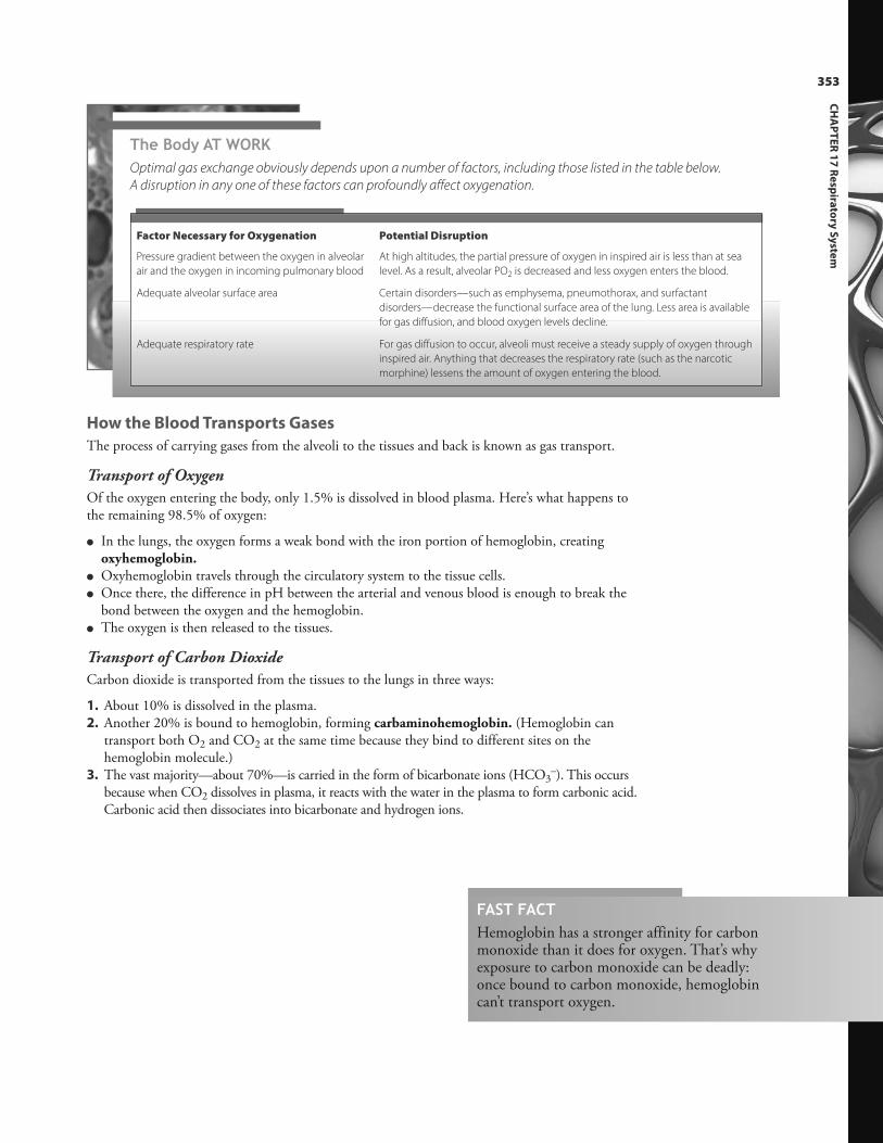
The Body AT WORKOptimal gas exchange obviously depends upon a number of factors, including those listed in the table below.
A disruption in any one of these factors can profoundly affect oxygenation.
FAST FACTHemoglobin has a stronger affinity for carbonmonoxide than it does for oxygen. That’s whyexposure to carbon monoxide can be deadly:once bound to carbon monoxide, hemoglobincan’t transport oxygen.
Factor Necessary for Oxygenation Potential Disruption
Pressure gradient between the oxygen in alveolar
air and the oxygen in incoming pulmonary blood
At high altitudes, the partial pressure of oxygen in inspired air is less than at sea
level. As a result, alveolar PO2 is decreased and less oxygen enters the blood.
Adequate alveolar surface area Certain disorders—such as emphysema, pneumothorax, and surfactant
disorders—decrease the functional surface area of the lung. Less area is available
for gas diffusion, and blood oxygen levels decline.
Adequate respiratory rate For gas diffusion to occur, alveoli must receive a steady supply of oxygen through
inspired air. Anything that decreases the respiratory rate (such as the narcotic
morphine) lessens the amount of oxygen entering the blood.

Review of Key Terms
Alveolus: Air sac in the lungs
Bronchi: The two main branchesleading from the trachea to the lungsthat serve as passageways for air
Bronchioles: One of the smallersubdivisions of the bronchial tubes
Epiglottis: The uppermost cartilage ofthe larynx; closes during swallowingto direct food and liquids into theesophagus
Glottis: The opening between thevocal cords
Hilum: Opening on the lung’s medialsurface through which primary bronchiand pulmonary blood vessels pass
Intrapleural pressure: The pressurebetween the visceral and parietalpleurae, which assists with lungexpansion
Larynx: Structure made of cartilageand muscle at the upper end of thetrachea; part of the airway and thevocal apparatus
Palate: Bony structure separating themouth from the nasal cavity
Partial pressure: The contribution of asingle gas in a mixture of gases towardthe total pressure of the gas mixture
Pharynx: Muscular tube behind theoral and nasal cavities; commonlycalled the throat
Pleura: Serous membrane covering thelungs and the thoracic cavity
Surfactant: Lipoprotein secreted byalveolar cells that decreases surfacetension of the fluid lining the alveoli,permitting expansion of alveoli
Tidal volume: The amount of airinhaled and exhaled during quietbreathing
Trachea: Portion of the respiratorytract that carries air through the neckand upper chest
Ventilation: The movement of air intoand out of the lungs
Vital capacity: The amount of air thatcan be inhaled and exhaled with thedeepest possible breath
PA
RT
IV M
ain
ten
an
ce o
f the
Bo
dy
Own the InformationTo make the information in this chapter part of your
working memory, take some time to reflect on what you’ve
learned. On a separate sheet of paper, write down
everything you recall from the chapter. After you’re done,
log on to the DavisPlus website, and check out the Study
Group podcast and Study Group Questions for the chapter.
Key Topics for Chapter 17:
• Structures of the upper respiratory tract
• Structures of the lower respiratory tract
• Structure and function of the muscles used in pulmonary
ventilation
• Neural control of breathing
• Factors influencing breathing
• How a pressure gradient influences the flow of air into
and out of the lungs
• How pulmonary compliance and alveolar surface tension
affect airflow
• Measurements of ventilation
• Variations in respiratory rhythm
• The process of gas exchange along the circulatory route
• How the blood transports oxygen and carbon dioxide
354

Test Your Knowledge1. Which of the following is a
function of the nasopharynx?a. Filter dustb. Warm and moisten inspired airc. Provide openings for the right
and left eustachian tubesd. Contain olfactory receptors
responsible for the sense ofsmell
2. Which structure is responsiblefor directing food and liquidsinto the esophagus during swallowing?a. Glottisb. Epiglottisc. Adam’s appled. Conchae
3. What purpose do the cartilaginousrings around the trachea serve?a. Keep the trachea from
collapsing during inhalationb. Attach the trachea firmly to
the esophagusc. Protect the trachea from
traumad. They serve no purpose
4. Inhaled food or foreign objectsare most likely to lodge in whichpart of the respiratory system?a. Pharynxb. Right bronchusc. Left bronchusd. Bronchioles
5. What is the purpose of surfactant?a. Facilitate in the diffusion of
oxygen across the respiratorymembrane
b. Transport oxygen to the alveolic. Purify the air entering the
respiratory tractd. Keep alveoli from collapsing
6. What is one of the purposes ofthe fluid in the pleural cavity?a. Lubricate the pleural surfaces
to allow them to glide painlessly during lung expansion and contraction
b. Warm and moisten lung tissuec. Prevent bacteria from entering
lung tissued. Assist in the diffusion of
oxygen across the respiratorymembrane
7. The main muscle responsible forpulmonary ventilation is:a. the abdominals.b. the external intercostals.c. the diaphragm.d. the internal intercostals.
8. Which gas is the primary regulatorof respiration?a. Oxygenb. Carbon dioxidec. Nitrogend. Bicarbonate
9. When pressure in the lungs dropslower than atmospheric pressure,what occurs?a. Air flows out of the lungs.b. Air flows into the lungs.c. A pneumothorax forms,
collapsing the lungs.d. The bronchioles constrict,
causing respiratory distress.
10. The primary way oxygen is transported in the blood is:a. in the form of bicarbonate.b. in the form of
carbaminohemoglobin.c. in the form of oxyhemoglobin.d. dissolved in plasma.
Answers: Chapter 171. Correct answer: c. The nasopharynx, part of the
pharynx, contains openings for the right and lefteustachian tubes. The nasal cavity contains ciliathat filter dust and foreign particles from inspiredair; the mucosa of the nasal cavity warms andmoistens inspired air; and branches of theolfactory nerve (responsible for the sense of smell)penetrate the upper nasal cavity and lead to thebrain.
2. Correct answer: b. The glottis is the space betweenthe vocal cords. The Adam’s apple—also called thethyroid cartilage—is the largest piece of cartilageforming the trachea and has no role in swallowing.Conchae are bones in the lateral walls of the nasalcavity.
3. Correct answer: a. Tracheal cartilage is C-shaped(with the opening in the posterior region), whichallows the esophagus to expand during swallowing,but the two structures are not attached to eachother. The primary purpose of the cartilage is notfor protection.
4. Correct answer: b. The right bronchus is slightlywider and more vertical than the left, making itthe most likely location for aspirated (inhaled)food particles and small objects to lodge.
5. Correct answer: d. Surfactant is a substance thathelps reduce surface tension (the force ofattraction between water molecules) to keep thealveolus from collapsing as air moves in and outduring respiration. It does not facilitate thediffusion of oxygen. Hemoglobin is the primarytransporter of oxygen in the bloodstream.Surfactant has no role in the purification of the airin the respiratory tract.
6. Correct answer: a. The fluid in the pleural cavitylubricates the pleural surfaces, allowing the twosurfaces to glide painlessly against each other asthe lungs expand and contract. It also it creates apressure gradient that assists in lung inflation.None of the other answers is correct.
7. Correct answer: c. The internal and externalintercostal muscles are used in the respiratoryprocess; however, the diaphragm is the mainmuscle responsible for pulmonary ventilation. Theabdominal muscles are accessory—not primary—muscles of respiration.
8. Correct answer: b. Carbon dioxide, not oxygen, isthe primary regulator of respiration. Nitrogen is amajor component of the atmosphere, but it is notinvolved in respiration. Carbon dioxide istransported through the bloodstream in the formof bicarbonate, but bicarbonate does not have arole in respiration.
9. Correct answer: b. Air flows from an area of higherto lower pressure; therefore, air flows out of thelungs when pressure in the lungs is higher thanatmospheric pressure. A pneumothorax resultswhen air accumulates in the pleural space. Asthmais a condition in which bronchioles spasm andconstrict, causing respiratory distress.
10. Correct answer: c. Carbaminohemoglobin andbicarbonate are two forms used to transportcarbon dioxide. Only 1.5% of the oxygen enteringthe body is dissolved in plasma; 98.5% travelsthrough the circulatory system in the form ofoxyhemoglobin.
Go to http://davisplus.fadavis.com Keyword:Thompson to see all of the resources availablewith this chapter.
355
CH
AP
TE
R 1
7 R
esp
irato
ry S
yste
m
CHAPTER OUTLINEOverview of the Urinary System
The Kidneys
Urine Formation
Composition of Urine
Storage and Elimination of Urine
LEARNING OUTCOMES1. Identify the location of the kidneys.
2. Name the internal and external structures of
the kidneys.
3. Trace the flow of blood through the kidney.
4. Describe the nerve supply to the kidney.
5. Trace the flow of fluid through the renal
tubule.
6. Describe the processes that occur in each
section of the renal tubule.
7. Discuss the mechanisms that drive glomerular
filtration.
8. Identify the mechanisms used by the kidneys
to ensure a steady glomerular filtration rate.
9. Describe the steps in the
renin‐angiotensin‐aldosterone system.
10. Discuss the tubular reabsorption and secretion
that occurs in the different parts of the renal
tubule.
11. Name the hormones that affect the urinary
system and identify their actions.
12. Describe the characteristics and components
of urine.
13. Identify the structure and function of the
ureters, urinary bladder, and urethra.
14. Describe how the structure of the urethra
varies between males and females.
15. Explain the process of urination.
18chapter URINARY SYSTEMEvery hour, the kidneys filter up to 12 pints (5.7 liters) of fluid
from the blood.Throughout the body, cells continually perform a variety of metabolic processes. Each of these processes produces waste as aby-product. Cleansing the blood of these toxic substances is the job of the kidneys—the principal organs of the urinarysystem. As blood filters through the kidneys, these mighty organs remove potential poisons, adjust the water content ofblood, tweak the levels of sodium and potassium, and adjust the pH level. What’s more, the kidneys also play a role in theregulation of blood pressure and the production of red blood cells.
Kidney
Ureter
Urinary bladder
Urethra
Diaphragm
Adrenal gland
Renal artery
Renal vein
Aorta
Inferior vena cava
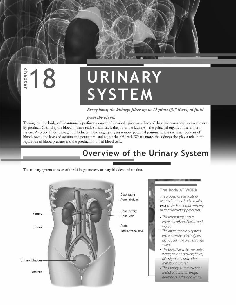
Overview of the Urinary SystemThe urinary system consists of the kidneys, ureters, urinary bladder, and urethra.
The Body AT WORKThe process of eliminating
wastes from the body is called
excretion. Four organ systems
perform excretory processes:
• The respiratory system
excretes carbon dioxide and
water.
• The integumentary system
excretes water, electrolytes,
lactic acid, and urea through
sweat.
• The digestive system excretes
water, carbon dioxide, lipids,
bile pigments, and other
metabolic wastes.
• The urinary system excretes
metabolic wastes, drugs,
hormones, salts, and water.

Ureter
Renal
papilla
Fibrous capsule
Hilum
11th rib
12th rib
Each kidney measures about 4 inches (10
cm) long, 2 inches (5 cm) wide, and 1 inch
(2.5 cm) thick; they extend from the level of
the T12 vertebra to the L3 vertebra.
Structures (such as blood vessels, the ureters,
and nerves) enter and leave the kidney
through a slit called the hilum—located in a
concave notch on the medial side.
The renal cortex forms the outer region of the kidney.
The renal medulla forms the inner region.
Extensions from the renal cortex, called renal columns,
divide the interior region into cone-shaped sections.
The cone-shaped sections are called renal pyramids.
Consisting of tubules for transporting urine away from
the cortex, the base of each pyramid faces outward
toward the cortex. The point of the pyramid, called the
renal papilla, faces the hilum.
The renal papilla extends into a cup called a minor
calyx. The calyx collects urine leaving the papilla.
Two or three minor calyces join together to form a
major calyx.
The major calyces converge to form the renal pelvis,
which receives urine from the major calyces. The renal
pelvis continues as the ureter, a tube-like structure that
channels urine to the urinary bladder.
PA
RT
IV M
ain
ten
an
ce o
f the
Bo
dy
358
The kidneys lie against the posterior abdominal wall and underneath the 12th rib. They are also retroperitoneal, meaningthat they are posterior to the parietal peritoneum. The ribs offer some protection to the kidneys, as does a heavy cushion offat encasing each organ.
A tough, fibrous capsule surrounds each kidney. The interior of the kidneyconsists of two regions: the renal cortex (the site of urine production) and therenal medulla (the site of urine collection).
FAST FACTThe right kidney sitslower than the leftbecause of the spaceoccupied by the liver justabove it.
The Kidneys

Nephron
Proximal
convoluted
tubule
Distal
convoluted
tubule
Collecting
duct
Cortex
Medulla
Loop of Henle
Nephrons—the filtration units of the kidney—primarily lie inthe kidney’s outer region; loops from the nephrons, however, dipinto the inner region of the medulla. Each nephron is wellsupplied with blood.
The renal artery—which branches off the abdominal aorta—
brings blood to the kidney.
As it enters the kidney, the renal artery divides, branching into
smaller and smaller arteries. The arteries pass through the
renal columns and extend into the renal cortex.
3 Blood leaves the glomerulus
through an efferent arteriole.
4 The efferent arteriole leads to a
network of capillaries around
the renal tubules called peritubular
capillaries. These capillaries pick up
water and solutes reabsorbed by the
renal tubules.
5 Blood flows from the
peritubular capillaries into larger
and larger veins that eventually feed
into the renal vein.
1 In the cortex, a series of afferent
arterioles arise from the smaller
arteries. Each afferent arteriole
supplies blood to one nephron.
2 Each afferent arteriole branches
into a cluster of capillaries
called a glomerulus. The glomerulus
is enclosed by Bowman’s capsule,
which will be discussed later in this
chapter.
Blood eventually leaves the kidney through the renal vein,
which empties into the inferior vena cava.
FAST FACTOver 20% of all theblood pumped by theheart each minute goesto the kidneys.
359
CH
AP
TE
R 1
8 U
rina
ry S
yste
m
Renal Circulation

� � � � � � � �� � � � � � � � � � � � � � �� � � � � � � � � � � � � � � � � � �� � � � � � � � � � � � �� � � � � � � � � � � �
� � � � � � �3 After returning to the cortex, the
ascending limb coils again,
forming the distal convoluted tubule.
4 The collecting duct
receives drainage
from the distal convoluted
tubules of several different
nephrons. The collecting
duct passes into a renal
pyramid, where it merges
with other collecting ducts
to form one tube. That
tube opens at a renal
papilla into a minor calyx.
1 Arising directly from Bowman’s capsule is the proximal
convoluted tubule: a winding, convoluted portion of the
renal tubule. Thousands of microvilli that allow absorption to
occur line the inside of the proximal convoluted tubule.
2 The renal tubule straightens out and dips into the
medulla before turning sharply and returning to the
cortex. This entire segment—which consists of a descending
limb and an ascending limb—is called the loop of Henle.
PA
RT
IV M
ain
ten
an
ce o
f the
Bo
dy
360
Renal Innervation
Along with blood vessels, nerves also enter the kidney at the hilum. These mainly sympathetic fibers stimulate the afferentand efferent arterioles, controlling the diameter of the vessels, which, in turn, regulate the rate of urine formation. Also, ifblood pressure drops, the nerves stimulate the release of renin, an enzyme that triggers processes for restoring blood pressure.
Nephron
The outer regions of the kidney are packed with over 1 million nephrons: the microscopic functional units of the kidney.These tiny structures consist of two main components: a renal corpuscle—which filters blood plasma—and a renal tubule—where urine is formed.
Renal Corpuscle
Known as the beginning of the nephron, a renal corpuscle consists of a glomerulus and Bowman’s capsule.
Renal Tubule
Leading away from the glomerulus are a series of tube-like structures that, collectively, are called the renal tubule. The renaltubule can be divided into four regions: the proximal convoluted tubule, nephron loop, distal convoluted tubule, andcollecting duct. The renal tubule has been stretched out in the following figure to more clearly show the different regions.
Proximal
tubule
Bowman’s capsule—also called a glomerular capsule—consists of
two layers of epithelial cells that envelop the glomerulus in an
open-ended covering. (To understand the structure of a renal
corpuscle, imagine pushing your fist into an inflated balloon. Your
fist represents the glomerulus. The balloon, which folds around
your fist in two layers, represents Bowman’s capsule.)
Fluid filters out of the glomerulus and collects in the space
between the two layers of Bowman’s capsule. From there, it flows
into the proximal renal tubule on the other side of the capsule.
FAST FACTThe renal corpuscle of all nephrons resides in therenal cortex. The loop of Henle dips into the renalmedulla; some dip in only slightly whereas othersextend deep into the medulla.

FAST FACTMost of the calcium, iron, andthyroid hormone in the blood isbound to plasma proteins, whichprevents these solutes from beingfiltered out of the blood in theglomerulus.
361
CH
AP
TE
R 1
8 U
rina
ry S
yste
m
� � � � � � � �� � � � � � � �! � � � � � � �� � � � � � � � " � # $ �% � � � � � �& � � � � � �
The creation of urine by the nephrons involves three processes: glomerular filtration, tubular reabsorption, and tubularsecretion.
Glomerular Filtration
The first step in the creation of urine from blood plasma occurs in the glomerulus as water and small solutes filter out of theblood and into the surrounding space of Bowman’s capsule. Filtration in the glomerulus occurs for the same reason filtrationoccurs in other blood capillaries: the existence of a pressure gradient.
1 Blood flows into the glomerulus
through the afferent arteriole, which is
much larger than the efferent arteriole.
Consequently, blood flows in faster than it
can leave, which contributes to higher
pressure within the glomerular capillaries.
2 The walls of glomerular capillaries are dotted
with pores, allowing water and small solutes
(such as electrolytes, glucose, amino acids,
vitamins, and nitrogenous wastes) to filter out of
the blood and into Bowman’s capsule. Blood cells
and most plasma proteins, however, are too large
to pass through the pores.
3 The fluid that has filtered into Bowman’s capsule
flows into the renal tubules. The amount of fluid
filtered by both kidneys—called the glomerular
filtration rate (GFR)—equals about 180 liters each
day, which is 60 times more than the body’s total
blood volume. The body reabsorbs about 99% of this
filtrate, leaving 1 to 2 liters to be excreted as urine.
The Body AT WORKSome kidney diseases damage the endothelium
of glomerular capillaries, allowing plasma
proteins to filter out into the urine. The presence of
protein in the urine—typically an abnormal
finding—is called proteinuria. Proteinuria may
also occur following intense physical exercise. In
this instance, researchers think the proteinuria
may result because hormones released during
exercise temporarily alter the permeability of the
filtration membrane.
Life lesson: Hypertension and kidney damageA common cause of kidney damage and kidneyfailure is uncontrolled hypertension. Systemichypertension pushes the normally high glomerularpressure even higher. As a result, the glomerularcapillaries can burst, causing scarring.Hypertension also causes atherosclerosis in bloodvessels throughout the body, including those inthe kidneys. This decreases blood flow to thekidneys, leading to further damage.
Urine Formation
ANIMATION

PA
RT
IV M
ain
ten
an
ce o
f the
Bo
dy
362
' ( ) * + , - . / 0 1 ( - + 12 0 - - 34 0 5 6 5
7 6 8 0 19 5 , 6 . * 0 5 3 6 5 :9 5 , 6 . * 0 5 3 6 5 : : 7 ( 5 ,; 6 < 5 0 =9 < 1 0 5 + -, - + 5 <9 - < . 3 * 0 1 . 5 0
9 > > 0 1 0 5 *+ 1 * 0 1 6 . - 0? > > 0 1 0 5 *+ 1 * 0 1 6 . - 0@ - . / 0 1 ( - ( 3A 1 . ) 6 / + -2 . 5 8 . - ( * 0 <* ( B ( - 0
C 6 3 * + -2 . 5 8 . - ( * 0 <* ( B ( - 01
2
3
4
5
Angiotensin I circulates to the
lungs, where angiotensin-
converting enzyme (ACE)
converts it into angiotensin II.
Angiotensin II stimulates the
adrenal glands to secrete
aldosterone.
Aldosterone causes the distal
convoluted tubule to retain
sodium, which leads to increased
retention of water. Blood volume
increases and blood pressure rises.
A drop in BP leads to decreased
blood flow to the kidneys.
Specialized cells found primarily
in the afferent arterioles—called
juxtaglomerular cells—respond
by releasing the enzyme renin.
Renin converts the inactive
plasma protein angiotensinogen
(made in the liver) into
angiotensin I.
1
2
3
45
FAST FACTDuring circulatory shock, the sympatheticnervous system constricts the afferent arteriolesto divert blood from the kidneys to the heartand brain. In this situation, the glomerularfiltration rate may slow to only a few millilitersper minute.
Regulation of the Glomerular Filtration Rate
For the body to maintain fluid and electrolyte balance, glomerular filtration should continue at a fairly constant rate despiteperiodic changes in blood pressure. If the flow rate is too high, the body will lose excessive amounts of water and nutrients;if it’s too slow, the tubules may reabsorb toxins that should be excreted.
The kidneys employ various mechanisms to control blood flow and ensure a steady glomerular filtration rate. Forexample, rising blood pressure stimulates the afferent arterioles to contract, preventing a surge of blood into the glomerulus.(The opposite is also true: falling blood pressure causes the afferent arterioles to relax.) In addition, specialized cells in thedistal convoluted tubule monitor the flow rate and composition of filtrate, allowing the renal tubules to make adjustmentsto alter flow as needed.
Finally, a key mechanism for maintaining blood pressure and, therefore, a steady glomerular filtration rate, is the renin-angiotensin-aldosterone system. The chain of events that occur in this system is outlined below. (See the section“Hormones Affecting the Urinary System” for more information on aldosterone.)
ANIMATION
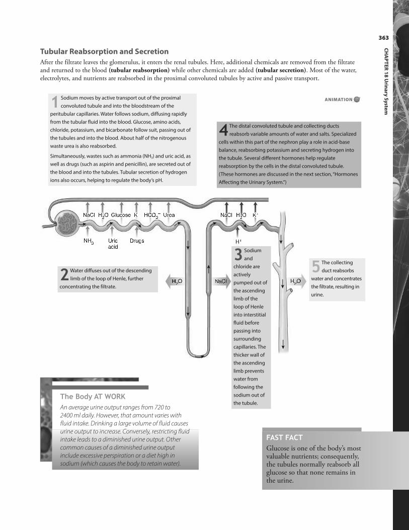
The Body AT WORKAn average urine output ranges from 720 to
2400 ml daily. However, that amount varies with
fluid intake. Drinking a large volume of fluid causes
urine output to increase. Conversely, restricting fluid
intake leads to a diminished urine output. Other
common causes of a diminished urine output
include excessive perspiration or a diet high in
sodium (which causes the body to retain water).
363
CH
AP
TE
R 1
8 U
rina
ry S
yste
m
D E F G H I J K LD E F G H I J M G N O P Q R S T R EK L H F J UVD E F G H I J M G N O P Q R W T N X Q S T R ES T Y OE O Y Z K L D E F G H I J K LH LD H U H F J U [H I J \ ] ^D E F G 5 The collecting
duct reabsorbs
water and concentrates
the filtrate, resulting in
urine.
1 Sodium moves by active transport out of the proximal
convoluted tubule and into the bloodstream of the
peritubular capillaries. Water follows sodium, diffusing rapidly
from the tubular fluid into the blood. Glucose, amino acids,
chloride, potassium, and bicarbonate follow suit, passing out of
the tubules and into the blood. About half of the nitrogenous
waste urea is also reabsorbed.
Simultaneously, wastes such as ammonia (NH3) and uric acid, as
well as drugs (such as aspirin and penicillin), are secreted out of
the blood and into the tubules. Tubular secretion of hydrogen
ions also occurs, helping to regulate the body’s pH.
2Water diffuses out of the descending
limb of the loop of Henle, further
concentrating the filtrate.
4 The distal convoluted tubule and collecting ducts
reabsorb variable amounts of water and salts. Specialized
cells within this part of the nephron play a role in acid-base
balance, reabsorbing potassium and secreting hydrogen into
the tubule. Several different hormones help regulate
reabsorption by the cells in the distal convoluted tubule.
(These hormones are discussed in the next section, “Hormones
Affecting the Urinary System.”)
3 Sodium
and
chloride are
actively
pumped out of
the ascending
limb of the
loop of Henle
into interstitial
fluid before
passing into
surrounding
capillaries. The
thicker wall of
the ascending
limb prevents
water from
following the
sodium out of
the tubule.
FAST FACTGlucose is one of the body’s mostvaluable nutrients; consequently,the tubules normally reabsorb allglucose so that none remains inthe urine.
Tubular Reabsorption and Secretion
After the filtrate leaves the glomerulus, it enters the renal tubules. Here, additional chemicals are removed from the filtrateand returned to the blood (tubular reabsorption) while other chemicals are added (tubular secretion). Most of the water,electrolytes, and nutrients are reabsorbed in the proximal convoluted tubules by active and passive transport.
ANIMATION
PA
RT
IV M
ain
ten
an
ce o
f the
Bo
dy
364
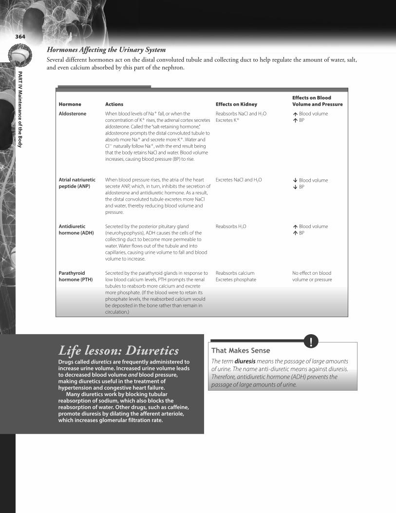
Hormones Affecting the Urinary System
Several different hormones act on the distal convoluted tubule and collecting duct to help regulate the amount of water, salt,and even calcium absorbed by this part of the nephron.
Hormone Actions Effects on Kidney
Effects on Blood
Volume and Pressure
Aldosterone When blood levels of Na1 fall, or when the
concentration of K1 rises, the adrenal cortex secretes
aldosterone. Called the “salt-retaining hormone,”
aldosterone prompts the distal convoluted tubule to
absorb more Na1 and secrete more K1. Water and
Cl2 naturally follow Na1, with the end result being
that the body retains NaCl and water. Blood volume
increases, causing blood pressure (BP) to rise.
Reabsorbs NaCl and H2O
Excretes K1
Blood volume
BP
Atrial natriuretic
peptide (ANP)
When blood pressure rises, the atria of the heart
secrete ANP, which, in turn, inhibits the secretion of
aldosterone and antidiuretic hormone. As a result,
the distal convoluted tubule excretes more NaCl
and water, thereby reducing blood volume and
pressure.
Excretes NaCl and H2O Blood volume
BP
Antidiuretic
hormone (ADH)
Secreted by the posterior pituitary gland
(neurohypophysis), ADH causes the cells of the
collecting duct to become more permeable to
water. Water flows out of the tubule and into
capillaries, causing urine volume to fall and blood
volume to increase.
Reabsorbs H2O Blood volume
BP
Parathyroid
hormone (PTH)
Secreted by the parathyroid glands in response to
low blood calcium levels, PTH prompts the renal
tubules to reabsorb more calcium and excrete
more phosphate. (If the blood were to retain its
phosphate levels, the reabsorbed calcium would
be deposited in the bone rather than remain in
circulation.)
Reabsorbs calcium
Excretes phosphate
No effect on blood
volume or pressure
That Makes SenseThe term diuresis means the passage of large amounts
of urine. The name anti-diuretic means against diuresis.
Therefore, antidiuretic hormone (ADH) prevents the
passage of large amounts of urine.
!Life lesson: DiureticsDrugs called diuretics are frequently administered toincrease urine volume. Increased urine volume leadsto decreased blood volume and blood pressure,making diuretics useful in the treatment ofhypertension and congestive heart failure.
Many diuretics work by blocking tubularreabsorption of sodium, which also blocks thereabsorption of water. Other drugs, such as caffeine,promote diuresis by dilating the afferent arteriole,which increases glomerular filtration rate.

365
CH
AP
TE
R 1
8 U
rina
ry S
yste
m
Urine consists of 95% water and 5% dissolved substances. The dissolved substances include nitrogenous wastes—such asurea, uric acid, ammonia, and creatinine—as well as other solutes, such as sodium, potassium, and sulfates.
The components of urine reveal a great deal about the health of the kidneys as well as other organs of the body. That’swhy a urinalysis (an examination of the characteristics of urine) is one of the most frequently prescribed medical tests.
The following table lists the normal characteristics of urine along with the possible implications of some commonabnormalities. Note: Glucose, blood, free hemoglobin, albumin, ketones, and bile pigments are not normally found in urine;their presence indicates a disease process.
FAST FACTComparing the amount of creatinine in the urineagainst the level of creatinine in the blood reflectsthe GFR; in turn, this reflects kidney function. Ahigh serum creatinine indicates a low GFR and poorkidney function.
FAST FACTUrine’s yellow color results from the pigmenturochrome, a by-product of the breakdown ofhemoglobin in worn-out red blood cells.
Characteristic Normal Finding Possible Abnormalities
Color Transparent pale yellow
to amber
• Darker urine usually results from poor hydration.
• Cloudy urine may result from bacteria, indicating an infection.
Odor Mild • A pungent smell (such as in a stale diaper) results when urine is
allowed to stand: bacteria multiplies and converts urea into
ammonia.
• A sweet, fruity odor (acetone) often occurs in diabetes.
• A rotten odor may indicate a urinary tract infection.
Specific gravity (Indicates the amount of
solid matter in a liquid)
1.001–1.035 • A high specific gravity could result from dehydration (reflecting a
low volume of water in relation to the amount of solids).
pH Average of 6.0 • A high pH reflects alkalosis.
• A low pH indicates acidosis.
The Body AT WORKMost adults produce 1 to 2 liters of urine a day. A
urine output of less than 400 ml/day (called
oliguria) is insufficient for clearing waste products
from the body.
Some diseases, particularly diabetes mellitus,
cause urine output to increase significantly. In this
instance, high levels of glucose oppose the
reabsorption of water, causing more water to pass
through the kidneys and exit the body as urine.
Another disorder that produces large volumes of
urine is diabetes insipidus. This disorder results
from hyposecretion of ADH. Without an adequate
supply of ADH, the collecting duct doesn’t reabsorb
much water and large volumes of water pass out of
the body as urine.
Composition of Urine

K Y Z _ R `a G E Z Z R TS T R b c T E
S T R b R Td R T Y b P _ R N eS T R b R T E GP f R _ Y _ X Q
Connecting the renal pelvis of each kidney with the bladder
are slender, muscular tubes called ureters. Each ureter
measures about 25 cm (9.8 inches) in length and has a very
narrow diameter. Peristaltic waves help propel urine from the
renal pelvis toward the bladder.
The wall of the bladder, called the detrusor muscle, consists of
three layers of smooth muscle.
Mucous transitional epithelium lines the bladder. When the
bladder is relaxed, this layer of tissue forms folds called rugae.
As urine fills the bladder, the rugae flatten and the epithelium
thins, allowing the bladder to expand. (Considered moderately
full when it contains 500 ml of urine, the bladder has a
maximum capacity of about 800 ml.)
The floor of the bladder has three openings: two from the
ureters (which pass behind the bladder to enter from below)
and one from the urethra. Together they form a triangular-
shaped, smooth area on the floor of the bladder called the
trigone. Infections commonly attack this area of the bladder.
At the point where the urethra leaves the bladder, a ring of
smooth muscle forms the internal urethral sphincter. This
sphincter contracts involuntarily to retain urine in the bladder.
A second sphincter, called the external urinary sphincter,
exists where the urethra passes through the pelvic floor. This
sphincter consists of skeletal muscle and is, therefore, under
voluntary control.
The urethra is a small tube that conveys urine away from the
bladder and out of the body. The opening of the urethra
leading to the outside of the body is called the external
urinary meatus.
Urinary Bladder
A collapsible muscular sac, the urinary bladder sits behind thesymphysis pubis and below the peritoneal membrane. Inwomen, it resides in front of the vagina and uterus; in men, itrests on top of the prostate gland.
The remaining structures of the urinary system are the ureters, urinary bladder, and urethra. The ureters and urethraserve as passageways for conducting urine away from the kidneys and out of the body while the bladder stores urine untilit can be eliminated.
Ureters
PA
RT
IV M
ain
ten
an
ce o
f the
Bo
dy
366
FAST FACTBecause the ureter is so narrow, kidney stones caneasily become lodged and obstruct the flow of urine.
Storage and Elimination of Urine

Bladder
Urethra Vagina
Rectum Bladder
Urethra
External urinarysphincter muscle
Rectum
Female urethra
In women, the urethra is 3 cm (1.2 inches) long and exits the body
just in front of the vaginal orifice.
Male urethra
In males, the urethra is much longer, measuring about 20 cm
(7.9 inches). From the bladder, the urethra passes through the
center of the prostate gland, curves around to enter the penis, and
then exits the body at the tip of the penis. In men, the urethra
performs a dual role. Besides conveying urine, it also conveys
semen. (For more information on the male reproductive system, see
Chapter 23, Reproductive Systems.)FAST FACTFemales are particularly prone to urinary tractinfections because bacteria such as Escherichia coli(found in the lower digestive tract) can easilymigrate up the short urethra and infect the bladder.
Renal calculi in
minor and major
calyces
Renal calculi
in the ureter
Life lesson: Kidney stonesKidney stones, or renal calculi, result when minerals (such ascalcium, phosphate, uric acid, or protein) crystallize in the renalpelvis. Many times these calculi are small enough to travel throughthe urinary tract and out of the body unnoticed. Sometimes, though,the stones become large enough to block the renal pelvis or ureter.When this occurs, excruciating pain results as the ureter contractsviolently as it attempts to dislodge the stone. If the stone remainslodged, urine may back up to the kidney, resulting in hydronephrosis.
Doctors often treat renal calculi with a technique calledlithotripsy. This technique uses ultrasound to disintegrate the stoneinto particles small enough to pass through the urinary tract.
367
CH
AP
TE
R 1
8 U
rina
ry S
yste
m
Urethra
The structure of the urethra varies between males and females.

PA
RT
IV M
ain
ten
an
ce o
f the
Bo
dy
368
3
1
2
4
When the bladder contains 200 ml or
more of urine, stretch receptors in the
bladder wall send impulses to the sacral
region of the spinal cord.
The brain has the ability to override the
impulse to void because the stretch
receptors in the bladder also send
impulses to the micturition center in the
pons. The pons integrates information
from the stretch receptors with information
from other parts of the brain, such as the
cerebrum, and evaluates whether the time
is appropriate to urinate.
If the time to urinate is not appropriate,
the brain sends impulses to inhibit
urination and to keep the external urinary
sphincter contracted. If the time to urinate
is appropriate, the brain sends signals to
the bladder wall to contract and to the
external urethral sphincter to relax, at
which point voluntary urination occurs.
1
3
4
The spinal cord then sends motor
impulses to the bladder wall to contract
and to the internal sphincter to relax.
When this happens, voiding will occur
involuntarily unless the brain overrides the
impulse.
2
Life lesson: Renal insufficiency and dialysisRenal insufficiency (also called renal failure) results when an extensive number of nephrons have been destroyedthrough disease or injury, impairing the ability of the kidneys to function. Loss of function may occur suddenly—as inacute renal failure—or over time—as in chronic renal insufficiency.
In acute renal failure, the kidneys often stop functioning as the result of an infection, drugs, or an injury. After thecause of the renal failure is treated, kidney function may return to normal.
In contrast, chronic renal insufficiency develops over years, perhaps even decades. Typically, a disease (such asdiabetes, hypertension, glomerulonephritis, or an autoimmune disorder) gradually destroys the nephrons. Eventually,damage becomes so extensive that the kidneys can no longer effectively clear blood plasma of waste. In this instance,the damage is irreversible. A variety of symptoms develop as wastes accumulate in the blood, making a kidneytransplant or dialysis necessary.
There are two forms of dialysis: hemodialysis and peritoneal dialysis. In hemodialysis, blood is pumped from thepatient’s radial artery to a dialysis machine (which is sometimes called an artificial kidney). In the machine, the bloodflows through a series of semi-permeable tubes immersed in dialysis fluid. Waste products such as urea, potassium,and creatinine, as well as excess water, diffuse out of the blood and into the dialysis fluid. The dialysis fluid is thendiscarded.
In peritoneal dialysis, the peritoneum serves as the semi-permeable membrane. In this procedure, dialysis fluid isintroduced into the peritoneal cavity through a catheter. The fluid is left in the body cavity for a few minutes to anhour, during which time waste products diffuse out of the blood and into the dialysate. At the end of the specifiedtime, the fluid is drained and discarded.
Urination
Also called micturition or voiding, urination begins when the external sphincter muscle of the bladder voluntarily relaxesand the detrusor muscle of the bladder contracts. The entire process involves several steps requiring input from the centralnervous system.
ANIMATION

Review of Key TermsAldosterone: Hormone that causes thedistal convoluted tubule to retainsodium, which leads to the retentionof water, resulting in increased bloodpressure
Angiotensin: A plasma protein produced when renin is released fromthe kidney; angiotensin II stimulatesthe adrenal glands to secrete aldosterone
Antidiuretic hormone: Hormone thatinhibits diuresis by stimulating thekidneys to conserve water
Bowman’s capsule: Two layers of epithelial cells that envelop theglomerulus in an open-ended covering; also called a glomerular capsule
Calyx: A cup-like structure that collectsurine leaving the papilla of the kidney
Collecting duct: Receives drainagefrom the distal convoluted tubules ofseveral different nephrons; eventuallydrains into a minor calyx
Detrusor muscle: Wall of the bladder
Diuresis: The secretion of largeamounts of urine
Glomerulus: Cluster of capillaries thatare part of the renal corpuscles in thenephrons
Hilum: Concave notch on medial sideof kidney; where blood vessels, theureters, and nerves enter and leave thekidney
Loop of Henle: U-shaped portion ofthe renal tubule
Micturition: Urination
Nephrons: The filtration units of thekidney
Peritubular capillaries: Network ofcapillaries surrounding the renaltubules
Renal corpuscles: One of the maincomponents of nephrons, consistingof a glomerulus and Bowman’s capsule, that filters blood plasma
Renal cortex: Outer region of the kidney; site of urine production
Renal medulla: Inner region of thekidney; site of urine collection
Renal tubules: Series of tube-likestructures within the nephron; whereurine is formed
Renin: Enzyme released by the kidneysin response to a drop in blood pressure that causes the conversion ofangiotensinogen into angiotensin I
Specific gravity: Measurement that indicates the amount of solid matterin a liquid
Tubular resorption: Process wherebychemicals are removed from filtrate inthe renal tubules and returned to theblood
Tubular secretion: Process wherebychemicals are added to the filtrate inthe renal tubules
Ureters: Muscular tubes connectingthe renal pelvis of each kidney withthe bladder
Urethra: Small tube that conveys urineaway from the bladder and out of thebody
Urinary bladder: Collapsible muscularsac that stores urine
369
CH
AP
TE
R 1
8 U
rina
ry S
yste
m
Own the InformationTo make the information in this chapter part of your
working memory, take some time to reflect on what you’ve
learned. On a separate sheet of paper, write down
everything you recall from the chapter. After you’re done,
log on to the DavisPlus website, and check out the Study
Group podcast and Study Group Questions for the chapter.
Key Topics for Chapter 18:
• Location of the kidney
• The internal and external structures of the kidney
• Blood flow through the kidney
• Nerve supply to the kidney
• Structure and function of the nephron
• Structure and function of the renal tubule
• Process of glomerular filtration
• Regulation of the glomerular filtration rate
• The renin-angiotensin-aldosterone system
• Tubular reabsorption and secretion occurring in the
different parts of the renal tubule
• Hormones affecting the urinary system
• The composition of urine
• The structure and function of the ureters, urinary
bladder, and urethra
• The process of urination

PA
RT
IV M
ain
ten
an
ce o
f the
Bo
dy
370
Answers: Chapter 181. Correct answer: d. The renal corpuscle consists of a
glomerulus and Bowman’s capsule. The loop ofHenle is the hairpin turn taken by the renal tubulebetween the proximal and distal convolutedtubule. The proximal convoluted tubule is awinding portion of the renal tubule arising fromBowman’s capsule.
2. Correct answer: b. The renal pelvis, formed by theconvergence of the major calyces, channels urineout of the kidney. The calyx collects urine leavingthe papilla. Renal pyramids are the cone-shapedsections in the kidney.
3. Correct answer: b. The renal corpuscle filters bloodplasma. Bowman’s capsule is part of the renalcorpuscle. An afferent arteriole supplies blood to anephron.
4. Correct answer: a. The distal convoluted tubuleand collecting ducts reabsorb variable amounts ofwater and salt. In the glomerulus, water and smallsolutes filter out of the blood and into thesurrounding space of Bowman’s capsule.
5. Correct answer: c. The other substances are allnormal constituents of urine.
6. Correct answer: a. Atrial natriuretic peptide (ANP)causes the distal convoluted tubule to excrete moresodium and water, thereby reducing bloodpressure. Antidiuretic hormone (ADH) causes thedistal and collecting tubules to become morepermeable to water. Parathyroid hormone (PTH)prompts the renal tubules to reabsorb morecalcium and excrete more phosphate.
7. Correct answer: c. None of the other answers iscorrect.
Test Your Knowledge1. The cluster of capillaries in the
nephron is the:a. renal corpuscle.b. loop of Henle.c. proximal convoluted tubule.d. glomerulus.
2. The location where blood vessels,nerves, and the ureter enter andleave the kidney is the:a. renal pelvis.b. hilum.c. calyx.d. renal pyramid.
3. The portion of the nephron incharge of making urine is the:a. renal corpuscle.b. renal tubule.c. Bowman’s capsule.d. afferent arteriole.
4. Most sodium is reabsorbed fromthe glomerular filtrate in the:a. proximal convoluted tubule.b. distal convoluted tubule.c. collecting duct.d. glomerulus.
5. Which of the following substances should never be found in urine?a. Sodiumb. Urea
c. Glucosed. Potassium
6. The hormone aldosterone:a. causes the distal convoluted
tubule to reabsorb moresodium, which leads to waterretention and a rise in bloodpressure.
b. causes the distal convolutedtubule to excrete more sodiumand water, thereby reducingblood pressure.
c. causes the distal and collectingtubules to become more permeable to water.
d. prompts the renal tubules toreabsorb more calcium and excrete more phosphate.
7. Many diuretics work by blockingtubular reabsorption of:a. water.b. glucose.c. sodium.d. potassium.

Go to http://davisplus.fadavis.com Keyword:Thompson to see all of the resources availablewith this chapter.
8. In women, the ______________is shorter than it is in men, which contributes to the higherincidence of urinary tract infections in women.a. urinary sphincterb. loop of Henlec. ureterd. urethra
9. Urination occurs when:a. the bladder and internal
urethral sphincter relax.b. the bladder and external
urethral sphincter contract.
c. the bladder contracts and theexternal urethral sphincter relaxes.
d. the bladder relaxes and the external urethral sphinctercontracts.
10. A triangular-shaped smooth areaon the floor of the bladder iscalled the:a. rugae.b. papilla.c. cortex.d. trigone.
8. Correct answer: d. In women, the urethra is muchshorter than it is in men; therefore, bacteria fromthe nearby rectal area can easily migrate up theshort tube to the bladder. The other structures aresimilar in both men and women.
9. Correct answer: c. None of the other answers iscorrect.
10. Correct answer: d. Rugae are folds of tissue on theinside of the bladder. Papillae are the blunt pointsof the renal pyramids. The cortex is the outer zoneof the kidney.
371
CH
AP
TE
R 1
8 U
rina
ry S
yste
m

CHAPTER OUTLINEWater Balance
Electrolyte Balance
Acid-Base Balance
Acid-Base Imbalances
LEARNING OUTCOMES1. Name the major fluid compartments.
2. Explain how fluid moves from one
compartment to another.
3. Identify the means by which the body
normally gains and loses fluid.
4. Describe the mechanisms for regulating intake
and output.
5. Differentiate between volume depletion and
dehydration.
6. Discuss the fluid shifts that occur in
dehydration.
7. Explain the physiological consequences of
fluid excess.
8. Explain how edema occurs.
9. Identify the main electrolytes found in
intracellular and extracellular fluid.
10. Explain the roles of sodium in the body.
11. Describe the mechanisms used by the body to
regulate the levels of sodium.
12. Identify common causes and consequences of
sodium, potassium, and calcium imbalances.
13. Define buffer.
14. Explain how chemical buffers can alter pH in
the body.
15. Explain how the respiratory system can alter
the pH in the body.
16. Describe how the kidneys help control pH.
17. Pinpoint the timeframe during which each
buffer system responds.
18. Identify the causes of respiratory acidosis and
alkalosis.
19. Identify the causes of metabolic acidosis and
alkalosis.
20. Describe how the lungs and kidneys
compensate for acid‐base imbalances.

19chapter FLUID, ELECTROLYTE,& ACID-BASEBALANCEApproximately two-thirds of the body—or about 50% to 70%
of an adult’s weight—consists of water.
The body is awash in fluid: it surrounds cells, fills tissues, and, of course, is the main component of blood and lymph. While thisfluid is primarily water, it also contains thousands of dissolved substances, such as electrolytes, that are used in the body’sbiochemical reactions. If the quantity of fluid, the concentration of electrolytes, or the pH strays outside precise boundaries, life-threatening disorders may occur. In other words, homeostasis depends upon balance—of water, electrolytes, and pH.
The maintenance of water balance involves more than the total volume of fluidwithin the body. How that fluid is divided among the various fluid compartments isalso important.
Fluid Compartments
Most of the body’s water (about 65%) resides inside cells; this is called intracellularfluid (ICF). The remaining 35% of the body’s water resides outside cells: this iscalled extracellular fluid (ECF). Extracellular fluid includes the fluid between thecells inside tissue, called interstitial fluid, as well as the fluid within vessels as bloodplasma and lymph. Various other extracellular fluids—such as cerebrospinal fluid,synovial fluid in the joints, vitreous and aqueous humors of the eye, and digestivesecretions—are called transcellular fluid.
That Makes SenseTo remember the names for the various body fluids, keep this
in mind: the prefix intra- means “within” (as in intravenous
fluids) while the prefix extra- means “outside of” (as in
extracurricular activities). Therefore, intracellular means
“within the cell,” while extracellular means “outside of the cell.”
Furthermore, the prefix inter- means “between.” (For
example, the word intercede means “to come between
two parties.”) Therefore, interstitial fluid refers to fluid
between cells.
!FAST FACTA man’s body weight is about 55% to 60%water, while women average slightly less because of an increased amount of body fat.(Adipose tissue contains less water than muscle.In fact, the weight of an obese person may beonly 45% water.) In contrast, the weight of anewborn infant is as much as 75% water.
Intracellular fluid (65%)
Interstitialfluid
Plasma
Lymph
Transcellular fluid
Extracellular fluid (35%)
Water Balance

PA
RT
IV M
ain
ten
an
ce o
f the
Bo
dy
374
Balancing Water Gain and Loss
Normally, the amount of water gained and lost by the body through the course of a day is equal.Specifically, an adult gains and loses about 2500 ml of fluid each day. Most fluid intake occursthrough eating and drinking; however, the cells themselves produce a fair amount of water as aby-product of metabolic reactions. (This is called metabolic water.) Fluid is lost through thekidneys (as urine), the intestines (as feces), the skin (by sweat as well as diffusion), and the lungs(through expired air).
g h i j k lm n o o p q r s t u v w i x w im n o o p q r s t uy z { t | } q ~ � pm o o p q� } } s� o o p q � � ~ � �� � o o p q � z � z �m o o p q� � ~ �n o o p q� � z t { � ~ � �� o o p q � � ~ � z� n o o p q
FAST FACTWater loss varies considerably withenvironmental temperature as well asphysical activity. For example, cold, dryair increases respiratory loss while hot,humid air increases the loss throughperspiration. Also, heavy physical exerciseincreases fluid loss through perspiration.
The Body AT WORKFluid doesn’t remain locked within a single compartment. Rather,
ICF and ECF continually mingle, as the fluid easily passes through
the semipermeable membrane surrounding each compartment.
The concentration of solutes (particularly electrolytes) within each
compartment determines the amount and direction of flow. For
example:
• If the concentration of
electrolytes (and therefore the
osmolarity) of tissue fluid rises,
water moves out of the cells
and into the tissues.
• If the osmolarity of tissue fluid falls, water moves out of the
tissues and into the cells. The
passage of fluid happens
within seconds so as to
maintain equilibrium. (The
various electrolytes and their
role in water balance are discussed later in this chapter.)
H2O
� � p } q t � ~ { uH2O
� � p } q t � ~ { u

375
CH
AP
TE
R 1
9 F
luid
, Ele
ctroly
te, &
Acid
-Ba
se B
ala
nce
Regulation of Intake and Output
To keep the total volume of water in the body in balance, the body uses mechanisms thatadjust fluid intake as well as urine output.
Mechanisms to fluid intake
Physical changes stimulate
the thirst center in the
hypothalamus.
Salivation decreases, causing a dry mouth
and the sensation of thirst.
Consumption of water
leads to a rise in total
water volume.
Physical changes stimulate
the hypothalamus, which, in
turn, stimulates the posterior
pituitary to secrete antidiuretic
hormone (ADH).
ADH prompts the collecting ducts
of the kidneys to reabsorb more water
and produce less urine.
The rate of fluid loss slows until water is ingested.
Mechanisms to urine output
Blood pressure drops, sodium concentration
rises, and osmolarity increases.
Various factors, including excessive sweating, cause the
volume of total body water to decline.
The Body AT WORKWhen blood volume and pressure are too high, or
blood osmolarity is too low, the hypothalamus
inhibits the release of ADH. This causes the renal
tubules to reabsorb less water, leading to an
increased urine output and a decline in total
body water.
FAST FACTElderly adults have a diminished sensation of thirst,placing them at risk for dehydration.
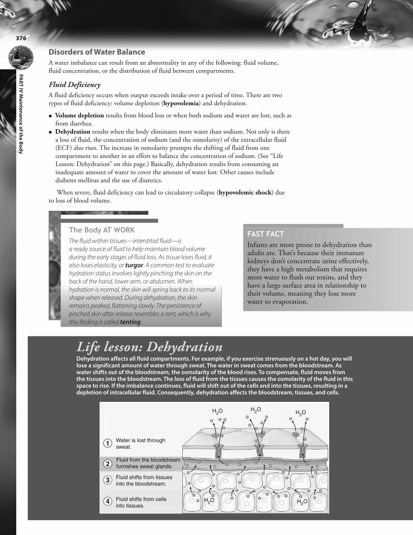
PA
RT
IV M
ain
ten
an
ce o
f the
Bo
dy
376
Water is lost through
sweat.
Fluid from the bloodstream
furnishes sweat glands.
Fluid shifts from tissues
into the bloodstream.
Fluid shifts from cells
into tissues.
H2OH
2O H
2O
H2O H
2O
1
2
3
4
The Body AT WORKThe fluid within tissues—interstitial fluid—is
a ready source of fluid to help maintain blood volume
during the early stages of fluid loss. As tissue loses fluid, it
also loses elasticity, or turgor. A common test to evaluate
hydration status involves lightly pinching the skin on the
back of the hand, lower arm, or abdomen. When
hydration is normal, the skin will spring back to its normal
shape when released. During dehydration, the skin
remains peaked, flattening slowly. The persistence of
pinched skin after release resembles a tent, which is why
this finding is called tenting.
Life lesson: DehydrationDehydration affects all fluid compartments. For example, if you exercise strenuously on a hot day, you willlose a significant amount of water through sweat. The water in sweat comes from the bloodstream. Aswater shifts out of the bloodstream, the osmolarity of the blood rises. To compensate, fluid moves fromthe tissues into the bloodstream. The loss of fluid from the tissues causes the osmolarity of the fluid in thisspace to rise. If the imbalance continues, fluid will shift out of the cells and into the tissues, resulting in adepletion of intracellular fluid. Consequently, dehydration affects the bloodstream, tissues, and cells.
FAST FACTInfants are more prone to dehydration thanadults are. That’s because their immaturekidneys don’t concentrate urine effectively,they have a high metabolism that requiresmore water to flush out toxins, and theyhave a large surface area in relationship totheir volume, meaning they lose morewater to evaporation.
Disorders of Water Balance
A water imbalance can result from an abnormality in any of the following: fluid volume,fluid concentration, or the distribution of fluid between compartments.
Fluid Deficiency
A fluid deficiency occurs when output exceeds intake over a period of time. There are twotypes of fluid deficiency: volume depletion (hypovolemia) and dehydration.
l Volume depletion results from blood loss or when both sodium and water are lost, such asfrom diarrhea.
l Dehydration results when the body eliminates more water than sodium. Not only is therea loss of fluid, the concentration of sodium (and the osmolarity) of the extracellular fluid(ECF) also rises. The increase in osmolarity prompts the shifting of fluid from onecompartment to another in an effort to balance the concentration of sodium. (See “LifeLesson: Dehydration” on this page.) Basically, dehydration results from consuming aninadequate amount of water to cover the amount of water lost. Other causes includediabetes mellitus and the use of diuretics.
When severe, fluid deficiency can lead to circulatory collapse (hypovolemic shock) dueto loss of blood volume.

Electrolytes are substances that break up into electrically charged particles called ions when dissolved in water. For example,sodium chloride (NaCl) breaks up into Na1, a cation carrying a positive charge, and Cl2, an anion carrying a negativecharge. A balance of electrolytes is crucial for the body to function properly: electrolytesdrive chemical reactions, affect distribution of the body’s water content, and determine acell’s electrical potential.
The major cations of the body are sodium (Na1), potassium (K1), calcium (Ca21), andhydrogen (H1). The major anions are chloride (Cl2), bicarbonate (HCO3
2), andphosphates (Pi). The following table shows the chief electrolytes, their concentration inblood plasma, and the terms used to describe imbalances. (Of the electrolytes listed, thebody can tolerate broad variations in phosphate, making homeostasis of this substance notas crucial as that of sodium, potassium, calcium, and chloride.)
FAST FACTAlthough sodium (Na1),potassium (K1), calcium (Ca21)and chloride (Cl2) are reallyions, they are commonly referredto as electrolytes.
377
CH
AP
TE
R 1
9 F
luid
, Ele
ctroly
te, &
Acid
-Ba
se B
ala
nce
Fluid Excess
The kidneys usually compensate for excessive fluid intake by producing more urine;consequently, fluid excess occurs less commonly than fluid deficit. However, when a fluidexcess does occur, it can be life-threatening.
One cause of fluid excess is renal failure. (In this instance, both sodium and water areretained, and the ECF remains isotonic.) Another type of fluid excess is called waterintoxication. Water intoxication can occur if someone consumes an excessive amount ofwater or if someone replaces heavy losses of water and sodium (such as from profusesweating) with just water. When this occurs, the amount of sodium in the ECF drops. Watermoves into the cells, causing them to swell. Possible complications of either type of fluidexcess include pulmonary or cerebral edema.
Fluid Accumulation
Another type of water imbalance involves the accumulation of fluid within a bodycompartment. For example, edema occurs when fluid accumulates in interstitial spaces,causing tissues to swell. Even though fluid can accumulate in any organ or tissue in thebody, it typically affects the lungs, brain, and dependent areas (such as the legs). Adisturbance in any of the factors regulating the movement of fluid between blood plasmaand the interstitial compartment—such as electrolyte imbalances, increased capillarypressure, and decreased concentration of plasma proteins—can trigger edema.
Electrolyte Balance
Electrolyte Plasma Level
Term for Imbalances
Deficiency Excess
Sodium (Na1) 136 to 146 mEq/l Hyponatremia Hypernatremia
Potassium (K1) 3.6 to 5.0 mEq/l Hypokalemia Hyperkalemia
Calcium (Ca21) 8.8 to 10.3 mEq/l Hypocalcemia Hypercalcemia
Chloride (Cl2) 96 to 106 mEq/l Hypochloremia Hyperchloremia
Phosphate (PO432) 2.4 to 4.1 mEq/l Hypophosphatemia Hyperphosphatemia
Electrolyte Concentrations
FAST FACTRemember: Cations refer to ions with a positivecharge; anions refer to ions with a negative charge.

PA
RT
IV M
ain
ten
an
ce o
f the
Bo
dy
378
Na+ Na+
Serum osmolarity
Serum Na+
(water excess)
Aldosterone prompts renal
tubules to reabsorb Na+.
(Water and Cl– passively follow
Na+ and are also reabsorbed.)
ADH release is suppressed,
causing the kidneys to
secrete more water.
Reabsorption of Na+
combined with secretion of
water causes serum Na+
levels to rise.
ADH stimulates the kidneys
to reabsorb water (which slows
the rise in serum Na+).
ADH stimulates thirst to
promote the consumption
of water.
Increased renal absorption
of water combined with
increased water intake
causes serum Na+ levels
to fall.
Serum osmolarity
Serum osmolarity normalizes.
Serum Na+
(water deficit)
Na+
H2O
H2O
Sodium
Sodium is the main electrolyte in extracellular fluid, accounting for 90% of its osmolarity.Sodium not only determines the volume of total body water but also influences how bodywater is distributed between fluid compartments. Furthermore, sodium plays a key role indepolarization, making it crucial for proper nerve and muscle function.
Sodium levels are primarily regulated by aldosterone and ADH: aldosterone adjusts theexcretion of sodium while ADH adjusts the excretion of water, as described below.

Potassium
Potassium is the chief cation of intracellular fluid, just as sodium is the chiefcation of extracellular fluid. Potassium works hand-in-hand with sodium. Forexample, potassium is crucial for proper nerve and muscle function (which alsodepends upon adequate levels of sodium). Furthermore, aldosterone regulatesserum levels of potassium, just as it does sodium. Rising potassium levelsstimulate the adrenal cortex to secrete aldosterone; aldosterone causes thekidneys to excrete potassium as they reabsorb sodium.
379
CH
AP
TE
R 1
9 F
luid
, Ele
ctroly
te, &
Acid
-Ba
se B
ala
nce
Na+ Na+
K+ K+
Hyperkalemial Hyperkalemia refers to a plasma concentration of K1
above 5.0 mEq/l.l It may develop suddenly following a crush injury or
severe burn (as damaged cells release large amounts ofK1 into the bloodstream).
l It may also develop gradually from the use of potassium-sparing diuretics or renal insufficiency.
l Hyperkalemia makes nerve and muscle cells irritable,leading to potentially fatal cardiac arrhythmias.
Hypokalemial Hypokalemia refers to a plasma concentration of K1
less than 3.5 mEq/l.l It often results from prolonged use of potassium-wasting
diuretics.l It may also result from chronic vomiting or diarrhea.l Hypokalemia causes K1 to move out of cells into
plasma, making cells less excitable.l Hypokalemia results in muscle weakness, depressed
reflexes, and cardiac arrhythmias.
Sodium Imbalances
Imbalances in sodium—the chief cation in extracellular fluid—affects total body water, thedistribution of water between compartments, and nerve and muscle function.
Potassium Imbalances
Imbalances in potassium—the chief electrolyte of intracellular fluid—can develop suddenlyor over a long period of time. Either way, imbalances can cause life-threatening cardiacarrhythmias.
Hypernatremial Hypernatremia refers to a plasma sodium concentration
greater than 146 mEq/l.l It usually indicates a water deficit (such as from a lack of
fluid intake).l Other possible causes include an excessive loss of body
water, such as from severe diarrhea or the use of certaintypes of diuretics.
l Hypernatremia usually self-corrects because even a smallrise in sodium triggers thirst. (An exception is elderlyadults, who tend to have an impaired thirst mechanism.)
l If uncorrected, hypernatremia can cause edema, lethargy,and weakness.
Hyponatremial Hyponatremia refers to a plasma sodium concentration
of less than 139 mEq/l.l It usually results from an excess of body water, such as
when someone drinks only water to replace heavy lossesof sodium and water through perspiration.
l Hyponatremia is usually corrected by excretion of excesswater.
l Uncorrected, hyponatremia can result in pulmonary orcerebral edema as fluid moves into cells.
FAST FACTPotassium imbalances are the mostdangerous of any electrolyteimbalance: very high levels areconsidered a medical emergency.

PA
RT
IV M
ain
ten
an
ce o
f the
Bo
dy
380
Hypercalcemial Hypercalcemia refers to a plasma concentration above
5.8 mEq/l.l It may result from hyperparathyroidism, hypothyroidism,
or alkalosis.l Hypercalcemia inhibits depolarization of nerve and
muscles cells, leading to muscle weakness, depressed reflexes, and cardiac arrhythmia.
Hypocalcemial Hypocalcemia refers to a plasma concentration below
4.5 mEq/l.l It may result from hypoparathyroidism, hyperthyroidism,
acidosis, or diarrhea.l Hypocalcemia increases excitation of nerves and muscles,
leading to muscle spasms and tetany.
Ca2+ Ca2+
Chloride and Phosphate
Chloride, the most abundant extracellular anion, is strongly linked to sodium. As sodium isretained or excreted, so is chloride. Therefore, chloride balance occurs along with sodiumbalance. Chloride contributes to the formation of stomach acid and also helps regulate fluidbalance and pH. Phosphate participates in carbohydrate metabolism, bone formation, and acid-base balance.
Acid-Base BalanceOne of the most important factors influencing homeostasis is the body’s balance between acids and bases. Even slightdeviations in pH can have profound, even fatal, consequences. Most enzymes used in the body’s metabolic reactions are verysensitive to pH; the slightest change can dramatically slow, or even halt, metabolic activity. Electrolyte activity, too, can beprofoundly affected by changes in pH, as can that of hormones.
Acids and Bases
The pH of a solution is determined by its concentration of hydrogen (H1) ions.l An acid is any chemical that releases H1 in solution.
• Strong acids dissociate freely in solution, releasing most of their H1 ions and can, consequently, markedly lower thepH of a solution.
• Weak acids release few H1 ions in solution; consequently, they don’t affect pH.
l A base is any chemical that accepts H1.• A strong base has a strong tendency to bind H1, removing
the H1 out of a solution and raising the pH.• A weak base binds few of the available H ions and has less
of an effect on pH.
(For further review of acids, bases, and pH, see Chapter 2,Chemistry of Life.)
FAST FACTAs the concentration of H1 ions increases, the pHdecreases and the solution becomes more acidic.
The Body AT WORKThe pH of blood and tissue fluid ranges from
7.35 to 7.45. Maintaining the pH within this very
narrow range is no small feat. Not only do we
introduce acids into our bodies through food,
the body’s metabolic processes also continually
produce acids. For example, anaerobic
metabolism produces lactic acid; the catabolism
of nucleic acids produces phosphoric acids; and
the catabolism of fat produces fatty acids and
ketones.
Calcium
Besides strengthening bones, calcium plays a key role in muscle contraction, nerve transmission,and blood clotting. Plasma calcium levels are regulated by parathyroid hormone, which affectsintestinal absorption of calcium and enhances the release of calcium from bones.
Calcium Imbalances
Calcium is a cation that exists mostly outside the cell.

FAST FACTIn the kidneys and red bloodcells, the enzyme carbonicanhydrase accelerates the reactionsof the bicarbonate buffer system,allowing them to occur atlightning speed.
381
CH
AP
TE
R 1
9 F
luid
, Ele
ctroly
te, &
Acid
-Ba
se B
ala
nce
Buffers
The body employs various mechanisms, called buffers, to keep acids and bases in balance. A buffer is any mechanism that resists changes in pH by converting a strong acid or base intoa weak one. There are two categories of buffers: chemical buffers and physiological buffers.
Chemical Buffers
Chemical buffers use a chemical to bind H1 and remove it from solution when levels risetoo high and to release H1 when levels fall. The three main chemical buffer systems are thebicarbonate buffer system, the phosphate buffer system, and the protein buffer system.Each system uses a pair of chemicals: a weak base to bind H1 ions and a weak acid to releasethem. The bicarbonate buffer system—the main buffering system of extracellular fluid—usesbicarbonate and carbonic acid. This reaction is detailed in the figure below. Note that thisreaction is reversible: it proceeds to the right when the body needs to lower pH, and itproceeds to the left when pH needs to be raised.
When carbon dioxide
combines with water, it forms
carbonic acid.
Carbonic acid dissociates
into hydrogen ions and
bicarbonate, thus lowering pH.pH
CO2 + H
2O H
2CO
3 H+ + HCO
3–
If more hydrogen ions are added to
the system, some of the added
hydrogen ions will react with the
bicarbonate ions to produce carbonic
acid. Binding with bicarbonate
removes the hydrogen ions from the
solution and raises pH.
The carbonic acid dissociates
into carbon dioxide and water.
H+ + HCO3
– H2CO
3 CO
2 + H
2O
When more hydrogen ions are
removed from the system, more
carbon dioxide will combine with
water to produce more carbonic acid.
The carbonic acid will then
dissociate, producing more
hydrogen and bicarbonate ions.
CO2 + H
2O H
2CO
3 H+ + HCO
3–
pH

PA
RT
IV M
ain
ten
an
ce o
f the
Bo
dy
382
� �� � � � z � � ~ � t { } � u� t { z t � s s z � { �� � � � �
� � �
FAST FACTDeath occurs when pH fallsbelow 6.8 or rises above 8.0.
The opposite is also true. When pH rises, the respiratory rate slows, which allows CO2 toaccumulate. The concentration of H1 ions increases and pH drops.
Since less CO2 is available to combine
with water to form carbonic acid, the
concentration of H1 ions falls and pH
rises.
Central chemoreceptors in the
brainstem detect a fall in pH resulting
from an accumulation of CO2.
The central chemoreceptors signal
the respiratory centers to increase the
rate and depth of breathing, resulting
in the expulsion of more CO2.
1 2 3
The Body AT WORKNot all the buffer systems begin work at the same time.
Chemical buffers respond first, followed by the respiratory
system and, finally, the renal system.
• When blood plasma pH rises above or falls below normal,
chemical buffers respond instantaneously. Chemical
buffers often restore blood plasma to a normal pH within
a fraction of a second.
• If pH remains outside the normal range for more than
1 to 2 minutes, the respiratory system changes the rate
and depth of breathing. This adjusts the amount of CO2,
which, in turn, alters H1 ion concentration and helps
stabilize pH.
• If the pH continues outside the normal range despite the
involvement of chemical buffers and the respiratory system,
the renal physiological buffer system becomes involved. The
renal system can neutralize more acids or bases than either
of the other systems; however, it’s the slowest to respond,
taking as long as 24 hours to be initiated.
Physiological Buffers
Physiological buffers use the respiratory and urinary systems to alter the output of acids,bases, or CO2. In turn, this stabilizes pH.
Respiratory Control of pHThe metabolism of food constantly produces CO2 as a byproduct. When CO2 increases, sodoes the concentration of H1 ions. (Recall the equation for the bicarbonate buffer system:when CO2 reacts with water, H1 ions result.) Normally, the lungs expel CO2 at the same ratethat metabolic processes produce it, keeping pH in balance. If CO2 begins to accumulate inthe bloodstream, the respiratory physiological buffer system begins to act.

383
CH
AP
TE
R 1
9 F
luid
, Ele
ctroly
te, &
Acid
-Ba
se B
ala
nce
Carbon dioxide leaves the blood and enters into
cells along the distal kidney tubules.
In these cells, CO2 combines with water to form
carbonic acid (H2CO
3).
The carbonic acid immediately dissociates to
yield hydrogen ions and bicarbonate ions.
• The hydrogen ions diffuse out of the cell and
into the tubular fluid, where they displace
sodium.
• The displaced sodium diffuses into the tubular
cell, where it combines with the bicarbonate to
form sodium bicarbonate.
Sodium bicarbonate is then reabsorbed into
the blood.
The end result is that H+ ions are excreted into the
urine (making the urine acidic) while sodium
bicarbonate is reabsorbed into the blood. Both
actions help raise plasma pH.
Peritubularcapillary
Renal tubularcells
Tubularfluid
CO2
CO2
H2CO
3
(Na)HCO3
–
Urine
H+
Na+
NaHCO3
H+
H+
H2CO
3
H2O+
1
2
3
4
5
Renal Control of pHThe kidneys are the only buffer system that actually expels H1 ions from the body. At thesame time, they also reabsorb bicarbonate, the predominant buffer in extracellular fluid. Thisdouble effect makes the renal system the most powerful of all the buffer systems. Thefollowing figure details how the kidneys help control pH.
That Makes SenseThe key to understanding the acid-base buffer system is to
realize that the body operates within a very narrow pH range.
Fighting against this are the body’s own cells. Cells constantly
produce CO2 as a by-product of metabolic reactions. In
turn, CO2 reacts with water to produce H+ (an acid). The
body combats this continual production of acid by:
• Binding with and “disguising” the acid (by way of the
chemical buffer system)
• Expelling CO2 (through the respiratory system)
• Excreting H+ ions (through the renal system)
!
ANIMATION
FAST FACTThe brush border of the renal tubule cells containthe enzyme carbonic anhydrase, which acceleratesthe breakdown of H2CO3 into CO2 and H2O.

PA
RT
IV M
ain
ten
an
ce o
f the
Bo
dy
384
� � � � � � � � � � � �� � � ¡ ¢ £ ¤ � � � � � � � � ¥ ¦� � � ¡ ¢ § £¨ © ª « ¬ ® ¯ °± ²³ ´ µ ¶· ¸ ¹ º » ¼ ½ ¾ ¿ À ÁÂ Ã Ä Å Æ ÇMaintaining the body’s normal pH range of 7.35 to 7.45 depends on a precise ratio of bicarbonate ions to carbonic acid.
Depending upon the type of disturbance causing the change in acid concentration, the imbalance isclassified as either respiratory or metabolic. Respiratory imbalances result from either an excess ordeficiency of CO2. (Remember: CO2 combines with water to produce carbonic acid, which quicklydissociates to produce H1. So, an excess of CO2 results in acidosis while a deficiency results inalkalosis.) On the other hand, metabolic imbalances result from an excess or deficiency of bicarbonate.(Remember: Bicarbonate is a buffer that helps bind H1.) Keep in mind that a deficiency of bicarbonatemay also result from a generation of acids that would use up the bicarbonate.
Either way, the end result is a gain or loss of acid, which affects pH. The following table outlinessome of the chief causes of acid gains and losses.
An excess of bicarbonate (resulting
in a loss of acid) causes the scale to
dip toward alkalosis.
An excess of carbonic acid
(resulting in a gain in acid) causes
the scale to dip toward acidosis.
Acid-Base Imbalances
Reaction Causes of Acid Gain (Acidosis) Causes of Acid Loss (Alkalosis)
Respiratory • Retention of CO2 (hypoventilation—such as from emphysema or
pneumonia—as well as apnea)
• Loss of CO2 (hyperventilation)
Metabolic • Increased production of acids (such as ketone bodies in diabetes
mellitus or lactic acid in anaerobic metabolism)
• Consumption of acidic drugs (such as aspirin)
• Inability of the kidneys to excrete H1 ions
• Loss of bicarbonate (such as chronic diarrhea or overuse of
laxatives)
• Loss of gastric juices (such as through vomiting or
suctioning)
• Excessive ingestion of bicarbonates (such as antacids)
The Body AT WORK
ACIDOSIS→ HYPERKALEMIA
In acidosis, plasma contains an excess concentration of H1. As
the body tries to achieve acid-base balance, H1 moves out of
the plasma and into the cells. The gain of cations inside the
cell changes the polarity of the cell. So, to restore polarity, K1
moves out of the cell as H1 moves in. The movement of K1
into plasma results in hyperkalemia.
ALKALOSIS→ HYPOKALEMIA
Just the opposite occurs in alkalosis. Because plasma contains a
low concentration of H1, H1 moves out of cells and into
plasma; at the same time, K1 moves out of the plasma and into
the cells. The movement of K1 out of plasma results in
hypokalemia.
Causes of Acid Gain and Losses
Acid-base imbalances directly effect blood potassium levels. As discussed previously, potassium imbalances can cause
life-threatening cardiac arrhythmias.
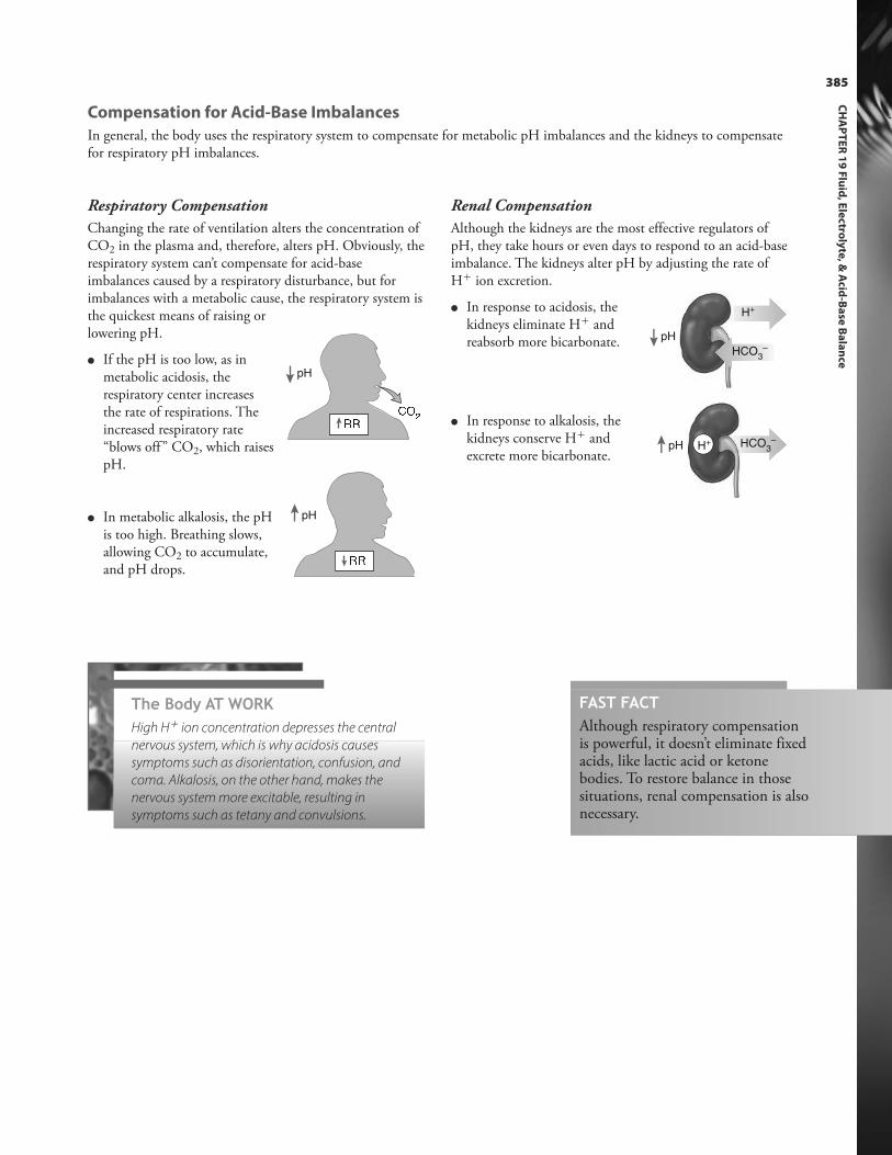
Respiratory Compensation
Changing the rate of ventilation alters the concentration ofCO2 in the plasma and, therefore, alters pH. Obviously, therespiratory system can’t compensate for acid-baseimbalances caused by a respiratory disturbance, but forimbalances with a metabolic cause, the respiratory system isthe quickest means of raising orlowering pH.
l If the pH is too low, as in metabolic acidosis, therespiratory center increasesthe rate of respirations. Theincreased respiratory rate“blows off ” CO2, which raisespH.
l In metabolic alkalosis, the pHis too high. Breathing slows, allowing CO2 to accumulate,and pH drops.
È ÈpH
Renal Compensation
Although the kidneys are the most effective regulators ofpH, they take hours or even days to respond to an acid-baseimbalance. The kidneys alter pH by adjusting the rate ofH1 ion excretion.
l In response to acidosis, thekidneys eliminate H1 and reabsorb more bicarbonate.
l In response to alkalosis, the kidneys conserve H1 and excrete more bicarbonate.
È È É Ê ËpH
pH
H+
HCO3
–
pH H+ HCO3
–
The Body AT WORKHigh H1 ion concentration depresses the central
nervous system, which is why acidosis causes
symptoms such as disorientation, confusion, and
coma. Alkalosis, on the other hand, makes the
nervous system more excitable, resulting in
symptoms such as tetany and convulsions.
385
CH
AP
TE
R 1
9 F
luid
, Ele
ctroly
te, &
Acid
-Ba
se B
ala
nce
Compensation for Acid-Base Imbalances
In general, the body uses the respiratory system to compensate for metabolic pH imbalances and the kidneys to compensatefor respiratory pH imbalances.
FAST FACTAlthough respiratory compensationis powerful, it doesn’t eliminate fixedacids, like lactic acid or ketone bodies. To restore balance in thosesituations, renal compensation is alsonecessary.

PA
RT
IV M
ain
ten
an
ce o
f the
Bo
dy
386386
Review of Key Terms
Acidosis: A decrease in the pH of theblood due to an accumulation ofacids; may result from respiratory ormetabolic disturbances
Alkalosis: An increase in the pH of theblood due to a loss of acid; may resultfrom respiratory or metabolicdisturbances
Anion: Ion with a negative charge
Buffers: Mechanisms employed by thebody to keep acids and bases inbalance
Cation: Ion with a positive charge
Dehydration: A fluid deficiency resulting from the loss of more waterthan sodium
Edema: Accumulation of fluid ininterstitial spaces
Electrolytes: Substances that break upinto electrically charged particlescalled ions when dissolved in water
Extracellular fluid: Body fluid residingoutside of cells
Hypercalcemia: An excessive concentration of calcium in the blood
Hyperkalemia: An excessive concentration of potassium in theblood
Hypernatremia: An excessive concentration of sodium in the blood
Hypocalcemia: Abnormally low bloodcalcium
Hypokalemia: Abnormally low bloodpotassium
Hyponatremia: Abnormally low bloodsodium
Hypovolemia: Decreased blood volume
Interstitial fluid: Fluid residingbetween cells inside tissues; acomponent of extracellular fluid
Intracellular fluid: Body fluid residinginside of cells
Transcellular fluid: Miscellaneousextracellular fluid that includescerebrospinal fluid, synovial fluid inthe joints, vitreous and aqueoushumors of the eye, and digestivesecretions
Turgor: Elasticity of the skin
Own the InformationTo make the information in this chapter part of your
working memory, take some time to reflect on what you’ve
learned. On a separate sheet of paper, write down
everything you recall from the chapter. After you’re done,
log on to the DavisPlus website, and check out the Study
Group podcast and Study Group Questions for the chapter.
Key Topics for Chapter 19:
• Body fluid compartments and the movement of fluid
between compartments
• Balancing water gain and loss
• Mechanisms for regulating intake and output
• Disorders of water balance
• The major cations and anions of the body
• Mechanisms for balancing the level of sodium in the body
• The causes and consequences of imbalances of sodium,
potassium, calcium, and chloride
• Acid-base balance
• How chemical buffers serve to stabilize pH
• How physiological buffers help to stabilize pH
• The time frame during which each buffer system
responds
• Types of acid-base imbalances
• Respiratory and renal compensation for acid-base
imbalances
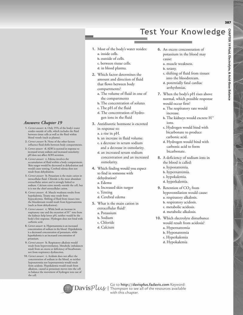
Test Your Knowledge387
CH
AP
TE
R 1
9 F
luid
, Ele
ctroly
te, &
Acid
-Ba
se B
ala
nce
Answers: Chapter 191. Correct answer: a. Only 35% of the body’s water
resides outside of cells, which includes the fluidbetween tissue cells as well as the fluid withinblood vessels (such as plasma).
2. Correct answer: b. None of the other factorsinfluence fluid shifts between body compartments.
3. Correct answer: d. ADH is secreted in response toincreased serum sodium and increased osmolarity.pH does not affect ADH secretion.
4. Correct answer: c. Edema involves theaccumulation of fluid within a body compartment.Skin turgor would be decreased in dehydration andwould cause tenting. Cerebral edema does notresult from dehydration.
5. Correct answer: b. Potassium is the main cation inintracellular fluid. Chloride is the most abundantextracellular anion and is strongly linked tosodium. Calcium exists mostly outside the cell, butit is not the chief extracellular cation.
6. Correct answer: d. Muscle weakness results fromhypokalemia. Tetany may result fromhypocalcemia. Shifting of fluid from tissues intothe bloodstream would result from hypernatremia(such as from dehydration).
7. Correct answer: c. While both an increase inrespiratory rate and the excretion of H1 ions fromthe kidneys help lower pH, neither would be thebody’s first response. Hydrogen does not bind withcarbonic acid.
8. Correct answer: a. Hypernatremia is an increasedconcentration of sodium in the blood. Hypokalemiais a decreased concentration of potassium, whilehyperkalemia is an increased concentration ofpotassium.
9. Correct answer: b. Respiratory alkalosis wouldresult from hyperventilation. Metabolic imbalancesresult from an excess or deficiency of bicarbonate,not from respiratory dysfunction.
10. Correct answer: c. Acidosis does not affect theconcentration of sodium in the blood, so neitherhyponatremia nor hypernatremia would resultfrom acidosis. Hypokalemia would result fromalkalosis, caused as potassium moves into the cellto balance the movement of hydrogen ions out ofthe cell.
1. Most of the body’s water resides:a. inside cells.b. outside of cells.c. between tissue cells.d. in blood plasma.
2. Which factor determines theamount and direction of fluidthat flows between body compartments?a. The volume of fluid in one of
the compartmentsb. The concentration of solutesc. The pH of the fluidd. The concentration of hydro-
gen ions in the fluid
3. Antidiuretic hormone is excretedin response to:a. a rise in pH.b. an increase in fluid volume.c. a decrease in serum sodium
and a decrease in osmolarity.d. an increased serum sodium
concentration and an increasedosmolarity.
4. Which finding would you expectto find in someone with dehydration?a. Edemab. Increased skin turgorc. Tentingd. Cerebral edema
5. What is the main cation in extracellular fluid?a. Potassiumb. Sodiumc. Chlorided. Calcium
6. An excess concentration of potassium in the blood maycause:a. muscle weakness.b. tetany.c. shifting of fluid from tissues
into the bloodstream.d. potentially fatal cardiac
arrhythmias.
7. When the body’s pH rises abovenormal, which possible responsewould occur first?a. The respiratory rate would
increase.b. The kidneys would excrete H1
ions.c. Hydrogen would bind with
bicarbonate to produce carbonic acid.
d. Hydrogen would bind withcarbonic acid to form bicarbonate.
8. A deficiency of sodium ions inthe blood is called:a. hyponatremia.b. hypernatremia.c. hypokalemia.d. hyperkalemia.
9. Retention of CO2 from hypoventilation would cause:a. respiratory alkalosis.b. respiratory acidosis.c. metabolic acidosis.d. metabolic alkalosis.
10. Which electrolyte disturbancewould result from acidosis?a. Hypernatremiab. Hyponatremiac. Hyperkalemiad. Hypokalemia
387
Go to http://davisplus.fadavis.com Keyword:Thompson to see all of the resources availablewith this chapter.

CHAPTER OUTLINEOverview of the Digestive System
Mouth
Pharynx
Esophagus
Stomach
Liver, Gallbladder, and Pancreas
Small Intestine
Chemical Digestion and Absorption
Large Intestine
LEARNING OUTCOMES1. Differentiate between mechanical and
chemical digestion.
2. Describe the tissue layers of the digestive tract.
3. Explain the role of the enteric nervous system.
4. Describe the structure and role of the
peritoneum.
5. Describe the anatomy of the mouth.
6. Explain the composition and function of saliva.
7. Describe the path that food takes as it travels
from the mouth to the stomach.
8. Describe the structure and function of the
stomach.
9. Identify the secretions of the stomach and
state the functions of each.
10. Describe the three phases of stimulating
gastric secretion.
11. Describe the anatomy of the liver, gallbladder,
and pancreas.
12. Identify and explain the function of the
digestive secretions of the liver, gallbladder,
and pancreas.
13. Name the divisions of the small intestine.
14. Describe the two types of movements that
occur in the small intestine.
15. Describe the lining of the small intestine and
explain the purpose it serves.
16. Explain the process of digestion for
carbohydrates, proteins, and fats.
17. Define contact digestion and explain where
this occurs.
18. Describe the anatomy of the large intestine.
19. Describe the types of contractions that occur
in the large intestine.
20. Explain the function of the bacteria found in
the large intestine.

20chapter DIGESTIVE SYSTEM
The digestive system performs the vital task of transforming food into chemicals that cells can absorb and use for energy. Itbreaks down food into its simplest components, after which it absorbs the components so they can be distributedthroughout the body. Converting food into fuel requires a dozen organs, a vast array of enzymes, and thousands of chemicalreactions.
Mouth
Pharynx
Esophagus
Stomach
Large intestine
Small intestine
Rectum
Anus
Salivary glands
Liver
Pancreas
Gallbladder
FAST FACTThe study of the digestive tract andthe diagnosis and treatment of itsdiseases is called gastroenterology.
The digestive system consists of the digestive tract (also called the alimentary canal) as well as accessory organs. Thedigestive tract is basically a tube that extends from the mouth to the anus; it contains several distinct sections, as shownbelow. The accessory organs are separate organs that aid digestion.
The bowel has been called the second brain. Not only does it contain
more neurons than the spinal cord, it secretes neurotransmitters and
hormones similar to those of the brain.
The digestive tract includes the:
l Mouthl Pharynxl Esophagusl Stomachl Large intestinel Small intestinel Rectuml Anus
The accessory organs include the:
l Teethl Tonguel Salivary glandsl Liverl Pancreasl Gallbladder
Overview of the Digestive System

PA
RT
IV M
ain
ten
an
ce o
f the
Bo
dy
390
Ì Í Í Î Ï Ð Ñ Ï Ð Ò Ó Ô ÏÕ Ò Ö Ð Ó Î Ó Ô × Î ÏÊ Ò Ø Î Ï Ó Ù Í Ú Ñ Ø Ò Û Ñ Í Ô ÓÕ Ò Ö Ð Ó Î Ó Ô × Î ÏÜ Ó Ô Í Û
Ý Ó Ù Ù Û Þ Î Ö Ö Î Ó Ö
ß × Î Í Ø Î Ï Ñ Ð Í Î Ï Þ Î à Ó Î á Ò Öâ ã ä å æ ç è ã å æ é ê ë ì ì í ì ä å îï â ð ñ ò óô Ò õ Õ Ò Ð Ù Ö Ô Ó Í Î Ï Þ Î à Ó Î á Ò Ö
Tissue Layers of the Digestive Tract
Four layers of tissue make up the walls of the digestive tract: the mucosa, the submucosa, themuscularis, and the serosa.
FAST FACTThe serosa is actually the visceral layer of theperitoneum. As you may recall, the visceral layer of theperitoneum covers the organs in the abdominopelviccavity; the parietal layer of the peritoneum attaches tothe walls of the cavity.
The mucosa—the innermost layer—consists
of three layers of its own: an inner layer of
epithelium, a layer of loose connective
tissue, and a thin layer of smooth muscle.
The submucosa is a thicker layer of
connective tissue. It contains glands, blood
vessels, lymphatic vessels, and nerves.
The muscularis consists of two layers of
smooth muscle: the inner layer encircles the
tract while the outer layer runs longitudinally.
This layer regularly contracts and relaxes to
propel food through the digestive tract.
The serosa—the outmost layer—covers
the portion of the digestive tract within the
peritoneal cavity. (The outer covering of
the esophagus and rectum is called the
adventitia.)
FAST FACTFood material insidethe digestive tract isconsidered to beexternal to the body.
The Body AT WORKDigestion breaks down food—both physically and chemically—and transforms it into a
substance that cells can use. Mechanical digestion and chemical digestion are two
distinct phases of this process.
• Mechanical digestion: This is the first phase of digestion. It involves physically breaking
down food into smaller pieces, beginning with chewing in the mouth and continuing
with contractions and churning in the stomach and small intestine.
• Chemical digestion: The second phase of digestion uses digestive enzymes produced in
the salivary glands, stomach, pancreas, and small intestines to break down food
particles into nutrients (such as glucose, amino acids, and fatty acids) that cells can use.
Once nutrients are released from food, they move into the epithelial cells lining the digestive
tract before passing into the bloodstream for distribution throughout the body. Food that
isn’t digested or absorbed becomes waste (feces) and is eliminated from the body.

391
The Body AT WORKA network of nerves called the enteric nervous system (ENS) innervates
the digestive system. Consisting of 100 million neurons, the enteric nervous
system—a part of the autonomic nervous system—regulates digestive
tract motility, secretion, and blood flow. One portion of the ENS (the
submucosal plexus) is embedded in the submucosa; another portion (the
myenteric plexus) resides between the two layers of the muscularis.
The enteric nervous system usually communicates with the central
nervous system (CNS). However, even if contact between the two systems is
severed, the ENS will continue to function. This amazing, self-contained
nervous system contains sensory neurons that monitor mechanical and
chemical conditions within the digestive tract, motor neurons that control
the churning and movement of the intestines, and still other neurons that
control the secretion of enzymes. It also contains support cells similar to the
astroglia of the brain and, finally, it uses more than 30 neurotransmitters,
most of which are identical to those found in the CNS.
Stomach
Transverse colonMesentery of thesmall intestine
Small intestine
Liver (retracted)
Gallbladder
Stomachö æ å ÷ ä å æê î å ã ä ë î
Extending from the greater curvature of the stomach and hanging down
over the small intestines like an apron is a portion of the mesentery
called the greater omentum. Deposits of fat scattered throughout the
omentum give it a lacy appearance. Besides helping to prevent friction,
the omentum also helps localize infection. If areas of inflammation
develop in the stomach or intestines, the omenta adheres to the area to
keep the infection from spreading to the rest of the abdomen.
The mesocolon
fixes the colon
to the posterior
abdominal wall.
The lesser omentum extends from the
lesser curve of the stomach to the liver.
Peritoneum
Because the stomach and intestines vigorously contract as they digest, they need to be free to move. At the same time, theyneed to be anchored enough so that the intestines don’t twist or kink when the body changes position. Layers of visceralperitoneum called mesenteries suspend the digestive organs within the abdominal cavity while anchoring them loosely tothe abdominal wall. Mesenteries also contain blood vessels, nerves, lymphatic vessels, and lymph nodes that supply thedigestive tract. (Although this figure shows the intestines separated from each other, in actuality, they are closely packed.)
FAST FACTBecause the peritoneal cavity contains emptyspace, it can fill with fluid from disease ortrauma. The accumulation of fluid in theperitoneal cavity is called ascites.
FAST FACTOrgans like the stomach and liver areenclosed by mesentery (or serosa) on bothsides, placing them within the peritonealcavity (intraperitoneal). Organs such asthe duodenum and most of the pancreaslie against the dorsal abdominal wall;mesentery covers only their ventral sides,so they are outside the peritoneal cavity(retroperitoneal).
CH
AP
TE
R 2
0 D
ige
stive
Sy
stem

PA
RT
IV M
ain
ten
an
ce o
f the
Bo
dy
392
Parotid duct
Masseter muscle
Sublingual ducts
Submandibular duct
Palatine tonsil Upper lip
Lower lip
Sublingualsalivary ductorifice
Submandibularsalivary ductorifice
The mouth is also called the oral, or buccal, cavity. It’s surrounded by lips and bordered on each side by the cheeks. Thepalate forms the roof of the mouth while the tongue and its muscles form the floor. The mouth is the entryway to thedigestive tract; it’s also where digestion begins.
Mouth
The tongue is a skeletal muscle covered by
a mucous membrane. It repositions food in
the mouth during chewing; it also contains
taste buds within projections called
lingual papillae. (For more information on
taste buds, see Chapter 11, Sense Organs.)
A fold of mucous membrane called the
lingual frenulum anchors the tongue to
the floor of the mouth. Numerous
superficial blood vessels populate the floor
of the mouth, making this an ideal site for
soluble drugs (such as nitroglycerine) to be
absorbed quickly into the circulation.
The hard palate—formed by
portions of the maxillae and
palatine bones—separates the
mouth from the nasal cavity.
The soft palate, which consists of
mostly skeletal muscle, forms an
arch between the mouth and
nasopharynx.
A cone-shaped process called the
uvula hangs downward from the
soft palate.
The parotid gland lies just underneath the
skin anterior to the ear. Its duct drains
saliva to an area near the second upper
molar. The mumps virus causes swelling
and inflammation of the parotid gland.
The submandibular gland
empties into the mouth on
either side of the lingual
frenulum.
The sublingual gland drains through
multiple ducts onto the floor of the mouth.
Salivary Glands
Salivary glands secrete saliva, a clear fluid consisting mostly ofwater, but also containing mucus, an enzyme that kills bacteria,antibacterial compounds, electrolytes, and two digestive enzymes.Besides the major salivary glands shown in the figure below, themouth also contains minor salivary glands in the tongue, inside thelips, and on the inside of the cheeks.
The Body AT WORKSaliva moistens the mouth and lubricates and
protects the teeth. It also plays an important role
in taste. One of its main roles, though, is to
moisten food and transform it into a mass called
a bolus that can be swallowed easily. Enzymes
contained in saliva begin the digestion process:
amylase breaks down starch while lipase begins
the digestion of fat.
Salivary glands secrete about 1 liter of saliva
daily. The pressure and taste of food in the mouth
stimulates the secretion of saliva. The smell or
sight of food—or even just the thought of
food—also stimulates salivation. On the other
hand, stimulation of the sympathetic nervous
system, such as through fear, inhibits the
secretion of saliva, causing the mouth to feel dry.

ø ù ú û ü ý þ ùÿ � � � ú ü û � ü ü � � �� � ú û ü ý þ ù � � �� � � ø� � � �� � �� � � �� �� � � û ü ý þ ù� � ú � ù � û ü ý þ ù� � � � ù � û ü ý þ ù
� � � � � � � � � � � � �� � � � � �
! � � � � � � � � "
Root canal
Gingiva
Teeth
Digestion begins when food enters the mouth and is chewed: a process called mastication. Besides breaking food into piecessmall enough to be swallowed, chewing allows food to become moistened with saliva. The adult mouth contains 32permanent teeth designed to cut, tear, and grind food.
Chisel-like incisors
have sharp edges for
cutting food.
Pointed canine teeth
are designed to tear
food.
Premolars and molars
have large, flat surfaces
for crushing or
grinding.
The crown is the
portion of the tooth
above the gum
(gingiva).
The root and crown
meet at the gum line—
a location called the
neck.
The root is embedded
in a socket in the
jawbone.
A hard tissue called enamel
covers the crown.
The bulk of the tooth consists of a
firm, yellowish tissue called
dentin.
The pulp cavity contains a mass
of connective tissue, blood and
lymphatic vessels, and nerves
called pulp. Pulp also fills the root
canals of each root.
A periodontal ligament lines the
socket; fibers from the ligament
infiltrate both the root and the
socket to anchor the tooth in place.
A connective tissue called
cementum covers the dentin in
the root.
Nerves and blood vessels enter
the tooth through an opening at
the end of the root canal.
The Body AT WORKAfter developing beneath the
gums, teeth erupt beginning at
about age six months. Twenty of
these early teeth, called
deciduous teeth (or baby teeth)
emerge in a predictable order
until about the age of 2 1/2 years.
Then, between the ages of 6 and
13 years, the deciduous teeth fall
out and are replaced by 32
permanent teeth. The figure on
this page shows the age at
eruption for the upper teeth. The
corresponding teeth on the
lower jaw erupt about the same
time, although some erupt
slightly later or even earlier than
those on the upper jaw.
FAST FACTDentin andcementum canregenerate if damaged;enamel cannot.
FAST FACTEnamel is thehardest substancein the body.
393
CH
AP
TE
R 2
0 D
ige
stive
Sy
stem
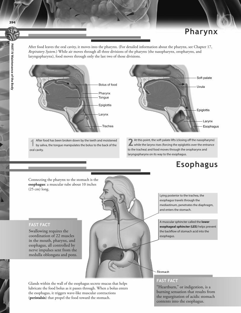
PA
RT
IV M
ain
ten
an
ce o
f the
Bo
dy
394
Bolus of food
Pharynx
Tongue
Epiglottis
Larynx
Trachea
# � ü û þ $ �
Uvula
Soft palate
Epiglottis
Esophagus
Larynx
After food leaves the oral cavity, it moves into the pharynx. (For detailed information about the pharynx, see Chapter 17,Respiratory System.) While air moves through all three divisions of the pharynx (the nasopharynx, oropharynx, andlaryngopharynx), food moves through only the last two of those divisions.
Pharynx
1 After food has been broken down by the teeth and moistened
by saliva, the tongue manipulates the bolus to the back of the
oral cavity.
2 At this point, the soft palate lifts (closing off the nasopharynx)
while the larynx rises (forcing the epiglottis over the entrance
to the trachea) and food moves through the oropharynx and
laryngopharynx on its way to the esophagus.
Connecting the pharynx to the stomach is theesophagus: a muscular tube about 10 inches(25 cm) long.
Esophagus
Glands within the wall of the esophagus secrete mucus that helpslubricate the food bolus as it passes through. When a bolus entersthe esophagus, it triggers wave-like muscular contractions(peristalsis) that propel the food toward the stomach.
FAST FACT“Heartburn,” or indigestion, is aburning sensation that results fromthe regurgitation of acidic stomachcontents into the esophagus.
A muscular sphincter called the lower
esophageal sphincter (LES) helps prevent
the backflow of stomach acid into the
esophagus.
Lying posterior to the trachea, the
esophagus travels through the
mediastinum, penetrates the diaphragm,
and enters the stomach.
FAST FACTSwallowing requires thecoordination of 22 musclesin the mouth, pharynx, andesophagus, all controlled bynerve impulses sent from themedulla oblongata and pons.

Duodenum
Esophagus
Pyloricsphincter
Greater c
urva
ture
cu r vatu
r e
L e sser
1. Cardia
2. Fundus
4. Pylorus
3. Body
Serosa
Longitudinalmuscle
Circular muscle
Oblique muscle
Submucosa
Mucosa
Esophagus
Pyloric sphincter
Lower esophagealsphincter (LES)
Just below the diaphragm, the digestive tube expands to form the stomach, a muscular sac whose primary function is tostore food. The stomach also prepares food for digestion (most of which occurs in the intestines). Specifically, the muscles of the stomach contract and churn to break food into small particles and to mix it with gastric juice. What results is a semi-fluid mixture called chyme. Chyme leaves the stomach and enters the duodenum by passing through the pyloricsphincter.
The normal curvature of the stomach creates two anatomical landmarks: the lesser curvature (along the upper surface ofthe stomach) and the greater curvature (along the lower surface). The stomach itself is divided into four regions, as shownin the figure below.
Stomach
The stomach contains the same layers as the rest of the digestive tract (mucosa, submucosa, muscularis, and serosa).However, the muscularis layer of the stomach has an extra layer of oblique muscle in addition to longitudinal and circularlayers.
The Body AT WORKWhile enzymes in the stomach partially digest
protein as well as small amounts of starch and
fat, most of the digestion process, and the
absorption of nutrients, takes place in the
intestine. Alcohol is also absorbed mostly by the
small intestine. However, the rate at which alcohol
is absorbed depends on how quickly the stomach
empties its contents into the intestine (which is
why eating before consuming alcohol slows the
effect of the alcohol).
The addition of the oblique muscle gives the muscles of
the stomach wall a crisscross pattern; this allows the
stomach to contract and churn vigorously as it
processes food.
When the stomach is empty, the mucosa and
submucosa are wrinkled into folds called rugae. As the
stomach fills with food, the rugae flatten and the
stomach expands. To illustrate: an adult’s empty
stomach has a volume of about 50 ml; after eating, it
typically expands to 1 to 1.5 liters.
395
CH
AP
TE
R 2
0 D
ige
stive
Sy
stem

396
Gastric pit
Mucosa
Submucosa
Muscularis
Serosa
The Body AT WORKSwallowing signals the stomach to relax so as to allow food to enter. Once the food has entered, the stomach goes to work,
mixing the food with gastric juices and propelling it toward the duodenum.
Mucous cells secrete mucus, which
protects the stomach lining and keeps the
stomach from digesting itself.
Parietal cells secrete hydrochloric acid and
intrinsic factor (which is necessary for the
absorption of vitamin B12). Hydrochloric
acid helps kill microbes in swallowed food.
Chief cells secrete digestive enzymes, such
as pepsinogen.
Enteroendocrine cells secrete the hormone
ghrelin (which stimulates the hypothalamus
to increase appetite) and gastrin (which
influences digestive function).
1 Contractions originate in the fundus and
move toward the pyloric region. Starting
out gently and growing progressively stronger,
wave-like peristaltic contractions break down
food and mix it with gastric juices to form chyme.
2 Chyme moves toward the pyloric
sphincter, propelled by the rhythm
of the contractions. Under the pressure
of a contraction, about 30 ml of chyme
squirts into the duodenum.
3 The stomach continues to churn and
mix its contents as it gradually releases
chyme into the duodenum. This continues
until the stomach empties, which takes
about 4 hours following a typical meal.
Chyme
Peristalticcontraction
Pyloricsphincter
Duodenum
The gastric mucosa contains depressions called gastric pits. Several different glands (calledgastric glands) open into the bottom of each gastric pit. These glands secrete the variouscomponents of gastric juice.
FAST FACTThe stomach empties in less than 4hours following a liquid meal, or itmay take as long as 6 hours toempty following a meal high in fat.
FAST FACTGastric glands produce up to 3liters of gastric juice each day.
PA
RT
IV M
ain
ten
an
ce o
f the
Bo
dy

397
CH
AP
TE
R 2
0 D
ige
stive
Sy
stem
The Body AT WORKThe stimulation of gastric secretion occurs in three phases. Each phase (the cephalic phase, gastric phase, and
intestinal phase) is activated by a different part of the body (the brain, the stomach, and the intestine, respectively).
% & ' ( ) * + , - . * / + &0 ) + 0 ) - 1 2 34 5 6 7 5 8 9 : ; <5 6 : < < 7 6 = 6 7 > 7 ? = 7@ A < ? = 5 6 : 8 B C : 8 7
D C @ E 7 ; C F : ; 9 : G : 5 =< ? = 5 6 : 8 = 7 8 6 7 5 : @ ;
1. The mere thought of food—as well as its sight, smell,
or taste—creates neural impulses that are relayed to
the brainstem. The parasympathetic nervous system
then signals the stomach to secrete gastric juice as well
as gastrin. Gastrin further stimulates gastric secretion.
Because the brain controls this phase of gastric
secretion, it’s called the cephalic phase.
2. The next phase—the gastric phase—begins when
food enters the stomach. The stretching of the
stomach as it fills with food triggers nerve reflexes that
increase the secretion of gastric juice and gastrin.
3. As chyme moves into the duodenum, the intestinal
phase begins. At this point, the duodenum triggers
nerve impulses and secretes hormones, both of which
inhibit gastric secretion.
Life lesson: Peptic ulcerCrater-like sores, or ulcerations, in the lining of the stomachor duodenum are called peptic ulcers. Typical symptomsinclude gnawing or burning pain in the middle or upperstomach between meals or at night, bloating, andheartburn. Untreated, ulcers can erode through the organwall, resulting in a potentially fatal hemorrhage.
While there is no single cause of ulcers, an acid-resistantbacterium called Helicobacter pylori (H. pylori) is oftenimplicated. By invading the mucosa of the stomach andduodenum, the bacterium causes chronic inflammationthat often leads to the development of an ulcer. Anothercommon cause of peptic ulcers is the use of nonsteroidalanti-inflammatory drugs (NSAIDs); these drugs interferewith the production of mucus that protects the lining of thestomach and duodenum, thus opening the door tochemical damage.
Ulcers caused by H. pylori are usually successfullytreated with antibiotics.
The Body AT WORKVomiting may result from a variety of stimuli:
overstretching of the stomach, chemical
irritants (such as alcohol or toxins), or even
pain or fear. When these types of stimuli occur,
the emetic center of the medulla oblongata
sends two types of impulses: one to the upper
esophageal sphincter to open and one to the
esophagus and body of the stomach to relax.
As this is happening, the abdominal muscles
forcefully contract to force chyme out of the
stomach and mouth.
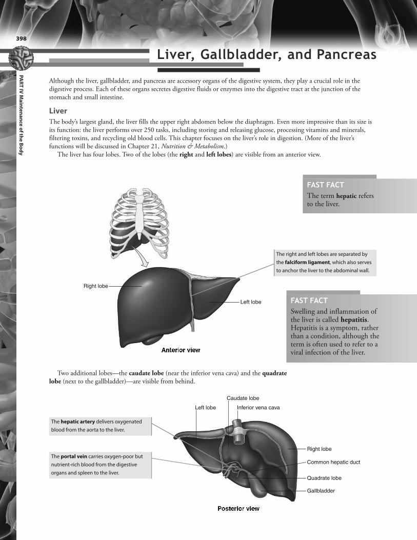
PA
RT
IV M
ain
ten
an
ce o
f the
Bo
dy
398
Right lobe
Left lobe
H I J K L M N L O M K PRight lobe
Caudate lobe
Inferior vena cavaLeft lobe
Q N R J K L M N L O M K P Gallbladder
Quadrate lobe
Common hepatic duct
Although the liver, gallbladder, and pancreas are accessory organs of the digestive system, they play a crucial role in thedigestive process. Each of these organs secretes digestive fluids or enzymes into the digestive tract at the junction of thestomach and small intestine.
Liver
The body’s largest gland, the liver fills the upper right abdomen below the diaphragm. Even more impressive than its size isits function: the liver performs over 250 tasks, including storing and releasing glucose, processing vitamins and minerals,filtering toxins, and recycling old blood cells. This chapter focuses on the liver’s role in digestion. (More of the liver’sfunctions will be discussed in Chapter 21, Nutrition & Metabolism.)
The liver has four lobes. Two of the lobes (the right and left lobes) are visible from an anterior view.
Liver, Gallbladder, and Pancreas
The right and left lobes are separated by
the falciform ligament, which also serves
to anchor the liver to the abdominal wall.
The hepatic artery delivers oxygenated
blood from the aorta to the liver.
The portal vein carries oxygen-poor but
nutrient-rich blood from the digestive
organs and spleen to the liver.
Two additional lobes—the caudate lobe (near the inferior vena cava) and the quadratelobe (next to the gallbladder)—are visible from behind.
FAST FACTThe term hepatic refersto the liver.
FAST FACTSwelling and inflammation ofthe liver is called hepatitis.Hepatitis is a symptom, ratherthan a condition, although theterm is often used to refer to aviral infection of the liver.

399
CH
AP
TE
R 2
0 D
ige
stive
Sy
stem
Liver Lobules
Tiny, six-sided cylinders called hepatic lobules fill the interior of the liver. These are the functionalunits of the liver.
A central vein passes through the core of
each lobule.
Sheets of hepatic cells (called hepatocytes)
fan out from the center of the lobule.
In between the sheets of cells are
passageways filled with blood called
sinusoids.
Tiny canals called canaliculi carry bile
secreted by hepatocytes.
1 Nutrient-rich blood from the stomach and
intestine enters the lobule through small
branches of the portal vein.
2Oxygen-rich blood enters the lobule
through small branches of the hepatic artery.
3 The blood filters through the sinusoids,
allowing the cells to remove nutrients (such as
glucose, amino acids, iron, and vitamins) as well as
hormones, toxins, and drugs. At the same time, the
liver secretes substances—such as clotting factors,
albumin, angiotensinogen, and glucose—into the
blood for distribution throughout the body.
4 Also, phagocytic cells called Kupffer cells
remove bacteria, worn-out red blood cells, and
debris from the bloodstream. 5 The central vein carries the processed
blood out of the liver.
6 Canaliculi carry bile secreted by hepatic cells and
ultimately drain into the right and left hepatic ducts.
Blood Flow Through the Liver
Each liver lobule receives nutrient-rich venous blood from the intestines as well as oxygenatedblood from the celiac trunk.

PA
RT
IV M
ain
ten
an
ce o
f the
Bo
dy
400
S @ F F @ ; 9 7 T ? 5 : 8 E C 8 5S U = 5 : 8 E C 8 5V ? ; 8 6 7 ? 5 : 8 E C 8 5W ? > > G > ? E E 7 6 V ? ; 8 6 7 ? =D C @ E 7 ; C F S @ F F @ ; G : > 7 E C 8 5X : < 9 5 ? ; E > 7 A 59 7 T ? 5 : 8 E C 8 5 =
Gallbladder
A sac attached to the underside of the liver, the gallbladder stores and concentrates bile. Thegallbladder is about 3 to 4 inches (7 to 10 cm) long and holds 30 to 50 ml of bile.
Bile reaches the gallbladder through a series of ducts. It leaves the liver
by the right and left hepatic ducts. These two ducts converge to form
the common hepatic duct, which goes on to become the common bile
duct. Bile from the liver first fills the common bile duct before backing up
into the gallbladder through the cystic duct.
The bile duct merges with the duct
of the pancreas to form the
hepatopancreatic ampulla
(ampulla of Vater).
The ampulla enters the duodenum
at a raised area called the major
duodenal papilla.
A sphincter called the hepatopancreatic
sphincter (sphincter of Oddi) controls the flow
of bile and pancreatic juice into the duodenum.
The Body AT WORKEach day, the liver secretes up to 1 liter of bile: a
yellow-green fluid containing minerals, cholesterol,
phospholipids, bile salts, bile pigments, and
cholesterol. The main bile pigment is bilirubin, which
results from the breakdown of hemoglobin. However,
the most important component of bile is bile salts.
Formed in the liver from cholesterol, bile salts aid in
the digestion and absorption of fat in the small
intestine. After secretion, 80% of bile salts are
reabsorbed in the final section of the small intestine
(the ileum). It is then returned to the liver, where
hepatocytes absorb and then secrete the bile once
again. The 20% of bile not reabsorbed is excreted in
feces. In fact, bile is responsible for giving feces its
brown color. If bile isn’t being secreted, or if the flow of
bile is obstructed, feces become grayish-white in color.
Life lesson: GallstonesHard masses, called gallstones, can form inside thegallbladder, often because of excessive cholesterol inthe bile. Gallstones may cause no signs or symptoms.However, if a gallstone lodges within a duct,excruciating pain may occur. If the gallstone blocks theflow of bile into the duodenum, bile will back up to theliver and be absorbed into the bloodstream, resultingin jaundice (a yellow discoloration of the skin, mucous,membranes, and sclerae). Treatment of symptomaticgallstones includes surgery to remove the gallbladderor medications to dissolve the gallstones.
A sphincter called the hepatopancreatic
sphincter (sphincter of Oddi) controls the flow
of bile and pancreatic juice into the duodenum.

401
CH
AP
TE
R 2
0 D
ige
stive
Sy
stemY Z [ \ Z ] ^ _ ` a b c d e ` b f
g ` b [ Z ] a b c d e ` b f
h b i j Z ] a b c d e ` b fk b c d e ` b ^ i d i f j ` ^ f
Pancreas
The pancreas lies behind the stomach; its head is nestled in the curve of the duodenum while itstapered tail sits below the spleen and above the left kidney.
The pancreas is both an endocrine and exocrine gland. Its endocrine function centers onpancreatic islets that secrete insulin and glucagon. (For more information on the endocrinefunction of the pancreas, see Chapter 12, Endocrine System.)
Most of the pancreas, though, consists of exocrine tissue. Each day, the pancreas secretes about1.5 liters of pancreatic juice—essentially digestive enzymes and an alkaline fluid—into the smallintestine.
Acinar cells secrete digestive enzymes in
an inactive form; once activated in the
duodenum, the enzymes help break down
lipids, proteins, and carbohydrates.
Epithelial cells in pancreatic ducts secrete
sodium bicarbonate, which buffers the
highly acidic chyme entering the
duodenum from the stomach.
Ducts converge on the main pancreatic
duct, which joins the bile duct at the
hepatopancreatic ampulla.
The Body AT WORKThe vagus nerve and the hormones cholecystokinin, gastrin, and secretin stimulate the secretion of
bile and pancreatic juice.
• Cholecystokinin (CCK): The arrival of chyme in the duodenum stimulates the duodenum to
secrete the hormone CCK. CCK causes gallbladder contraction—forcing bile into the bile duct—
and the release of pancreatic enzymes. At the same time, it prompts the hepatopancreatic
sphincter to relax, which allows bile and pancreatic juice to enter the duodenum.
• Gastrin: This hormone from the stomach and duodenum triggers both gallbladder contraction
and pancreatic enzyme secretion. However, it exerts a much less powerful effect than does CCK.
• Secretin: The acid in chyme also triggers the duodenum to release secretin. Secretin causes the
bile and pancreatic ducts to release bicarbonate, which helps neutralize the stomach acid
entering the duodenum.
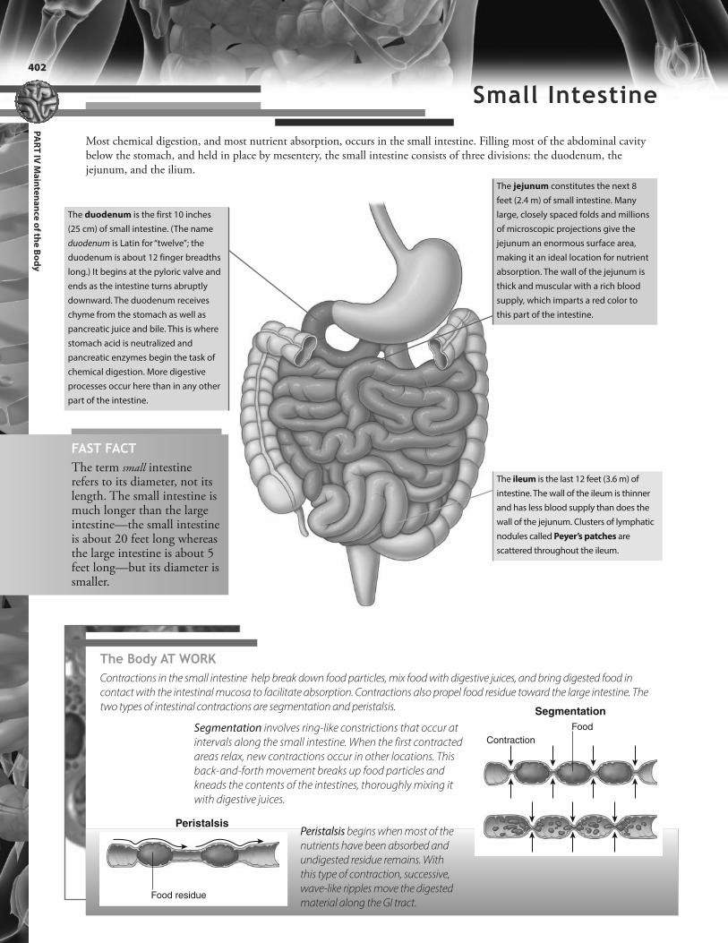
PA
RT
IV M
ain
ten
an
ce o
f the
Bo
dy
402
The Body AT WORKContractions in the small intestine help break down food particles, mix food with digestive juices, and bring digested food in
contact with the intestinal mucosa to facilitate absorption. Contractions also propel food residue toward the large intestine. The
two types of intestinal contractions are segmentation and peristalsis.
Food
Contraction
Food residue
Most chemical digestion, and most nutrient absorption, occurs in the small intestine. Filling most of the abdominal cavitybelow the stomach, and held in place by mesentery, the small intestine consists of three divisions: the duodenum, thejejunum, and the ilium.
Small Intestine
The duodenum is the first 10 inches
(25 cm) of small intestine. (The name
duodenum is Latin for “twelve”; the
duodenum is about 12 finger breadths
long.) It begins at the pyloric valve and
ends as the intestine turns abruptly
downward. The duodenum receives
chyme from the stomach as well as
pancreatic juice and bile. This is where
stomach acid is neutralized and
pancreatic enzymes begin the task of
chemical digestion. More digestive
processes occur here than in any other
part of the intestine.
The jejunum constitutes the next 8
feet (2.4 m) of small intestine. Many
large, closely spaced folds and millions
of microscopic projections give the
jejunum an enormous surface area,
making it an ideal location for nutrient
absorption. The wall of the jejunum is
thick and muscular with a rich blood
supply, which imparts a red color to
this part of the intestine.
The ileum is the last 12 feet (3.6 m) of
intestine. The wall of the ileum is thinner
and has less blood supply than does the
wall of the jejunum. Clusters of lymphatic
nodules called Peyer’s patches are
scattered throughout the ileum.
FAST FACTThe term small intestinerefers to its diameter, not itslength. The small intestine ismuch longer than the largeintestine—the small intestineis about 20 feet long whereasthe large intestine is about 5feet long—but its diameter issmaller.
Peristalsis begins when most of the
nutrients have been absorbed and
undigested residue remains. With
this type of contraction, successive,
wave-like ripples move the digested
material along the GI tract.
Segmentation involves ring-like constrictions that occur at
intervals along the small intestine. When the first contracted
areas relax, new contractions occur in other locations. This
back-and-forth movement breaks up food particles and
kneads the contents of the intestines, thoroughly mixing it
with digestive juices.
Segmentation
Peristalsis

FAST FACTThe folds, villi, andmicrovilli increase the surfacearea of the small intestinemore than 500 times.
l : 8 6 @ m : > > :n : > > :o T : 5 9 7 > : C Fl : 8 6 @ m : > > :W @ G > 7 5 8 7 > >p ? 8 5 7 ? >q ; 5 7 = 5 : ; ? > 8 6 U T 5 =
n 7 ; C > 7p U F T 9 ? 5 : 8 m 7 = = 7 >r 6 5 7 6 : @ > 7
l C 8 @ = ?4 C G F C 8 @ = ?l C = 8 C > ? 6 : =4 7 6 @ = ?
The Body AT WORKMost nutrients pass into the
bloodstream through
capillaries, and then travel
to the liver. Most fat,
however, is absorbed by the
lacteal.
The intestinal lining contains multiple projections that increase its surface area and enhancethe absorption of nutrients.
The intestinal lining contains circular folds that
slow the progress of chyme and increase its
contact with the mucosa. On top of the circular
folds are projections called villi (singular: villus).
The villi are covered by absorptive
epithelial cells as well as mucus-
secreting goblet cells. An arteriole, a
venule, and a lymph vessel called a
lacteal fill the core of each villus.
Epithelial cells covering the villi have a brush
border of ultrafine microvilli. Besides further
increasing the absorptive area, the microvilli
produce digestive enzymes.
403
CH
AP
TE
R 2
0 D
ige
stive
Sy
stem
Pores at the bases of the villi, called
intestinal crypts, contain goblet cells
that secrete mucus that helps the
passage of food. They also serve as
sites for rapid cellular growth,
producing new cells to replace those
shed from the villi.

PA
RT
IV M
ain
ten
an
ce o
f the
Bo
dy
404
Once food has been mechanically broken down into minute particles, chemical digestion takes control. Using digestiveenzymes, chemical digestion transforms food molecules into particles that can be absorbed into the bloodstream. By thetime food residue leaves the small intestine, most nutrients have been absorbed. The process of digestion and absorptionvaries between classes of nutrients, with the major classes being carbohydrates, proteins, and fats.
Carbohydrates
Carbohydrates consist of sugars called saccharides:
l Polysaccharides contain many saccharide groups linked together; these include starches and glycogen.l Disaccharides contain two groups linked together; these include sucrose, lactose, and maltose.l Monosaccharides contain only one saccharide group; these include glucose,
fructose, and galactose.
Most foods contain polysaccharides, which must be broken into smallerparticles (namely, disaccharides and monosaccharides). To do so, the digestivesystem uses enzymes.
The process begins
in the mouth,
where salivary
glands secrete the
enzyme amylase.
Salivary amylase
works to hydrolyze
polysaccharides
into disaccharides.
Once the food is
swallowed, the low pH
of stomach acid
inactivates the salivary
amylase. By this point,
though, about 50% of
the starch has already
been digested.When the resultant
chyme reaches the
small intestine, it
mixes with
pancreatic
amylase, and the
process of starch
digestion resumes.
The final steps in carbohydrate
digestion take place at the level of the
villi. The membranes of the epithelial
cells covering the villi contain the
enzymes sucrase, lactase, and
maltase. As the chyme slides against
the brush border of the epithelial
cells, the enzymes bind with the
disaccharides. (This is called contact
digestion.) This final step in the
digestion process produces glucose,
which is immediately absorbed.
Salivary amylasesecreted
Salivary amylaseinactivated
Pancreatic amylasesecreted
Sucrase Lactase
Glucose absorbed
Maltase
FAST FACTCellulose is an indigestiblecarbohydrate found in the diet.Even though cellulose doesn’tprovide the body with nutrients,it is an important source ofdietary fiber. (Cellulose will bediscussed further in Chapter 21,Nutrition & Metabolism.)
Chemical Digestion and Absorption

Glycerol and short-chain fatty acids are
absorbed into the bloodstream of the villi.
Long-chain fatty acids and
monoglycerides, which are too large to be
absorbed by the capillaries, are absorbed
into the walls of the villi. There, they are
changed into triglycerides, which
eventually enter the lacteal of the villi.
After travelling through the lymphatic
system, the triglycerides enter the
bloodstream at the left subclavian vein.
Once the fat is emulsified,
pancreatic lipase (the
main fat-digesting
enzyme) begins to digest
the fat. In the process, fats
are broken down into a
mixture of glycerol,
short-chain fatty acids,
long-chain fatty acids,
and monoglycerides.
When a fat globule
enters the
duodenum, two
substances in bile
(lecithin and bile
salts) break up the fat
into small droplets.
Finally, brush border
enzymes called
peptidases break the
remaining chains into
individual amino acids,
which are then
absorbed into the
bloodstream.
In the stomach, the
enzyme pepsin
hydrolyzes the
peptide bonds
between certain
amino acids.
Proteins
Proteins are complex molecules made up of folded and twisted chains of amino acids linked together by peptide bonds.Before the body can absorb amino acids, it must break the peptide bonds. To do so, it uses enzymes called proteases. Absentfrom saliva, proteases work in the stomach and small intestine.
Pepsinsecreted
Fats
Although fat digestion begins in the mouth, most fat digestion occurs in the small intestine. Because fat doesn’t dissolve inwater, it enters the duodenum as a congealed mass. To facilitate digestion, the fat must first be broken into small droplets, aprocess called emulsification.
Peptidases
Amino acid chain
Aminoacidsabsorbed
BilePancreaticlipase
GlycerolShort-chain fatty acids
Long-chain fatty acidsMonoglycerides
Capillary
Lacteal
As soon as the chyme
enters the duodenum,
pepsin is inactivated. At
that point, the
pancreatic enzymes
trypsin and
chymotrypsin assume
the task of breaking the
peptide bonds.
Pancreatic enzymessecreted
FAST FACTAmino acids arrange themselves in awide variety of combinations, witheach combination employing aslightly different peptide bond. As aresult, the body calls upon a vast arrayof proteases to digest protein.
405
CH
AP
TE
R 2
0 D
ige
stive
Sy
stem

PA
RT
IV M
ain
ten
an
ce o
f the
Bo
dy
406
Once the food has been processed and its nutrients absorbed, the remaining residue is ready to leave the small intestine forthe large intestine. In fact, about 500 ml of residue—consisting of undigested food, sloughed off epithelial cells, minerals,salts, and bacteria—enter the large intestine each day. The large intestine absorbs large amounts of water from the residuebefore passing the resulting waste material (feces) out of the body.
The following figure shows the regions of the colon, beginning with the cecum, where food residue enters the colon, andending with the anal canal, through which waste material leaves the body. All along the way are pouches called haustra,which result from the increased tone of the smooth muscle of the muscularis layer.
Appendix
Ileum
Ileocecal valve
Right colic(hepatic) flexure
Left colic(splenic) flexure
Haustra
External anal sphincter
1 The large intestine begins in a blind
pouch called the cecum. Attached to
the lower end of the cecum is a tubular
organ called the appendix. The
appendix contains masses of lymphoid
tissue and serves as a source for immune
cells. At the point where the ileum meets
the large intestine is the ileocecal valve,
which helps insure that material moves
in a forward direction only.
2 The ascending colon
extends up toward the liver.
The colon makes a sharp turn at
the right colic (hepatic) flexure.
3 The transverse colon passes below the liver,
stomach, and spleen. The colon turns sharply
downward at the left colic (splenic) flexure.
4 The descending colon
extends downward along
the left side of the abdominal
cavity.
5 The sigmoid colon forms an
S shape down to the rectum.
6 The rectum is the last 7 to 8 inches
(17 to 20 cm) of intestine. The
mucosa of the rectum forms longitudinal
folds called anal columns.7 The anal canal, which makes up the last
inch (2.5 cm) of the rectum, opens to the
exterior through the anus. The anus contains
two sphincters: an internal sphincter
(composed of involuntary smooth muscle)
and an external sphincter (composed of
voluntary skeletal muscle).
FAST FACTAppendicitis, an inflammation of theappendix, results when a blockage trapsinfectious material inside the appendix.Surgical removal of the appendix(appendectomy) is one of the mostcommonly performed emergency surgeries.
Large Intestine
ANIMATION

The Body AT WORKThe large intestine houses over 700 species of
bacteria; these are called bacterial flora. These
bacterial flora, also called the normal flora,
perform a variety of functions beneficial to the
body’s health. For example, some of the bacteria
digest cellulose and other materials for which we
have no digestive enzymes. Other bacteria
produce significant amounts of vitamins,
particularly vitamin K and a B vitamin.
One of the products of bacterial action is
intestinal gas, or flatus. Although flatus partially
results from swallowed air, it also results from
bacterial fermentation of undigested
polysaccharides. Flatus mainly consists of
nitrogen and carbon dioxide, although it also
contains hydrogen, methane, and hydrogen
sulfide. When a large amount of undigested
nutrients pass into the colon, painful cramping
can result as the bacterial action produces an
increased volume of intestinal gas.
The Body AT WORKIntestinal contractions move feces through the
colon and toward the rectum.
• Haustral contractions: As a haustrum fills with
residue, it becomes distended, which
stimulates the haustrum to contract. Besides
mixing the residue, the contraction pushes the
residue into the next haustrum. Haustral
contractions generally occur every 30 minutes.
• Stronger peristaltic contractions called mass
movements, move larger amounts of
material several inches at a time along the colon.
In particular, these movements are triggered by
the filling of the stomach or duodenum.
The mass movement of feces into the rectum
stimulates the defecation reflex: the rectum
contracts and the internal anal sphincter relaxes.
When the external anal sphincter relaxes (which
is under voluntary control to prevent the
uncontrolled release of feces), feces is expelled
from the body (defecation).
Organ Enzyme Substance Digested
Salivary glands Amylase Starch Lipase Fat
Stomach Pepsin Protein
Pancreas • Proteases (trypsin, chymotrypsin)
• Lipase
• Amylase
• Protein
• Fats
• Starch
Intestine • Peptidases
• Sucrase
• Lactase
• Maltase
• Peptides
• Sucrose (cane sugar)
• Lactose (milk sugar)
• Maltose (malt sugar)
FAST FACTHemorrhoids are swollen,engorged veins in the anal canal.
407
CH
AP
TE
R 2
0 D
ige
stive
Sy
stem
Life lesson: Changes with AgingEfficiency of the digestive system decreases with age. Over time, the muscular wall of the digestive tract weakens andintestinal peristalsis slows, often resulting in constipation and hemorrhoids. Peristalsis also slows in the esophagus,making swallowing more difficult. The cardiac sphincter—the sphincter that regulates the flow of food from theesophagus into the stomach—also weakens, often leading to esophageal reflux and “heartburn.” Tooth loss is common,making eating difficult. Factoring in diminished sensations of smell and taste, it’s easy to understand why many elderlyindividuals have poor appetites.
Aging also interferes with the absorption of nutrients. Specifically, the stomach has decreased production ofhydrochloric acid and other digestive enzymes, leading to poor absorption of vitamin B12. Efficiency of the liver alsodeclines, slowing an elderly individual’s ability to metabolize drugs, placing them at greater risk for drug overdose.
Key Digestive Enzymes
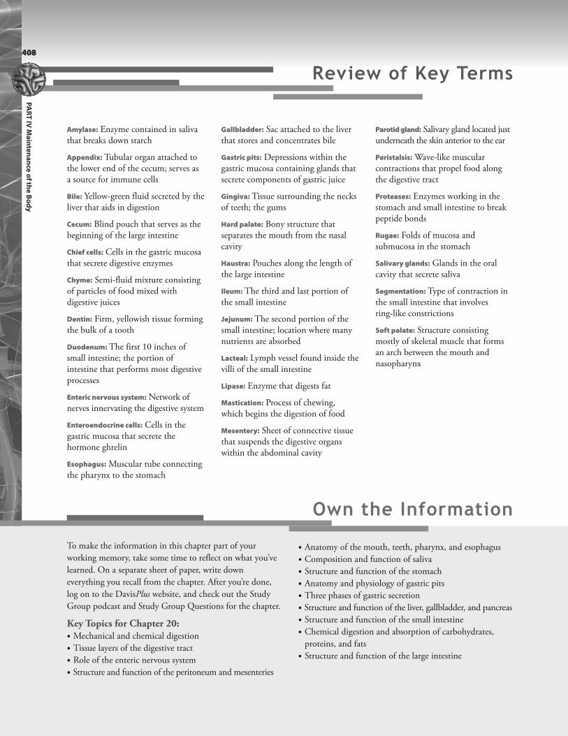
PA
RT
IV M
ain
ten
an
ce o
f the
Bo
dy
408408
Amylase: Enzyme contained in salivathat breaks down starch
Appendix: Tubular organ attached tothe lower end of the cecum; serves asa source for immune cells
Bile: Yellow-green fluid secreted by theliver that aids in digestion
Cecum: Blind pouch that serves as thebeginning of the large intestine
Chief cells: Cells in the gastric mucosathat secrete digestive enzymes
Chyme: Semi-fluid mixture consistingof particles of food mixed with digestive juices
Dentin: Firm, yellowish tissue formingthe bulk of a tooth
Duodenum: The first 10 inches ofsmall intestine; the portion of intestine that performs most digestiveprocesses
Enteric nervous system: Network ofnerves innervating the digestive system
Enteroendocrine cells: Cells in the gastric mucosa that secrete the hormone ghrelin
Esophagus: Muscular tube connectingthe pharynx to the stomach
Gallbladder: Sac attached to the liverthat stores and concentrates bile
Gastric pits: Depressions within thegastric mucosa containing glands thatsecrete components of gastric juice
Gingiva: Tissue surrounding the necksof teeth; the gums
Hard palate: Bony structure that separates the mouth from the nasalcavity
Haustra: Pouches along the length ofthe large intestine
Ileum: The third and last portion ofthe small intestine
Jejunum: The second portion of thesmall intestine; location where manynutrients are absorbed
Lacteal: Lymph vessel found inside thevilli of the small intestine
Lipase: Enzyme that digests fat
Mastication: Process of chewing,which begins the digestion of food
Mesentery: Sheet of connective tissuethat suspends the digestive organswithin the abdominal cavity
Parotid gland: Salivary gland located justunderneath the skin anterior to the ear
Peristalsis: Wave-like muscular contractions that propel food alongthe digestive tract
Proteases: Enzymes working in thestomach and small intestine to breakpeptide bonds
Rugae: Folds of mucosa and submucosa in the stomach
Salivary glands: Glands in the oralcavity that secrete saliva
Segmentation: Type of contraction inthe small intestine that involves ring-like constrictions
Soft palate: Structure consistingmostly of skeletal muscle that formsan arch between the mouth and nasopharynx
Review of Key Terms
Own the InformationTo make the information in this chapter part of your
working memory, take some time to reflect on what you’ve
learned. On a separate sheet of paper, write down
everything you recall from the chapter. After you’re done,
log on to the DavisPlus website, and check out the Study
Group podcast and Study Group Questions for the chapter.
Key Topics for Chapter 20:
• Mechanical and chemical digestion
• Tissue layers of the digestive tract
• Role of the enteric nervous system
• Structure and function of the peritoneum and mesenteries
• Anatomy of the mouth, teeth, pharynx, and esophagus
• Composition and function of saliva
• Structure and function of the stomach
• Anatomy and physiology of gastric pits
• Three phases of gastric secretion
• Structure and function of the liver, gallbladder, and pancreas
• Structure and function of the small intestine
• Chemical digestion and absorption of carbohydrates,
proteins, and fats
• Structure and function of the large intestine

Answers: Chapter 201. Correct answer: c. Mechanical digestion involves
breaking the food into smaller pieces, but thepieces are not small enough for cells to absorb.Absorption is the phase in which the cells absorbthe nutrients. Ingestion is the process of consuming food.
2. Correct answer: b. The mucosa is the innermostlayer. The submucosa is a layer of connective tissue containing glands, blood vessels, lymphaticvessels, and nerves. The serosa is the outermostlayer that covers the portion of the digestive tractwithin the peritoneal cavity.
3. Correct answer: a. The lesser omentum extendsfrom the lesser curve of the stomach to the liver.The mesocolon fixes the colon to the posterior abdominal wall. The jejunum is a portion of thesmall intestine.
4. Correct answer: a. Pepsin acts in the stomach to digest protein. Trypsin is a pancreatic enzyme thatbreaks down protein in the duodenum. Sucraseacts in the intestine to digest sucrose.
5. Correct answer: d. The duodenum is the first 10inches of small intestine. The ileum is the thirdand last portion of the intestine. The sigmoidcolon is the S-shaped portion of the large intestineleading to the rectum.
6. Correct answer: b. The brush border of the epithelialcells lining the small intestine secretes enzymes thatbreak down carbohydrates into glucose and proteininto individual amino acids; the brush border doesnot participate in the digestion or absorption of fat.Long-chain fatty acids and monoglycerides areabsorbed into the walls of the villi; there, they arechanged into triglycerides. Triglycerides do enterthe bloodstream after travelling through thelymphatic system.
7. Correct answer: c. All of the other answers are incorrect.
8. Correct answer: a. The stretching of the stomach asit fills with food increases the secretion of gastricjuice and gastrin, but it is not the initial trigger.The movement of chyme into the duodenum initiates the intestinal phase of digestion, and gastric secretion is inhibited. Contractions withinthe stomach mix food particles with gastric juice,but they do not stimulate the secretion of gastricjuice.
9. Correct answer: a. The islets of Langerhans secreteinsulin. Duct cells in the pancreas secrete sodiumbicarbonate. Kupffer cells are phagocytic cells inhepatic lobules.
10. Correct answer: b. All of the other answers are incorrect.
11. Correct answer: c. All of the other answers are incorrect.
Test Your Knowledge1. In which phase of digestion do
enzymes break down food intoparticles cells can absorb?a. Mechanical digestionb. Absorptionc. Chemical digestiond. Ingestion
2. The layer of the digestive tractthat contracts and relaxes to propel food forward is the:a. mucosa.b. muscularis.c. submucosa.d. serosa.
3. Which mesentery hangs over thesmall intestine like an apron?a. Greater omentumb. Lesser omentumc. Mesocolond. Jejunum
4. Which enzyme acts in the mouthto begin the breakdown ofstarch?a. Amylaseb. Pepsinc. Trypsind. Sucrase
5. The muscular tube that connectsthe pharynx to the stomach isthe:a. duodenum.b. ileum.c. sigmoid colon.d. esophagus.
6. How do triglycerides enter thebloodstream?a. They pass directly into the
bloodstream after passingthrough the intestinal brushborder.
b. They enter the lacteal and travelthrough the lymphatic systemto enter the bloodstream.
c. They are absorbed into thewall of the villi, after whichthey pass into the bloodstream.
d. Triglycerides don’t enter thebloodstream.
7. What is the purpose of rugae?a. Secrete gastric juiceb. Propel food residue through
the small intestine.c. Allow the stomach to expand.d. Help break food into small
particles.
8. Which of the following acts as aninitial trigger to the stomach tobegin secreting gastric juice?a. The smell of foodb. The stretching of the stomach
as it fills with foodc. The movement of chyme into
the duodenumd. Contractions within the
stomach
9. Which pancreatic cells secrete digestive enzymes?a. Acinar cellsb. Islets of Langerhansc. Duct cellsd. Kupffer cells
10. Most nutrients are absorbed inthe:a. stomach.b. small intestine.c. large intestine.d. mouth.
11. Where is most fat digested?a. Mouthb. Stomachc. Duodenumd. Large intestine
409
CH
AP
TE
R 2
0 D
ige
stive
Sy
stem
Go to http://davisplus.fadavis.com Keyword:Thompson to see all of the resources availablewith this chapter.

CHAPTER OUTLINEUnits of Energy
Hunger and Satiety
Nutrients
Metabolism
Thermoregulation
LEARNING OUTCOMES1. Define metabolism and calorie.
2. Explain basal metabolic rate and identify the
factors that influence it.
3. Discuss the five hormones that affect hunger
and satiety.
4. Define nutrient and identify the five categories
of nutrients.
5. Name the three forms of dietary
carbohydrates.
6. Describe three roles of fat in the body.
7. Differentiate between saturated and
unsaturated fats.
8. Differentiate between complete and
incomplete proteins.
9. Name the fat‐soluble and water‐soluble
vitamins, and explain the difference between
them.
10. Identify the major minerals in the body and
describe their functions.
11. Summarize the process of carbohydrate
metabolism, including glycolysis, anaerobic
fermentation, and aerobic fermentation.
12. Explain what occurs in glycogenesis,
glycogenolysis, and gluconeogenesis.
13. Describe the process of lipid metabolism.
14. Describe the process of protein metabolism.
15. Explain what is meant by protein balance,
differentiating between negative and positive
nitrogen balance.
16. Identify the three means by which the body
loses heat.
17. Explain the negative feedback mechanisms
that help regulate the body’s temperature.

21chapter NUTRITION &METABOLISM
The body—even at rest—consumes a tremendous amount of energy. Obviously, walking across a room or even picking up abook requires energy, but so does the beating of the heart, the conduction of nervous impulses, the movement of the lungsduring respiration, and even the manufacture of new blood cells. In fact, the body needs a steady supply of fuel just tosurvive.
As was discussed in the previous chapter, the body’s source of fuel is food. Once consumed, the digestive system breaksfood into usable food components (called nutrients), which are then absorbed into the bloodstream. That’s just thebeginning, however. The nutrients then enter the cells and undergo further chemical reactions, called metabolism. It’sthrough the process of metabolism that nutrients are transformed into energy or materials the body can either use or store.
As shown in the figure below, some body systems demand a greater supply of energy than others.s t u v t w x y z t { | t w t u z } t ~ � t w � t �� } � y u � { � � { u z y w �� � � � � � � � � � � � � �� � � � � � � � � �� �� � � � � � � � �� � �� � � � �� � �� � � � �� �¡ � � � �¢ �£ � � � � ¤ �¥ �
About two-thirds of an adult’s energy expenditure is used simply to
maintain normal body function.
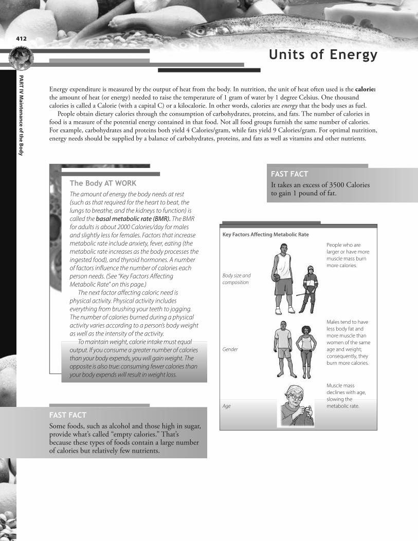
PA
RT
IV M
ain
ten
an
ce o
f the
Bo
dy
412
Energy expenditure is measured by the output of heat from the body. In nutrition, the unit of heat often used is the calorie:the amount of heat (or energy) needed to raise the temperature of 1 gram of water by 1 degree Celsius. One thousandcalories is called a Calorie (with a capital C) or a kilocalorie. In other words, calories are energy that the body uses as fuel.
People obtain dietary calories through the consumption of carbohydrates, proteins, and fats. The number of calories infood is a measure of the potential energy contained in that food. Not all food groups furnish the same number of calories.For example, carbohydrates and proteins both yield 4 Calories/gram, while fats yield 9 Calories/gram. For optimal nutrition,energy needs should be supplied by a balance of carbohydrates, proteins, and fats as well as vitamins and other nutrients.
FAST FACTIt takes an excess of 3500 Caloriesto gain 1 pound of fat.
The Body AT WORKThe amount of energy the body needs at rest
(such as that required for the heart to beat, the
lungs to breathe, and the kidneys to function) is
called the basal metabolic rate (BMR). The BMR
for adults is about 2000 Calories/day for males
and slightly less for females. Factors that increase
metabolic rate include anxiety, fever, eating (the
metabolic rate increases as the body processes the
ingested food), and thyroid hormones. A number
of factors influence the number of calories each
person needs. (See “Key Factors Affecting
Metabolic Rate” on this page.)
The next factor affecting caloric need is
physical activity. Physical activity includes
everything from brushing your teeth to jogging.
The number of calories burned during a physical
activity varies according to a person’s body weight
as well as the intensity of the activity.
To maintain weight, calorie intake must equal
output. If you consume a greater number of calories
than your body expends, you will gain weight. The
opposite is also true: consuming fewer calories than
your body expends will result in weight loss.
Key Factors Affecting Metabolic Rate
Body size and
composition
People who are
larger or have more
muscle mass burn
more calories.
Gender
Males tend to have
less body fat and
more muscle than
women of the same
age and weight;
consequently, they
burn more calories.
Age
Muscle mass
declines with age,
slowing the
metabolic rate.
FAST FACTSome foods, such as alcohol and those high in sugar,provide what’s called “empty calories.” That’sbecause these types of foods contain a large numberof calories but relatively few nutrients.
Units of Energy

413
CH
AP
TE
R 2
1 N
utritio
n a
nd
Me
tab
olism
The hypothalamus contains control centers for both hunger and satiety (the feeling of being satisfied after eating). Recentresearch has identified a number of hormones that profoundly affect these centers. The following figure illustrates five keyhormones affecting appetite: one of the hormones—ghrelin—stimulates appetite while the other four suppress it.
¦ § ¨ © ª « ¬ ¬ ® ¯ ª « ¬ ° ° ±² ³ ³´ © µ ¶ « ¬
Secreted by adipose tissue, leptin signals the
brain that the body has enough energy
stores, such as body fat, and, therefore,
suppresses appetite. Although leptin levels
are higher in people who are overweight,
obese people have built up a resistance to
the appetite-suppressing effects of leptin.
Peptide YY (PYY)—which is secreted by
enteroendocrine cells in the ileum and
colon—signals satiety and, therefore,
serves as a prompt to stop eating.
Insulin levels increase rapidly after eating
to counteract the rise in glucose levels. The
amount of insulin released varies
according to the amount of body fat. Like
leptin, insulin alerts the brain as to the
amount of body fat and, therefore,
suppresses appetite.
Cholecystokinin (CCK) is secreted by cells
in the duodenum and jejunum; in addition
to stimulating the secretion of bile and
pancreatic enzymes, this hormone also
suppresses appetite.
FAST FACTGhrelin levels fall dramatically following gastric bypass surgery, suggesting that this may be one ofthe factors contributing to weight loss after the procedure.
Secreted by parietal cells in the stomach,
especially when the stomach is empty,
ghrelin produces the sensation of hunger.
Ghrelin secretions decrease for about
3 hours after a meal.
Hunger and Satiety

PA
RT
IV M
ain
ten
an
ce o
f the
Bo
dy
414
Nutrients used by the body for growth, repair, and maintenance include carbohydrates, lipids, proteins, and water as well asvitamins and minerals. Because the body requires fairly large amounts of carbohydrates, lipids, proteins, and water, these arecalled macronutrients. On the other hand, the body needs only small quantities of vitamins and minerals; therefore, theseare called micronutrients.
The body must obtain most of its nutrients—including most vitamins, minerals, eight amino acids, and some fattyacids—through diet. These are called essential nutrients because they’re essential in the diet. Some nutrients, however, thebody can synthesize; because they’re not essential in the diet, these nutrients are called nonessential nutrients.
(Basic concepts of nutrition and metabolism were discussed in Chapter 2, Chemistry of Life. It may be helpful to reviewthese topics before proceeding with this chapter.)
Carbohydrates
Carbohydrates are the body’s primary energy source. We obtain carbohydrates by eating foods (particularly plants) that contain them. In plants, carbohydrates result from photosynthesis: the process by which plants use energy from sunlight to take up carbon dioxide and release oxygen. In essence, the carbohydrates in plants represent stored energy. By consuming carbohydrates, the body has access to that energy.
Dietary carbohydrates occur in three forms: monosaccharides, disaccharides, and polysaccharides.
Monosaccharides
• Known as simple sugars
• Taste sweet
• Absorbed through the small intestine
without being broken down
• Examples include glucose, fructose (fruit
sugar), and galactose
Disaccharides
• Also called simple sugars
• Broken down into monosaccharides
during the digestive process
• Examples include sucrose (table sugar),
lactose (milk sugar), and maltose (a sugar
found in germinating cereals, such as barley)
Polysaccharides
• Known as complex carbohydrates
• Consist of the starches found in vegetables,
grains, potatoes, rice, and legumes
• Includes cellulose as an important
polysaccharide (See “The Body at Work”
on this page)
Because the body continually burns carbohydrates forfuel, they should form the greatest percentage of the diet.Specifically, average adults should obtain 40% to 50% oftheir calories from carbohydrates; very active people mayneed to obtain up to 60% of their calories fromcarbohydrates.
Most of the carbohydrates consumed should be complexcarbohydrates. That’s because these foods contain othervital nutrients in addition to carbohydrates. Also, the bodyabsorbs complex carbohydrates more slowly than simplesugars, which helps prevent unhealthy spikes in bloodglucose levels.
The Body AT WORKBecause the body can’t digest cellulose, it’s not
considered a nutrient. Nevertheless, cellulose still
makes an important contribution to the diet in
the form of dietary fiber. A major component of
plant tissues, cellulose absorbs water in the
intestines and swells. This adds bulk to the stool,
which stretches the colon and increases
peristalsis. In turn, this allows stool to pass more
quickly out of the body.
Nutrients

415
Lipids
Lipids, or fats, act as a reservoir of excess energy. Specifically, excess carbohydrates areconverted to fat. Then, during periods of decreased food intake, fat reserves can be mobilizedand broken down to release energy.
Fat is an ideal substance for storing energy: not only is it more compact thancarbohydrates, it’s also more energy dense. Each gram of fat contains 9 Calories per gram,compared to 4 Calories for each gram of carbohydrate. Fat fulfills other key roles in the bodyas well. For example:
Saturated fats
• Derived mainly from animals
• Tend to be solid at room temperature
• Found in meat, egg yolks, and dairy products
• Include hydrogenated oils (oils that are
processed so as to make them solid at room
temperature), such as vegetable shortening
and margarine
Unsaturated fats
• Occur in nuts, seeds, and vegetable oils
• Tend to be in liquid form at room temperature
Ideally, fat should comprise no more than 30% of an adult’s daily intake,with no more than 20% being saturated fat. For example, based on a 2000-calorie-a-day diet, fat intake should be limited to 400 to 700 calories, or 44to 78 grams. Of that, less than 10% should be saturated fat.
A specific type of saturated fat commonly found in commercial bakedgoods and snack foods is trans fat. Trans fat results when hydrogen is addedto liquid vegetable shortening during food processing to make it more solid.Trans fat increases the risk for heart attack and stroke, which is why theAmerican Heart Association recommends limiting trans fat consumption tono more than 1% of daily calorie intake.
The body can synthesize most of the fatty acids it needs. However, thereare a few (such as linolenic acid, which is a necessary part of the cellmembrane) it cannot synthesize. Called essential fatty acids, these fats mustbe obtained through the diet. They are found in such foods as vegetable oils,whole grains, and vegetables.
l Fat enables absorption of certain vitamins. Vitamins A, D, E, and K are fat-soluble, meaning that they depend on dietary fat to be absorbed by the intestine.
l Fat contributes to cellular structure. Dietary fats can be converted to otherlipids (such as phospholipids and cholesterol), which are major structural components of cell membranes and myelin. Cholesterol also acts as a precursorto steroid hormones, bile acids, and vitamin D.
l Fat insulates and protects the body. Besides insulating the body to conserveheat, fat also surrounds delicate internal organs to protect them from damage.
As was discussed in Chapter 2, fats can be saturated or unsaturated depending onthe structure of their carbon chains.
FAST FACTExcessive consumption ofsaturated fat has been linked toarteriosclerosis, heart disease,diabetes mellitus, and even breastand colon cancers.
FAST FACTProcessed foods typically containsaturated fats because they resistbecoming rancid and becausethey tend to remain solid atroom temperature.
CH
AP
TE
R 2
1 N
utritio
n a
nd
Me
tab
olism
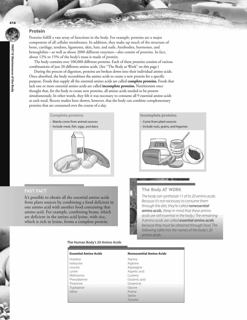
PA
RT
IV M
ain
ten
an
ce o
f the
Bo
dy
416
Protein
Proteins fulfill a vast array of functions in the body. For example, proteins are a majorcomponent of all cellular membranes. In addition, they make up much of the structure ofbone, cartilage, tendons, ligaments, skin, hair, and nails. Antibodies, hormones, andhemoglobin—as well as about 2000 different enzymes—also consist of proteins. In fact,about 12% to 15% of the body’s mass is made of protein.
The body contains over 100,000 different proteins. Each of these proteins consists of variouscombinations of just 20 different amino acids. (See “The Body at Work” on this page.)
During the process of digestion, proteins are broken down into their individual amino acids.Once absorbed, the body recombines the amino acids to create a new protein for a specificpurpose. Foods that supply all the essential amino acids are called complete proteins. Foods thatlack one or more essential amino acids are called incomplete proteins. Nutritionists oncethought that, for the body to create new proteins, all amino acids needed to be presentsimultaneously. In other words, they felt it was necessary to consume all 9 essential amino acidsat each meal. Recent studies have shown, however, that the body can combine complementaryproteins that are consumed over the course of a day.
Complete proteins
• Mainly come from animal sources
• Include meat, fish, eggs, and dairy
Incomplete proteins
• Come from plant sources
• Include nuts, grains, and legumes
The Body AT WORKThe body can synthesize 11 of its 20 amino acids.
Because it’s not necessary to consume them
through the diet, they’re called nonessential
amino acids. (Keep in mind that these amino
acids are still essential in the body.) The remaining
9 amino acids are called essential amino acids
because they must be obtained through food. The
following table lists the names of the body’s 20
amino acids.
Essential Amino Acids Nonessential Amino Acids
Histidine
Isoleucine
Leucine
Lysine
Methionine
Phenylalanine
Threonine
Tryptophan
Valine
Alanine
Arginine
Asparagine
Aspartic acid
Cysteine
Glutamic acid
Glutamine
Glycine
Proline
Serine
Tyrosine
FAST FACTIt’s possible to obtain all the essential amino acidsfrom plant sources by combining a food deficient inone amino acid with another food containing thatamino acid. For example, combining beans, whichare deficient in the amino acid lysine, with rice,which is rich in lysine, forms a complete protein.
The Human Body’s 20 Amino Acids

417
CH
AP
TE
R 2
1 N
utritio
n a
nd
Me
tab
olism
Major Vitamins
Vitamin Dietary Source Functions Deficiency Symptoms
Water-Soluble Vitamins
Ascorbic acid (C) Citrus fruits, green vegetables Synthesis of collagen, antioxidant, red blood cell
formation, wound healing, aids in iron absorption
Scurvy; degeneration of skin,
bone, and blood vessels
Thiamine (B1) Red meat, eggs, legumes Carbohydrate metabolism, blood formation,
circulation, central nervous system maintenance
Beriberi (nervous system
disorder), edema
Riboflavin (B2) Meat, eggs, whole grains,
legumes
Energy metabolism; cell respiration; epithelial,
eye, and mucosal tissue maintenance
Dry, scaly skin; corneal opacities;
keratitis
Niacin (B3) Meat, fish, legumes, grains Circulation; cholesterol level reduction; growth; stimulates
central nervous system; metabolism of carbohydrate;
protein, and fat
Dermatitis, dementia, diarrhea
Pyridoxine (B6) Red meat, fish, green vegetables,
whole grains
Helps in metabolism of amino acids and unsaturated
fatty acids, antibody formation, DNA and RNA synthesis
Dermatitis, anemia, muscle
weakness
Folic acid (B9) Vegetables, liver Synthesis of DNA, catabolism of lipids and amino acids,
cell growth and reproduction, red blood cell formation
Anemia
Cyanocobalamin (B12) Red meat, eggs, milk Formation and maturation of red blood cells, nerve cell
maintenance
Pernicious anemia
Biotin Raw egg yolks, liver, peanuts,
tomatoes
Necessary for cell growth, the production of fatty
acids, and the metabolism of fats and amino acids
Dermatitis, hair loss, neurological
symptoms
Fat-Soluble Vitamins
Vitamin A
(retinol)
Fish oil, eggs, green and yellow
vegetables
Growth, night vision, maintenance of epithelial tissue Night blindness; dry, scaly skin
Vitamin D
(calciferol)
Fish oils, eggs, milk, liver Calcium and phosphorus absorption and metabolism,
development of bones and teeth, normal blood
clotting
Rickets (in children), osteomalacia
(in adults)
Vitamin E
(tocopherol)
Fish oils, eggs, milk, seed oils
(such as corn and soybean oil)
Antioxidant, normal muscle function, fetal
development, myocardial perfusion
Reproductive disorders
Vitamin K Vegetables such as cabbage,
spinach, and tomatoes
Essential for normal blood clotting Blood coagulation disorders,
hemorrhage
Vitamins
Besides carbohydrates, lipids, and proteins, the body also requires small amounts of organiccompounds called vitamins. Vitamins are mostly incorporated into coenzymes: moleculesthat support enzymes in metabolic processes. Other than vitamin D (which can be formedby exposure of the skin to the sun), the body cannot manufacture vitamins. Therefore,vitamins must be obtained regularly through food.
Vitamins are classified as either water-soluble or fat-soluble. Water-soluble vitamins areabsorbed with water in the small intestine, after which they are dissolved in body fluids andthen excreted by the kidneys. Because water-soluble vitamins aren’t stored in the body, therisk of toxicity (hypervitaminosis) is slight. (However, toxicity can still occur from megadosesof water-soluble vitamins, such as vitamin C.)
Fat-soluble vitamins are absorbed with dietary fat, after which they are stored in the liverand fat tissues of the body until needed. Consequently, megadosing may lead to toxicity.Vitamins A, D, E, and K are fat-soluble vitamins. The following table outlines the majorvitamins along with their dietary sources, their function, and the symptoms of deficiency.
FAST FACTVitamins were originally named with letters according to theorder of their discovery. However, vitamins also havechemically derived names, such as ascorbic acid for vitamin C.

PA
RT
IV M
ain
ten
an
ce o
f the
Bo
dy
418
Minerals
The body also needs various inorganic substances called minerals. Minerals are needed fornumerous vital functions, including nerve conduction, brain function, heart contraction, andred blood cell formation. Good sources of minerals include vegetables, legumes, milk, eggs,fish, shellfish, and some meats. Most cereals and grains are a poor source of minerals(although minerals are typically added to processed cereals).
Major & Trace Minerals
Minerals Dietary Source Functions Deficiency Symptoms
Major Minerals
Calcium Milk and other dairy
products, greens, legumes,
sardines
Formation of bones and teeth, blood clotting,
muscle contraction and relaxation, nerve
function
Numbness and tingling, muscle
spasms, arrhythmias
Phosphorus Meat, fish, poultry, eggs, milk,
grains
Formation of bones and teeth, cell growth
and repair, aids in acid-base balance
Bone degeneration
Magnesium Meat, seafood, nuts, legumes,
grains
Muscle relaxation and nerve impulse
transmission, protein synthesis
Nerve disorders, heart
arrhythmias
Sodium Table salt, meat, seafood, milk Fluid balance, nerve and muscle function,
cell permeability
Weakness
Potassium Potatoes, melons, citrus fruit,
bananas, meat, most fruits and
vegetables
Nerve and muscle function, acid-base
balance, cardiac rhythm
Muscle weakness, nerve
disorders
Trace Minerals
Iron Liver, red meat, egg yolks, nuts,
legumes
Hemoglobin production Anemia, fatigue
Iodine Seafood, iodized salt Thyroid hormone production, physical and
mental development
Goiter, hypothyroidism
Selenium Grains, meat, fish, dairy products Antioxidant, immune mechanisms Anemia
Zinc Meat, seafood, whole grains Wound healing, nutrient metabolism,
carbohydrate digestion
Delayed growth, rough skin,
poor appetite
Copper Legumes, grains, nuts, organ meats Component of many enzymes Fatigue, anemia
Fluoride Fluoridated drinking water, seafood,
seaweed
Formation of bones and teeth Dental caries
Manganese Greens, legumes, grains Formation of bone, activation of some enzymes Delayed wound healing,
dizziness, menstrual disorders
FAST FACTAbout 4% of the body’s mass consists of minerals.Of this, three-quarters is calcium and phosphorus,which are found in the bones and teeth.

419
Once inside the cell, nutrients are transformed through metabolism into energy that thebody can use immediately or store for later use. Metabolism involves two processes:
l Catabolism: Used in the metabolism of carbohydrates and lipids, this process breaksdown complex substances into simpler ones or into energy.
l Anabolism: Used in protein metabolism, this process forms complex substances out ofsimpler ones.
Carbohydrate Metabolism
All ingested carbohydrates are converted to glucose, most of which is immediately burned asenergy. If the body doesn’t need the glucose for immediate energy, it stores it as glycogen orconverts it to lipids.
The primary goal of glucose catabolism is to generate adenosine triphosphate (ATP),which cells use for energy. This crucial task occurs in three distinct phases: glycolysis,anaerobic fermentation, and aerobic respiration.
Glycolysis ¦ ª ¯² · ¨ ¯ ¸ « ¹º ¹ « » ² · ¨ ¯ ¸ « ¹º ¹ « »¹ ¼ ® © Energy
½ § © ¬ ¼ ¾ · ¿ © ¬ « ® « ¬ ® § ¼ ¨ ¶ ® ¯ µ µ ª · Àµ · ¨ ¯ ¸ « ¹ º ¹ « » « ® ¹ ¼ ¬ ¸ © ¨ ¶ © » « ¬ ¶ ¼ ª º ¹ ¶ « ¹ º ¹ « » Á § © ª º ¹ ¶ « ¹ º ¹ « » ¶ § © ¬ ¶ ¨ º ¸ © ª ® ¶ § ¨ ¼ ¯ ¿ § ¶ § ©Ã ª ¼ ¼ » ® ¶ ¨ © º Ä ¶ ¼ ¶ § © ª « ¸ © ¨ À Å § © ¨ © « ¶ « ® ® ¶ ¼ ¨ © » Á´ º ¶ © ¨ À » ¯ ¨ « ¬ ¿ º µ © ¨ « ¼ » ¼ Æ ¨ © ® ¶ Å § © ¬¼ ¾ · ¿ © ¬ à © ¹ ¼ Ä © ® º ¸ º « ª º à ª © À ¶ § © ª º ¹ ¶ « ¹º ¹ « » « ® ¹ ¼ ¬ ¸ © ¨ ¶ © » à º ¹ Ç « ¬ ¶ ¼ µ · ¨ ¯ ¸ « ¹º ¹ « » Á È ¶ ¶ § º ¶ µ ¼ « ¬ ¶ À ¶ § © µ · ¨ ¯ ¸ « ¹ º ¹ « »¹ º ¬ © ¬ ¶ © ¨ ¶ § © ® © ¨ « © ® ¼ Æ ¨ © º ¹ ¶ « ¼ ¬ ®« ¬ º © ¨ ¼ à « ¹ ¨ © ® µ « ¨ º ¶ « ¼ ¬ Á
´ º ¹ ¶ « ¹º ¹ « »² · ¨ ¯ ¸ « ¹º ¹ « »² · ¨ ¯ ¸ « ¹º ¹ « »
´ º ¹ ¶ « ¹º ¹ « »´ º ¹ ¶ « ¹º ¹ « »
Anaerobic Fermentation
Taking place without oxygen, glycolysis splits one glucose molecule
into two molecules of pyruvic acid. Glycolysis releases only a fraction of
the available energy; further reactions are needed to release the rest of
the energy. Which reactions occur depend upon whether oxygen is
available. When oxygen is not available—such as during intense
exercise when the demand for energy exceeds the supply of oxygen—
anaerobic fermentation occurs. Most ATP, however, is generated in
mitochondria, which require oxygen. So, when oxygen is available,
aerobic respiration is the pathway of choice.
FAST FACTProcesses that occurwithout oxygen are calledanaerobic processes; thosethat require oxygen arecalled aerobic.
FAST FACTBecause glycolysis doesn’trequire oxygen, it can supply the cell with energy (although limitedenergy) even when oxygenis absent.
Metabolism CH
AP
TE
R 2
1 N
utritio
n a
nd
Me
tab
olism

PA
RT
IV M
ain
ten
an
ce o
f the
Bo
dy
420
 § « ® à © ¿ « ¬ ® º ® © ¨ « © ® ¼ Æ ¨ © º ¹ ¶ « ¼ ¬ ® Ç ¬ ¼ Å ¬º ® ¶ § © É Ê Ë Ì Ê É Í É Ê Î É Ï É Ð Ñ Á § « ® « ® Æ ¼ ª ª ¼ Å © » à · º ¬ ¼ ¶ § © ¨ ® © ¨ « © ® ¼ Æ ¨ © º ¹ ¶ « ¼ ¬ ®¹ º ª ª © » ¶ § © Ñ Ð Ñ É Ë Ì Ò Ó Ô Ë Ì Í Ó Õ Ö Ò Ì Ë É × Í Ê Ó Á § © © ¬ » ¨ © ® ¯ ª ¶ ¼ Æ º ª ª ¶ § © ® © ¨ © º ¹ ¶ « ¼ ¬ ® « ® ¶ § ©¹ ¼ Ä µ ª © ¶ © à ¨ © º Ç » ¼ Å ¬ ¼ Æ ¿ ª ¯ ¹ ¼ ® © « ¬ ¶ ¼ ¹ º ¨ à ¼ ¬» « ¼ ¾ « » © À Å º ¶ © ¨ À º ¬ » Ä ¼ ® ¶ « Ä µ ¼ ¨ ¶ º ¬ ¶ ª · À ª º ¨ ¿ ©º Ä ¼ ¯ ¬ ¶ ® ¼ Æ È Â ² ÁØ Ê Ë Ì Ê É Í É Ê Î É Ï É Ð Ñ
² · ¨ ¯ ¸ « ¹º ¹ « » È ¹ © ¶ · ª° ¼ ÈÙ Ð Ñ É Ë Ì Ò Ó Ô Ë Ì Í Ó Õ Ö Ò Ì Ë É × Í Ê ÓØ Ú Û Ü Ý Û Ú Ü
Energy
½ § © ¬ ¼ ¾ · ¿ © ¬ « ® º ¸ º « ª º à ª © Àµ · ¨ ¯ ¸ « ¹ º ¹ « » © ¬ ¶ © ¨ ® ¶ § ©Ä « ¶ ¼ ¹ § ¼ ¬ » ¨ « º º ¬ » « ® ¹ ¼ ¬ ¸ © ¨ ¶ © »« ¬ ¶ ¼ º ¹ © ¶ · ª ¹ ¼ © ¬ Þ · Ä © Èß º ¹ © ¶ · ª ° ¼ È à Á
That Makes SenseTo remember the terms related for the formation and
breakdown of glycogen and glucose, keep the following in
mind:
• Genesis means “to create.”
• Lysis means “to break down.”
• Neo- means “new.”
Therefore, glycogenesis means “the creation of glycogen.”
Glyconeogenesis means “the creation of new glycogen.”
Glycogenolysis means “the breakdown of glycogen.”
!
FAST FACTMost body cells draw on energy suppliedby both carbohydrates and fat; some cells,though—such as neurons anderythrocytes—depend exclusively oncarbohydrates for energy.
Aerobic Respiration
The Body AT WORKBecause most cells depend on a steady supply of
glucose for fuel, the body uses several
mechanisms to maintain a consistent level of
glucose in the blood. These mechanisms include
glycogenesis, glycogenolysis, and
gluconeogenesis.
• Glycogenesis: When the amount of glucose in
the blood exceeds the body’s needs, hormones
stimulate the liver to convert glucose into
glycogen, which is then stored in the liver.
• Glycogenolysis: When blood glucose levels fall,
such as between meals, glycogen stored in the
liver is broken down into glucose. The glucose
diffuses into the bloodstream for use by the
body’s cells.
• Gluconeogenesis: The body also has the ability
to create glucose from noncarbohydrates, such
as fats and amino acids. This mainly occurs in
the liver.
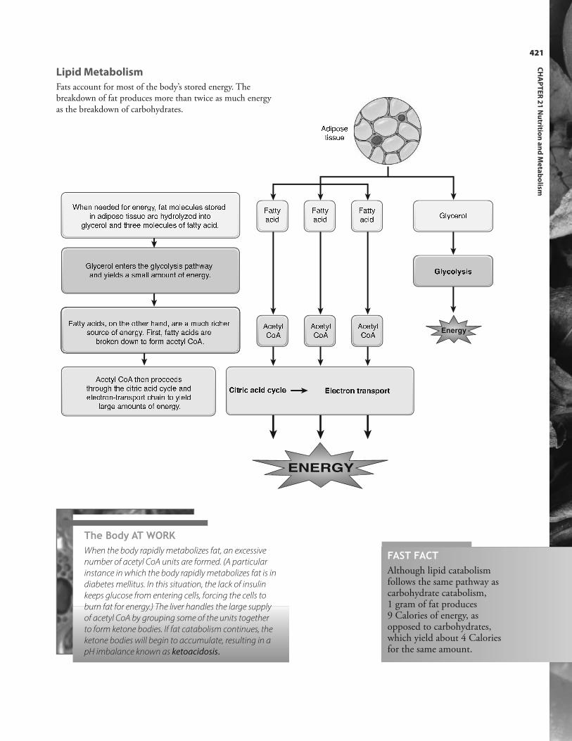
½ § © ¬ ¬ © © » © » Æ ¼ ¨ © ¬ © ¨ ¿ · À Æ º ¶ Ä ¼ ª © ¹ ¯ ª © ® ® ¶ ¼ ¨ © »« ¬ º » « µ ¼ ® © ¶ « ® ® ¯ © º ¨ © § · » ¨ ¼ ª · Þ © » « ¬ ¶ ¼¿ ª · ¹ © ¨ ¼ ª º ¬ » ¶ § ¨ © © Ä ¼ ª © ¹ ¯ ª © ® ¼ Æ Æ º ¶ ¶ · º ¹ « » Á¦ ª · ¹ © ¨ ¼ ª © ¬ ¶ © ¨ ® ¶ § © ¿ ª · ¹ ¼ ª · ® « ® µ º ¶ § Å º ·º ¬ » · « © ª » ® º ® Ä º ª ª º Ä ¼ ¯ ¬ ¶ ¼ Æ © ¬ © ¨ ¿ · Áá º ¶ ¶ · º ¹ « » ® À ¼ ¬ ¶ § © ¼ ¶ § © ¨ § º ¬ » À º ¨ © º Ä ¯ ¹ § ¨ « ¹ § © ¨® ¼ ¯ ¨ ¹ © ¼ Æ © ¬ © ¨ ¿ · Á á « ¨ ® ¶ À Æ º ¶ ¶ · º ¹ « » ® º ¨ ©Ã ¨ ¼ Ç © ¬ » ¼ Å ¬ ¶ ¼ Æ ¼ ¨ Ä º ¹ © ¶ · ª ° ¼ È ÁÈ ¹ © ¶ · ª ° ¼ È ¶ § © ¬ µ ¨ ¼ ¹ © © » ®¶ § ¨ ¼ ¯ ¿ § ¶ § © ¹ « ¶ ¨ « ¹ º ¹ « » ¹ · ¹ ª © º ¬ »© ª © ¹ ¶ ¨ ¼ ¬ â ¶ ¨ º ¬ ® µ ¼ ¨ ¶ ¹ § º « ¬ ¶ ¼ · « © ª »ª º ¨ ¿ © º Ä ¼ ¯ ¬ ¶ ® ¼ Æ © ¬ © ¨ ¿ · Á
á º ¶ ¶ ·º ¹ « » á º ¶ ¶ ·º ¹ « » á º ¶ ¶ ·º ¹ « »È ¹ © ¶ · ª° ¼ È È ¹ © ¶ · ª° ¼ È È ¹ © ¶ · ª° ¼ È
¦ ª · ¹ © ¨ ¼ ªã Ð Ï É Ò Ð Ï Õ Ê ÕEnergy
ENERGY
Ø Ê Ë Ì Ê É Í É Ê Î É Ï É Ð Ñ Ù Ð Ñ É Ë Ì Ò Ó Ë Ì Í Ó Õ Ö Ò Ì Ë
È » « µ ¼ ® ©¶ « ® ® ¯ ©421
CH
AP
TE
R 2
1 N
utritio
n a
nd
Me
tab
olism
Lipid Metabolism
Fats account for most of the body’s stored energy. Thebreakdown of fat produces more than twice as much energyas the breakdown of carbohydrates.
The Body AT WORKWhen the body rapidly metabolizes fat, an excessive
number of acetyl CoA units are formed. (A particular
instance in which the body rapidly metabolizes fat is in
diabetes mellitus. In this situation, the lack of insulin
keeps glucose from entering cells, forcing the cells to
burn fat for energy.) The liver handles the large supply
of acetyl CoA by grouping some of the units together
to form ketone bodies. If fat catabolism continues, the
ketone bodies will begin to accumulate, resulting in a
pH imbalance known as ketoacidosis.
FAST FACTAlthough lipid catabolismfollows the same pathway ascarbohydrate catabolism, 1 gram of fat produces 9 Calories of energy, asopposed to carbohydrates,which yield about 4 Caloriesfor the same amount.

PA
RT
IV M
ain
ten
an
ce o
f the
Bo
dy
422
Protein Metabolism
Whereas carbohydrates and fat are mainly used to supply energy, proteins are primarily used to build tissue. Proteinanabolism plays a significant role in the growth and maintenance of the body: new proteins are used to repair tissue, replaceworn-out cells (including red blood cells), create enzymes, grow muscle, and synthesize antibodies, among many otherthings.
Proteins may also be catabolized: converted to glucose and fat or used directly as fuel. Before that happens, however, theamino acids must be altered in the liver.
When food containing
protein is digested...
...the proteins are broken down
into individual amino acids to
create a pool of amino acids.Within the cells, ribosomes pull amino
acids from the amino acid pool and
recombine them to form new proteins.
The liver splits an amino group (NH2)
from an amino acid (called deamination),
forming a molecule of ammonia and a
molecule of keto acid.
È Ä « ¬ ¼º ¹ « » È Ä Ä ¼ ¬ « º± © ¶ ¼ º ¹ « »The keto acid may then enter the
citric acid cycle, where it is
oxidized for energy; it may also be
converted to glucose or fat.
¦ ª ¯ ¹ ¼ ® ©± © ¶ ¼ º ¹ « » á º ¶Energy
The liver converts the ammonia, which is toxic
to cells, to urea, a nitrogenous waste product
that is excreted through urine by the kidneys.
È Ä Ä ¼ ¬ « º ä ¨ © º ä ¨ « ¬ ©FAST FACTAmmonia is so toxicthat, if a diseasedliver fails to removethis nitrogenouswaste product, comaand death will soonresult.
The Body AT WORKThe body doesn’t store amino acids or proteins like it does fat or carbohydrates; therefore,
protein turnover must occur continually. Remarkably, in healthy adults, the amount of
protein being broken down equals the amount of protein being created. This state—in
which the rate of protein anabolism equals the rate of protein catabolism—is called
protein balance.
Recall that the catabolism of protein yields nitrogenous waste products. Therefore, if
the body is in protein balance, it’s also in nitrogen balance: the amount of nitrogen being
consumed in the form of protein-containing foods equals the amount of nitrogen being
excreted in urine, feces, and sweat as a waste product.
When more proteins are being catabolized than are being created—such as during
starvation or a wasting illness—the amount of nitrogen in the urine exceeds the amount
of nitrogen in the protein foods ingested. In this instance, the body is said to be in a state
of negative nitrogen balance.
On the other hand, if the amount of nitrogen being consumed through foods is greater
than the nitrogen output in urine, the body is in a state of positive nitrogen balance.
This situation—in which protein anabolism outpaces protein catabolism—occurs when
large amounts of tissue are being synthesized, such as during periods of growth, during
pregnancy, or when recovering from a wasting illness.

423
CH
AP
TE
R 2
1 N
utritio
n a
nd
Me
tab
olism
A key factor in homeostasis is the maintenance of body temperature within a narrow range. For this to occur, heatproduction must balance heat loss. This is known as thermoregulation.
Normal body temperature is not static. Rather, it fluctuates about 1.8° F (1° C) through the course of a day, being lowestin the early morning and highest in the late afternoon. Body temperature also varies from one region of the body to another.Specifically, temperature of the deeper body regions—such as that of organs in the cranial, thoracic, and abdominalcavities—ranges from 99° F to 99.7° F (37.2° C to 37.6° C). Temperature closer to the body’s surface—such as that of theskin and mouth—ranges from 97.9° F to 98.6° F (36.6° C to 37° C). The temperature of the body’s deeper regions, knownas core temperature, is obtained with a rectal thermometer. The temperature near the surface, called the shell temperature,is obtained with an oral thermometer.
Heat Production and Loss
Most of the body’s heat results as a by-product of the chemical reactions occurring in cells.The cells of the muscles, brain, liver, and endocrine glands provide the greatest portion ofheat when the body is at rest. While skeletal muscles provide 20% to 30% of the heat whenthe body is at rest, this percentage increases dramatically during vigorous exercise.
To balance the production of heat, the body loses heat by three means: radiation, conduction, and evaporation.
Radiation involves the transfer of heat through the air by electromagnetic heat waves. An
example of gaining heat by radiation would be standing in bright sunlight. Because our
bodies are typically warmer than the air around us, we tend to lose heat through radiation.
Conduction involves the transfer of heat between two materials that touch each other. In
other words, heat passes out of our bodies and into objects that we touch, such as our
clothes. (The reason a chair is warm immediately after someone has been sitting in it is
because conduction has caused heat to pass out of the body and into the chair.) A
practical example is a spoon placed in a bowl of hot soup; after a while, the spoon will
become hot through the process of conduction. Conductive heat loss can be accelerated
by convection,which is the transfer of heat by the actual movement of the warmed
matter. For example, heat rises from the body just as steam rises from hot coffee. As the
hot air rises, it is replaced by cooler air.
Evaporation requires heat to change water into a gas. That’s why perspiration can
effectively cool the body. Sweat causes the skin to become wet; then, as evaporation
occurs, heat is drawn from the body.
Thermoregulation
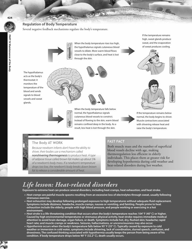
PA
RT
IV M
ain
ten
an
ce o
f the
Bo
dy
424
The hypothalamus
acts as the body’s
thermostat: it
monitors the
temperature of the
blood and sends
signals to blood
vessels and sweat
glands.
When the body temperature rises too high,
the hypothalamus signals cutaneous blood
vessels to dilate. More warm blood flows
close to the body’s surface, and heat is lost
through the skin.
If the temperature remains
high, sweat glands produce
sweat, and the evaporation
of sweat produces cooling.
When the body temperature falls below
normal, the hypothalamus signals
cutaneous blood vessels to constrict.
Instead of flowing to the skin, warm blood
remains confined deep in the body. As a
result, less heat is lost through the skin.
If the temperature remains below
normal, the body begins to shiver.
Muscle contractions associated
with shivering release heat and
raise the body’s temperature.
FAST FACTBoth muscle mass and the number of superficialblood vessels decline with age, makingthermoregulation less efficient in elderlyindividuals. This places them at greater risk fordeveloping hypothermia during cold weather andheat-related disorders during hot weather.
Life lesson: Heat-related disordersExposure to extreme heat can produce several disorders, including heat cramps, heat exhaustion, and heat stroke.
• Heat cramps are painful muscle spasms resulting from an excessive loss of electrolytes through sweat, usually followingstrenuous exercise.
• Heat exhaustion may develop following prolonged exposure to high temperatures without adequate fluid replacement.Symptoms include dizziness, headache, muscle cramps, nausea or vomiting, and fainting. People prone to heatexhaustion include the elderly, people with high blood pressure, and people working or exercising in a hotenvironment.
• Heat stroke is a life-threatening condition that occurs when the body’s temperature reaches 104° F (40° C) or higher.Caused by high environmental temperatures or strenuous physical activity, heat stroke requires immediate medicalattention to avoid brain damage, organ failure, or death. Symptoms include hot, dry, flushed skin; headache; rapidheart rate; and muscle cramps or weakness. Seizures, hallucinations, or unconsciousness may also occur.
• Hypothermia occurs when the body’s temperature falls below 95° F (35° C). Typically caused by exposure to coldweather or immersion in cold water, symptoms include shivering, lack of coordination, slurred speech, confusion, anddrowsiness. The confused thinking associated with hypothermia usually keeps the person from being aware of hiscondition. If body temperature drops below 90° F (32.2° C), death usually occurs.
Regulation of Body Temperature
Several negative feedback mechanisms regulate the body’s temperature.
The Body AT WORKBecause newborn infants don’t have the ability to
shiver, their bodies use a mechanism called
nonshivering thermogenesis to produce heat. A type
of adipose tissue called brown fat makes up about 5%
of a newborn’s body mass. If a newborn’s temperature
drops too low, the newborn’s body breaks down brown
fat to release a considerable amount of heat.

425
CH
AP
TE
R 2
1 N
utritio
n a
nd
Me
tab
olism
Review of Key TermsAerobic: Processes that require oxygen
Anaerobic: Processes occurring withoutoxygen
Basal metabolic rate: The amount ofenergy the body needs at rest
Calorie: The amount of heat needed toraise the temperature of 1 gram ofwater by 1° Celsius
Carbohydrates: The body’s primaryenergy source
Complete protein: Foods that supplyall the essential amino acids
Essential fatty acid: Fat that must beobtained through the diet
Essential nutrients: Nutrients essentialin the diet because they cannot besynthesized by the body
Ghrelin: Hormone secreted by cells inthe stomach that stimulates appetite.
Glycolysis: The breakdown of carbohydrates for energy
Incomplete proteins: Foods that lackone or more essential amino acids
Lipids: Fats that act as a reservoir ofexcess energy
Metabolism: Chemical processes occurring in cells that transform nutrients into energy or into materialsthe body can use or store
Mineral: An inorganic element orcompound needed for numerous vitalfunctions
Nonessential amino acids: Amino acidsthe body can synthesize, making themnonessential in the diet
Nutrients: Usable food componentsthat supply the body with chemicalsnecessary for energy
Vitamins: Vital nutrients, obtainedmostly through food and needed formetabolism
Own the InformationTo make the information in this chapter part of your
working memory, take some time to reflect on what you’ve
learned. On a separate sheet of paper, write down
everything you recall from the chapter. After you’re done,
log on to the DavisPlus website, and check out the Study
Group podcast and Study Group Questions for the chapter.
Key Topics for Chapter 21:
• Units of energy
• Hunger and satiety
• Nutrients
• Metabolism of carbohydrates, lipids, and proteins
• Thermoregulation

PA
RT
IV M
ain
ten
an
ce o
f the
Bo
dy
426
Test Your Knowledge1. Which statement about basal
metabolic (BMR) rate is accurate?a. BMR is fixed at birth.b. BMR tends to be higher in
females.c. A person's size affects his
BMR.d. Eating a large meal
temporarily slows BMR.
2. Which hormone stimulates theappetite?a. Ghrelinb. Leptinc. Peptide YYd. Cholecystokinin
3. In the body, what stores thegreatest amount of energy in thesmallest amount of space?a. Vitaminsb. Proteinc. Fatd. Glucose
4. Nonessential amino acids are:a. not necessary for metabolic
processes.b. synthesized by the body.c. not necessary for the synthesis
of new proteins.d. unavailable in most foods.
5. Which of the following is a digestible polysaccharide?a. Starchb. Glucosec. Cellulosed. Maltose
6. An incomplete protein is one that:a. cannot be broken down
completely.b. lacks one or more essential
amino acids.c. comes mainly from animal
sources.d. is manufactured by the body
and, therefore, not necessary inthe diet.
7. The major nitrogenous waste resulting from protein metabolismis:a. carbon dioxide.b. keto acid.c. fluoride.d. urea.
Go to http://davisplus.fadavis.com Keyword:Thompson to see all of the resources availablewith this chapter.
8. Which statement about water-soluble vitamins is true?a. A deficiency may result from a
very-low-fat diet.b. They include the vitamins A,
D, E, and K.c. They are excreted by the
kidney through urine andmust be replaced daily.
d. They are stored in the liverand fat tissues of the body.
9. Sitting on the cold ground causesheat loss through:a. convection.b. radiation.c. conduction.d. evaporation.
10. All ingested carbohydrates arefirst converted to:a. glucose.b. starch.c. protein.d. fat.
Answers: Chapter 211. Correct answer: c. BMR is higher in those who are
larger or have more muscle mass. It is not fixed atbirth. BMR is higher in males. Eating increasesBMR as the body processes the ingested food.
2. Correct answer: a. All the other hormones listedsuppress the appetite.
3. Correct answer: c. All of the other answers areincorrect.
4. Correct answer: b. Nonessential amino acids arenecessary for metabolic processes, but because theyare synthesized by the body, they are not necessaryin the diet. Nonessential amino acids are necessaryfor the synthesis of new proteins.
5. Correct answer: a. Glucose is a monosaccharide.Cellulose is a polysaccharide, but it is notdigestible by humans. Maltose is a disaccharide.
6. Correct answer: b. Incomplete proteins are brokendown into individual amino acids, just likecomplete proteins. Incomplete proteins are mainlyfound in plant sources. Incomplete proteins areobtained through food, as opposed to beingmanufactured by the body.
7. Correct answer: d. Carbon dioxide results fromprotein metabolism; however, it is not anitrogenous waste. Keto acid results (along withammonia) when the liver splits an amino groupfrom an amino acid during protein catabolism.Fluoride is a mineral found in the diet.
8. Correct answer: c. All of the other statementspertain to fat-soluble vitamins.
9. Correct answer: c. Convection is the transfer ofheat by the actual movement of the warmedmatter, such as heat rising from the body. Heatloss through radiation occurs as we lose warmth tothe air around us. Evaporation involves theevaporation of water, which carries heat with it.
10. Correct answer: a. Starches are complexcarbohydrates and are obtained through food.Carbohydrates are not converted to proteins.Excess carbohydrates are stored in the body as fat.
427
CH
AP
TE
R 2
1 N
utritio
n a
nd
Me
tab
olism

CHAPTER OUTLINEBuilding a Microbiome
Components of the Microbiome
LEARNING OUTCOMES1. Describe the findings of the Human
Microbiome Project.
2. Explain how an individual acquires a
microbiome.
3. Describe the components of a microbiome,
including how microbiomes differ across sites
on the body as well as between individuals.
4. Identify the structural components of bacteria.
5. Identify the structural components of viruses.
6. Explain how a disruption in the microbiome
can occur.
7. Describe some of the ways a disruption in the
microbiome can affect health.

22chapter HUMAN MICROBIOMENinety percent of the cells in the human body are bacterial,
fungal, or otherwise nonhuman.
For decades, bacteria have been viewed as forerunners of disease, something to be avoided or eradicated. In actuality, everyhealthy adult houses more than 100 trillion microorganisms. In other words, microbes outnumber human cells by 10 to 1.This community of microbes—known as the human microbiome—is essential for human life, so much so that manyexperts say it should be considered an organ system in its own right.
Although scientists have long been aware that bacteria live on the human body, many of these microbes resist beingcultured and grown in a laboratory. It wasn’t until the advent of sophisticated DNA sequencing technology, and thesubsequent completion of the Human Microbiome Project, that scientists caught a glimpse of this unseen world. (See “Lifelesson: The Human Microbiome Project” on the next page.)
FAST FACTIf gathered together, the microorganisms that inhabit the human body would occupy a space about the size of the liver and weighapproximately 3 pounds.
The individual microorganisms found within the microbiomework constantly on our behalf: They digest food, synthesizevitamins, and form a barricade against disease-causing bacteria.Recent research suggests that bacteria even alter brain chemistry,which could affect mood and behavior.
Furthermore, when the composition of the microbiome isdisrupted, such as by an excess of a specific bacteria or, moreoften, through the use of broad-spectrum antibiotics, disease canresult. In fact, imbalances in the microbiome are being linked tosuch disorders as diabetes, heart disease, asthma, multiplesclerosis, and even obesity.
This view of the body as a vast, changeable ecosystem isgradually altering how medicine is practiced. Instead of simplycombating bacteria, practitioners are recognizing the need tocultivate and nurture the bacterial communities within ourbodies.

PA
RT
IV M
ain
ten
an
ce o
f the
Bo
dy
430
Life lesson: The Human Microbiome ProjectAfter successful completion of the Human Genome Project, the National Institutes of Health decided to use DNAsequencing technology to study the microbial population of healthy adults. So, for 5 years, scientists followed 242healthy adults, periodically sampling bacteria from 15 or more sites on the subjects’ bodies, including their mouths,nasal passages, skin, stool, and, in women, vaginas. The study, known as the Human Microbiome Project, wascompleted in 2012, and scientists are still coming to terms with the results.
Specifically, the study revealed the existence of a vast, organized system of microbes within healthy adults. Thissystem, known as a microbiome, consists of more than 100 trillion microscopic life-forms. Most appear to be bacteria,although viruses and even fungi are included. The diversity of bacteria within the microbiome is staggering; thus far,more than 10,000 species have been identified, and scientists are still analyzing microorganisms that have never beforebeen successfully cultured or identified. One surprising finding was that nearly everyone carried bacteria known tocause disease. However, instead of causing illness, they coexisted peacefully with the rest of the microbiome,prompting scientists to rethink current concepts of how disease occurs.
What is clear is that every person’s body contains numerous microbial communities. Each community seems to be charged with a distinct set of metabolic tasks, such as the digestion of sugars in the mouth or of complexcarbohydrates in the intestines. The components of each community vary widely across locations. For example, themouth contains a rich diversity of bacteria, whereas the microbiome in the vagina contains far fewer species.
Adding complexity, the specific inhabitants of each community vary between individuals: Bacteria found inabundant numbers in one person’s mouth, for example, may be scarce in another person’s. Even more interesting,different microbes appear to perform the same tasks in different individuals. In other words, the intestines need apopulation of bacteria to digest fat; however, the specific species of bacteria performing that job can vary betweenpersons.
Although researchers are just beginning to understand what all of these microbes do, it is certain they play animportant role in maintaining human health. Preliminary evidence suggests that when we eradicate a certain species ofbacteria or alter its relative population, we can open the door to the development of any number of diseases, rangingfrom asthma to obesity.
The Body AT WORKEach person carries a unique
mix of pathogens. Consider
this: A study analyzed bacteria
on the hands of 51
undergraduate students
leaving an examination room.
Each student carried
approximately 3200 bacteria
from 150 species on his or her
hands. However, only five
species were found on all
students’ hands; in addition,
any two hands (even those
belonging to the same person)
had only 13% of their bacteria
in common.
FAST FACTThe microbial communitiesthat live on the teeth aredifferent from those thatlive in saliva.
Vagina
300 species
10,000 genes
Gastrointestinal tract
4000 species
800,000 genes
Nostrils
900 species
30,000 genes
Mouth
800 species
70,000 genes
Teeth
1300 species
20,000 genes
Microbial species and genes inhabiting the body

431
CH
AP
TE
R 2
2 H
um
an
Micro
bio
me
The first, crucial step in the development of a microbiome occurs when a newborn passes through the birth canal. Thenewborn leaves the womb without a single microbe. Then, during the birth process, bacteria from the mother’s vagina coatthe newborn. This allows microbes to pass from mother to child, forming the basis of the newborn’s microbiome. (See “TheBody at Work” on the next page.)
After birth, the microbiome expands as the newborn picks up bacteria from his or her immediate environment—otherpeople, food, clothing, furniture, pets, and even the air. As the child grows, the microbiome becomes more complex. At thesame time, the microbiome seems to boost development of the immune system. If something disrupts the vibrancy of themicrobiome, such as the use of broad-spectrum antibiotics, health seems to suffer. According to researchers, children whotake high levels of antibiotics have a higher risk for the development of allergies and asthma.
Furniture
Breast-feeding
Pets
Birthprocess
Other people
Skin contact
Trees
Building a Microbiome

PA
RT
IV M
ain
ten
an
ce o
f the
Bo
dy
432
The Body AT WORKDuring pregnancy, the bacterial community within a woman’s vagina changes significantly as it prepares for the
newborn’s passage. Normally abundant species become rare, while once-rare species begin to proliferate. Bacteria
not normally found in the vagina also take up residence.
For example, one recent study discovered a sizable population of a bacterium called Lactobacillus johnsonii. This
bacterium normally resides in the gut, where it produces enzymes that digest milk. Its appearance in the vagina
seemed unusual until researchers considered that the neonate would be coated with, and ingest, the bacteria during
the birth process. They then concluded that the L. johnsonii would inoculate the newborn and prepare him or her to
digest milk.
Mothers further contribute to their children’s microbiomes when they breastfeed. A recent study of lactating
women showed that the subjects’ breast milk contained 1600 species of bacteria along with sugars
(oligosaccharides) that infants cannot digest. The sugar nourishes beneficial gut bacteria, helping good bacteria
proliferate, which, in turn, inhibits the growth of harmful bacterial species.
Life lesson: Bacteria may prevent sinusitisThe sinus passages of a person with sinusitis are typically inhabited by some 900 strains of bacteria. Remarkably,a healthy person has even more—1200 species. Experts think that the other members of the bacterialcommunity help keep the infection in check.
Specifically, one study found that the bacterium Lactobacillus sakei may be a key player in warding off thecondition: Persons with this particular microbe had a far lower incidence of sinusitis. Unfortunately, L. sakei isdestroyed by antibiotics, leading some to speculate that frequent antibiotic use may actually set the stage for thedevelopment of sinusitis.
FAST FACTInfants delivered by cesarean section—and who, therefore, lack many microbesroutinely passed from mother to child—have a much higher incidence ofallergies and asthma than do childrendelivered by vaginal birth.
FAST FACTBecause children acquire significantcomponents of their microbiomes fromtheir mothers, some experts theorize thatdiseases that appear to be genetic, butwhose causative genes can’t be located,really are heritable; it’s just that thegenes causing the disease are bacterial.
FAST FACTBy the age of 18 years, the averageAmerican child has received from 10 to20 courses of antibiotics.

433
CH
AP
TE
R 2
2 H
um
an
Micro
bio
me
Bacteria make up the bulk of the human microbiome, although viruses and fungi have also been identified. We also knowthat bacteria, viruses, and fungi can cause illness. Currently, scientists don’t clearly understand how the body determineswhich microbes to kill and which microbes to nourish.
Bacteria
Bacteria—the chief inhabitants of the microbiome—are single-celled microscopic organisms. They come in a variety ofshapes and sizes and are found practically everywhere on earth. Indeed, bacteria can be found in such extreme environmentsas volcanic vents and Antarctic ice.
Components of the Microbiome
The cytoplasmic membrane,
composed of phospholipids and
proteins, regulates the flow of materials
into and out of the cell.
Bacteria lack a membrane-enclosed nucleus
and organelles. Rather, bacterial DNA is
grouped within the cell’s central region.
Some species of bacteria have a
capsule: a gelatinous covering that
keeps the bacterium from drying out.
The capsule also helps ward off attack
by larger microorganisms, including the
body’s white blood cells. For this reason,
bacteria that have a capsule are more
likely to cause disease compared to
those without a capsule.
Many species of bacteria have small,
hair-like projections called pili. These
outgrowths allow bacteria to attach
to other cells and surfaces (such as
your teeth).
Some bacteria have flagella (singular:
flagellum), which aid in motility.
A rigid cell wall composed of a
polysaccharide molecule called
peptidoglycan encloses the
bacterium and gives the cell its shape.
The composition of the cell wall varies
widely between species, making it an
important distinguishing factor
between bacterial types. (See “Life
lesson: Identifying bacteria” on
page 7.)
Bacteria contain small loops of
DNA called plasmids that can be
transmitted from one bacterium to
another, either directly or through
viruses. This ability to trade genes
allows bacteria to be extremely
adaptable. For example, genes that
resist an antibiotic can spread rapidly
through a bacterial population.
Ribosomes synthesize proteins.

PA
RT
IV M
ain
ten
an
ce o
f the
Bo
dy
434
Bacterial Shapes
Most bacteria have one of three shapes:
Some bacteria live singly; others exist in aggregates or clusters.
Life lesson: Bacterial transplantsBroad-spectrum antibiotics can be lifesavers. Unfortunately, they also annihilate good bacteria along with the bad. Oncetreatment stops, there is no guarantee that the microbiome will return to normal. The disrupted bacterial ecosystemthen allows harmful bacteria to invade. Once in place, they can proliferate with abandon, unchecked by the goodbacteria.
One particularly vicious bacterium that may invade a person’s gut following a course of antibiotics is Clostridiumdifficile. According to the Centers for Disease Control and Prevention, C. difficile afflicts more than 330,000 persons in theUnited States each year and results in 14,000 deaths. The infection, which usually afflicts hospitalized patients, is verydifficult to treat, and patients are left to suffer from intense diarrhea and abdominal pain.
A major breakthrough in the treatment of this serious infection recently occurred when researchers transplantedfeces from a healthy individual into the intestines of patients with C. difficile. Once delivered (by way of an enema orcolonoscopy), the good bacteria multiplied rapidly, squeezing out the C. difficile. Most patients felt significantimprovement almost immediately. In fact, one recent study involving 77 patients had an initial success rate of 91%.When the seven who didn’t respond the first time were given a second transplant, six were cured.
Cocci
Cocci are round or spherical.
Bacilli
Bacilli are rod-shaped.
Spirilla
Spirilla are spiral-shaped.
Diplococci
Diplococci are cocci that exist in
sets of two, whereas monococci
live singly.
Staphylococci
Staphylococci are cocci that
occur in clusters.
Streptococci
Streptococci are cocci that exist
in chain form.

435
CH
AP
TE
R 2
2 H
um
an
Micro
bio
me
The Body AT WORKThe organisms gathered during the Human
Microbiome Project contained about 8 million
genes; this dwarfs the 22,000 contained in the
human genome. Put another way, for every
human gene in your body, you also have 360
microbial genes.
The function of half the microbial genes remains
a mystery. What is certain, though, is that microbial
genes have just as great an influence on health and
the development of disease as human genes do.
Human microbiome8,000,000 genes
Human genome22,000 genes
FAST FACTPenicillin interferes with a bacterium’s ability tomanufacture peptidoglycan. As a result, the cellwall becomes fragile and bursts, killing thebacterium. Because human cells don’t containpeptidoglycan, they are not harmed.
Life lesson: Identifying bacteriaGram staining—which involves applying dye to a bacterial sample—is almost always the first step in identifying thebacterial cause of an illness. Whether or not the bacteria retain the dye determines whether the bacteria will beclassified as gram negative or gram positive. Although the technique can’t identify the species of bacteria causing anillness, the fact that it provides immediate results can be useful when making treatment decisions.
Because antibiotics such as penicillin work by attacking the peptidoglycan in the bacterial cell wall, they are more
effective against Gram-positive bacteria.
Peptidoglycan (cell wall)
Cytoplasmic membrane
Outer membrane
Peptidoglycan (cell wall)
Cytoplasmic membrane
Gram-positive bacteria
• Have a thick layer of
peptidoglycan in their cell
walls, which retains the dye
• Stain purple
Gram-negative bacteria
• Have a cell wall consisting of
a thin layer of peptidoglycan
and an outer membrane
• Lose the dye when rinsed
• Appear red or pink after a
counterstain is applied

Helical capsid
Helical viruses consist of a strand of RNA
spiraled within a protein cylinder. The rabies
virus and Ebola virus are both helical viruses.
The influenza virus is a helical virus with an
envelope.
Polyhedral capsid
In polyhedral viruses, the capsid consists of
many triangular faces that surround a strand
of DNA. Adenovirus is a polyhedral virus.
Herpes virus is a polyhedral virus with an
envelope.
Complex capsid
Complex viruses are neither helical nor
polyhedral. Bacteriophages (which infect
bacterial cells) are complex viruses, consisting
of a helical sheath and a complex head
containing DNA or RNA. A bacteriophage
uses the tail fibers to attach to the surface of
its host. It then uses the sheath like a syringe
to inject its nucleic acid into the target cell.
PA
RT
IV M
ain
ten
an
ce o
f the
Bo
dy
436
Viruses
Viruses are extremely small infectious agents, too small in most cases even to be seen under a light microscope. Unlikebacteria, viruses are not cells. They can’t metabolize nutrients, produce or excrete wastes, or move around independently.They can’t even reproduce on their own; to do so, they must be inside a host cell. Even so, viruses spark many humandiseases, including smallpox, AIDS, influenza, certain types of cancer, and the common cold.
Nucleic acid
(DNA or RNA)
Spikes
Viral Shapes
The capsid may assume one of three basic shapes: helical, polyhedral, or complex. In each case, an envelope may, or maynot, surround the capsid.
Viruses are, simply, a bundle of genetic
material (either DNA or RNA)
surrounded by a protein shell called a
capsid.
Individual structural units called
capsomeres join together to form the
capsid.
Some viruses have an additional layer
surrounding the capsid: a spikey lipid
membrane called an envelope. The spikes
help the viruses grip their target cell.
Capsomere
Capsid
RNA
Capsomere
Capsid
DNA
Sheath
DNACapsid
(head)
Tail fiber

437
CH
AP
TE
R 2
2 H
um
an
Micro
bio
me
1
1
1
1
Nucleic acid
(DNA or RNA)
Virus
Nucleus
2
3
4
1
Viral Replication
Left to themselves, viruses are inert. To replicate, they must invade a host cell and hijack that cell’s metabolic chemicals andribosomes.
When a virus encounters a host cell, it
binds to the cell’s surface.
The virus then commandeers the host
cell’s machinery, and the host begins
producing viral nucleic acids instead of
its own. Each nucleic acid is then
encased in a capsid manufactured from
the host’s amino acids.
The virus then
penetrates the cell,
either by fusing with the
cell’s membrane or by
causing the cell to engulf
it. Once inside, the virus
releases its DNA into the
cell.
The completed viruses are
released from the cell,
often destroying the host
in the process.
FAST FACTA single cell mayproduce from 10,000 to50,000 new viruses in aslittle as 48 hours.
FAST FACTViruses can mutate rapidly. These frequent changes make it difficultto create effective vaccines to protect humans against infection.

PA
RT
IV M
ain
ten
an
ce o
f the
Bo
dy
438
Fungi
Fungi, which include molds and yeasts, live in the soil, on plants, and even in the air. Some fungi reproduce through tinyspores in the air, making it possible to inhale the spores or for them to land on your skin. Consequently, many fungalinfections begin on the skin or in the lungs.
Persons most likely to experience fungal infections include those with weakened immune systems or those takingantibiotics. For example, when the microbiome becomes disrupted by a course of antibiotics, a yeast-like fungus calledCandida albicans—which normally resides on the skin as well as in the mouth, intestines, and vagina—can overgrow,resulting in vaginitis or oral thrush. Athlete’s foot and ringworm are also fungal infections.
The Body AT WORKBacteria are microscopic, ranging in size from
1 to 10 micrometers. In contrast, viruses are
ultramicroscopic; they are measured in nanometers
(nm). In fact, 2000 bacteriophages would fit into a
single bacterial cell.
Life lesson: A bacterial link to obesityThe incidence of obesity has become epidemic in the United States, particularly among children. Recent scientificfindings suggest that in some instances, obesity may result from disruption of the microbiome in the gut. Onestudy, for example, found that mice gained weight rapidly after their intestinal microbiomes were disrupted byantibiotic medication. Given in doses comparable to those used to treat children with ear infections, theantibiotics eradicated bacteria needed to metabolize calories efficiently. Without the microbes, the mice absorbedmore calories from the same amount of food and gained weight.
The link between antibiotic ingestion and weight gain has been a well-known fact in the ranching communityfor years. In fact, approximately three quarters of antibiotics consumed in the United States are given to livestock,not to treat illness but rather to promote rapid growth and weight gain.
Although antibiotics save lives and are a key weapon against bacterial infections, it is becoming evident thatantibiotics also disrupt the microbiome in ways still not completely understood.
The Body AT WORKExperts have long maintained that infectious disease occurs when a microorganism known to cause disease (called
a pathogen) invades the human body through a portal of entry (such as a break in the skin or the respiratory,
gastrointestinal, or genitourinary tract). However, the focus on one pathogen as the cause of a particular disease is
beginning to change.
It’s now clear that nearly everyone carries pathogens within the mix of the microbiome. In healthy individuals,
potential pathogens coexist peacefully within the microbiome and produce no ill effects. Research is ongoing as
scientists seek to discover why, and under what conditions, some pathogens trigger illness. Findings are beginning to
show that what matters is not a particular bacterium, but the function of the microbiome as a whole.
Red blood cell
Bacterium
Adenovirus
Bacteriophage
Rhinovirus
Poliovirus
Review of Key TermsBacilli: Rod-shaped bacteria
Bacteria: Single-celled microscopicorganisms that are the chiefinhabitants of the microbiome
Capsid: Protein shell that surroundsviral genetic material
Capsomeres: Individual structuralunits forming the viral capsid
Capsule: Gelatinous covering thatkeeps the bacterium from drying out
Cocci: Round- or spherical-shapedbacteria
Gram stain: Staining technique used toclassify bacteria into one of two groups
Human microbiome: Microbialmakeup of healthy humans
Microbe: Microscopic organism,including bacteria, viruses, and fungi
Pathogen: Disease-causingmicroorganism
Plasmids: Small loops of DNAcontained within bacteria that allowthem to transmit DNA from one cellto another
Portal of entry: Pathway by whichinfectious organisms gain access to thebody
Spirilla: Spiral-shaped bacteria
Virus: Ultramicroscopic pathogenconsisting of a nucleic acid within aprotein shell
Own the InformationTo make the information in this chapter part of your
working memory, take some time to reflect on what you’ve
learned. On a separate sheet of paper, write down
everything you recall from the chapter. After you’re done,
log on to the DavisPlus website, and check out the Study
Group podcast and Study Group Questions for the chapter.
Key Topics for Chapter 22:
• Findings of the Human Microbiome Project
• How individuals acquire their microbiomes
• Role of the microbiome in health and disease
• Components of the microbiome
• Effect of antibiotics on the microbiome
• Structural components of bacteria
• Structural components of viruses
• How viruses replicate
439
CH
AP
TE
R 2
2 H
um
an
Micro
bio
me
PA
RT
IV M
ain
ten
an
ce o
f the
Bo
dy
440
Test Your Knowledge11. What was the goal of the Human
Microbiome Project?a. To sequence the human
genomeb. To identify disease-causing
microorganismsc. To identify microorganisms
residing within and on healthyadults
d. To discover whether bacteriacontain DNA
12. The Human Microbiome Projectdiscovered that healthy adults:a. harbor more bacterial cells than
they have human cells.b. harbor a significant number of
bacterial cells but still havemore human cells than bacterialcells.
c. have no bacterial cells,confirming that bacteria causedisease.
d. harbor very few bacterial cells.
13. How does a person’s microbiomenormally develop?a. The microbiome begins to
develop in the womb, asbacterial cells cross theplacenta.
b. The microbiome begins todevelop at age 3 months.
c. Immunizations are necessaryto trigger the development ofthe microbiome.
d. The microbiome begins todevelop as the infant passesthrough the birth canal.
14. Which statement about thehuman microbiome is mostaccurate?a. Every healthy adult carries a
mix of microorganisms that isbasically similar, except for afew minor variations.
b. The components of themicrobiome are basically thesame from one part of thebody to another.
c. The components of themicrobiome vary considerablybetween sites on the body andbetween individuals.
d. A healthy microbiome should be free from anydisease-causing bacteria.
15. Which statement about bacteriais most accurate?a. Bacteria are microscopic cells
that contain a nucleus andorganelles.
b. The one consistent featureamong all bacterial species isthe composition of the cellwall.
c. Bacteria have the ability totransmit DNA from onebacterium to another.
d. All bacteria have the samebasic shape.
16. What effect do bacterial geneshave on human health?a. Bacterial genes exert some
effect, although human DNAexerts a greater effect.
b. Bacterial genes have just asgreat an influence on humanhealth as human genes do.
c. Bacteria within themicrobiome stay within theirown community; therefore,their genes do not influencehealth.
d. Bacteria do not have genes.