ultraviolet radiation suppresses obesity and ... -...
TRANSCRIPT

Sian Geldenhuys,1 Prue H. Hart,1 Raelene Endersby,1 Peter Jacoby,1 Martin Feelisch,2 Richard B. Weller,3
Vance Matthews,4 and Shelley Gorman1
Ultraviolet RadiationSuppresses Obesity andSymptoms of MetabolicSyndrome Independentlyof Vitamin D in Mice Fed aHigh-Fat DietDiabetes 2014;63:3759–3769 | DOI: 10.2337/db13-1675
The role of vitamin D in curtailing the development of obe-sity and comorbidities such as the metabolic syndrome(MetS) and type 2 diabetes has received much attentionrecently. However, clinical trials have failed to conclu-sively demonstrate the benefits of vitamin D supple-mentation. In most studies, serum 25-hydroxyvitamin D[25(OH)D] decreases with increasing BMI above nor-mal weight. These low 25(OH)D levels may also bea proxy for reduced exposure to sunlight-derived ultra-violet radiation (UVR). Here we investigate whether UVRand/or vitamin D supplementation modifies the devel-opment of obesity and type 2 diabetes in a murine modelof obesity. Long-term suberythemal and erythemal UVRsignificantly suppressed weight gain, glucose intoler-ance, insulin resistance, nonalcoholic fatty liver diseasemeasures; and serum levels of fasting insulin, glucose,and cholesterol in C57BL/6 male mice fed a high-fatdiet. However, many of the benefits of UVR were notreproduced by vitamin D supplementation. In furthermechanistic studies, skin induction of the UVR-inducedmediator nitric oxide (NO) reproduced many of theeffects of UVR. These studies suggest that UVR (sun-light exposure) may be an effective means of suppress-ing the development of obesity and MetS, through
mechanisms that are independent of vitamin D butdependent on other UVR-induced mediators such as NO.
Obesity has significant effects on our health and well-being: obese people have increased comorbidities resultingfrom cardiovascular disease, type 2 diabetes, breast andcolon cancers, dementia, and depression. Vitamin D defi-ciency is recognized as a health problem affecting manyindividuals worldwide (1) and may contribute to the devel-opment of obesity. Insufficient levels of vitamin D are as-sociated with obesity, and obese people are more likely thanothers to be vitamin D deficient (reviewed in Earthmanet al. [2] and Autier et al. [3]). Vitamin D is synthesizedfrom dermal 7-dehydrocholesterol after cutaneous ex-posure to the ultraviolet radiation (UVR) of sunlight.Vitamin D is transported to the liver bound to the vita-min D–binding protein for conversion into the storageform 25-hydroxyvitamin D [25(OH)D], before furtherconversion into the active form 1,25-dihydroxyvitamin D[1,25(OH)2D] in the kidneys. Many cells in other tissuesexpress the enzymatic machinery required to convert 25(OH)D into active 1,25(OH)2D (2).
1Telethon Kids Institute, The University of Western Australia, Perth, WesternAustralia, Australia2Clinical and Experimental Sciences, Faculty of Medicine, University of South-ampton, Southampton General Hospital, Southampton, U.K.3University of Edinburgh, MRC Centre for Inflammation Research, Edinburgh,Scotland4Laboratory for Metabolic Dysfunction, Harry Perkins Institute of Medical Research,Centre for Medical Research, The University of Western Australia, Perth, WesternAustralia, Australia
Corresponding author: Shelley Gorman, [email protected].
Received 30 October 2013 and accepted 27 May 2014.
This article contains Supplementary Data online at http://diabetes.diabetesjournals.org/lookup/suppl/doi:10.2337/db13-1675/-/DC1.
© 2014 by the American Diabetes Association. Readers may use this article aslong as the work is properly cited, the use is educational and not for profit, andthe work is not altered.
Diabetes Volume 63, November 2014 3759
OBESITYSTUDIES

It is not known whether vitamin D deficiency is a causalpathway for the development of obesity and the metabolicsyndrome (MetS). Serum 25(OH)D levels generally de-crease with increasing BMI above normal weight (4), andresults from a genetic association study (5) suggest thata higher BMI leads to reduced circulating 25(OH)D levels.Furthermore, randomized controlled trials that test theefficacy of vitamin D supplementation for weight loss (2)or for curbing MetS-related diseases like type 2 diabetesand cardiovascular disease (3,6,7) have had little success.Even so, there is currently much interest in vitamin Dsupplementation as a clinical means of controlling obesityand MetS, with .100 clinical trials underway assessingvitamin D supplementation (ClinicalTrials.gov).
Increased storage of fat-soluble vitamin D in obeseindividuals may reduce circulating 25(OH)D levels (8). Also,obese people exercise less and spend less time in the sun(9). Our increasingly “indoor” lifestyles, coupled with con-cerns about rising skin cancer rates for light-skinned pop-ulations, have resulted in concomitant decreases in sunexposure (10) and increased prevalence of vitamin D de-ficiency (11) worldwide, including countries like Australia,which experiences some of the highest obesity rates in theworld. Long-term sunlight exposure (particularly suberythe-mal UVR) itself may be beneficial for obesity and MetSoutcomes like type 2 diabetes (12) and nonalcoholic fattyliver disease (NAFLD) (13).
In this article, we present data further defining the roleof sunlight-induced vitamin D in modulating the de-velopment of obesity and aberrant metabolic outputs,including glucose intolerance, insulin resistance, andNAFLD. We directly compared the abilities of long-termUVR and/or dietary vitamin D to alter the development ofobesity using a physiologically relevant model induced byfeeding a high-fat diet to C57BL/6 male mice. Ourprevious studies have shown that long-term UVR expo-sure does not modify serum 25(OH)D levels in male mice(14), allowing us to investigate the ability of UVR to modu-late obesity and MetS independent of circulating 25(OH)Dlevels. Here, long-term UVR exposure but not dietary vi-tamin D suppressed weight gain and various measures ofMetS (circulating cholesterol levels, glucose intolerance,and insulin resistance). Further, while vitamin D supple-mentation did improve NAFLD, UVR suppressed its de-velopment even more effectively. Vitamin D supplementationsuppressed circulating tumor necrosis factor-a (TNF-a)levels, identifying a possible mechanism for the controlof NAFLD. In further mechanistic studies, UVR-inducednitric oxide (NO) significantly suppressed some measuresof obesity and MetS development, including weight, whiteadipose tissue (WAT) accumulation, fasting glucose level,the development of insulin resistance, and NAFLD. Thesestudies suggest that while vitamin D supplementationmay be useful for preventing NAFLD development, sun-light exposure may be more effective, and have the addedbenefits of suppressing obesity and MetS through NO-dependent pathways.
RESEARCH DESIGN AND METHODS
MiceAll experiments were performed according to the ethicalguidelines of the National Health and Medical ResearchCouncil of Australia and with approval from the TelethonInstitute for Child Health Research Animal Ethics Com-mittee. C57BL/6 male mice were purchased from theAnimal Resources Centre (Murdoch, Western Australia,Australia). The temperature and lighting were controlled,with a normal 12-h light/dark cycle to mimic day andnight. Mice were housed under Perspex-filtered fluorescentlighting, which emitted no detectable UVR B as measuredusing an ultraviolet (UV) radiometer (UVX Digital Radio-meter; Ultraviolet Products Inc., Upland, CA). Mice wereallowed access to food and acidified water ad libitum.
DietAll diets were obtained from Specialty Feeds (Glen Forrest,Western Australia, Australia) and included two semipurelow-fat diets (5% fat; canola oil), which were supplementedwith vitamin D3 (2,280 or 0 IU vitamin D3/kg) (LF-D
+) ornot (LF-D2) and two high-fat diets (23%; lard [20.7%] andcanola oil [2.9%]) that were supplemented with vitamin D3
(2,280 or 0 IU vitamin D3/kg) (HF-D+) or were not (HF-D2).
Mice that started on a vitamin D3–supplemented diet werecontinued on diets supplemented with vitamin D3 through-out. The LF-D2 and HF-D2 were also supplemented with2% calcium (vs. 1% for the LF-D+ and HF-D+) to ensurenormocalcemia.
UVR and Topical Skin TreatmentsA bank of six 40-W lamps (TL UV-B; Philips, Eindhoven,the Netherlands) emitting broadband UVR (250–360 nm),with 65% of the output in the UVB range (280–315 nm),was used to irradiate mice to deliver suberythemal (1 kJ/m2)(15) or erythemal (4 or 8 kJ/m2) UVR onto a clean-shaven8-cm2 dorsal skin area, as previously described (16). Al-ternatively, skin was treated with 0.1 mmoles S-nitroso-N-acetylpenicillamine (SNAP; Sigma-Aldrich) (17), a NOdonor. In other treatments, a NO scavenger, carboxy-PTIOpotassium salt (cPTIO; 0.1 mmoles; Sigma-Aldrich) (18), or1,25(OH)2D (11.4 pmol/cm2; Sigma-Aldrich) (19) wereapplied immediately after delivery of suberythemal UVR(1 kJ/m2). This dose of 1,25(OH)2D was previously reportedto not induce hypercalcemia (19). All topical reagents werediluted with a vehicle consisting of ethanol, propylene glycol,and water (2:1:1) (20). All topical treatments were per-formed in the morning.
Measuring Weight GainMice were weighed weekly on the same day in themorning using a digital scale (.0.1 g sensitivity; Scout;Ohaus). The percentage weight gain was calculated from8 weeks of age.
Glucose and Insulin Tolerance TestsMice were fasted for 5 h and then intraperitoneallychallenged with either 1 g/kg glucose (Phebra, Lane Cove,New South Wales, Australia), for glucose tolerance tests
3760 UV Inhibits Obesity Independently of Vitamin D Diabetes Volume 63, November 2014

(GTTs), or 0.5–0.75 IU/kg insulin (Lilly, Indianapolis, IN),for insulin tolerance tests (ITTs). Glucose levels wererecorded at 0, 15, 30, 45, 60, and 90 min postinjectionusing the Accu-Chek Performa glucometer (Roche).
Serum MetabolitesSerum 25(OH)D levels were measured using IDS EIA kits(Immunodiagnostic Systems Ltd., Fountain Hills, AZ) asdescribed by the manufacturer (limit of detection 5–7nmol/L; coefficient of variation 0.08 for internal controls).For confirmation, 25(OH)D levels in selected samples weremeasured using a liquid chromatography-tandem mass spec-trometry method (21), which significantly correlated withimmunoassay 25(OH)D levels (n = 8; r = 0.99, P #0.0001). Serum calcium, cholesterol, HDL cholesterol, LDLcholesterol, and triglyceride levels were measured by standardcolorimetric reactions using the Architect c16000 Analyzer(Abbott Diagnostics, Abbott Park, IL). Glucose, insulin, adipo-nectin, and leptin levels were measured in serum after fastingmice for 5 h. Fasting glucose level was measured using theAccu-Chek Performa glucometer (Roche, Castle Hill, NewSouth Wales, Australia). Fasting insulin, adiponectin, and lep-tin levels were measured using rat/mouse insulin, mouse adi-ponectin, and mouse leptin ELISA kits, respectively, asdescribed by the manufacturer (EMD Millipore Corporation,Billerica, MA). Serum interleukin (IL)-6, TNF-a, and IL-10concentrations were measured in serum using ELISA as pre-viously described (15,22) with antibody pairs supplied by
Figure 1—The experimental approach. The 4-week-old C57BL/6male mice were fed a low-fat diet (either LF-D+ or LF-D2) for 4weeks. At 8 weeks of age, mice were either continued on thesediets or switched to an HF-D+ or an HF-D2. At the same time,each dietary group was further divided into three treatment groupsof mice that received long-term irradiation with suberythemal UVR(1 kJ/m2 twice a week [biweekly]), erythemal UVR (4 kJ/m2 oncea fortnight [fortnightly]), or no UVR. Mice were fed these diets andirradiated with these UVR regimens for a further 12 weeks until micewere 20 weeks of age. There were a total of 12 treatments, with 18mice per treatment. The experiment was performed two times.
Figure 2—The effects of long-term skin exposure to UVR, dietaryvitamin D, and a high-fat diet on serum 25(OH)D levels. A: The 4-week-old C57BL/6 male mice were fed a low-fat diet (either LF-D+
or LF-D2) for 4 weeks. B–D: At 8 weeks of age (week 0), mice wereeither continued on these diets or switched to an HF-D+ or anHF-D2. At the same time, each dietary group was further dividedinto three treatment groups of mice that received long-term irradi-ation with no UVR (B), suberythemal UVR (1 kJ/m2 twice a week)(C), or erythemal UVR (4 kJ/m2 once a fortnight) (D) for a further12 weeks. In B–D, serum 25(OH)D levels are depicted for mice thatunderwent these UVR/dietary interventions for 12 weeks. Data areshown as the mean 6 SEM for n = 4–9 mice at each time, pooledfrom two independent experiments (*P < 0.05).
diabetes.diabetesjournals.org Geldenhuys and Associates 3761
BD Biosciences (Franklin Lakes, NJ). The levels of detec-tion for the IL-6, TNF-a, and IL-10 assays were 12, 3, and14 pg/mL, respectively. Serum nitrite and nitrate levelswere measured as previously described (23).
Histopathological Assessment of Liver PathologyThe severity of NAFLD was assessed by grading formalin-fixed and hematoxylin-eosin–stained liver sections. Steato-sis and hepatocellular ballooning were scored using a scoringsystem based on the nonalcoholic steatohepatitis (NASH)scoring system (24). A separate score was given for stea-tosis (0–3) and hepatocellular ballooning (0–3). Thesescores were added together for an overall score (#6).
Measurement of Skin NO LevelsFormation of NO in the skin was measured by a non-invasive in vivo assay using the substrate DAF-2 (appliedin the form of the membrane-permeable precursor 4,5-diaminofluorescein diacetate [DAF-2DA]; Millipore [cleavedby intracellular esterases to generate DAF-2, which thenchemically reacts with NO to form the highly fluorescentcompound DAF-2T) (25). DAF-2DA [1 mmole in an etha-nol, propylene glycol, and water (2:1:1) vehicle (20)] wasapplied to shaved dorsal skin for absorption for 1 h priorto skin treatment with UVR and/or the topical reagent.Serial images of skin fluorescence (excitation at 488 nm,emission at 515 nm) were taken every 5 min over 20 minusing the IVIS Spectrum Bioimager (PerkinElmer).
Statistical AnalysesArea under the curve (AUC) was calculated for GTT andITT using GraphPad Prism (version 5) using 0 as thebaseline. Student t tests and ANOVA were used to com-pare treatments with Tukey post hoc analyses. Because ofa significantly greater variance in weight gain among high-fat diet–fed mice, the effects of vitamin D intake and UVRtreatment (and their interaction) on weight gain wereanalyzed separately from the low-fat diet–fed mice usingSPSS (version 21.0.0). Results were considered to be sta-tistically significant for P values ,0.05.
RESULTS
Tracking the Effects of Long-term UVR Exposure andDietary Fat on Serum 25(OH)DTo confirm our previous findings that UVR does notmodify serum 25(OH)D levels in male mice (14), vitaminD–deficient male or female C57BL/6 mice were exposed toa single erythemal dose (4 or 8 kJ/m2) of UVR, and serum25(OH)D levels were tracked over 17 days. Serum 25(OH)Dlevels were raised in a dose-related fashion by skin ex-posure to erythemal UVR in female but not male mice
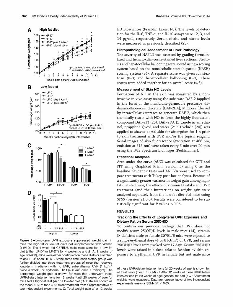
Figure 3—Long-term UVR exposure suppressed weight gain inmice fed high-fat or low-fat diets not supplemented with vitaminD (VitD). The 4-week-old C57BL/6 male mice were fed a low-fatdiet (either LF-D+ or LF-D2) for 4 weeks. A and B: At 8 weeks ofage (week 0), mice were either continued on these diets or switchedto an HF-D+ or an HF-D2. At the same time, each dietary group wasfurther divided into three treatment groups of mice that receivedlong-term irradiation with no UVR, suberythemal UVR (1 kJ/m2
twice a week), or erythemal UVR (4 kJ/m2 once a fortnight). Thepercentage weight gain is shown for mice that underwent theseUVR/dietary interventions for 12 weeks (until 20 weeks of age) formice fed a high-fat diet (A) or a low-fat diet (B). Data are shown asthe mean6 SEM for n = 18 mice/treatment from a representative oftwo independent experiments. C: Total weight gain after 12 weeks
of these UVR/dietary interventions (at 20 weeks of age) is shown forall treatments (mean 6 SEM). D: After 12 weeks of these UVR/dietaryinterventions (at 20 weeks of age) gonadal fat-pad (n = 18/treatment)weights were measured. Data are representative of two independentexperiments (mean + SEM). *P < 0.05.
3762 UV Inhibits Obesity Independently of Vitamin D Diabetes Volume 63, November 2014

(Supplementary Fig. 1). To determine the relative roles ofdietary vitamin D and/or UVR-induced vitamin D in theregulation of obesity and related cardiometabolic diseaseoutcomes, we performed the following experiment usingC57BL/6 mice (Fig. 1). Male mice were fed a vitamin D–supplemented or nonsupplemented (low-fat) diet from 4to 8 weeks of age to establish vitamin D sufficiency ordeficiency (Fig. 2A). From 8 weeks of age, mice were con-tinued on the supplemented or nonsupplemented diets,but some were switched to a diet that was high in fat.Each of these four dietary treatments were further di-vided into three treatments, with the shaved skin ofmice exposed to long-term irradiation with no UVR,
suberythemal UVR (1 kJ/m2 twice a week) or erythemalUVR (4 kJ/m2 once a fortnight), as indicated in Fig. 1.Mice were treated from 8 to 20 weeks of age with theseUVR and dietary interventions. A high-fat diet signifi-cantly increased serum 25(OH)D levels in mice fed dietsnot specifically supplemented with vitamin D (HF-D2,LF-D2) (Fig. 2B). Mice fed either diet that was furthersupplemented with vitamin D (HF-D+, LF-D+) had signif-icantly higher serum 25(OH)D levels than those mice feda diet that was not supplemented with vitamin D (Fig. 2B).There was no additive effect of a high-fat diet and vitamin Dsupplementation on serum 25(OH)D level (Fig. 2B). Al-though not observed in our preliminary (Supplementary
Table 1—AUC values for GTTs and ITTs, and fasting glucose, insulin, leptin, and adiponectin levels measured 9–11 weeks afterUVR/dietary intervention
Treatment DietUVR
(kJ/m2)
GTT (AUC,% basalglucose)
ITT (AUC,% basalglucose)
Fastingglucose(mmol/L)
Fastinginsulin(ng/mL)
Fastingleptin(ng/mL)
Fastingadiponectin(ng/mL)
1 HF-D+ 0 2,190 6 83 1,200 6 63 9.8 6 0.5 8.2 6 3.5 36.7 6 3.0 10.4 6 0.3
2 HF-D+ 1 1,770 6 49* 1,060 6 46 8.8 6 0.4 7.1 6 0.4 29.8 6 5.7 11.9 6 1.8
3 HF-D+ 4 1,880 6 180 1,370 6 34 10.2 6 0.4 3.6 6 1.1 19.7 6 7.3 15.8 6 3.9
4 LF-D+ 0 1,470 6 67 800 6 38 7.9 6 0.3 1.0 6 0.4 1.5 6 0.6 12.9 6 2.8
5 LF-D+ 1 1,510 6 65 760 6 37 8.0 6 0.4 4.9 6 2.8 2.6 6 1.1 8.8 6 2.5
6 LF-D+ 4 1,390 6 56 770 6 79 7.8 6 0.4 1.8 6 1.0 2.2 6 0.7 11.9 6 1.0
7 HF-D2 0 2,120 6 130 1,230 6 15 9.8 6 0.3 11.1 6 1.9 29.8 6 3.5 13.0 6 2.6
8 HF-D2 1 1,760 6 65† 1,050 6 43† 8.7 6 0.3† 3.8 6 1.1† 32.6 6 5.6 11.3 6 0.9
9 HF-D2 4 1,690 6 73† 960 6 72† 8.1 6 0.4† 3.9 6 2.8† 14.0 6 5.3† 13.0 6 1.1
10 LF-D2 0 1,260 6 51 680 6 48 6.3 6 0.2 3.4 6 1.6 5.9 6 2.5 16.6 6 6.2
11 LF-D2 1 1,280 6 102 600 6 27 6.0 6 0.2 1.6 6 1.1 1.0 6 0.5 10.8 6 0.6
12 LF-D2 4 1,480 6 36 760 6 60 7.7 6 0.4 4.3 6 1.8 1.9 6 0.2 11.7 6 1.9
Data are the mean 6 SEM; n = 4–8 mice/treatment. *P , 0.05 vs. no UVR and HF-D+ with data representative of two experiments.†P , 0.05 relative to no UVR and HF-D2 with data representative of two experiments.
Table 2—Circulating triglyceride and cholesterol levels at 12 weeks after dietary and UVR interventions
Treatment DietUVR
(kJ/m2)Triglycerides(mmol/L)
HDL cholesterol(mmol/L)
LDL cholesterol(mmol/L)
Total cholesterol(mmol/L)
1 HF-D+ 0 0.7 6 0.1 2.1 6 0.2 0.3 6 0.0 4.2 6 0.4
2 HF-D+ 1 0.6 6 0.0 2.0 6 0.2 0.2 6 0.0 3.8 6 0.4
3 HF-D+ 4 0.8 6 0.1 2.1 6 0.1 0.2 6 0.0 4.3 6 0.2
4 LF-D+ 0 1.0 6 0.1 1.5 6 0.1 0.2 6 0.0 2.5 6 0.2
5 LF-D+ 1 1.2 6 0.1 1.8 6 0.1 0.2 6 0.0 2.9 6 0.1
6 LF-D+ 4 1.1 6 0.3 1.3 6 0.2 0.1 6 0.0 2.2 6 0.3
7 HF-D2 0 0.9 6 0.1 2.1 6 0.1 0.4 6 0.0 4.3 6 0.1
8 HF-D2 1 0.6 6 0.0 2.1 6 0.0 0.3 6 0.0 4.2 6 0.2
9 HF-D2 4 0.9 6 0.1 1.5 6 0.2* 0.2 6 0.0* 2.6 6 0.3*
10 LF-D2 0 1.2 6 0.1 1.6 6 0.3 0.1 6 0.0 2.4 6 0.4
11 LF-D2 1 0.9 6 0.1 1.4 6 0.1 0.1 6 0.0 2.0 6 0.1
12 LF-D2 4 1.1 6 0.1 1.5 6 0.1 0.1 6 0.0 2.3 6 0.1
n = 4 mice/treatment. *P , 0.05 relative to no UVR and HF-D2 with data representative of two experiments.
diabetes.diabetesjournals.org Geldenhuys and Associates 3763

Fig. 1) and past investigations (14), long-term suberythe-mal (Fig. 2C) or erythemal (Fig. 2D) UVR exposure signif-icantly (but transiently) enhanced serum 25(OH)D levels,when administered to mice fed an LF-D+ (but not HF-D+,LF-D2, or HF-D2) (Supplementary Fig. 2). The effectswere more pronounced for mice administered the long-term erythemal UVR, but returned to baseline levels after6 weeks of UVR/dietary intervention (Fig. 2D and Supple-mentary Fig. 2B).
Long-term UVR Exposure Suppressed Weight Gain inMice Fed a Vitamin D–Nonsupplemented DietThere was no effect of vitamin D supplementation onweight gain (Fig. 3A and B). Both long-term suberythemalUVR (1 kJ/m2 twice a week) and erythemal UVR (4 kJ/m2
once a fortnight) treatment suppressed weight gain in micefed the HF-D2 (Fig. 3A) by $40%. Long-term erythemalUVR exposure also suppressed weight gain in mice fed theLF-D2 (Fig. 3B). The effects of long-term skin exposureto UVR were less apparent for mice fed the vitaminD–supplemented diet, where UVR exposure suppressedweight gain in a transient fashion in mice fed the HF-D+
(Supplementary Fig. 3A). At the end of the UVR/dietaryintervention period (12 weeks), gonadal fat-pad weightswere not affected by dietary vitamin D supplementationbut were significantly suppressed in mice irradiated withUVR and fed the HF-D2 (Fig. 3D).
Long-term UVR Exposure Suppressed GlucoseIntolerance and Insulin Resistance in Mice Feda Vitamin D–Nonsupplemented DietAfter 10 and 11 weeks of UVR/dietary intervention, GTTsand ITTs were performed (Table 1). Mice fed the high-fatdiets developed glucose intolerance (Supplementary Fig.3B) and insulin resistance (Supplementary Fig. 3C), withno suppressive effect of vitamin D supplementation (Sup-plementary Fig. 3B and C; Table 1 for AUC). Both mea-sures were suppressed in mice receiving long-term irradiationwith UVR (either suberythemal or erythemal) and fed theHF-D2 (Table 1). Glucose intolerance was significantlysuppressed by long-term suberythemal UVR in mice fedthe HF-D+ only (Table 1). In addition, fasting glucose andinsulin levels were also reduced by UVR treatment inmice fed the HF-D2, with fasting leptin levels also sup-pressed in mice that received long-term irradiation witherythemal UVR (Table 1). There were no effects of long-term UVR (or dietary vitamin D) on fasting adiponectinlevels (Table 1).
Long-term Erythemal UVR Exposure SuppressedCirculating Cholesterol Levels in Mice Fed a High-FatDiet Not Supplemented With Vitamin DAfter 12 weeks of UVR/dietary intervention, circulatinglevels of triglycerides and cholesterol (HDL, LDL, andtotal) were measured (Table 2). Triglyceride levels were
Figure 4—Long-term UVR significantly reduced the extent of liver steatosis and lobular ballooning in mice fed a high-fat diet. The 4-week-old C57BL/6 male mice were fed a low-fat diet (either LF-D+ or LF-D2) for 4 weeks. At 8 weeks of age, mice were either continued on thesediets or switched to an HF-D+ or an HF-D2. At the same time, each dietary group was further divided into three treatment groups of micethat received long-term irradiation with no UVR (A, D, G, and J), suberythemal UVR (1 kJ/m2 twice a week; B, E, H, K), or erythemal UVR(4 kJ/m2 once a fortnight; C, F, I, and L). After 12 weeks of these UVR/dietary interventions (at 20 weeks of age), the extent of liverhistopathology was measured in liver specimens (n = 10/treatment for data pooled from two independent experiments). A–L: Represen-tative hematoxylin-eosin–stained sections of liver for each treatment (B and C, original magnification 320 [equivalent to 150 mm]).Examples of liver steatosis (blue arrow) and lobular ballooning (red arrow) are shown in G.
3764 UV Inhibits Obesity Independently of Vitamin D Diabetes Volume 63, November 2014

not modified by vitamin D supplementation or long-termUVR (Table 2). HDL, LDL, and total cholesterol levelswere suppressed in mice fed the HF-D2 and also receivinglong-term irradiation with erythemal UVR (Table 2).
Long-term UVR Exposure More Effectively Suppressedthe Development of NAFLD Than Vitamin DSupplementationThe development of markers of NAFLD was measured byanalyzing the degree of liver steatosis and lobularballooning after 12 weeks of UVR/dietary intervention(Figs. 4 and 5A). Long-term skin exposure to UVR sub-stantially suppressed liver histopathology in mice fed thehigh-fat diets (Fig. 4A–C, HF-D+; Fig. 4G–I, HF-D2; Fig.5A) to a greater degree than that achieved by dietaryvitamin D supplementation alone (Fig. 4A, HF-D+; Fig.4G, HF-D2; Fig. 5A). Vitamin D supplementation hadno effect on liver weight, whereas long-term erythemalUVR suppressed liver weight in mice fed the HF-D2
(Fig. 5B).
Vitamin D Supplementation Prevented the SuppressiveEffects of UVR Upon Weight Gain and Markers of MetSThe results presented above suggest that many of theeffects of UVR were more prominent in mice not furthersupplemented with vitamin D. We used a general linearmodel to assess whether there may be interactions withinthe high-fat diet treatments, such that dietary vitamin Dmay have inhibited the suppressive ability of UVR.Significant interactions between dietary vitamin D andlong-term UVR exposure were detected for weight gain(Fig. 3C) (P = 0.05), gonadal fat-pad weights (Fig. 3D) (P =0.03), and fasting glucose levels (Table 1) (P = 0.01), butnot the other measures, including liver histopathology(Figs. 4 and 5A) (P . 0.05).
Serum Vitamin D or Calcium Levels Were Not Relatedto Weight Loss or Suppression of MetS in UVR-Irradiated MiceLong-term UVR exposure suppressed aspects of weightgain and measures of MetS, independently of changes tocirculating 25(OH)D levels (Fig. 2 and Supplementary Fig.2). Therefore, it is unlikely that the mechanism throughwhich UVR acted was dependent on vitamin D. As calciumlevels can be modified by vitamin D and have been asso-ciated with weight loss (26), we also assessed circulatingcalcium levels after 12 weeks of UVR/dietary intervention,but observed no significant effects of dietary vitamin D orlong-term skin exposure to UVR in mice fed the high-fatdiets (Fig. 5C). Long-term skin exposure to UVR reducedcalcium levels in mice fed a low-fat diet (Fig. 5C).
Figure 5—Long-term UVR exposure significantly reduced the ex-tent of liver histopathology in mice fed a high-fat diet. The 4-week-old C57BL/6 male mice were fed a low-fat diet (either LF-D+ orLF-D2) for 4 weeks. At 8 weeks of age, mice were either continuedon these diets or switched to an HF-D+ or HF-D2. At the sametime, each dietary group was further divided into three treatmentgroups of mice that received long-term irradiation with no UVR,suberythemal UVR (1 kJ/m2 twice a week), or erythemal UVR(4 kJ/m2 once a fortnight). After 12 weeks of these UVR/dietaryinterventions (at 20 weeks of age), the extent of liver histopathology
(n = 10/treatment for data pooled from two independent experi-ments) (A), liver weights (n = 18/treatment for data from a repre-sentative experiment) (B), and serum levels of calcium (n = 4–8/treatment for data pooled from two independent experiments) (C )and TNF-a (n = 12–18/treatment for data pooled from two in-dependent experiments) (D) are shown. Data are shown as themean 6 SEM. *P < 0.05. VitD, vitamin D.
diabetes.diabetesjournals.org Geldenhuys and Associates 3765

Figure 6—The UVR-induced mediator NO may regulate body weight, WAT accumulation, glucose metabolism, and the development ofNAFLD in mice fed a high-fat diet. A and B: Using the DAF-2DA substrate, skin NO levels are shown for adult C57BL/6 male mice fed a low-fat diet (LF-D2), 5 min after skin treatment with vehicle, 1 kJ/m2 UVR, or the NO donor SNAP, with a quantitative measure (in photons persecond) (A) and representative skin fluorescence (B) shown. The 4-week-old C57BL/6 male mice were fed an LF-D2 for 4 weeks. At 8weeks of age, mice were either continued on these diets or switched to the HF-D2. Within the HF-D2 treatments, mice were further dividedinto five treatment groups. The shaved dorsal skin of these mice 1) was treated with vehicle only, 2) received long-term irradiation withsuberythemal UVR (1 kJ/m2 twice a week) and then vehicle, 3) was topically treated with SNAP, 4) received long-term irradiation with
3766 UV Inhibits Obesity Independently of Vitamin D Diabetes Volume 63, November 2014

Circulating TNF-a Level Was Linked With ImprovedMarkers of NAFLD in the Absence of Dietary Vitamin DSupplementation But Not Skin Exposure to UVRThe ability of phototherapy to suppress the development ofNAFLD has been associated with reduced expression of TNF-a(13). However, long-term UVR did not modify serum TNF-alevels after 12 weeks of UVR/dietary intervention in mice feda high-fat diet (Fig. 5D). Vitamin D supplementation reducedcirculating TNF-a levels in mice fed an HF-D+ when comparedwith those fed an HF-D2 (Fig. 5D). Serum levels of IL-6 andIL-10 were below the level of detection of the ELISA.
UV-Induced NO Suppresses the Development ofObesity and Symptoms of MetSA role for NO, an alternate (non–vitamin D) mediatorinduced by UVR, was examined. Skin levels of NO in-creased from as early as 5 min after UVR/SNAP (Fig. 6Aand B) treatment as determined using DAF-2. To examinea role for UVR-induced NO in modulating obesity andMetS symptoms, 4-week-old C57BL/6 male mice werefed an LF-D2 for 4 weeks. From 8 weeks of age, micewere either continued on this diet or switched to theHF-D2, with mice fed an HF-D2 further divided intogroups receiving the following five dorsal skin treatments:1) vehicle only; 2) suberythemal UVR (1 kJ/m2) and thenvehicle; 3) SNAP; 4) suberythemal UVR and then cPTIO;or 5) suberythemal UVR and then 1,25(OH)2D. This finaltreatment was selected to test whether active 1,25(OH)2Dcould prevent the suppressive effects of UVR on obesityand MetS development (like dietary vitamin D in Supple-mentary Fig. 3A) through inhibition of skin-induced NO.Indeed, vitamin D may repair UV-induced DNA damage inskin by suppressing NO (27).
After 12 weeks of feeding mice the HF-D2, skin NOlevels were assessed 10 min after a final treatment withone of the five topical treatments detailed above. Skin NOlevels increased with UVR or SNAP (Fig. 6C). The NOscavenger cPTIO reduced levels of NO in skin after UVRtreatment, but, unexpectedly, 1,25(OH)2D did not. Serumnitrite/nitrate concentrations, measured 20 min after thefinal skin treatment, were not altered by treatment withlong-term low-dose UVR or SNAP (data not shown). Long-term UVR suppressed weight gain and the accumulationof WAT after 12 weeks of the HF-D2 (Fig. 6D). Long-termSNAP treatment also effectively suppressed mouse weights(although not weight gain) and WAT accumulation (Fig. 6D).However, neither the NO scavenger cPTIO nor 1,25(OH)2Dreversed the suppressive effects of UVR on weight gainor WAT accumulation. Indeed, the UVR and 1,25(OH)2Dtreatment was more effective than UVR treatment alone,
but this observation may reflect the hypercalcemia observedearly on with topical 1,25(OH)2D treatment (4 weeks post-UVR [2.4 6 0.03 mmol/L] vs. post-UVR+1,25(OH)2D[3.5 6 0.07]; *P , 0.001 for serum calcium). In responseto these observations, we halved the dose of 1,25(OH)2D ad-ministered, and mice were treated only once per week after 4weeks of intervention. Despite this change, 1,25(OH)2D-treated mice were still modestly hypercalcemic at the endof the experiment (12 weeks post-UVR [2.4 6 0.03] vs.post-UVR+1,25(OH)2D [2.7 6 0.07]; *P , 0.001 for serumcalcium).
As observed previously, long-term UVR exposure sup-pressed fasting glucose and insulin levels, and the de-velopment of glucose intolerance and insulin resistance(Fig. 6E and F). Here, long-term SNAP treatment alsosuppressed the development of insulin resistance (Fig.6F). Furthermore, cPTIO treatment after UVR reversedthe suppressive effects of UVR alone upon fasting glucoselevels (Fig. 6E). Finally, both long-term UVR and SNAPtreatment suppressed the development of NAFLD, whilecPTIO reversed the effects of UVR upon liver histopathol-ogy (Fig. 6G). Cumulatively, these data suggest that UVR-induced NO may play an important role in modulating thedevelopment of obesity and MetS through effects onweight, WAT accumulation, fasting glucose level, andthe development of insulin resistance and NAFLD.
DISCUSSION
Here we present evidence that long-term skin exposure tolow-dose (suberythemal) and high-dose (erythemal) UVRsuppresses the development of obesity and measures ofMetS in mice fed a high-fat diet. Vitamin D supplemen-tation alone did not reproduce these effects. In addition,the suppressive effects of UVR on obesity and MetSdevelopment were not observed to the same degree inmice that were further supplemented with vitamin D (i.e.,HF-D+). For mice fed a high-fat diet, serum 25(OH)Dlevels were not enhanced by long-term UVR exposure,suggesting that any effects induced by UVR in thesemice were independent of circulating 25(OH)D levels.The HF-D2 increased circulating 25(OH)D levels; it islikely that this diet contains vitamin D, perhaps withinthe lard-derived fat fraction. Supplementation of this dietwith vitamin D (i.e., the HF-D+) further increased serum25(OH)D levels. Both UV irradiation and vitamin D sup-plementation reduced the severity of NAFLD, suggestingthat vitamin D can recapitulate the effects of UVR for theprevention of certain obesity-related pathologies. We alsoshowed that some of the effects of UVR may occurthrough NO production. In particular, it is likely that
suberythemal UVR and then cPTIO, or 5) received long-term irradiation with suberythemal UVR and then 1,25(OH)2D. Mice were treated for12 weeks with these skin/dietary interventions until 20 weeks of age. C: Skin NO levels, 10 min after skin treatment (n = 8 mice/treatment).D: Mouse weights, weight gain, and WAT weights (n = 18 mice/treatment). E: Fasting glucose and GTT AUC (n = 8 mice/treatment). F:Fasting insulin and ITT AUC (n = 8 mice/treatment). G: Liver histopathology scores (n = 8 mice/treatment). Data are shown as the mean 6SEM from one experiment. *P < 0.05. VitD, vitamin D.
diabetes.diabetesjournals.org Geldenhuys and Associates 3767

UVR-induced NO may have profound effects on the de-velopment of NAFLD, as topical SNAP suppressed liverpathology, and cPTIO antagonized the effects of UVR.Various non–vitamin D immunomodulators induced byUVR, like NO (28), may be important for the regulationof immunity (29) and obesity/MetS development (30).Skin exposure to UVR releases NO from skin (28) andcould control obesity through NO-dependent effects onmitochondria biogenesis within brown adipose tissue (31).We have recently shown that UVR-induced NO reducesblood pressure in healthy human volunteers (28). NO mayalso be a crucial modulator of insulin and glucose transport,and inhibition of NO may cause insulin resistance (32).Combined with our results, these studies point to topicallyinduced NO as a potentially important clinical means tosuppress obesity and type 2 diabetes development.
The capacity of long-term UVR to suppress the devel-opment of obesity and metrics of MetS was less effectivein mice orally supplemented with vitamin D [but not withtopical 1,25(OH)2D]. This was an unexpected finding butcould be explained by potential interactions of UVR-inducedmediators and dietary vitamin D, including NO (27). Thedifferent effects of dietary vitamin D and topical 1,25(OH)2D could be accounted for by the hypercalcemia in-duced by long-term topical 1,25(OH)2D. In addition, after12 weeks of treatment, serum 25(OH)D levels were signif-icantly reduced by topical 1,25(OH)2D but not by the othertreatments (data not shown). Others have also observed(33) that vitamin D suppressed weight gain in vivo afterintraperitoneal injections of 1,25(OH)2D (5 mg/kg every 2days), although the effects on circulating levels of calcium[and 25(OH)D] were not reported. Others have shown (34)that UVR may increase cortisol production in skin, whichhas the potential to impact the hypothalamic-pituitary-adrenal axis. While this might be hypothesized to alterphysical activity, no obvious behavioral effects were ob-served in this study. However, we cannot exclude thepossibility that UVR alters neuroendocrine signaling net-works in the skin (35) that might have a systemic impact.
Nakano et al. (13) showed that phototherapy sup-pressed NAFLD but failed to reduce obesity, steatosis,and blood glucose levels in Zucker fa-fa rats. These resultsmay differ from our own through significant differencesin the phototherapies delivered and the mouse model ofobesity. Dietary vitamin D has also previously been shownto suppress the development of NAFLD in Sprague-Dawley rats fed a “westernized” (high-fat/fructose) diet(36), and in Lewis rats fed a choline-deficient and iron-supplemented L-amino acid–defined diet (13). We alsoobserved that dietary vitamin D suppressed circulatingTNF-a levels in mice fed a high-fat diet. UVR did notsuppress serum TNF-a levels, suggesting that dietary vi-tamin D and UVR may suppress NAFLD through differingmechanisms. For control of NAFLD, the role of otherplayers within the vitamin D pathway is worthy of furtherconsideration. For example, circulating levels of the vita-min D binding protein GC inversely correlate with liver
steatosis, and may determine the ability of vitamin D tomodulate the development of NAFLD (37). In addition,1,25(OH)2D may act through the vitamin D receptor toimprove insulin sensitivity (38).
Our observations suggest that not all of the effects ofUVR on disease prevention can be achieved throughdietary vitamin D and that the role of other UV-inducedmediators like NO deserve further consideration. Fur-thermore, by using a mouse modeling approach we wereable to remove the confounding effects of activity out ofdoors, which might explain the observed associations ofreduced obesity and increased serum 25(OH)D levels. Acaveat is that while mice have conserved the ability tosynthesize vitamin D and NO in the skin and systemicallypost-UVR, as fur-covered nocturnal animals they are notusually exposed to much sunlight. Further studies arerequired to translate the findings of our murine studies tohumans. However, our results support recent calls forclinical trials that test the efficacy of skin exposure tosunlight or UVR for the control of chronic diseases likemultiple sclerosis (39) and depression (40), which, likeobesity and MetS, may take years to develop. In conclu-sion, our studies show that long-term low-dose sunlightexposure may be an effective means of suppressing obe-sity and MetS in mice fed a high-fat diet, through path-ways that are independent of vitamin D and at leastpartially dependent on skin-derived NO.
Acknowledgments. The authors thank Drs. Bernadette Fernandez andMagda Minnion for measuring serum nitrite/nitrate levels; Professor MichaelClarke at the Centre for Metabolomics (University of Western Australia) forperforming the liquid chromatography-mass spectrometry detection of serum25(OH)D; Linda Gregory at the PathWest Laboratory at Royal Perth Hospital(Perth, Western Australia, Australia) for performing the serum calcium,cholesterol, HDL cholesterol, LDL cholesterol, and triglyceride analyses; andMaxine Crook at Princess Margaret Hospital Pathology (Subiaco, WesternAustralia, Australia) for embedding, sectioning, and staining the liver specimens.Funding. This research was supported by the BrightSpark Foundation and theTelethon Institute for Child Health Research.Duality of Interest. No potential conflicts of interest relevant to this articlewere reported.Author Contributions. S.Ge. performed the majority of the experimentsand statistical analyses, and reviewed and edited the manuscript. P.H.H.contributed to the discussion, and reviewed and edited the manuscript. R.E.helped to optimize the skin NO assay and reviewed and edited the manuscript.P.J. provided the statistical expertise for the experimental design and dataanalysis. M.F. helped to design the study, supervised the analysis of serum NOmetabolites, and reviewed and edited the manuscript. R.B.W. helped to designthe study and reviewed and edited the manuscript. V.M. helped to design thestudy, contributed to the discussion, and reviewed and edited the manuscript.S.Go. envisaged and designed the study and wrote the manuscript. S.Go. is theguarantor of this work and, as such, had full access to all the data in the studyand takes responsibility for the integrity of the data and the accuracy of the dataanalysis.Prior Presentation. Parts of this study were presented in abstract form atthe Australian Society for Medical Research Western Australia Scientific Symposium,Perth, Western Australia, Australia, 5 June 2013; the Murdoch Children’s ResearchInstitute Molecular Medicine Series, Melbourne, Victoria, Australia, 12 July 2013;
3768 UV Inhibits Obesity Independently of Vitamin D Diabetes Volume 63, November 2014

and the 6th Asia and Oceania Conference on Photobiology, Sydney, New SouthWales, Australia, 10–13 November 2013.
References1. Holick MF. Vitamin D deficiency in 2010: health benefits of vitamin D andsunlight: a D-bate. Nat Rev Endocrinol 2011;7:73–752. Earthman CP, Beckman LM, Masodkar K, Sibley SD. The link betweenobesity and low circulating 25-hydroxyvitamin D concentrations: considerationsand implications. Int J Obes (Lond) 2012;36:387–3963. Autier P, Boniol M, Pizot C, Mullie P. Vitamin D status and ill health:a systematic review. Lancet Diabetes Endocrinol 2014;2:76–894. Daly RM, Gagnon C, Lu ZX, et al. Prevalence of vitamin D deficiency and itsdeterminants in Australian adults aged 25 years and older: a national, population-based study. Clin Endocrinol (Oxf) 2012;77:26–355. Vimaleswaran KS, Berry DJ, Lu C, et al.; Genetic Investigation of Anthro-pometric Traits-GIANT Consortium. Causal relationship between obesity andvitamin D status: bi-directional Mendelian randomization analysis of multiplecohorts. PLoS Med 2013;10:e10013836. Lavie CJ, Lee JH, Milani RV. Vitamin D and cardiovascular disease will it liveup to its hype? J Am Coll Cardiol 2011;58:1547–15567. Maxwell CS, Wood RJ. Update on vitamin D and type 2 diabetes. Nutr Rev2011;69:291–2958. Brouwer DA, van Beek J, Ferwerda H, et al. Rat adipose tissue rapidlyaccumulates and slowly releases an orally-administered high vitamin D dose. BrJ Nutr 1998;79:527–5329. Kull M, Kallikorm R, Lember M. Body mass index determines sunbathinghabits: implications on vitamin D levels. Intern Med J 2009;39:256–25810. Lucas RM, Ponsonby AL, Dear K, et al. Vitamin D status: multifactorialcontribution of environment, genes and other factors in healthy Australian adultsacross a latitude gradient. J Steroid Biochem Mol Biol 2013;136:300–30811. Ginde AA, Liu MC, Camargo CA Jr. Demographic differences and trends ofvitamin D insufficiency in the US population, 1988-2004. Arch Intern Med 2009;169:626–63212. Lindqvist PG, Olsson H, Landin-Olsson M. Are active sun exposure habitsrelated to lowering risk of type 2 diabetes mellitus in women, a prospectivecohort study? Diabetes Res Clin Pract 2010;90:109–11413. Nakano T, Cheng YF, Lai CY, et al. Impact of artificial sunlight therapy on theprogress of non-alcoholic fatty liver disease in rats. J Hepatol 2011;55:415–42514. Gorman S, Scott NM, Tan DH, et al. Acute erythemal ultraviolet radiation causessystemic immunosuppression in the absence of increased 25-hydroxyvitaminD3 levels in male mice. PLoS One 2012;7:e4600615. McGlade JP, Gorman S, Zosky GR, et al. Suppression of the asthmaticphenotype by ultraviolet B-induced, antigen-specific regulatory cells. Clin ExpAllergy 2007;37:1267–127616. McGlade JP, Gorman S, Lenzo JC, et al. Effect of both ultraviolet B irra-diation and histamine receptor function on allergic responses to an inhaled an-tigen. J Immunol 2007;178:2794–280217. Ikeyama K, Fuziwara S, Denda M. Topical application of neuronal nitricoxide synthase inhibitor accelerates cutaneous barrier recovery and preventsepidermal hyperplasia induced by barrier disruption. J Invest Dermatol 2007;127:1713–171918. Yasukawa K, Tokuda H, Tun X, Utsumi H, Yamada K. The detrimental effectof nitric oxide on tissue is associated with inflammatory events in the vascularendothelium and neutrophils in mice with dextran sodium sulfate-induced colitis.Free Radic Res 2012;46:1427–143619. Dixon KM, Norman AW, Sequeira VB, et al. 1a,25(OH)₂-vitamin D anda nongenomic vitamin D analogue inhibit ultraviolet radiation-induced skin car-cinogenesis. Cancer Prev Res (Phila) 2011;4:1485–149420. Gorman S, Kuritzky LA, Judge MA, et al. Topically applied 1,25-dihydroxyvitaminD3 enhances the suppressive activity of CD4+CD25+ cells in the draining lymphnodes. J Immunol 2007;179:6273–6283
21. Clarke MW, Tuckey RC, Gorman S, Holt B, Hart PH. Optimized 25 hydroxy
vitamin D analysis using liquid–liquid extraction with 2D separation with LCMS/
MS detection, provides superior precision compared to conventional assays.
Metabolomics 2013;9:1031–104022. Gorman S, Judge MA, Hart PH. Immune-modifying properties of topical
vitamin D: focus on dendritic cells and T cells. J Steroid Biochem Mol Biol 2010;
121:247–24923. Milsom AB, Fernandez BO, Garcia-Saura MF, Rodriguez J, Feelisch M.
Contributions of nitric oxide synthases, dietary nitrite/nitrate, and other sources
to the formation of NO signaling products. Antioxid Redox Signal 2012;17:422–
43224. Kleiner DE, Brunt EM, Van Natta M, et al.; Nonalcoholic Steatohepatitis
Clinical Research Network. Design and validation of a histological scoring system
for nonalcoholic fatty liver disease. Hepatology 2005;41:1313–132125. Rodriguez J, Specian V, Maloney R, Jourd’heuil D, Feelisch M. Performance
of diamino fluorophores for the localization of sources and targets of nitric oxide.
Free Radic Biol Med 2005;38:356–36826. Zemel MB, Shi H, Greer B, Dirienzo D, Zemel PC. Regulation of adiposity by
dietary calcium. FASEB J 2000;14:1132–113827. Mason RS, Sequeira VB, Dixon KM, et al. Photoprotection by 1alpha,25-
dihydroxyvitamin D and analogs: further studies on mechanisms and implications
for UV-damage. J Steroid Biochem Mol Biol 2010;121:164–16828. Liu D, Fernandez BO, Hamilton MB, et al. UVA irradiation of human skin
vasodilates arterial vasculature and lowers blood pressure independently of nitric
oxide synthase. J Invest Dermatol 2014;134:1839–184629. Zosky GR, Berry LJ, Elliot JG, James AL, Gorman S, Hart PH. Vitamin D
deficiency causes deficits in lung function and alters lung structure. Am J Respir
Crit Care Med 2011;183:1336–134330. Sessa WC. A new approach to weight loss: just activate endothelial NO
synthase! Circ Res 2012;111:1111–111231. Knott AB, Bossy-Wetzel E. Impact of nitric oxide on metabolism in
health and age-related disease. Diabetes Obes Metab 2010;12(Suppl. 2):
126–13332. Sydow K, Mondon CE, Cooke JP. Insulin resistance: potential role of the
endogenous nitric oxide synthase inhibitor ADMA. Vasc Med 2005;10(Suppl. 1):
S35–S4333. Yin Y, Yu Z, Xia M, Luo X, Lu X, Ling W. Vitamin D attenuates high fat diet-
induced hepatic steatosis in rats by modulating lipid metabolism. Eur J Clin Invest
2012;42:1189–119634. Skobowiat C, Sayre RM, Dowdy JC, Slominski AT. Ultraviolet radiation
regulates cortisol activity in a waveband-dependent manner in human skin ex
vivo. Br J Dermatol 2013;168:595–60135. Zmijewski MA, Slominski AT. Neuroendocrinology of the skin: an overview
and selective analysis. Dermatoendocrinol 2011;3:3–1036. Roth CL, Elfers CT, Figlewicz DP, et al. Vitamin D deficiency in obese rats
exacerbates nonalcoholic fatty liver disease and increases hepatic resistin and
Toll-like receptor activation. Hepatology 2012;55:1103–111137. Adams LA, White SW, Marsh JA, et al. Association between liver-specific
gene polymorphisms and their expression levels with nonalcoholic fatty liver
disease. Hepatology 2013;57:590–60038. Takiishi T, Gysemans C, Bouillon R, Mathieu C. Vitamin D and diabetes.
Endocrinol Metab Clin North Am 2010;39:419–44639. Correale J, Farez MF. Modulation of multiple sclerosis by sunlight exposure:
role of cis-urocanic acid. J Neuroimmunol 2013;261:134–14040. Knippenberg S, Damoiseaux J, Bol Y, et al. Higher levels of reported sun
exposure, and not vitamin D status, are associated with less depressive
symptoms and fatigue in multiple sclerosis. Acta Neurol Scand 2014;129:
123–131
diabetes.diabetesjournals.org Geldenhuys and Associates 3769