ultrasound in obstetrics - children's memorial hermann hospital
TRANSCRIPT
Ultrasound in Obstetrics Who, Where, When and How Many?
Anthony Johnson, D.O.
Visiting Professor
Departments of Obstetrics, Gynecology and
Reproductive Sciences and Pediatric Surgery
Co-Director, Texas Fetal Center
Clinical Considerations
• Should all patients be offered ultrasound?
• How many ultrasounds does a low risk patient need?
• What is the sensitivity for detecting fetal anomalies?
• What is the optimal gestational age for an obstetrical
examination?
• What impact does maternal BMI play in antenatal
ultrasound screening?
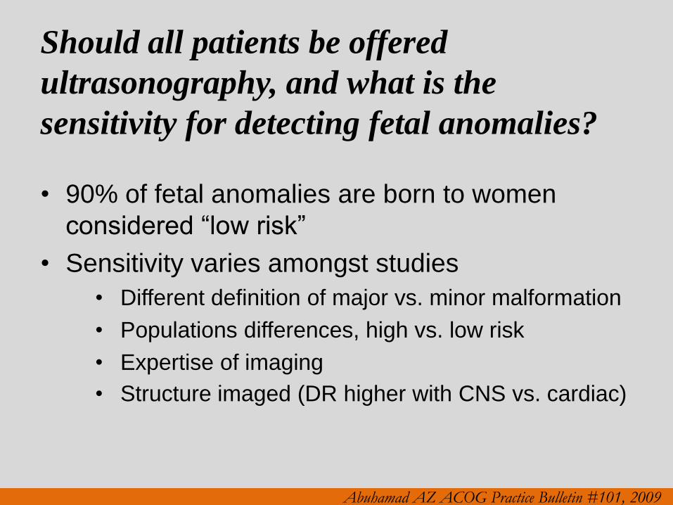
Should all patients be offered
ultrasonography, and what is the
sensitivity for detecting fetal anomalies?
• 90% of fetal anomalies are born to women
considered “low risk”
• Sensitivity varies amongst studies
• Different definition of major vs. minor malformation
• Populations differences, high vs. low risk
• Expertise of imaging
• Structure imaged (DR higher with CNS vs. cardiac)
Abuhamad AZ ACOG Practice Bulletin #101, 2009
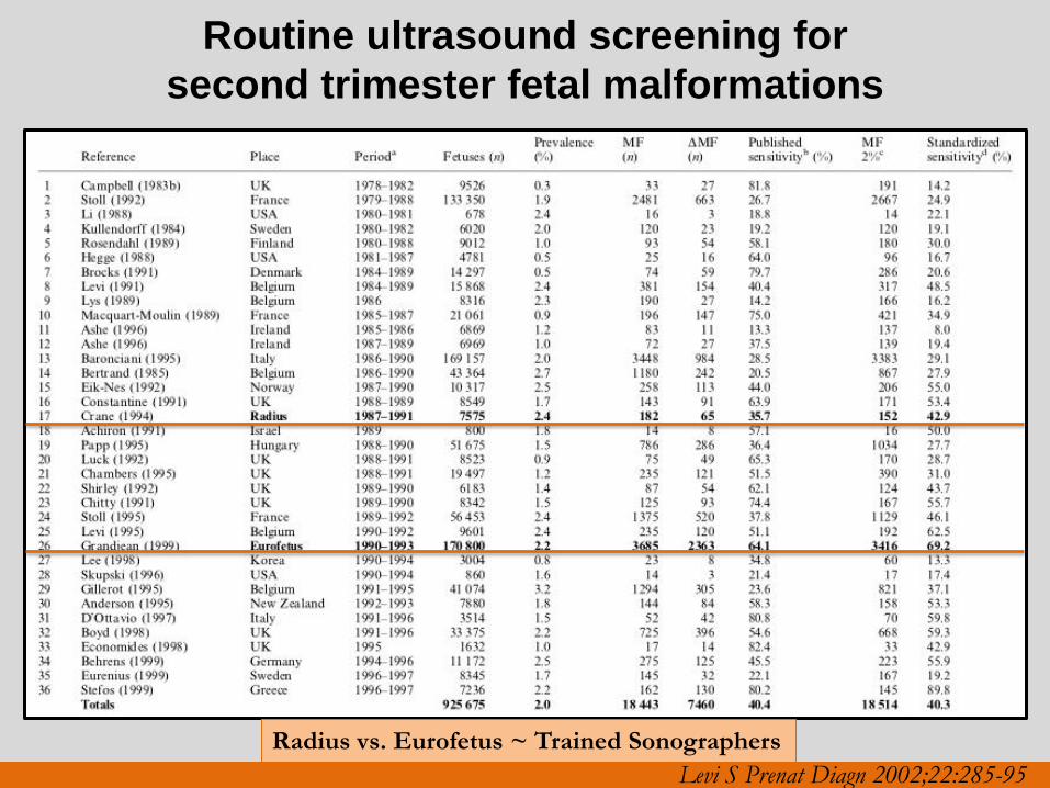
Levi S Prenat Diagn 2002;22:285-95
Routine ultrasound screening for
second trimester fetal malformations
Radius vs. Eurofetus ~ Trained Sonographers
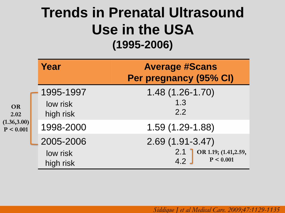
Trends in Prenatal Ultrasound
Use in the USA (1995-2006)
Year Average #Scans
Per pregnancy (95% CI)
1995-1997
low risk
high risk
1.48 (1.26-1.70) 1.3
2.2
1998-2000 1.59 (1.29-1.88)
2005-2006
low risk
high risk
2.69 (1.91-3.47) 2.1
4.2
OR
2.02
(1.36,3.00)
P < 0.001
OR 1.19; (1.41,2.59,
P < 0.001
Siddique J et al Medical Care. 2009;47:1129-1135
REAFFIRMED 2011
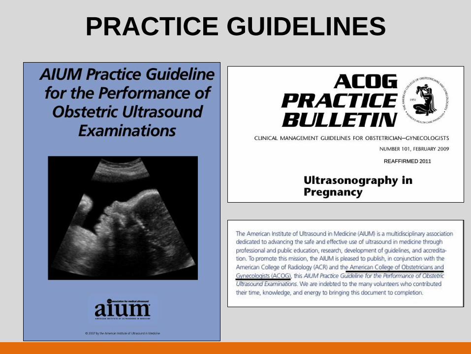
PRACTICE GUIDELINES
Practice Guidelines
• Performance and recording of high-quality ultrasound
examinations
• Minimum criteria for complete examination
• Not intended to establish a legal standard of care (SOC)
• Deviation from or exceeding guidelines will be needed in
some cases
ACR –ACOG-AIUM Reston (VA), 2007;1025-1033
ACOG Practice Bulletin 101, 2009,
AIUM J Ultrasound Med 2010;29:157-166,
ISUOG Ultrasound Obstet Gynecol 2011;37 116-126
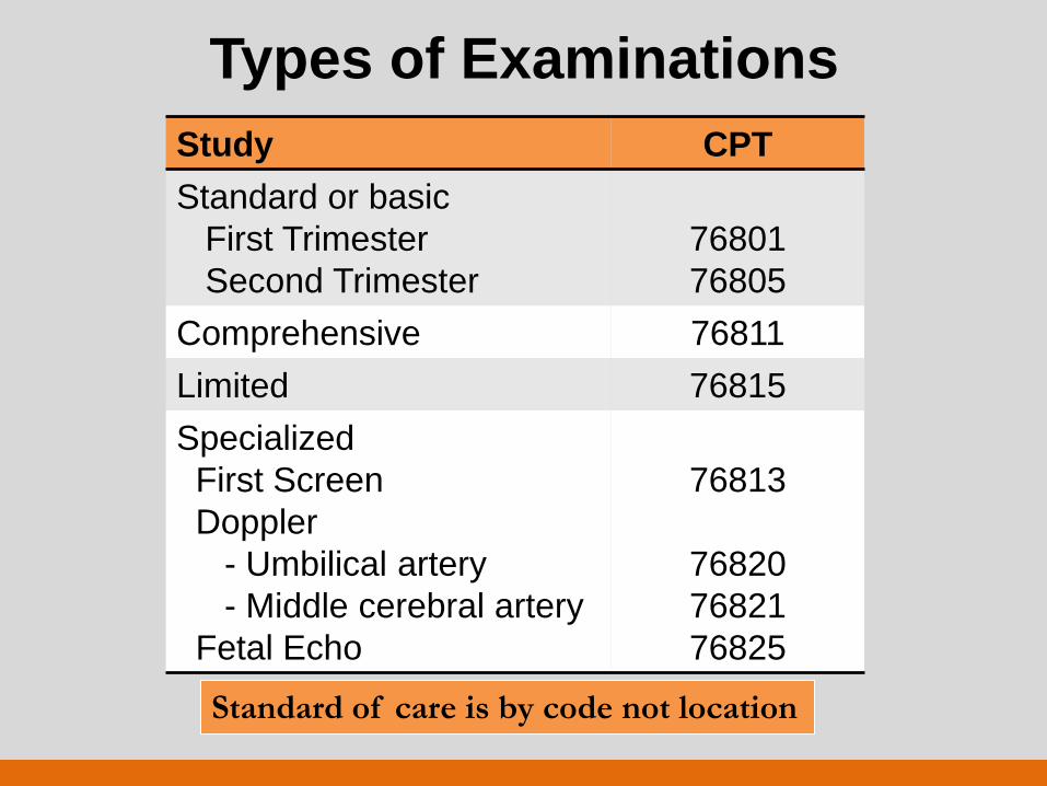
Types of Examinations
Study CPT
Standard or basic
First Trimester
Second Trimester
76801
76805
Comprehensive 76811
Limited 76815
Specialized
First Screen
Doppler
- Umbilical artery
- Middle cerebral artery
Fetal Echo
76813
76820
76821
76825
Standard of care is by code not location
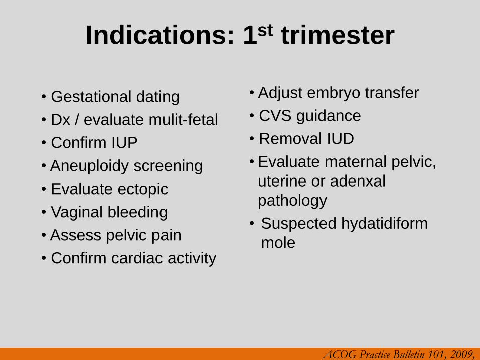
Indications: 1st trimester
• Gestational dating
• Dx / evaluate mulit-fetal
• Confirm IUP
• Aneuploidy screening
• Evaluate ectopic
• Vaginal bleeding
• Assess pelvic pain
• Confirm cardiac activity
• Adjust embryo transfer
• CVS guidance
• Removal IUD
• Evaluate maternal pelvic,
uterine or adenxal
pathology
• Suspected hydatidiform
mole
ACOG Practice Bulletin 101, 2009,
Standard Examination Essential
Elements 1st trimester Scan
• Gestational sac
• Location
• Yolk sac / embryo
• Anembyronic ~ MGSD
• Crown rump length (CRL)
• Cardiac activity
• TV ~ > 5 mm embryo
• < 5 mm w/o FHR repeat
• Fetal number
• Multi-fetal
• Chorionicity
• Amnionicity
• Uterus, adnexa & cul-de-sac
• Aneuploidy screening
• Nuchal translucency
• NTQR
• Fetal Medicine Foundation
• Additional observation
• Nasal bone
• Ductus venosus
• Tricuspid regurgitation
Not
SOC • Embryonic/fetal anatomy
“Appropriate for 1st trimester
assessment”?
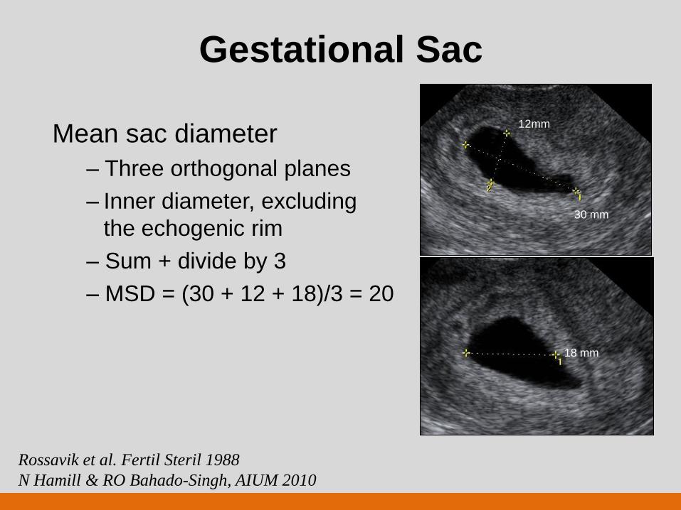
Gestational Sac
Mean sac diameter
– Three orthogonal planes
– Inner diameter, excluding
the echogenic rim
– Sum + divide by 3
– MSD = (30 + 12 + 18)/3 = 20
18 mm
12mm
30 mm
Rossavik et al. Fertil Steril 1988
N Hamill & RO Bahado-Singh, AIUM 2010
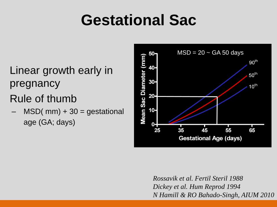
Gestational Sac
Linear growth early in
pregnancy
Rule of thumb – MSD( mm) + 30 = gestational
age (GA; days)
MSD = 20 ~ GA 50 days
Rossavik et al. Fertil Steril 1988
Dickey et al. Hum Reprod 1994
N Hamill & RO Bahado-Singh, AIUM 2010
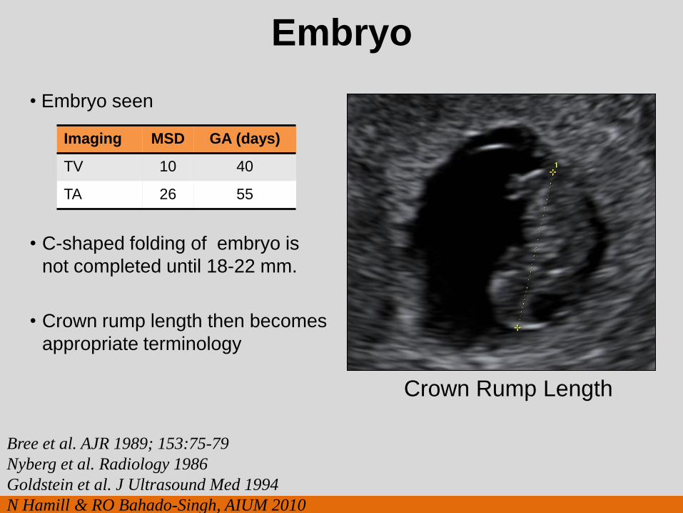
Embryo
• Embryo seen
• C-shaped folding of embryo is
not completed until 18-22 mm.
• Crown rump length then becomes
appropriate terminology
Bree et al. AJR 1989; 153:75-79
Nyberg et al. Radiology 1986
Goldstein et al. J Ultrasound Med 1994
N Hamill & RO Bahado-Singh, AIUM 2010
Imaging MSD GA (days)
TV 10 40
TA 26 55
Crown Rump Length
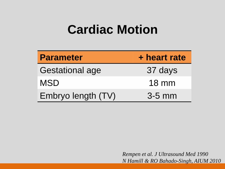
Cardiac Motion
Parameter + heart rate
Gestational age 37 days
MSD 18 mm
Embryo length (TV) 3-5 mm
Rempen et al. J Ultrasound Med 1990
N Hamill & RO Bahado-Singh, AIUM 2010
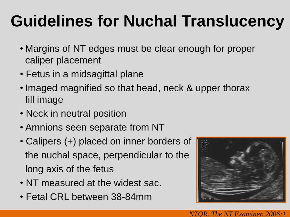
Guidelines for Nuchal Translucency
• Margins of NT edges must be clear enough for proper
caliper placement
• Fetus in a midsagittal plane
• Imaged magnified so that head, neck & upper thorax
fill image
• Neck in neutral position
• Amnions seen separate from NT
• Calipers (+) placed on inner borders of
the nuchal space, perpendicular to the
long axis of the fetus
• NT measured at the widest sac.
• Fetal CRL between 38-84mm
NTQR. The NT Examiner. 2006;1
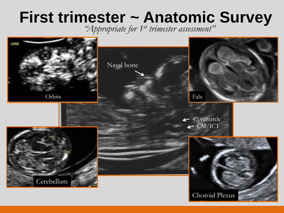
First trimester ~ Anatomic Survey “Appropriate for 1st trimester assessment”
Nasal bone
4th ventricle CM/ICT
Orbits
Cerebellum
Falx
Choroid Plexus
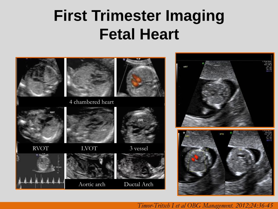
First Trimester Imaging
Fetal Heart
4 chambered heart
RVOT LVOT 3 vessel
Aortic arch Ductal Arch
Timor-Tritsch I et al OBG Management. 2012;24:36-45
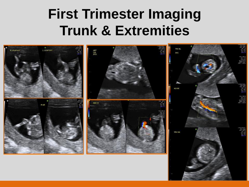
First Trimester Imaging
Trunk & Extremities
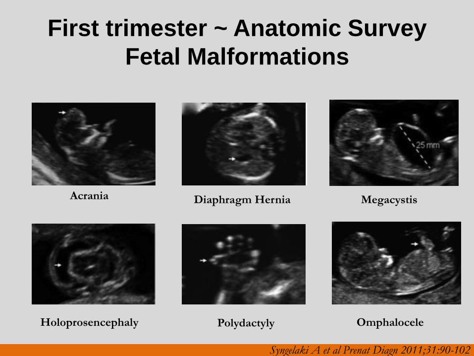
First trimester ~ Anatomic Survey
Fetal Malformations
Acrania
Holoprosencephaly
Diaphragm Hernia
Polydactyly
Megacystis
Omphalocele
Syngelaki A et al Prenat Diagn 2011;31:90-102
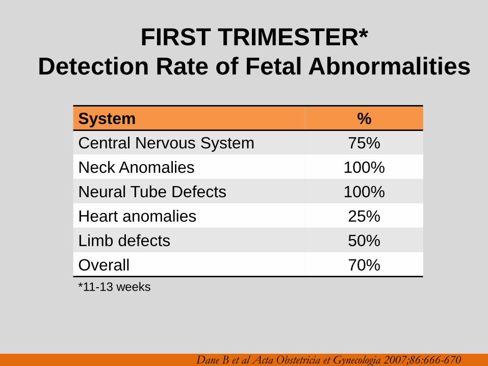
FIRST TRIMESTER*
Detection Rate of Fetal Abnormalities
System %
Central Nervous System 75%
Neck Anomalies 100%
Neural Tube Defects 100%
Heart anomalies 25%
Limb defects 50%
Overall 70%
Dane B et al Acta Obstetricia et Gynecologia 2007;86:666-670
*11-13 weeks
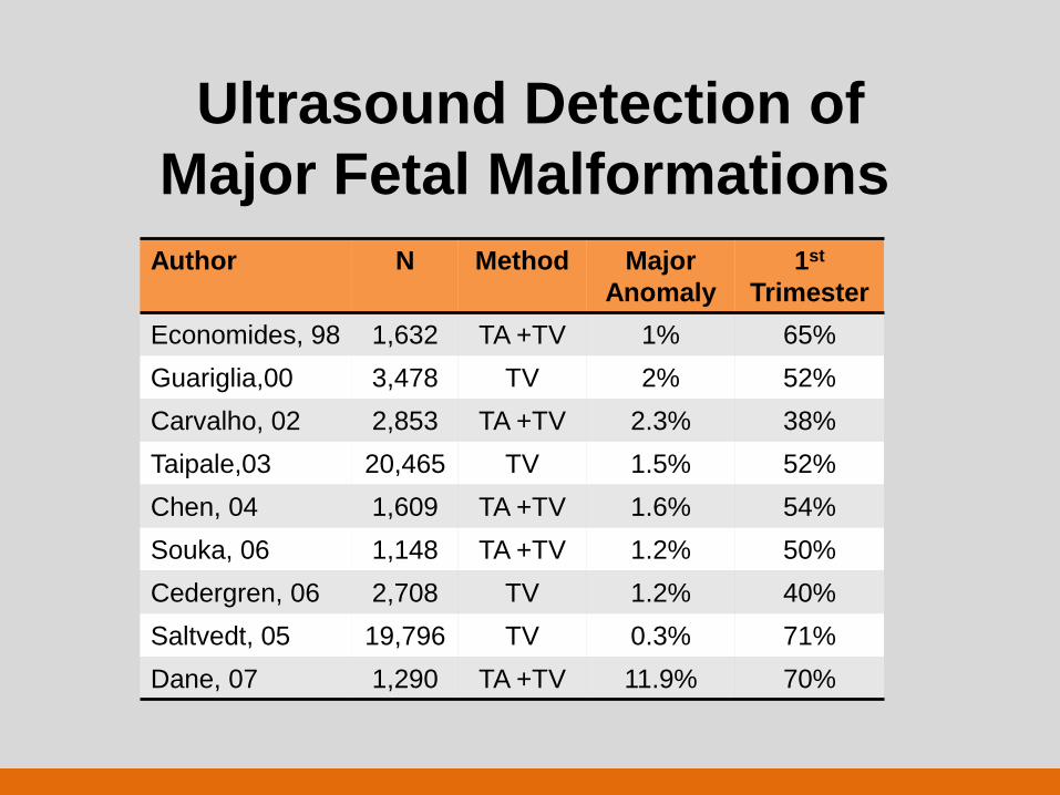
Ultrasound Detection of
Major Fetal Malformations
Author N Method Major
Anomaly
1st
Trimester
Economides, 98 1,632 TA +TV 1% 65%
Guariglia,00 3,478 TV 2% 52%
Carvalho, 02 2,853 TA +TV 2.3% 38%
Taipale,03 20,465 TV 1.5% 52%
Chen, 04 1,609 TA +TV 1.6% 54%
Souka, 06 1,148 TA +TV 1.2% 50%
Cedergren, 06 2,708 TV 1.2% 40%
Saltvedt, 05 19,796 TV 0.3% 71%
Dane, 07 1,290 TA +TV 11.9% 70%
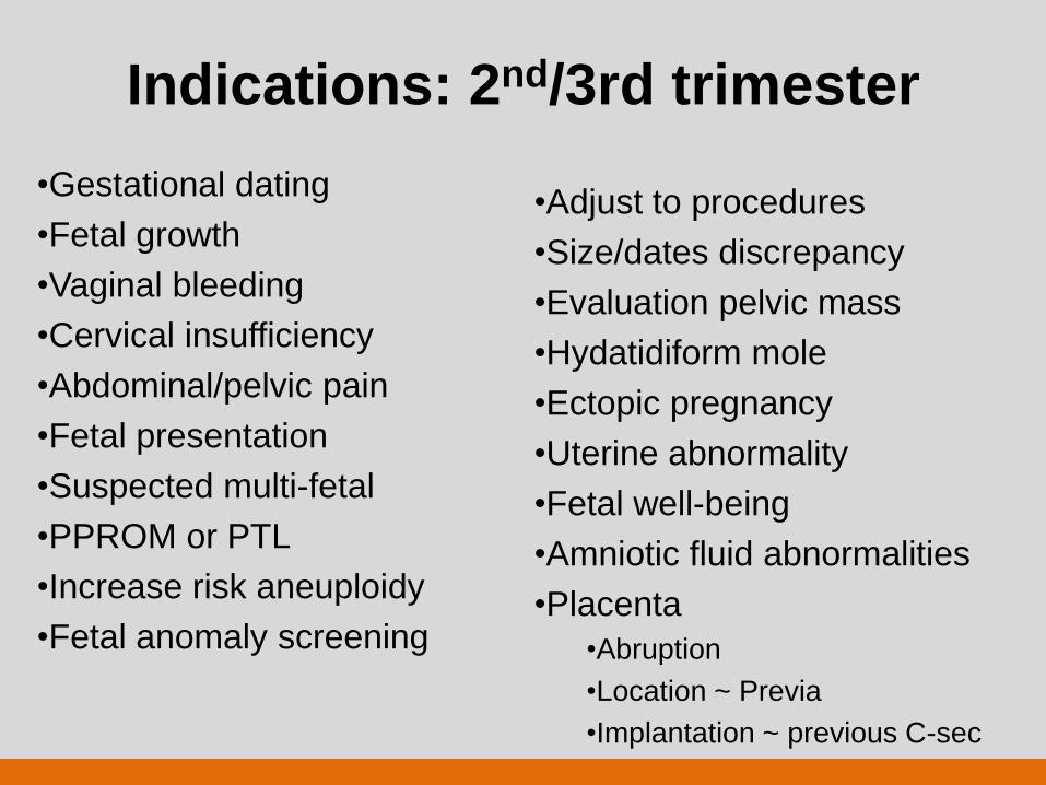
Indications: 2nd/3rd trimester
•Gestational dating
•Fetal growth
•Vaginal bleeding
•Cervical insufficiency
•Abdominal/pelvic pain
•Fetal presentation
•Suspected multi-fetal
•PPROM or PTL
•Increase risk aneuploidy
•Fetal anomaly screening
•Adjust to procedures
•Size/dates discrepancy
•Evaluation pelvic mass
•Hydatidiform mole
•Ectopic pregnancy
•Uterine abnormality
•Fetal well-being
•Amniotic fluid abnormalities
•Placenta
•Abruption
•Location ~ Previa
•Implantation ~ previous C-sec
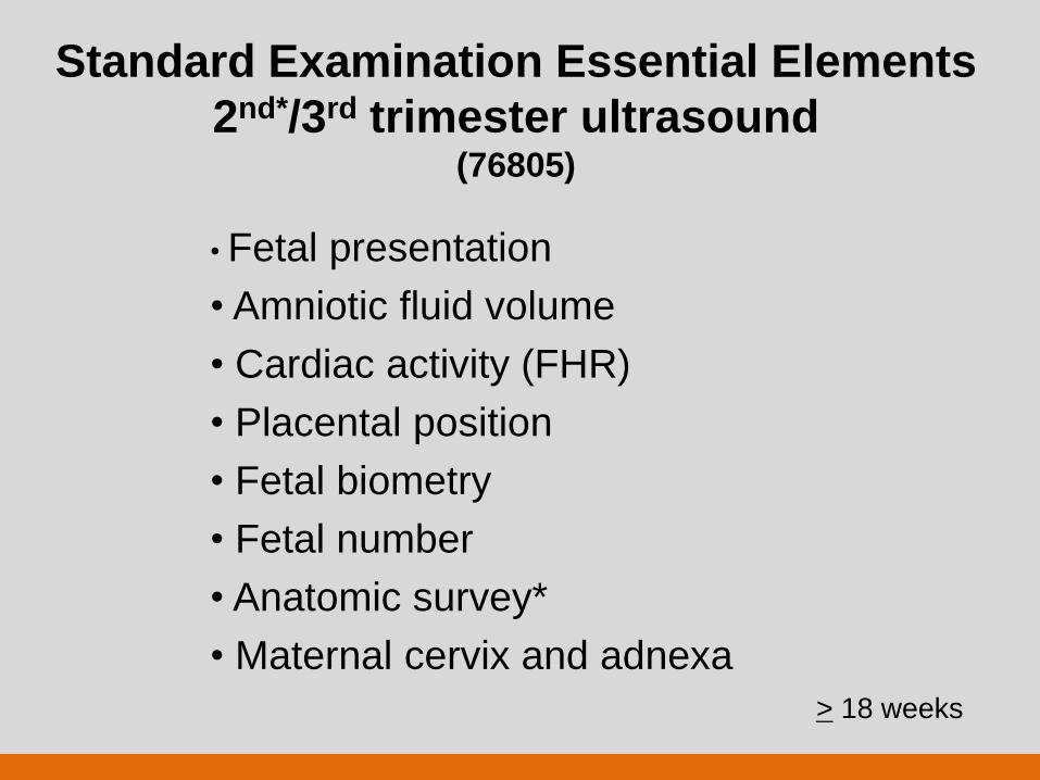
Standard Examination Essential Elements
2nd*/3rd trimester ultrasound (76805)
• Fetal presentation
• Amniotic fluid volume
• Cardiac activity (FHR)
• Placental position
• Fetal biometry
• Fetal number
• Anatomic survey*
• Maternal cervix and adnexa
> 18 weeks
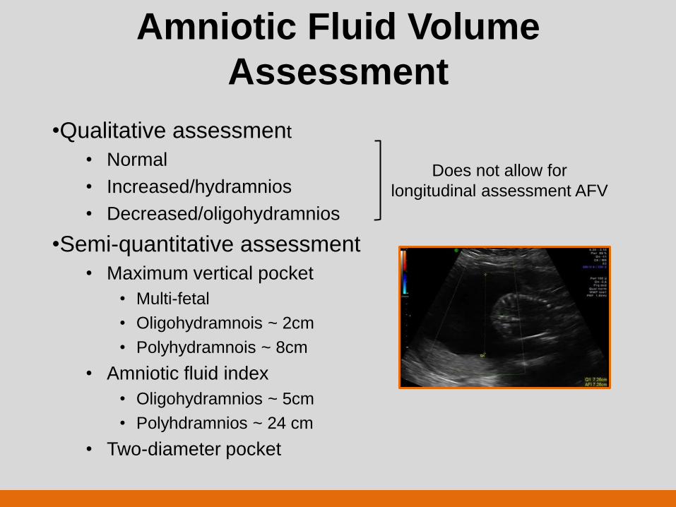
Amniotic Fluid Volume
Assessment
•Qualitative assessment
• Normal
• Increased/hydramnios
• Decreased/oligohydramnios
•Semi-quantitative assessment
• Maximum vertical pocket
• Multi-fetal
• Oligohydramnois ~ 2cm
• Polyhydramnois ~ 8cm
• Amniotic fluid index
• Oligohydramnios ~ 5cm
• Polyhdramnios ~ 24 cm
• Two-diameter pocket
Does not allow for
longitudinal assessment AFV
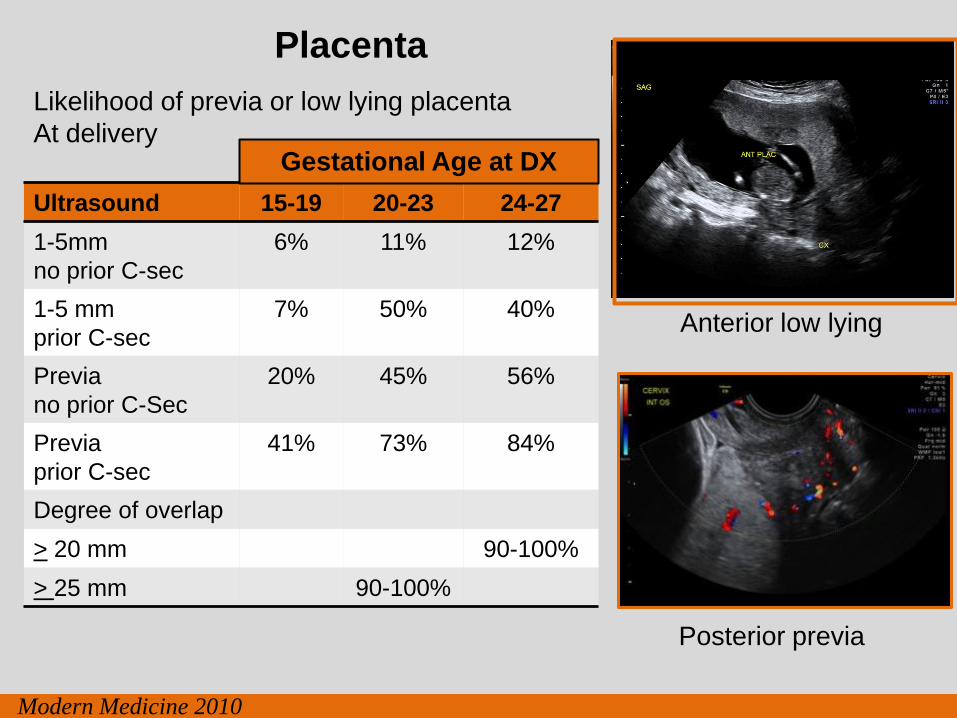
Placenta
Posterior previa
Anterior low lying
Ultrasound 15-19 20-23 24-27
1-5mm
no prior C-sec
6% 11% 12%
1-5 mm
prior C-sec
7% 50% 40%
Previa
no prior C-Sec
20% 45% 56%
Previa
prior C-sec
41% 73% 84%
Degree of overlap
> 20 mm 90-100%
> 25 mm 90-100%
Gestational Age at DX
Likelihood of previa or low lying placenta
At delivery
Modern Medicine 2010
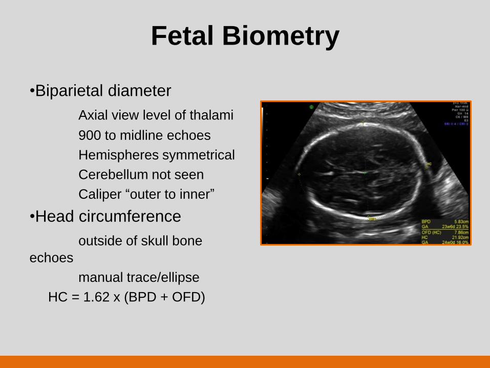
Fetal Biometry
•Biparietal diameter
Axial view level of thalami
900 to midline echoes
Hemispheres symmetrical
Cerebellum not seen
Caliper “outer to inner”
•Head circumference
outside of skull bone
echoes
manual trace/ellipse
HC = 1.62 x (BPD + OFD)
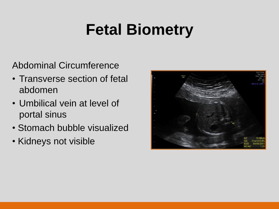
Fetal Biometry
Abdominal Circumference
• Transverse section of fetal
abdomen
• Umbilical vein at level of
portal sinus
• Stomach bubble visualized
• Kidneys not visible
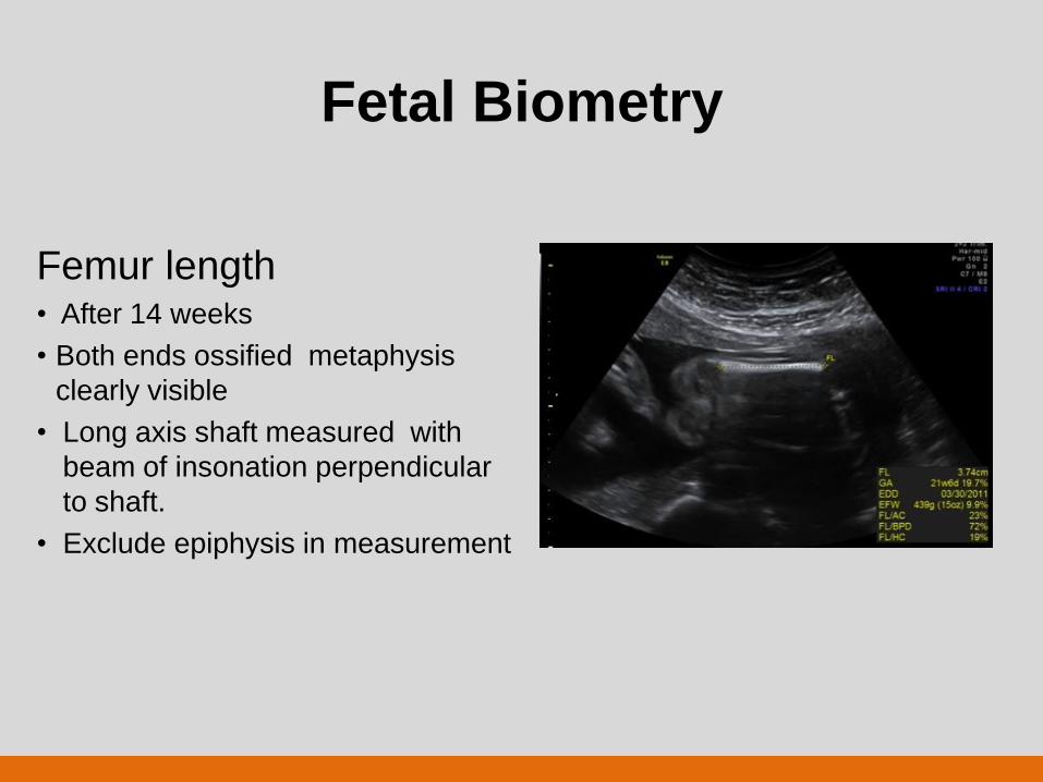
Fetal Biometry
Femur length • After 14 weeks
• Both ends ossified metaphysis
clearly visible
• Long axis shaft measured with
beam of insonation perpendicular
to shaft.
• Exclude epiphysis in measurement
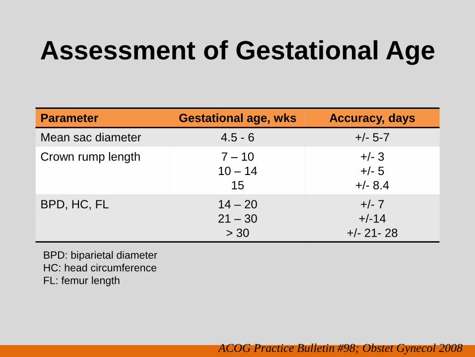
Assessment of Gestational Age
Parameter Gestational age, wks Accuracy, days
Mean sac diameter 4.5 - 6 +/- 5-7
Crown rump length
7 – 10
10 – 14
15
+/- 3
+/- 5
+/- 8.4
BPD, HC, FL 14 – 20
21 – 30
> 30
+/- 7
+/-14
+/- 21- 28
ACOG Practice Bulletin #98; Obstet Gynecol 2008
BPD: biparietal diameter
HC: head circumference
FL: femur length
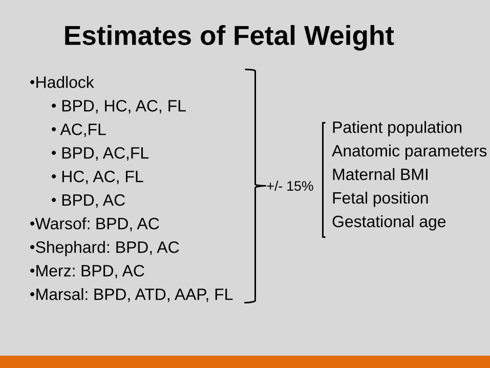
Estimates of Fetal Weight
•Hadlock
• BPD, HC, AC, FL
• AC,FL
• BPD, AC,FL
• HC, AC, FL
• BPD, AC
•Warsof: BPD, AC
•Shephard: BPD, AC
•Merz: BPD, AC
•Marsal: BPD, ATD, AAP, FL
Patient population
Anatomic parameters
Maternal BMI
Fetal position
Gestational age
+/- 15%
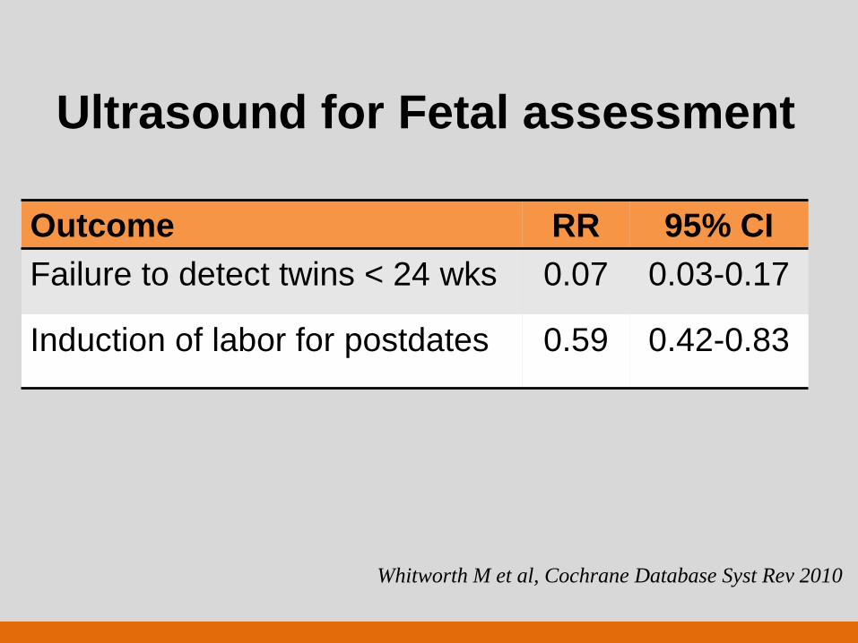
Ultrasound for Fetal assessment
Outcome RR 95% CI
Failure to detect twins < 24 wks 0.07 0.03-0.17
Induction of labor for postdates 0.59 0.42-0.83
Whitworth M et al, Cochrane Database Syst Rev 2010
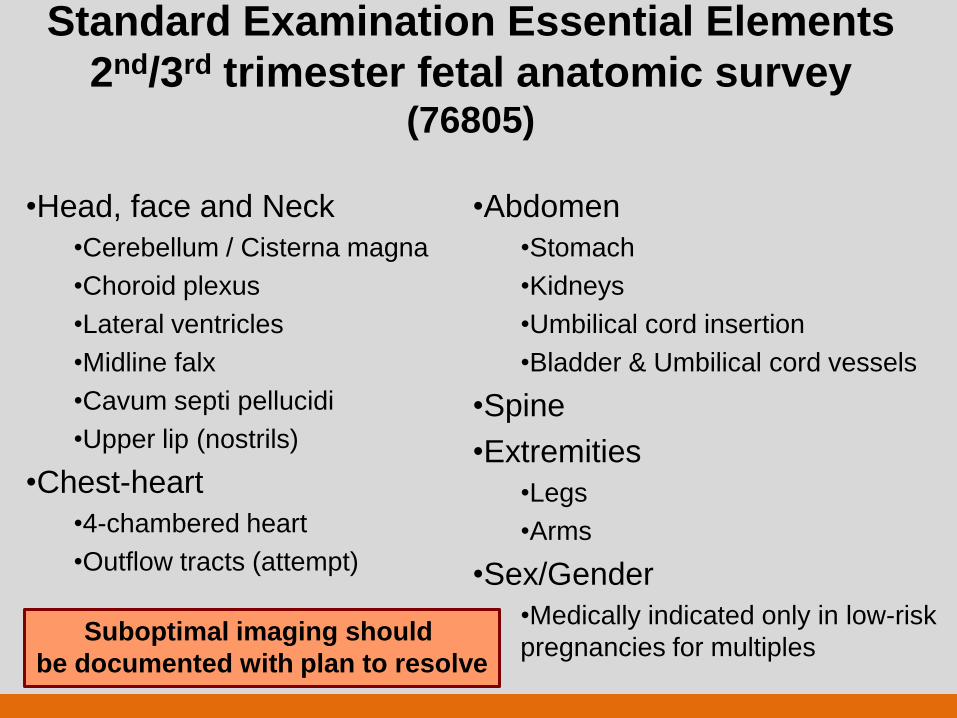
Standard Examination Essential Elements
2nd/3rd trimester fetal anatomic survey (76805)
•Head, face and Neck
•Cerebellum / Cisterna magna
•Choroid plexus
•Lateral ventricles
•Midline falx
•Cavum septi pellucidi
•Upper lip (nostrils)
•Chest-heart
•4-chambered heart
•Outflow tracts (attempt)
•Abdomen
•Stomach
•Kidneys
•Umbilical cord insertion
•Bladder & Umbilical cord vessels
•Spine
•Extremities
•Legs
•Arms
•Sex/Gender
•Medically indicated only in low-risk
pregnancies for multiples
Suboptimal imaging should
be documented with plan to resolve
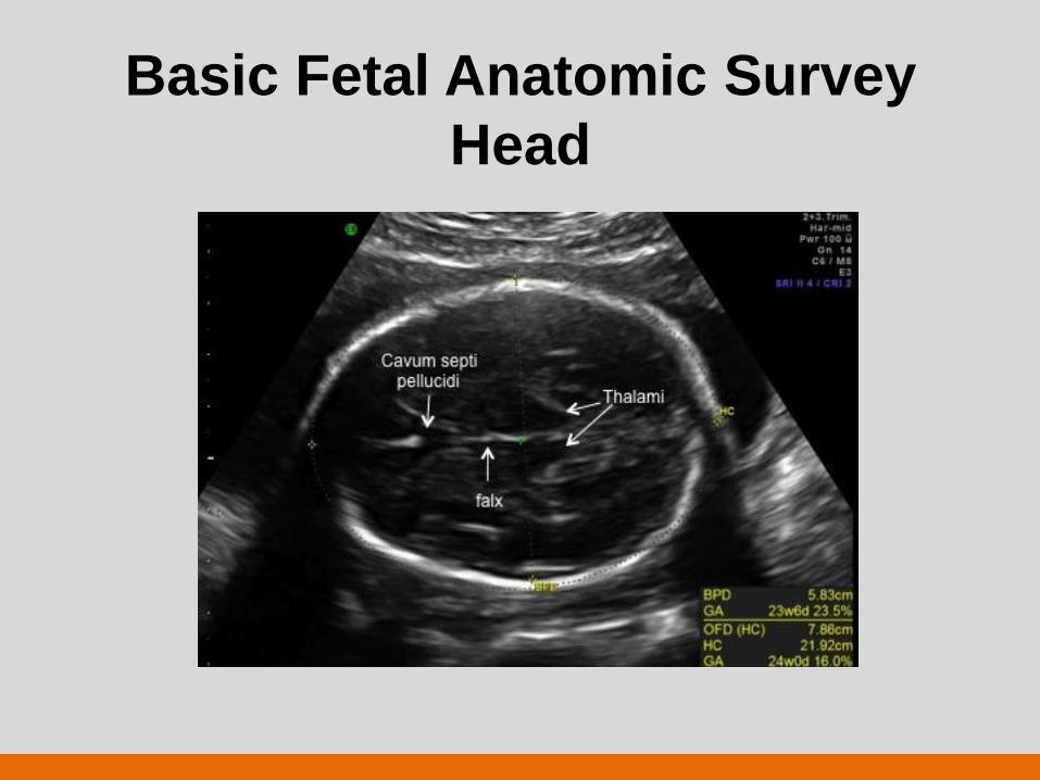
Basic Fetal Anatomic Survey
Head
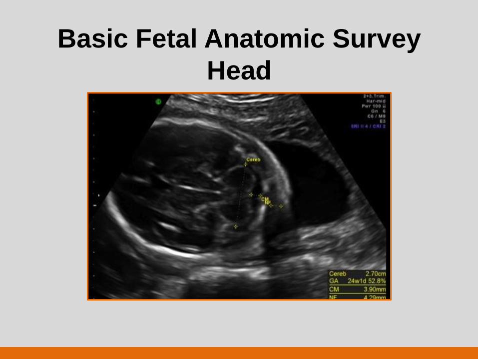
Basic Fetal Anatomic Survey
Head
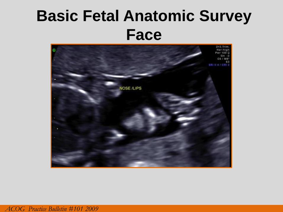
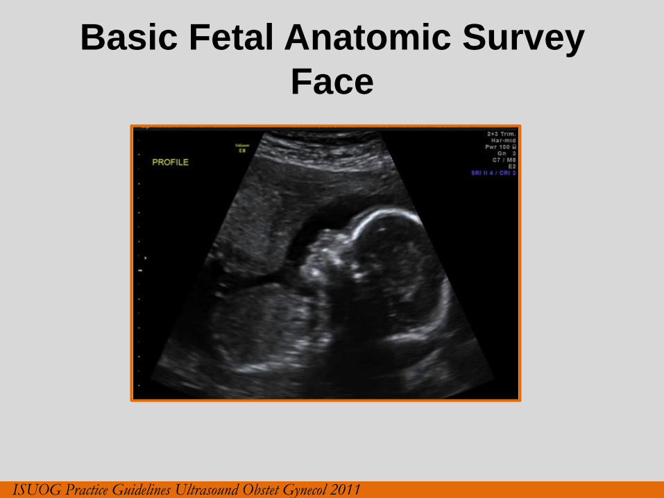
Basic Fetal Anatomic Survey
Face
ACOG Practice Bulletin #101 2009
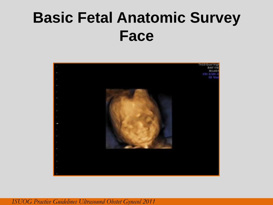
Basic Fetal Anatomic Survey
Face
ISUOG Practice Guidelines Ultrasound Obstet Gynecol 2011
Basic Fetal Anatomic Survey
Face
ISUOG Practice Guidelines Ultrasound Obstet Gynecol 2011
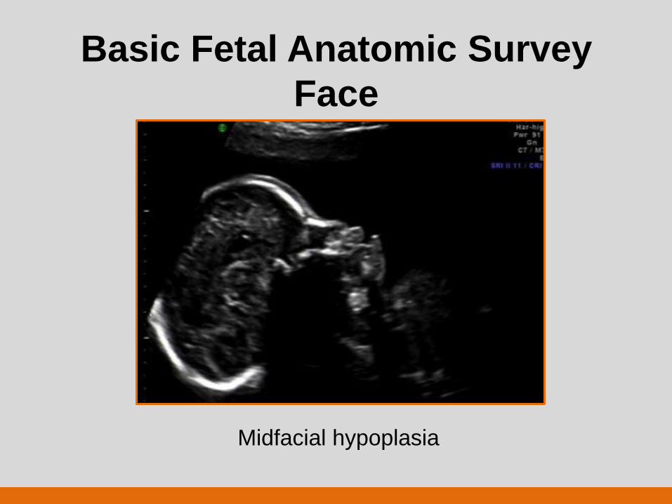
Basic Fetal Anatomic Survey
Face
Midfacial hypoplasia
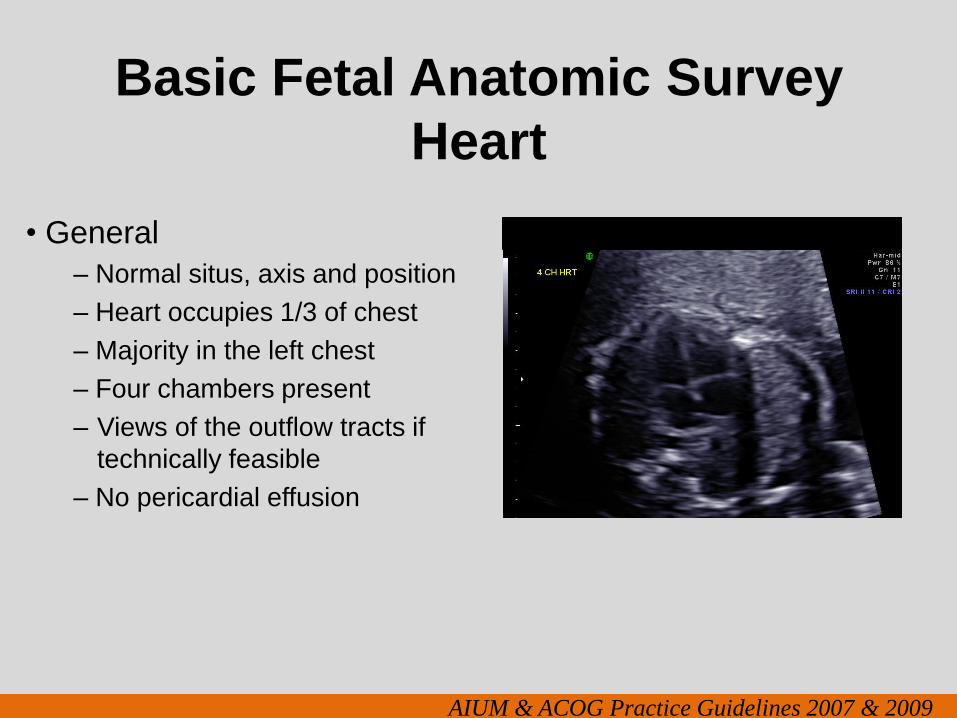
Basic Fetal Anatomic Survey
Heart
• General
– Normal situs, axis and position
– Heart occupies 1/3 of chest
– Majority in the left chest
– Four chambers present
– Views of the outflow tracts if
technically feasible
– No pericardial effusion
AIUM & ACOG Practice Guidelines 2007 & 2009
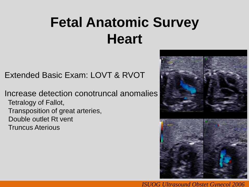
Fetal Anatomic Survey
Heart
Extended Basic Exam: LOVT & RVOT
Increase detection conotruncal anomalies Tetralogy of Fallot,
Transposition of great arteries,
Double outlet Rt vent
Truncus Aterious
ISUOG Ultrasound Obstet Gynecol 2006
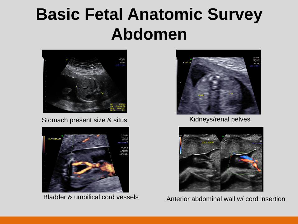
Basic Fetal Anatomic Survey
Abdomen
Stomach present size & situs Kidneys/renal pelves
Bladder & umbilical cord vessels Anterior abdominal wall w/ cord insertion
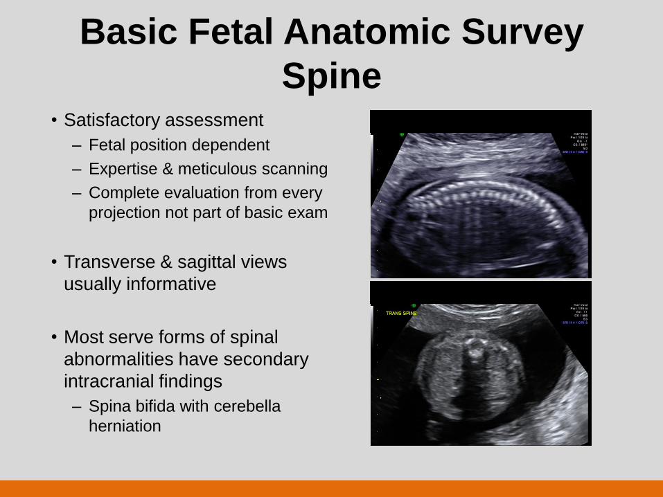
Basic Fetal Anatomic Survey
Spine • Satisfactory assessment
– Fetal position dependent
– Expertise & meticulous scanning
– Complete evaluation from every
projection not part of basic exam
• Transverse & sagittal views
usually informative
• Most serve forms of spinal
abnormalities have secondary
intracranial findings
– Spina bifida with cerebella
herniation
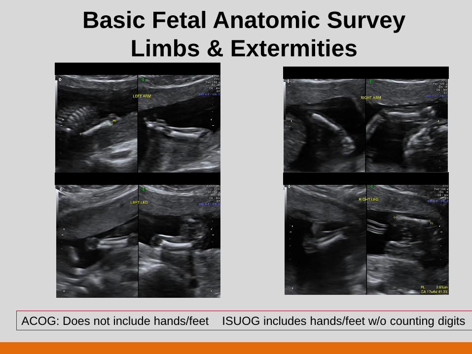
Basic Fetal Anatomic Survey
Limbs & Extermities
ACOG: Does not include hands/feet ISUOG includes hands/feet w/o counting digits
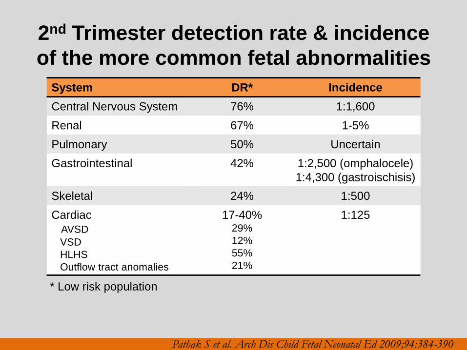
2nd Trimester detection rate & incidence
of the more common fetal abnormalities
System DR* Incidence
Central Nervous System 76% 1:1,600
Renal 67% 1-5%
Pulmonary 50% Uncertain
Gastrointestinal 42% 1:2,500 (omphalocele)
1:4,300 (gastroischisis)
Skeletal 24% 1:500
Cardiac
AVSD
VSD
HLHS
Outflow tract anomalies
17-40% 29%
12%
55%
21%
1:125
Pathak S et al. Arch Dis Child Fetal Neonatal Ed 2009;94:384-390
* Low risk population
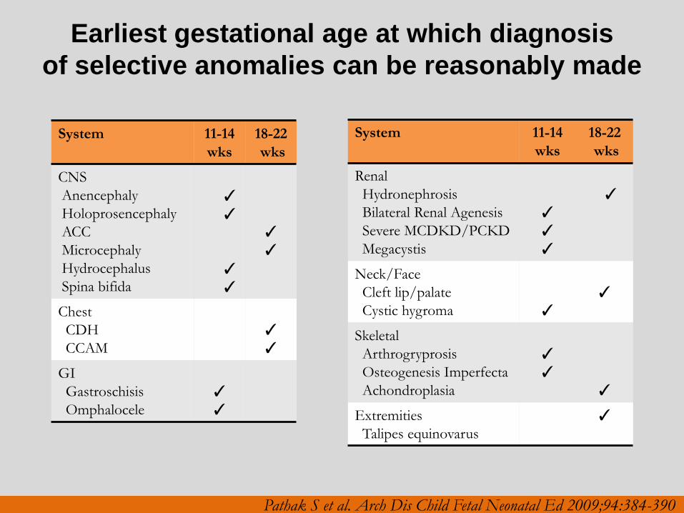
Earliest gestational age at which diagnosis
of selective anomalies can be reasonably made
System 11-14
wks
18-22
wks
CNS
Anencephaly
Holoprosencephaly
ACC
Microcephaly
Hydrocephalus
Spina bifida
✓
✓
✓
✓
✓
✓
Chest
CDH
CCAM
✓
✓
GI
Gastroschisis
Omphalocele
✓
✓
System 11-14
wks
18-22
wks
Renal
Hydronephrosis
Bilateral Renal Agenesis
Severe MCDKD/PCKD
Megacystis
✓
✓
✓
✓
Neck/Face
Cleft lip/palate
Cystic hygroma
✓
✓
Skeletal
Arthrogryprosis
Osteogenesis Imperfecta
Achondroplasia
✓
✓
✓
Extremities
Talipes equinovarus
✓
Pathak S et al. Arch Dis Child Fetal Neonatal Ed 2009;94:384-390
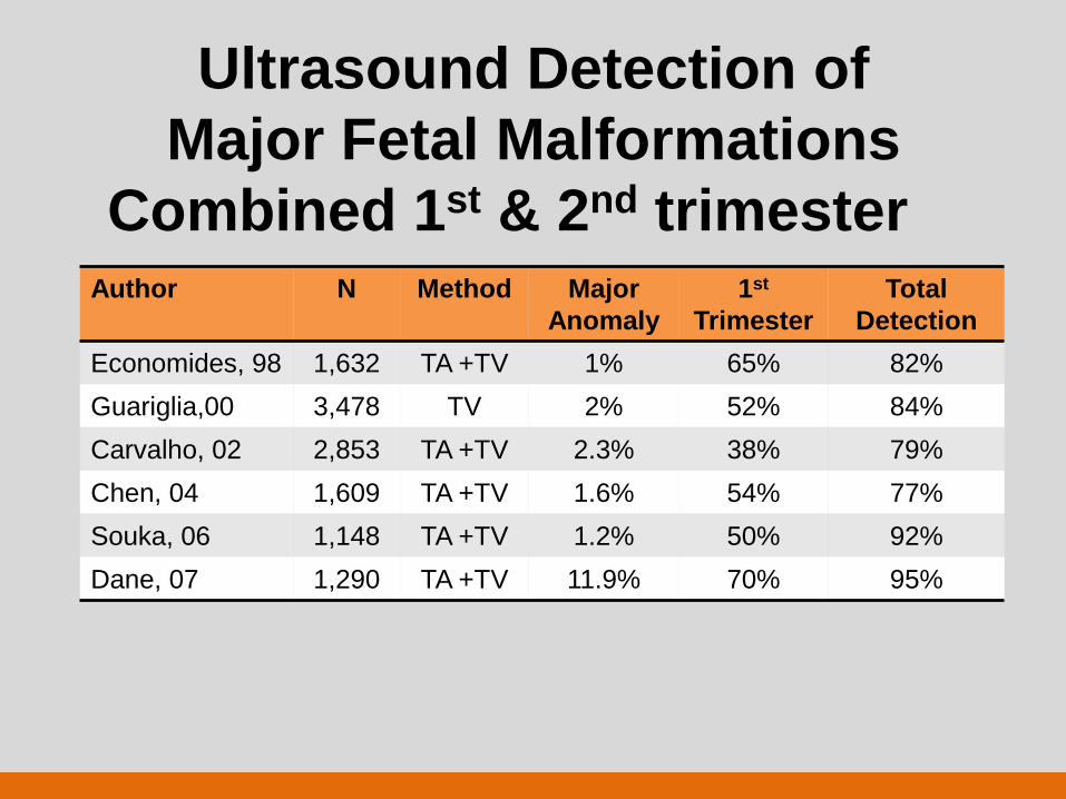
Ultrasound Detection of
Major Fetal Malformations
Combined 1st & 2nd trimester Author N Method Major
Anomaly
1st
Trimester
Total
Detection
Economides, 98 1,632 TA +TV 1% 65% 82%
Guariglia,00 3,478 TV 2% 52% 84%
Carvalho, 02 2,853 TA +TV 2.3% 38% 79%
Chen, 04 1,609 TA +TV 1.6% 54% 77%
Souka, 06 1,148 TA +TV 1.2% 50% 92%
Dane, 07 1,290 TA +TV 11.9% 70% 95%
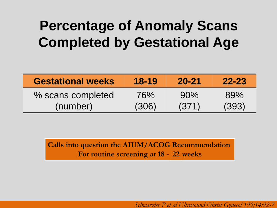
Percentage of Anomaly Scans
Completed by Gestational Age
Gestational weeks 18-19 20-21 22-23
% scans completed
(number)
76%
(306)
90%
(371)
89%
(393)
Schwarzler P et al Ultrasound Obstet Gynecol 199;14:92-7
Calls into question the AIUM/ACOG Recommendation
For routine screening at 18 - 20 weeks 22
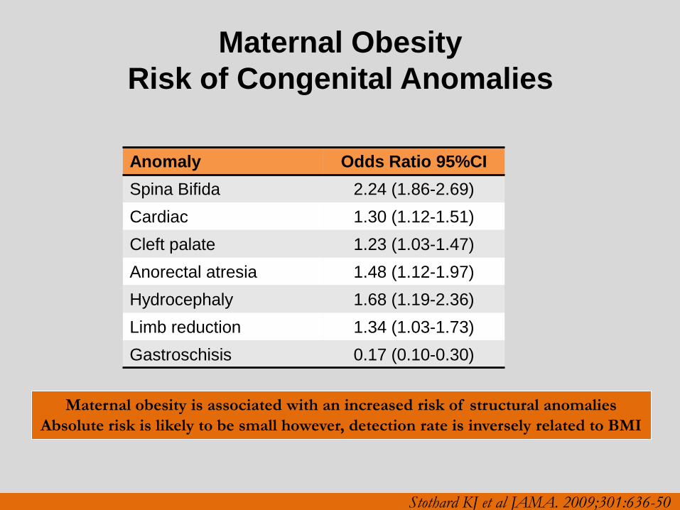
Maternal Obesity
Risk of Congenital Anomalies
Anomaly Odds Ratio 95%CI
Spina Bifida 2.24 (1.86-2.69)
Cardiac 1.30 (1.12-1.51)
Cleft palate 1.23 (1.03-1.47)
Anorectal atresia 1.48 (1.12-1.97)
Hydrocephaly 1.68 (1.19-2.36)
Limb reduction 1.34 (1.03-1.73)
Gastroschisis 0.17 (0.10-0.30)
Maternal obesity is associated with an increased risk of structural anomalies
Absolute risk is likely to be small however, detection rate is inversely related to BMI
Stothard KJ et al JAMA. 2009;301:636-50
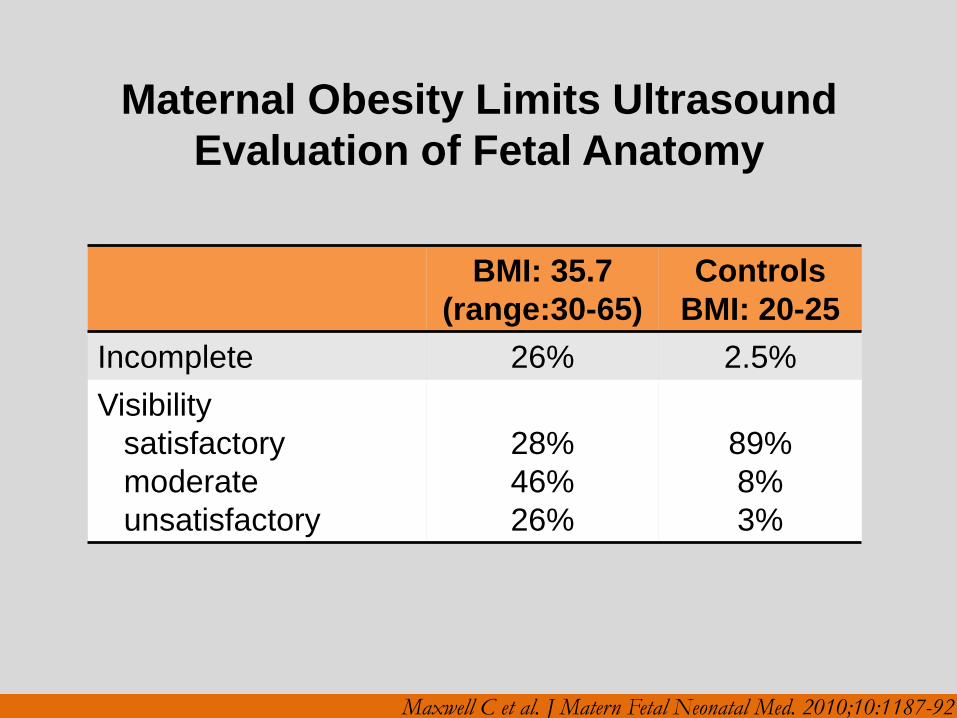
Maternal Obesity Limits Ultrasound
Evaluation of Fetal Anatomy
BMI: 35.7
(range:30-65)
Controls
BMI: 20-25
Incomplete 26% 2.5%
Visibility
satisfactory
moderate
unsatisfactory
28%
46%
26%
89%
8%
3%
Maxwell C et al. J Matern Fetal Neonatal Med. 2010;10:1187-92
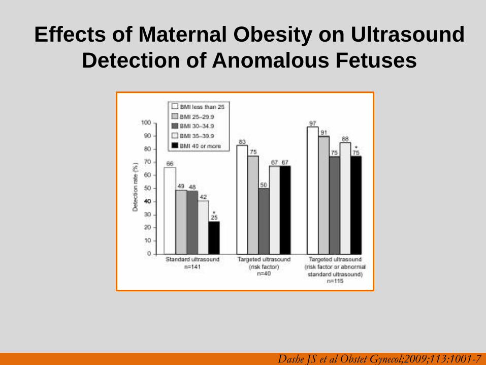
Effects of Maternal Obesity on Ultrasound
Detection of Anomalous Fetuses
Dashe JS et al Obstet Gynecol;2009;113:1001-7
Comprehensive/Level II/Targeted/Genetic
Ultrasound
• Not intended as a routine scan performed for all
pregnancies
• Indicated for suspected or increased risk of fetal or genetic
abnormalities
• Expected to be rarely performed outside of referral
practices
• Only one medically indicated 76811 per pregnancy per
practice
• Includes all of the components of 76805 with detailed fetal
anatomic survey
76811
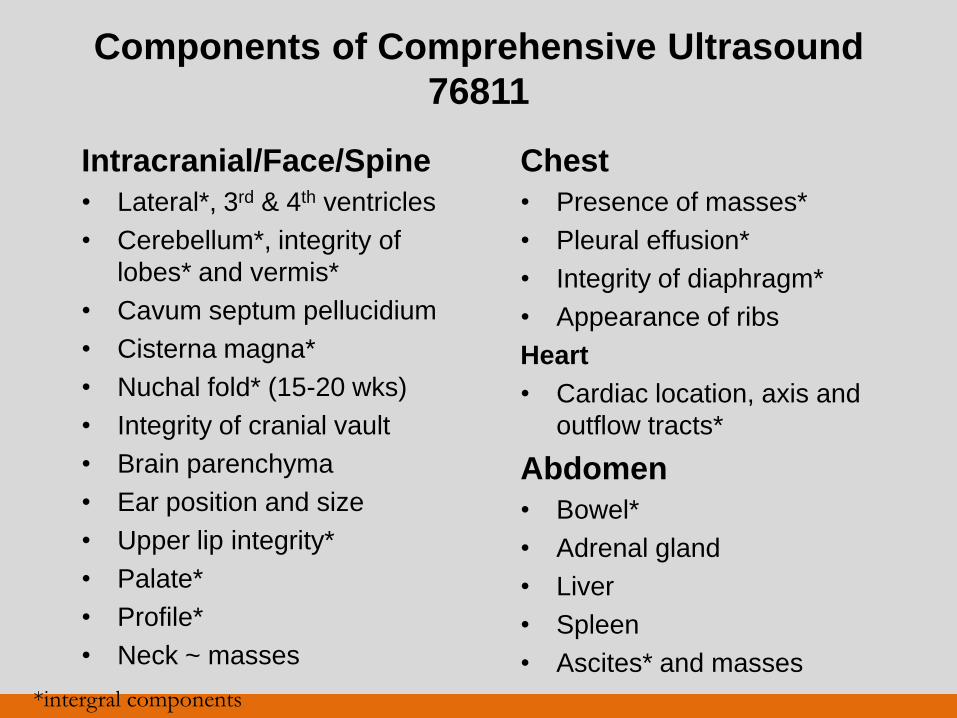
Components of Comprehensive Ultrasound
76811
Intracranial/Face/Spine
• Lateral*, 3rd & 4th ventricles
• Cerebellum*, integrity of
lobes* and vermis*
• Cavum septum pellucidium
• Cisterna magna*
• Nuchal fold* (15-20 wks)
• Integrity of cranial vault
• Brain parenchyma
• Ear position and size
• Upper lip integrity*
• Palate*
• Profile*
• Neck ~ masses
Chest
• Presence of masses*
• Pleural effusion*
• Integrity of diaphragm*
• Appearance of ribs
Heart
• Cardiac location, axis and
outflow tracts*
Abdomen
• Bowel*
• Adrenal gland
• Liver
• Spleen
• Ascites* and masses
*intergral components

Fetal Anatomic Survey
Heart
• General
– Normal situs, axis and position
– Heart occupies 1/3 of chest
– Majority in the left chest
– Four chambers present
– Views of the outflow tracts if
technically feasible
–No pericardial effusion
• Atria
– Equal in size
– Foramen ovale flap in lt atrium
– Atrial septum primum present
• Ventricles
– Equal in size
– No cardiac wall hypertrophy
– Moderator band at Rt
ventricular apex
– Ventricular septum intact
• AV Valves
– Valves open freely
– Tricuspid valves inserts
closer to the apex
ISUOG Practice Guidelines Cardiac Scan Ultrasound Obstet Gynecol 2006
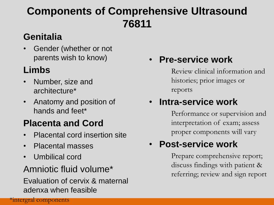
Components of Comprehensive Ultrasound
76811 Genitalia
• Gender (whether or not
parents wish to know)
Limbs
• Number, size and
architecture*
• Anatomy and position of
hands and feet*
Placenta and Cord
• Placental cord insertion site
• Placental masses
• Umbilical cord
Amniotic fluid volume*
Evaluation of cervix & maternal
adenxa when feasible
• Pre-service work
Review clinical information and
histories; prior images or
reports
• Intra-service work
Performance or supervision and
interpretation of exam; assess
proper components will vary
• Post-service work
Prepare comprehensive report;
discuss findings with patient &
referring; review and sign report
*intergral components
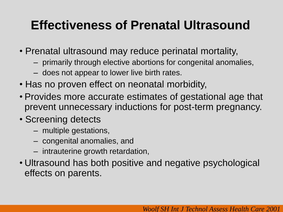
Effectiveness of Prenatal Ultrasound
• Prenatal ultrasound may reduce perinatal mortality, – primarily through elective abortions for congenital anomalies,
– does not appear to lower live birth rates.
• Has no proven effect on neonatal morbidity,
• Provides more accurate estimates of gestational age that prevent unnecessary inductions for post-term pregnancy.
• Screening detects – multiple gestations,
– congenital anomalies, and
– intrauterine growth retardation,
• Ultrasound has both positive and negative psychological effects on parents.
Woolf SH Int J Technol Assess Health Care 2001
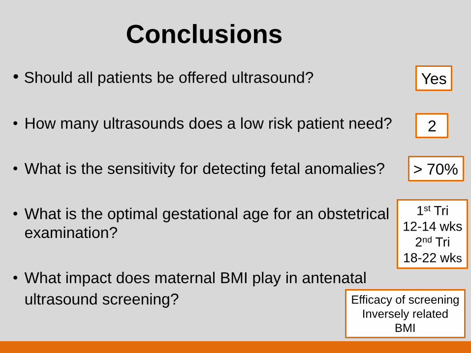
Conclusions
• Should all patients be offered ultrasound?
• How many ultrasounds does a low risk patient need?
• What is the sensitivity for detecting fetal anomalies?
• What is the optimal gestational age for an obstetrical
examination?
• What impact does maternal BMI play in antenatal
ultrasound screening?
Yes
2
1st Tri
12-14 wks
2nd Tri
18-22 wks
> 70%
Efficacy of screening
Inversely related
BMI
Thank you for
your attention