uk standards for microbiology investigations november · clostridium histolyticum and clostridium...
TRANSCRIPT
Issued by the Standards Unit, Microbiology Services PHE
Bacteriology --- Identification | ID 8 | Issue no: dp+| Issue date: dd.mm.yy <tab+enter> | Page: 1 of 22
UK Standards for Microbiology Investigations
Identification of Clostridium species
DRAFT -
THIS D
OCUMENT WAS C
ONSULTED O
N BETW
EEN 4 O
CTOBER - 1
NOVEMBER 201
3

Identification of Clostridium species
Bacteriology --- Identification | ID 8 | Issue no: dp+| Issue date: dd.mm.yy <tab+enter> | Page: 2 of 22 UK Standards for Microbiology Investigations | Issued by the Standards Unit, Public Health England
Acknowledgments
UK Standards for Microbiology Investigations (SMIs) are developed under the auspices of Public Health England (PHE) working in partnership with the National Health Service (NHS), Public Health Wales and with the professional organisations whose logos are displayed below and listed on the website http://www.hpa.org.uk/SMI/Partnerships. SMIs are developed, reviewed and revised by various working groups which are overseen by a steering committee (see http://www.hpa.org.uk/SMI/WorkingGroups).
The contributions of many individuals in clinical, specialist and reference laboratories who have provided information and comments during the development of this document are acknowledged. We are grateful to the Medical Editors for editing the medical content.
For further information please contact us at:
Standards Unit Microbiology Services Public Health England 61 Colindale Avenue London NW9 5EQ E-mail: [email protected]
Website: http://www.hpa.org.uk/SMI
UK Standards for Microbiology Investigations are produced in association with:
DRAFT -
THIS D
OCUMENT WAS C
ONSULTED O
N BETW
EEN 4 O
CTOBER - 1
NOVEMBER 201
3
Identification of Clostridium species
Bacteriology --- Identification | ID 8 | Issue no: dp+| Issue date: dd.mm.yy <tab+enter> | Page: 3 of 22 UK Standards for Microbiology Investigations | Issued by the Standards Unit, Public Health England
UK Standards for Microbiology Investigations#: Status
Users of SMIs Three groups of users have been identified for whom SMIs are especially relevant:
• SMIs are primarily intended as a general resource for practising professionals in the field operating in the field of laboratory medicine in the UK. Specialist advice should be obtained where necessary.
• SMIs provide clinicians with information about the standard of laboratory services they should expect for the investigation of infection in their patients and the documents provide information that aids the electronic ordering of appropriate tests from hospital wards.
• SMIs also provide commissioners of healthcare services with the standard of microbiology investigations they should be seeking as part of the clinical and public health care package for their population.
Background to SMIs SMIs comprise a collection of recommended algorithms and procedures covering all stages of the investigative process in microbiology from the pre-analytical (clinical syndrome) stage to the analytical (laboratory testing) and post analytical (result interpretation and reporting) stages.
Syndromic algorithms are supported by more detailed documents containing advice on the investigation of specific diseases and infections. Guidance notes cover the clinical background, differential diagnosis, and appropriate investigation of particular clinical conditions. Quality guidance notes describe essential laboratory methodologies which underpin quality, for example assay validation, quality assurance, and understanding uncertainty of measurement.
Standardisation of the diagnostic process through the application of SMIs helps to assure the equivalence of investigation strategies in different laboratories across the UK and is essential for public health interventions, surveillance, and research and development activities. SMIs align advice on testing strategies with the UK diagnostic and public health agendas.
Involvement of Professional Organisations The development of SMIs is undertaken within PHE in partnership with the NHS, Public Health Wales and with professional organisations.
The list of participating organisations may be found at http://www.hpa.org.uk/SMI/Partnerships. Inclusion of an organisation’s logo in an SMI implies support for the objectives and process of preparing SMIs. Representatives of professional organisations are members of the steering committee and working groups which develop SMIs, although the views of participants are not necessarily those of the entire organisation they represent.
SMIs are developed, reviewed and updated through a wide consultation process. The resulting documents reflect the majority view of contributors. SMIs are freely available to view at http://www.hpa.org.uk/SMI as controlled documents in Adobe PDF format.
# UK Standards for Microbiology Investigations were formerly known as National Standard Methods.
Microbiology is used as a generic term to include the two GMC-recognised specialties of Medical Microbiology (which includes Bacteriology, Mycology and Parasitology) and Medical Virology.
DRAFT -
THIS D
OCUMENT WAS C
ONSULTED O
N BETW
EEN 4 O
CTOBER - 1
NOVEMBER 201
3

Identification of Clostridium species
Bacteriology --- Identification | ID 8 | Issue no: dp+| Issue date: dd.mm.yy <tab+enter> | Page: 4 of 22 UK Standards for Microbiology Investigations | Issued by the Standards Unit, Public Health England
Quality Assurance The process for the development of SMIs is certified to ISO 9001:2008.
NHS Evidence has accredited the process used by PHE to produce SMIs. Accreditation is valid for three years from July 2011. The accreditation is applicable to all guidance produced since October 2009 using the processes described in PHE’s Standard Operating Procedure SW3026 (2009) version 6.
SMIs represent a good standard of practice to which all clinical and public health microbiology laboratories in the UK are expected to work. SMIs are well referenced and represent neither minimum standards of practice nor the highest level of complex laboratory investigation possible. In using SMIs, laboratories should take account of local requirements and undertake additional investigations where appropriate. SMIs help laboratories to meet accreditation requirements by promoting high quality practices which are auditable. SMIs also provide a reference point for method development. SMIs should be used in conjunction with other SMIs.
UK microbiology laboratories that do not use SMIs should be able to demonstrate at least equivalence in their testing methodologies.
The performance of SMIs depends on well trained staff and the quality of reagents and equipment used. Laboratories should ensure that all commercial and in-house tests have been validated and shown to be fit for purpose. Laboratories should participate in external quality assessment schemes and undertake relevant internal quality control procedures.
Whilst every care has been taken in the preparation of SMIs, PHE, its successor organisation(s) and any supporting organisation, shall, to the greatest extent possible under any applicable law, exclude liability for all losses, costs, claims, damages or expenses arising out of or connected with the use of an SMI or any information contained therein. If alterations are made to an SMI, it must be made clear where and by whom such changes have been made.
SMIs are the copyright of PHE which should be acknowledged where appropriate.
Microbial taxonomy is up to date at the time of full review.
Equality and Information Governance An Equality Impact Assessment on SMIs is available at http://www.hpa.org.uk/SMI.
PHE is a Caldicott compliant organisation. It seeks to take every possible precaution to prevent unauthorised disclosure of patient details and to ensure that patient-related records are kept under secure conditions.
Suggested citation for this document: Public Health England. (YYYY <tab+enter>). Identification of Clostridium species. UK Standards for Microbiology Investigations. ID 8 Issue xxx. http://www.hpa.org.uk/SMI/pdf.
DRAFT -
THIS D
OCUMENT WAS C
ONSULTED O
N BETW
EEN 4 O
CTOBER - 1
NOVEMBER 201
3
Identification of Clostridium species
Bacteriology --- Identification | ID 8 | Issue no: dp+| Issue date: dd.mm.yy <tab+enter> | Page: 5 of 22 UK Standards for Microbiology Investigations | Issued by the Standards Unit, Public Health England
Contents
ACKNOWLEDGMENTS ............................................................................................................. 2
UK STANDARDS FOR MICROBIOLOGY INVESTIGATIONS: STATUS .................................................. 3
AMENDMENT TABLE ............................................................................................................... 6
SCOPE OF DOCUMENT ........................................................................................................... 7
INTRODUCTION ..................................................................................................................... 7
TECHNICAL INFORMATION/LIMITATIONS ................................................................................. 10
1 SAFETY CONSIDERATIONS .......................................................................................... 11
2 TARGET ORGANISMS ................................................................................................. 11
3 IDENTIFICATION ........................................................................................................ 11
4 IDENTIFICATION OF CLOSTRIDIUM SPECIES FLOWCHART ................................................ 17
5 REPORTING .............................................................................................................. 18
6 REFERRALS ............................................................................................................... 19
REFERENCES ........................................................................................................................ 20
DRAFT -
THIS D
OCUMENT WAS C
ONSULTED O
N BETW
EEN 4 O
CTOBER - 1
NOVEMBER 201
3
Identification of Clostridium species
Bacteriology --- Identification | ID 8 | Issue no: dp+| Issue date: dd.mm.yy <tab+enter> | Page: 6 of 22 UK Standards for Microbiology Investigations | Issued by the Standards Unit, Public Health England
Amendment Table
Each SMI method has an individual record of amendments. The current amendments are listed on this page. The amendment history is available from [email protected].
New or revised documents should be controlled within the laboratory in accordance with the local quality management system.
Amendment No/Date. 4/dd.mm.yy <tab+enter>
Issue no. discarded. 3
Insert Issue no. xxx
Section(s) involved. Amendment.
Whole document.
Document presented in a new format.
Reorganisation of some text.
Edited for clarity.
Information regarding Clostridium difficile updated.
Test procedures updated.
Removal of Reference Laboratory contact details.
References. Some references updated.
Amendment No/Date. 3/14.07.08
Issue no. discarded. 2.1
Insert Issue no. 3
Section(s) involved. Amendment.
Front page. NIMAG logo added.
All. PDF links amended to read reference document title.
References. References reviewed and updated.
DRAFT -
THIS D
OCUMENT WAS C
ONSULTED O
N BETW
EEN 4 O
CTOBER - 1
NOVEMBER 201
3
Identification of Clostridium species
Bacteriology --- Identification | ID 8 | Issue no: dp+| Issue date: dd.mm.yy <tab+enter> | Page: 7 of 22 UK Standards for Microbiology Investigations | Issued by the Standards Unit, Public Health England
Scope of Document
This SMI describes the identification of Clostridium species.
There are many species of clostridia which may be found naturally in the environment and animal faeces. Only species associated with human infection will be discussed in this SMI.
This SMI should be used in conjunction with other SMIs.
Introduction
Taxonomy The genus Clostridium belongs to the family Clostridiaceae and it currently contains 203 species and 5 subspecies, with only a few species being pathogenic to humans. Of these species, 21 have been reclassified to other genera, 5 have been reclassified within the genus and 1 has been de-accessioned1.
In 1994 the heterogeneity of this species was confirmed by 16S rRNA gene sequencing2. This has been reaffirmed by the work of Yutin et al that 16S rRNA and ribosomal protein sequences are better indicators of evolutionary proximity than phenotypic traits. This genus like several others has undergone a number of revisions with the increasing availability of genomic data. An analysis of a number of proteins from a number of members of this genus suggested another revision 3. The main findings from the proposal suggested that:
- The Selenomonas-Megasphaera-Sporomusa group are still members of the genus Clostridium
- Clostridium difficile and its close relatives are placed within the family Peptostreptococcaceae. Under this proposal, the species Clostridium difficile would become Peptoclostridium difficile
- Members of the family Ruminococcaceae belong to the genus Clostridium
- It was also proposed to create six new genera to accommodate the 78 validly described species that fell outside the family Clostridiaceae. These genera are: Erysipelatoclostridium, Gottschalkia, Lachnoclostridium, Peptoclostridium, Ruminiclostridium and Tyzzerella
The type species is Clostridium butyricum.
Characteristics of Clostridium species Clostridium are phylogenetically heterogeneous and genus now includes both Gram positive and Gram negative bacteria, spore formers and non spore formers, rods and cocci and anaerobic and non-anaerobic bacteria4.
Medically significant Clostridium strains tend to be Gram positive rods (some are Gram variable), 0.3 --- 2.0 x 1.5 --- 20.0µm which are often arranged in pairs or short chains, with rounded or sometimes pointed or square ends. They are commonly pleomorphic and vary considerably in their oxygen tolerance. Some species such as Clostridium novyi type A and Clostridium haemolyticum are may require extended incubation on pre-reduced or freshly prepared plates and total handling in an anaerobic chamber. Conversely, Clostridium tertium, Clostridium histolyticum and Clostridium carnis are aerotolerant and will form colonies on blood agar plates incubated in an atmosphere of air with 5-10% added CO2
5.
Virtually all of the members of the genus, except Clostridium perfringens, are motile with peritrichous flagellae and form oval or spherical endospores that may distend the cell. They
DRAFT -
THIS D
OCUMENT WAS C
ONSULTED O
N BETW
EEN 4 O
CTOBER - 1
NOVEMBER 201
3
Identification of Clostridium species
Bacteriology --- Identification | ID 8 | Issue no: dp+| Issue date: dd.mm.yy <tab+enter> | Page: 8 of 22 UK Standards for Microbiology Investigations | Issued by the Standards Unit, Public Health England
may be saccharolytic or proteolytic and are usually catalase negative. Many species produce potent exotoxins.
Toxins of Clostridium species
Clinically significant Clostridium species produce a variety of toxins. It is the production of these toxins which leads to the distinctive clinical features of the diseases they cause, eg tetanus and botulism result from the production of neurotoxins that are amongst the most lethal substances known to man6. Clostridial toxins are biologically active proteins that are antigenic in nature and can therefore be neutralised with specific antisera.
Detection of particular toxins directly from some clinical samples may render the isolation of the organism unnecessary for primary investigation eg C. difficile (refer to B 10 - Investigation of faecal specimens for Clostridium difficile). Culture is required for typing (outbreaks and incidents) and susceptibility testing.
Clostridium perfringens is the most commonly isolated Clostridium species. Five types (A-E) may be distinguished by the combinations of major lethal toxins they produce5.
Clostridium tetani produces two exotoxins, tetanolysin and tetanospasmin --- Tetanolysin causes lysis of RBCs and serves no known benefit to C. tetani infections while tetanospasmin is a neurotoxin that causes the clinical manifestations of tetanus6.
Clostridium botulinum also produces neurotoxins (which are the most potent natural poisons known) that cause botulism, a disease characterized by a symmetrical, descending paralysis 7. There are seven toxin types (A-G), man is susceptible to type A, B, E, F and G toxins; types A, B, C and D cause intoxication in animals. Although less common, bivalent strains that express two different toxin types exist and are designated by the predominant toxin produced. Strains of C. baratii and C. butyricum have been implicated as causative agents of botulism as they also produce the types F and E respectively6,8. C. argentinense (formerly C. botulinum type G) produce botulinum neurotoxin.
Clostridium difficile is the most common toxigenic Clostridium species. They produce two potent exotoxins namely --- Toxin A (enterotoxin) and toxin B (cytotoxic activity)6.
Clostridium novyi comes in three types, labelled A, B, and a non-pathogenic type C distinguished by the range of toxins they produce. The toxins are designated by Greek letters6.
Clostridium sordellii produces three toxins in common with non-pathogenic C. bifermentans namely; a lecithinase, an oxygen-labile haemolysin and a fibrinolysin. It also has a major lethal toxin referred to as beta-toxin that distinguishes it from C. bifermentans. This beta-toxin actually contains two toxins: lethal toxin (LT) and haemorrhagic toxin (HT)6.
Other Clostridium species produce similar toxins to that produced by C. perfringens.
The medically important species are;
Clostridium perfringens
They are non-motile straight-sided gram-variable rods with blunt ends that occur singly or in pairs, 0.6 --- 2.4µm wide by 1.3 --- 19.0µm long, and rarely produce spores. They grow vigorously at temperatures between 20 and 50°C, with an optimum of 45°C for most strains. On blood agar, large discrete colonies are produced after overnight incubation. They may be flat and rough-edged or smooth and domed, and either non-haemolytic or with a narrow zone of complete haemolysis inside a larger zone of partial haemolysis. Haemolysis is more pronounced on sheep blood agar than on horse blood agar9. They are positive for lecithinase, nitrate, and fermentation of sugars but negative for lipase, indole and urease tests.
DRAFT -
THIS D
OCUMENT WAS C
ONSULTED O
N BETW
EEN 4 O
CTOBER - 1
NOVEMBER 201
3

Identification of Clostridium species
Bacteriology --- Identification | ID 8 | Issue no: dp+| Issue date: dd.mm.yy <tab+enter> | Page: 9 of 22 UK Standards for Microbiology Investigations | Issued by the Standards Unit, Public Health England
Clostridium tetani
Cells are 0.5 --- 1.7 by 2.1 --- 18.1µm and often possess terminal endospores that give a ‘‘drumstick’’ appearance. Cells in culture older than 24hr. may appear Gram negative. They are also motile by peritrichous flagella. The optimal growth temperature is 37°C and little or no growth takes place at 25 or 42°C.
Growth may appear as a film rather than discrete colonies because of swarming due to the vigorous motility after 48hr incubation. On blood agar, the colonies are flat, translucent, and grey with a matte surface, showing a zone of β- haemolysis and are 4 to 6mm in diameter. Colonies have irregular and rhizoid margins. They are negative for fermentation of sugars, lecithinase, lipase, urease, nitrate reduction tests but give variable results for indole test.
Clostridium botulinum
Cells are gram variable bacilli that show profuse sub-terminal and free spores. The proteolytic types A, B and F initially produce discrete rhizoidal colonies that spread and coalesce. Haemolysis is variable, but the odour is strong and redolent of rotten eggs due to production of H2S. They are positive for lipase but negative for indole and urease tests. They give variable test results for lecithinase reaction.
They have been isolated from clinical samples such as --- faeces, wounds, tissue, and pus as well as from foods.
Clostridium difficile
Cells are motile rods, with dimensions of 0.5 -1.9 by 3.0 --- 16.9µm, which forms oval sub-terminal spores and show optimum growth on blood agar at human body temperatures in the absence of oxygen. Colonies of C. difficile are 4 - 6mm in diameter, irregular, raised, opaque, and grey-white after 48hr incubation. They may be isolated from faecal specimens using cycloserine cefoxitin fructose agar (CCFA) or cycloserine cefoxitin egg yolk agar (CCEY). They ferment sugars but are negative for lecithinase, lipase and indole tests.
Clostridium novyi
Cells are motile, gram variable rods with occasional sub-terminal spores. Cell dimensions are 0.5 --- 1.6 by 1.6 --- 18µm except for C. novyi type B, which are larger, 1.1 --- 2.5 by 3.3 --- 22.5µm. Isolating and identifying C. novyi is difficult due to its extreme anaerobic nature. Because of their fastidious nature and difficulty in culturing, they require the presence of thiols for growth10. Growth is stimulated by fermentable carbohydrates, serum or peptic digest of blood. On blood agar after overnight incubation anaerobically, colonies appear as small, flat, rough or rhizoidal, translucent, haemolytic colonies with a spreading edge and 1 - 5mm in diameter and after incubation for 48---72hr, colonies will often coalesce to give a fine spreading growth that may cover the entire plate, often with a marked β-haemolysis so as to make the blood agar plate completely transparent. There is poor growth in nutrient broth or cooked meat broth. They ferment glucose and liquefy gelatin. Proteolytic activity is variable. They are positive for lecithinase and lipase reactions but give variable results for indole test.
C. novyi type A is usually unreactive in commercial anaerobe identification kits and commonly is not identified by this approach. C. novyi type B has different phenotypic characteristics and can be distinguished by its biochemical reactions9.
Clostridium sordellii
Colonies are large, grey-white and irregular, sometimes with a ‘‘fern-leaf’’ edge. They produce indole and lecithinase as well as ferment sugars. They are also urease positive, which differentiates them from C. bifermentans, generally regarded as a non-pathogen.
DRAFT -
THIS D
OCUMENT WAS C
ONSULTED O
N BETW
EEN 4 O
CTOBER - 1
NOVEMBER 201
3
Identification of Clostridium species
Bacteriology --- Identification | ID 8 | Issue no: dp+| Issue date: dd.mm.yy <tab+enter> | Page: 10 of 22 UK Standards for Microbiology Investigations | Issued by the Standards Unit, Public Health England
Clostridium septicum
Cells are gram variable rods with numerous sub-terminal spores. On blood agar, they grow rapidly and usually produce a thick haemolytic swarming growth. In culture, it has no characteristic odour. They are negative for lecithinase, lipase, indole and urease tests.
They are easily recognised by use of commercially available identification kits. The most common source of C. septicum isolates seen in recent years has been from blood cultures from patients with malignancies of the colon or caecum9.
Principles of Identification Clues to the identity of certain pathogenic species may be obtained by observing characteristics such as colonial appearance, Gram stain appearance and the presence or absence of β-haemolysis. Other phenotypic tests may also be applied to obtain a presumptive identification11. It is important to ensure the culture is pure, as the fine spreading growth of some Clostridium species may mask contaminating organisms.
If confirmation of Clostridium species is required, isolates should be referred to the Anaerobe Reference Unit, Public Health Wales, Cardiff.
If C. difficile confirmation is required, refer to B 10 - Investigation of faecal specimens for Clostridium difficile.
If C. botulinum is suspected, samples of patient’s serum, faeces and implicated foodstuff should be referred directly to the Foodborne Pathogens Reference Section, Colindale.
Technical Information/Limitations
Antibiotic susceptibility
Reduced susceptibility of C. difficile to metronidazole has been demonstrated12.
Sporulation
Several species of Clostridium, including C. carnis, C. histolyticum and C. tertium can grow, but not sporulate, in air13.
Gram stain
It is important to ensure the culture is pure, as the fine spreading growth of some Clostridium species may mask contaminating organisms.
There can be failure to determine the Gram reaction correctly (many anaerobes over decolourise and appear Gram negative). Eg Gram negatively staining Clostridium species, especially C. clostridioforme, can be misidentified as Bacteriodes11.
Commercial identification kit
The use of commercially available anaerobe identification kits alone may not give an accurate identification eg C. novyi type A is usually unreactive in commercial anaerobe identification kits and commonly is not identified by this approach. C. novyi type B has different phenotypic characteristics and can be distinguished by its biochemical reactions9.
DRAFT -
THIS D
OCUMENT WAS C
ONSULTED O
N BETW
EEN 4 O
CTOBER - 1
NOVEMBER 201
3
Identification of Clostridium species
Bacteriology --- Identification | ID 8 | Issue no: dp+| Issue date: dd.mm.yy <tab+enter> | Page: 11 of 22 UK Standards for Microbiology Investigations | Issued by the Standards Unit, Public Health England
1 Safety Considerations14-22
Hazard Group 2 organisms
Laboratory acquired infections have been reported23,24.
Refer to current guidance on the safe handling of all Hazard Group 2 organisms documented in this SMI.
Laboratory procedures that give rise to infectious aerosols must be conducted in a microbiological safety cabinet16.
The above guidance should be supplemented with local COSHH and risk assessments.
Compliance with postal and transport regulations is essential.
2 Target Organisms
Clostridium species reported to have caused human disease5,11
C. perfringens, C. septicum, C. novyii type A, C. sordellii, C. tetani, C. difficile, C. botulinum, C. butyricum, C. baratii, C. tertium, C. histolyticum,
“Non-pathogenic” Clostridium species commonly isolated that may have caused human infections13,25,26
C. sporogenes, C. ramosum. C. innocuum, C. paraputrificum, C. cadaveris, C. bifermentans, C. fallax, C. clostridioforme
3 Identification
3.1 Microscopic Appearance
(TP 39 - Staining Procedures)
Gram stain
Clostridium species are Gram positive rods, which may possess a single endospore. Some species may be Gram variable.
Spore stain
This is used to determine the shape and position of the spore (phase contrast microscopy is an alternative option).
C. perfringens Oval, subterminal
Note: C. perfringens spores are rarely seen in vivo or usual in vitro conditions. They do not sporulate on normal agar media. C. perfringens also have non-spore forming strains.
C. botulinum Oval, subterminal
C. difficile Oval, subterminal
C. novyi Oval, central or subterminal
C. sordellii Oval, subterminal
C. septicum Oval, subterminal
C. tetani Round, terminal (giving a drumstick appearance)
DRAFT -
THIS D
OCUMENT WAS C
ONSULTED O
N BETW
EEN 4 O
CTOBER - 1
NOVEMBER 201
3
Identification of Clostridium species
Bacteriology --- Identification | ID 8 | Issue no: dp+| Issue date: dd.mm.yy <tab+enter> | Page: 12 of 22 UK Standards for Microbiology Investigations | Issued by the Standards Unit, Public Health England
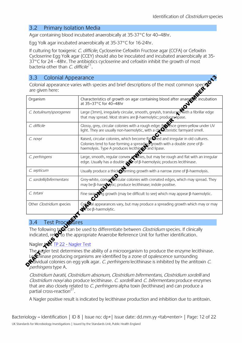
3.2 Primary Isolation Media Agar containing blood incubated anaerobically at 35-37°C for 40---48hr.
Egg Yolk agar incubated anaerobically at 35-37°C for 16-24hr.
If culturing for toxigenic C. difficile, Cycloserine Cefoxitin Fructose agar (CCFA) or Cefoxitin Cycloserine Egg Yolk agar (CCEY) should also be inoculated and incubated anaerobically at 35-37°C for 24 - 48hr. The antibiotics cycloserine and cefoxitin inhibit the growth of most bacteria other than C. difficile11.
3.3 Colonial Appearance Colonial appearance varies with species and brief descriptions of the most common species are given here:
Organism Characteristics of growth on agar containing blood after anaerobic incubation at 35---37°C for 40---48hr
C. botulinum/sporogenes Large (3mm), irregularly circular, smooth, greyish, translucent with a fibrillar edge that may spread. Most strains are β-haemolytic; produces lipase.
C. difficile Glossy, grey, circular colonies with a rough edge; fluoresce green-yellow under UV light. They are usually non-haemolytic, with a characteristic farmyard smell.
C. novyi Raised, circular colonies, which become flattened and irregular in old cultures. Colonies tend to fuse forming a spreading growth with a double zone of β-haemolysis. Type A produces lecithinase and lipase.
C. perfringens Large, smooth, regular convex colonies, but may be rough and flat with an irregular edge. Usually has a double zone of β-haemolysis; produces lecithinase.
C. septicum Usually produce a thick swarming growth with a narrow zone of β-haemolysis.
C. sordellii/bifermentans Grey-white, convex, circular colonies with crenated edges, which may spread. They may be β-haemolytic; produce lecithinase; indole positive.
C. tetani Fine swarming growth (may be difficult to see) which may appear β-haemolytic.
Other Clostridium species Colonial appearances vary, but may produce a spreading growth which may or may not be β-haemolytic.
3.4 Test Procedures The following tests can be used to differentiate between Clostridium species. If clinically indicated, refer to the appropriate Anaerobe Reference Unit for further identification.
Nagler test TP 22 - Nagler Test The nagler test determines the ability of a microorganism to produce the enzyme lecithinase. Lecithinase producing organisms are identified by a zone of opalescence surrounding individual colonies on egg yolk agar. C. perfringens lecithinase is inhibited by the antitoxin C. perfringens type A.
Clostridium baratii, Clostridium absonum, Clostridium bifermentans, Clostridium sordelli and Clostridium novyi also produce lecithinase. C. sordelli and C. bifermentans produce enzymes that are also closely related to C. perfringens alpha toxin (lecithinase) and can produce a partial cross-reaction27.
A Nagler positive result is indicated by lecithinase production and inhibition due to antitoxin.
DRAFT -
THIS D
OCUMENT WAS C
ONSULTED O
N BETW
EEN 4 O
CTOBER - 1
NOVEMBER 201
3
Identification of Clostridium species
Bacteriology --- Identification | ID 8 | Issue no: dp+| Issue date: dd.mm.yy <tab+enter> | Page: 13 of 22 UK Standards for Microbiology Investigations | Issued by the Standards Unit, Public Health England
Note: In recent years, popularity of the Nagler test has declined because the antitoxin has not been widely available. An alternative to the Nagler test used in some laboratories is called reverse CAMP test.
Reverse CAMP test Reverse CAMP test can be used for differentiation of C. perfringens from other Clostridium species. Alpha toxin producing C. perfringens and group B, β-haemolytic streptococci grow in a characteristic pattern on blood agar; however care must be taken to ensure pure cultures are used28.
Indole test TP 19 - Indole Test The indole test determines the ability of an organism to produce indole from the degradation of the amino acid tryptophan.
Anaerobes, particularly Clostridium species, form indole but can rapidly break it down as it is produced; therefore, false negative reactions may occur29.
C. novyi A strains give variable indole test results but are usually indole negative.
Lipase test
The lipase test determines the ability of microorganisms to produce the enzyme lipase that catalyses the hydrolysis of triglycerides and diglycerides to fatty acids and glycerol. This is shown by the iridescent sheen on and surrounding colonies on plate medium. This aids in differentiation of Clostridium species.
C. botulinum, C. sporogenes, C. novyi A, C. ghonii and C. cochlearium produce lipase. C. leptum give variable lipase reactions but are usually lipase negative.
Urease test TP 36 - Urease Test
The urease test is used to determine the ability of an organism to split urea, through the production of the enzyme urease.
C. sordellii are urease positive which distinguishes it from C. bifermentans, which it resembles and are urease negative.
Commercial identification kit
Laboratories must follow manufacturer’s instructions and rapid tests and kits and must be validated and be shown to be fit for purpose prior to use.
3.5 Further Identification Rapid Methods
A variety of current rapid identification and sensitivity methods have been developed for isolates from clinical samples; these include molecular techniques such as Real-time Polymerase Chain reaction (PCR), Pulsed- Field Gel Electrophoresis (PFGE), Fluorescent Amplified Fragment Length Polymorphism (AFLP),16S rDNA gene sequencing, PCR- restriction fragment length Polymorphism (PCR-RFLP), Microarray analysis, Multiple-Locus Variable-Number Tandem-Repeat Analysis (MVLA), Matrix Assisted Laser Desorption Ionisation Time-of-Flight (MALDI-TOF) and even whole-genome sequencing (WGS). All of these approaches enable subtyping of strains, but do so with different accuracy, discriminatory power, and reproducibility.
However, these methods remain accessible to reference laboratories only and are difficult to implement for routine bacterial identification in a clinical laboratory.
DRAFT -
THIS D
OCUMENT WAS C
ONSULTED O
N BETW
EEN 4 O
CTOBER - 1
NOVEMBER 201
3

Identification of Clostridium species
Bacteriology --- Identification | ID 8 | Issue no: dp+| Issue date: dd.mm.yy <tab+enter> | Page: 14 of 22 UK Standards for Microbiology Investigations | Issued by the Standards Unit, Public Health England
Matrix-Assisted Laser Desorption/Ionisation - Time of Flight (MALDI-TOF)
Matrix-assisted laser desorption ionization---time-of-flight mass spectrometry (MALDI-TOF MS), which can be used to analyse the protein composition of a bacterial cell, has emerged as a new technology for species identification. This has been shown to be a rapid and powerful tool because of its reproducibility, speed and sensitivity of analysis. The advantage of MALDI-TOF as compared with other identification methods is that the results of the analysis are available within a few hours rather than several days. The speed and the simplicity of sample preparation and result acquisition associated with minimal consumable costs make this method well suited for routine and high-throughput use30.
This has been used for the identification of Clostridium species however, an extensive database is essential to identify species reliably and available databases needs to be optimised31.
Real-time Polymerase Chain reaction (RT-PCR)
PCR is usually considered to be a good method for bacterial detection as it is simple, rapid, sensitive and specific. The basis for PCR diagnostic applications in microbiology is the detection of infectious agents and the discrimination of non-pathogenic from pathogenic strains by virtue of specific genes. However, it does have limitations. Although the 16S rRNA gene is generally targeted for the design of species-specific PCR primers for identification, designing primers is difficult when the sequences of the homologous genes have high similarity.
This has been used successfully in the identification of Clostridium species eg C. perfringens, C. botulinum, C. baratii and C. butyricum, C. novyi, C. difficile32-36.
Fluorescent Amplified Fragment Length Polymorphism (AFLP)
Fluorescent Amplified Fragment Length Polymorphism is a high-resolution whole genome methodology used as a tool for rapid and cost-effective analysis of genetic diversity within bacterial genomes. It is useful for a broad range of applications such as identification and subtyping of microorganisms from clinical samples, for identification of outbreak genotypes, for studies of micro and macro-variation, and for population genetics.
FAFLP has numerous advantages over other DNA fingerprinting techniques because it assesses the whole genome for both conserved and rapidly evolving sequences in a relatively unbiased way. The number of fragments obtained for comparative purposes between isolates is significantly greater than pulsed-field gel electrophoresis (PFGE), thus making it more discriminatory than PFGE and the FAFLP results are highly reproducible due to stringent PCR cycling parameters.
This relatively fast method can be applied to different clostridia and used for the generation of identification libraries. Libraries of AFLP profiles of well-defined Clostridium strains provide a valuable additional tool in the identification of Clostridium species.
This technique has been used to genotype C. botulinum37, C. difficile38, C. novyi39 and C. perfringens40. (It has equally being used to differentiate between C. bifermentans and C. sordellii strains (which closely resemble phenotypically) and between strains of C. perfringens41.
Pulsed Field Gel Electrophoresis (PFGE)
PFGE detects genetic variation between strains using rare-cutting restriction endonucleases, followed by separation of the resulting large genomic fragments on an agarose gel. PFGE is known to be highly discriminatory and a frequently used technique for outbreak investigations. However, the stability of PFGE may be insufficient for reliable application in long-term epidemiological studies. Due to its time-consuming nature (30hr or longer to perform) and its
DRAFT -
THIS D
OCUMENT WAS C
ONSULTED O
N BETW
EEN 4 O
CTOBER - 1
NOVEMBER 201
3
Identification of Clostridium species
Bacteriology --- Identification | ID 8 | Issue no: dp+| Issue date: dd.mm.yy <tab+enter> | Page: 15 of 22 UK Standards for Microbiology Investigations | Issued by the Standards Unit, Public Health England
requirement for special equipment and the interpretation of its results often being subjective, PFGE is not used widely outside reference laboratories42,43.
PFGE is considered a very useful tool for molecular epidemiological analysis of proteolytic C. botulinum types A and B as it enabled discrimination between them but this has not been very successful with non-proteolytic C. botulinum44. It has been used for typing C. difficile although a considerable proportion of strains are non-typable by this technique due to degradation of the DNA during the procedure; making un-interpretable gel smears45 or likely spore formation.
PFGE has also been used to establish C. perfringens as the etiological agent in food-borne outbreaks and to reveal its wide genetic diversity from different sources46.
16S rDNA gene sequencing analysis
A genotypic identification method, 16S rRNA gene sequencing is used for phylogenetic studies and has subsequently been found to be capable of re-classifying bacteria into completely new species, or even genera. It has also been used to describe new species that have never been successfully cultured.
This has been used for to differentiate between Clostridium species eg between C. novyi type A and C. botulinum type C that are closely related9.
Microarrays
DNA microarray technology can provide detailed, clinically relevant information on the isolate by detecting the presence or absence of a large number of virulence-associated genes simultaneously in a single assay; however, their clinical value has been limited by a complicated methodology that is unsuitable for routine use in diagnostic microbiology laboratories.
This technique has been used and it demonstrates the high-throughput detection and identification of pathogenic Clostridium species and it has advantages over the conventional traditional methods. This has also been particularly useful in efficiently and specifically identifying all Clostridium species present in a mixed bacterial population. The high- throughput feature of this technique is very useful in the detection and analysis of outbreak strains and for epidemiologic studies of Clostridium infections7.
Multiple-Locus Variable-Number Tandem-Repeat Analysis (MVLA)
Multiple-Locus Variable number tandem repeat Analysis (MLVA) is a method used to perform molecular typing of particular microorganisms. It utilizes the naturally occurring variation in the number of tandem repeated DNA sequences found in many different loci in the genome of a variety of organisms. The molecular typing profiles are used to study transmission routes, to assess sources of infection and also to assess the impact of human intervention such as vaccination and use of antibiotics on the composition of bacterial populations.
This has been used successfully in the typing of C. perfringens and newly emerging variants of C. difficile47,48.
Whole Genome Sequencing (WGS)
This is also known as full genome sequencing, complete genome sequencing, or entire genome sequencing. It is a laboratory process that determines the complete DNA sequence of an organism's genome at a single time. There are several high-throughput techniques that are available and used to sequence an entire genome such as Pyrosequencing, nanopore technology, IIIumina sequencing, Ion Torrent sequencing, etc. This sequencing method holds great promise for rapid, accurate, and comprehensive identification of bacterial transmission pathways in hospital and community settings, with concomitant reductions in infections, morbidity, and costs.
DRAFT -
THIS D
OCUMENT WAS C
ONSULTED O
N BETW
EEN 4 O
CTOBER - 1
NOVEMBER 201
3

Identification of Clostridium species
Bacteriology --- Identification | ID 8 | Issue no: dp+| Issue date: dd.mm.yy <tab+enter> | Page: 16 of 22 UK Standards for Microbiology Investigations | Issued by the Standards Unit, Public Health England
This has been used successfully to explore the phylogeny, horizontal gene transfer, recombination, and micro- and macroevolution of the major hospital-acquired pathogen, C. difficile49 as well as proteolytic C. botulinum and C. perfringens50,51.
3.6 Storage and Referral If further identification is required, refer to the appropriate reference laboratory user manual for details on referral.
Frozen storage (-20°C) of toxin positive faecal samples is recommended for retrospective culture should the need for further investigation arise52,53.
DRAFT -
THIS D
OCUMENT WAS C
ONSULTED O
N BETW
EEN 4 O
CTOBER - 1
NOVEMBER 201
3

Identification of Clostridium species
Bacteriology --- Identification | ID 8 | Issue no: dp+| Issue date: dd.mm.yy <tab+enter> | Page: 17 of 22 UK Standards for Microbiology Investigations | Issued by the Standards Unit, Public Health England
4 Identification of Clostridium species Flowchart
Subculture to egg yolk agar and incubate anaerobically at 35-37°C for 16-24hr
Nagler Test (TP 22)
Positive Negative
Clinical specimensPrimary isolation plate
Gram stain on pure cultureGram positive rods which may possess a single endoscope. Some species
may be gram variable.Spore stain/phase contrast microscopy
used to determine the shape and position of the spore
Blood containing agar incubated anaerobically at 35-37°C for 40-48hr
β or non haemolytic colonies which may spread all round the agar plate (swarming)
Lipase Test No ReactionIndole Test
(TP 19)
C. perfringensC. sordellii
C. novyi Type AC. novyi Type BC. botulinum*
C. baratiiC. bifermentansC. haemolyticum
C. ghonii
C. botulinum*C. difficile
C. histolyticumC. novyi Type C
C. cadaverisC. septicum
C. tetaniC. butyricum
C. tertiumC. sporogenesC. innocuum
C. novyi Type AC. sporogenesC. botulinum
C. tetani*C. ghonii
C. tetani*C. difficile
C. novyi Type BC. novyi Type CC. perfringensC. cadaveris
C. histolyticumC. baratii
C. haemolyticumC. bifermentans
C. butyricumC. sordellii
C. septicumC. tertium
C. innocuum
C. novyi Type A*C. sordelliiC. tetani*
C. bifermentans
C. perfringensC. novyi Type A
C. septicumC. sporogenesC. histolyticum
C. tetani*C. difficile
C. botulinumC. butyricum
C. baratii
C. botulinumC. sporogenes
C. novyiC. septicum
C. tetaniC. sordeilii
C. bifermentans
C. perfringensC. difficile
Positive Negative Positive NegativeSwarming
Yes No
* These give variable test results
Further identification if clinically indicated, send to the appropriate reference laboratory
Optional
Urease Test(TP 36)
C. sordellii
C. bifermentansC. novyi Type B*C. novyi Type D*C. novyi Type AC. perfringensC. septicumC. botulinum
C. tetani
Positive Negative
The flowchart is for guidance only.
DRAFT -
THIS D
OCUMENT WAS C
ONSULTED O
N BETW
EEN 4 O
CTOBER - 1
NOVEMBER 201
3
Identification of Clostridium species
Bacteriology --- Identification | ID 8 | Issue no: dp+| Issue date: dd.mm.yy <tab+enter> | Page: 18 of 22 UK Standards for Microbiology Investigations | Issued by the Standards Unit, Public Health England
5 Reporting
5.1 Presumptive Identification If appropriate growth characteristics, colonial appearances and Gram stain of the culture are demonstrated and the isolate is metronidazole susceptible.
5.2 Confirmation of Identification Following identification processes as outlined in this document and/or Reference Laboratory report.
5.3 Medical Microbiologist Inform the medical microbiologist of all positive cultures from normally sterile sites.
According to local protocols, the medical microbiologist should also be informed of a presumptive and confirmed Clostridium species. When the request card bears relevant information eg:
• Cases of trauma, penetrating injury, compound fracture or retained foreign body, or known injecting drug abuse (especially heroin)
• Septic abortion
• Suspicion of clostridial myonecrosis, (necrotising) myofasciitis, surgical wound infection (especially in cases with occlusive peripheral vascular disease and/or diabetes mellitus)
• Other serious medical conditions eg alcohol or substance abuse, immunodeficiency, cancer, or persons receiving treatment for cancer (including neutropenia and/or mucositis)
• Food poisoning (especially involving descending paralysis with cranial nerve involvement) and/or consumption of unusual or imported foods (suspicion of botulism)
• Investigation of outbreaks
• Pseudomembranous colitis or antibiotic related diarrhoea
• Suspicion of tetanus
Follow local protocols for reporting to clinician.
5.4 CCDC Refer to local Memorandum of Understanding.
5.5 Public Health England54 Refer to current guidelines on CDSC and COSURV reporting.
5.6 Infection Control Team Inform the infection control team of presumptive and confirmed isolates of C. botulinum and C. difficile according to local protocols.
DRAFT -
THIS D
OCUMENT WAS C
ONSULTED O
N BETW
EEN 4 O
CTOBER - 1
NOVEMBER 201
3
Identification of Clostridium species
Bacteriology --- Identification | ID 8 | Issue no: dp+| Issue date: dd.mm.yy <tab+enter> | Page: 19 of 22 UK Standards for Microbiology Investigations | Issued by the Standards Unit, Public Health England
6 Referrals
6.1 Reference Laboratory For information on the tests offered, turnaround times, transport procedure and the other requirements of the reference laboratory refer to the appropriate reference laboratory.
http://www.hpa.org.uk/ProductsServices/InfectiousDiseases/LaboratoriesAndReferenceFacilities
DRAFT -
THIS D
OCUMENT WAS C
ONSULTED O
N BETW
EEN 4 O
CTOBER - 1
NOVEMBER 201
3

Identification of Clostridium species
Bacteriology --- Identification | ID 8 | Issue no: dp+| Issue date: dd.mm.yy <tab+enter> | Page: 20 of 22 UK Standards for Microbiology Investigations | Issued by the Standards Unit, Public Health England
References
1. Euzeby,JP. Genus Clostridium.
2. Collins MD, Lawson PA, Willems A, Cordoba JJ, Fernandez-Garayzabal J, Garcia P, et al. The phylogeny of the genus Clostridium: proposal of five new genera and eleven new species combinations. Int J Syst Bacteriol 1994;44:812-26.
3. Yutin N, Galperin MY. A genomic update on clostridial phylogeny: Gram-negative spore formers and other misplaced clostridia. Environ Microbiol 2013.
4. Finegold SM, Song Y, Liu C. TaxonomyΓÇöGeneral Comments and Update on Taxonomy of Clostridia and Anaerobic cocci. Anaerobe 2002;8:283-5.
5. Koneman EW, Allen S D, Janda W M, Schreckenberger P C, Winn W J, editors. Color Atlas and Textbook of Diagnostic Microbiology. 5th ed. Philadelphia: Lippincott Williams and Wilkins; 1997. p. 709-84
6. Hatheway CL. Toxigenic clostridia. Clin Microbiol Rev 1990;3:66-98.
7. Janvilisri T, Scaria J, Gleed R, Fubini S, Bonkosky MM, Gr+Âhn YT, et al. Development of a microarray for identification of pathogenic Clostridium spp. Diagnostic Microbiology and Infectious Disease 2010;66:140-7.
8. Kalia VC, Mukherjee T, Bhushan A, Joshi J, Shankar P, Huma N. Analysis of the unexplored features of rrs (16S rDNA) of the Genus Clostridium. BMC Genomics 2011;12:18.
9. Brazier JS, Duerden BI, Hall V, Salmon JE, Hood J, Brett MM, et al. Isolation and identification of Clostridium spp. from infections associated with the injection of drugs: experiences of a microbiological investigation team. J Med Microbiol 2002;51:985-9.
10. Moore WB. Solidified media suitable for the cultivation of Clostridium novyi type B. J Gen Microbiol 1968;53:415-23.
11. Jousimies-Somer H, Summanen P, Citron D. Preliminary Identification Methods, Identification using pre-formed enzyme tests, Advanced Identification Methods, Laboratory tests for diagnosis of C. difficile Enteric Disease. Anaerobic Bacteriology Manual. 6th ed. Star Publishing Company; 2002. p. 54-141.
12. Baines SD, O'Connor R, Freeman J, Fawley WN, Harmanus C, Mastrantonio P, et al. Emergence of reduced susceptibility to metronidazole in Clostridium difficile. J Antimicrob Chemother 2008;62:1046-52.
13. Koneman EW, Allen S D, Janda W M, Schreckenberger P C, Winn W J, editors. Color Atlas and Textbook of Diagnostic Microbiology. 5th ed. Philadelphia: Lippincott Williams and Wilkins; 1997. p. 709-84
14. Advisory Committee on Dangerous Pathogens. The Approved List of Biological Agents. Health and Safety Executive. 2013. p. 1-32
15. Advisory Committee on Dangerous Pathogens. Infections at work: Controlling the risks. Her Majesty's Stationery Office. 2003.
16. Advisory Committee on Dangerous Pathogens. Biological agents: Managing the risks in laboratories and healthcare premises. Health and Safety Executive. 2005.
17. Health and Safety Executive. Control of Substances Hazardous to Health Regulations. The Control of Substances Hazardous to Health Regulations 2002. 5th ed. HSE Books; 2002.
18. Health and Safety Executive. Five Steps to Risk Assessment: A Step by Step Guide to a Safer and Healthier Workplace. HSE Books. 2002.
19. Health and Safety Executive. A Guide to Risk Assessment Requirements: Common Provisions in Health and Safety Law. HSE Books. 2002.
DRAFT -
THIS D
OCUMENT WAS C
ONSULTED O
N BETW
EEN 4 O
CTOBER - 1
NOVEMBER 201
3
Identification of Clostridium species
Bacteriology --- Identification | ID 8 | Issue no: dp+| Issue date: dd.mm.yy <tab+enter> | Page: 21 of 22 UK Standards for Microbiology Investigations | Issued by the Standards Unit, Public Health England
20. British Standards Institution (BSI). BS EN12469 - Biotechnology - performance criteria for microbiological safety cabinets. 2000.
21. Health Services Advisory Committee. Safe Working and the Prevention of Infection in Clinical Laboratories and Similar Facilities. HSE Books. 2003.
22. Department for transport. Transport of Infectious Substances, 2011 Revision 5. 2011.
23. Pike RM. Laboratory-associated infections: summary and analysis of 3921 cases. Health Lab Sci 1976;13:105-14.
24. Baron EJ, Miller JM. Bacterial and fungal infections among diagnostic laboratory workers: evaluating the risks. 3 2008;60:241-6.
25. Poduval RD, Mohandas R, Unnikrishnan D, Corpuz M. Clostridium cadaveris bacteremia in an immunocompetent host. Clin Infect Dis 1999;29:1354-5.
26. Gucalp R, Motyl M, Carlisle P, Dutcher J, Fuks J, Wiernik PH. Clostridium cadaveris bacteremia in the immunocompromised host. Med Pediatr Oncol 1993;21:70-2.
27. Collee JG, Fraser AG, Marmion BP, editors. Mackie & McCartney Practical Medical Microbiology. 14th ed. Edinburgh: Churchill Livingstone; 1996. p. 531
28. Buchanan AG. Clinical laboratory evaluation of a reverse CAMP test for presumptive identification of Clostridium perfringens. J Clin Microbiol 1982;16:761-2.
29. Reed RW. Nitrate, Nitrite and Indole Reactions of Gas Gangrene Anaerobes. J Bacteriol 1942;44:425-31.
30. Barbuddhe SB, Maier T, Schwarz G, Kostrzewa M, Hof H, Domann E, et al. Rapid identification and typing of listeria species by matrix-assisted laser desorption ionization-time of flight mass spectrometry. Appl Environ Microbiol 2008;74:5402-7.
31. Veloo AC, Welling GW, Degener JE. The identification of anaerobic bacteria using MALDI-TOF MS. Anaerobe 2011;17:211-2.
32. Grant KA, Kenyon S, Nwafor I, Plowman J, Ohai C, Halford-Maw R, et al. The identification and characterization of Clostridium perfringens by real-time PCR, location of enterotoxin gene, and heat resistance. Foodborne Pathog Dis 2008;5:629-39.
33. Satterfield BA, Stewart AF, Lew CS, Pickett DO, Cohen MN, Moore EA, et al. A quadruplex real-time PCR assay for rapid detection and differentiation of the Clostridium botulinum toxin genes A, B, E and F. J Med Microbiol 2010;59:55-64.
34. Fach P, Micheau P, Mazuet C, Perelle S, Popoff M. Development of real-time PCR tests for detecting botulinum neurotoxins A, B, E, F producing Clostridium botulinum, Clostridium baratii and Clostridium butyricum. J Appl Microbiol 2009;107:465-73.
35. Akbulut D, Grant KA, McLauchlin J. Development and application of Real-Time PCR assays to detect fragments of the Clostridium botulinum types A, B, and E neurotoxin genes for investigation of human foodborne and infant botulism. Foodborne Pathog Dis 2004;1:247-57.
36. Belanger SD, Boissinot M, Clairoux N, Picard FJ, Bergeron MG. Rapid detection of Clostridium difficile in feces by real-time PCR. J Clin Microbiol 2003;41:730-4.
37. Keto-Timonen R, Nevas M, Korkeala H. Efficient DNA fingerprinting of Clostridium botulinum types A, B, E, and F by amplified fragment length polymorphism analysis. Appl Environ Microbiol 2005;71:1148-54.
38. Klaassen CH, van Haren HA, Horrevorts AM. Molecular fingerprinting of Clostridium difficile isolates: pulsed-field gel electrophoresis versus amplified fragment length polymorphism. J Clin Microbiol 2002;40:101-4.
DRAFT -
THIS D
OCUMENT WAS C
ONSULTED O
N BETW
EEN 4 O
CTOBER - 1
NOVEMBER 201
3

Identification of Clostridium species
Bacteriology --- Identification | ID 8 | Issue no: dp+| Issue date: dd.mm.yy <tab+enter> | Page: 22 of 22 UK Standards for Microbiology Investigations | Issued by the Standards Unit, Public Health England
39. McLauchlin J, Salmon JE, Ahmed S, Brazier JS, Brett MM, George RC, et al. Amplified fragment length polymorphism (AFLP) analysis of Clostridium novyi, C. perfringens and Bacillus cereus isolated from injecting drug users during 2000. J Med Microbiol 2002;51:990-1000.
40. McLauchlin JC, Ripabelli G, Brett MM, Threlfall EJ. Amplified fragment length polymorphism (AFLP) analysis of Clostridium perfringens for epidemiological typing. International Journal of Food Microbiology 2000;56:21-8.
41. Keto-Timonen R, Heikinheimo A, Eerola E, Korkeala H. Identification of Clostridium species and DNA fingerprinting of Clostridium perfringens by amplified fragment length polymorphism analysis. J Clin Microbiol 2006;44:4057-65.
42. Liu D. Identification, subtyping and virulence determination of Listeria monocytogenes, an important foodborne pathogen. J Med Microbiol 2006;55:645-59.
43. Brosch R, Brett M, Catimel B, Luchansky JB, Ojeniyi B, Rocourt J. Genomic fingerprinting of 80 strains from the WHO multicenter international typing study of listeria monocytogenes via pulsed-field gel electrophoresis (PFGE). Int J Food Microbiol 1996;32:343-55.
44. Nevas M, Lindstrom M, Hielm S, Bjorkroth KJ, Peck MW, Korkeala H. Diversity of proteolytic Clostridium botulinum strains, determined by a pulsed-field gel electrophoresis approach. Appl Environ Microbiol 2005;71:1311-7.
45. Alonso R, Martin A, Pelaez T, Marin M, Rodriguez-Creixems M, Bouza E. An improved protocol for pulsed-field gel electrophoresis typing of Clostridium difficile. J Med Microbiol 2005;54:155-7.
46. Johansson A, Aspan A, Bagge E, Baverud V, Engstrom BE, Johansson KE. Genetic diversity of Clostridium perfringens type A isolates from animals, food poisoning outbreaks and sludge. BMC Microbiol 2006;6:47.
47. Youhanna S, Sawires S, Songer JG. Multiple-locus variable-number tandem repeat analysis for strain typing of Clostridium perfringens. Anaerobe 2005;11:262-72.
48. van den Berg RJ, Schaap I, Templeton KE, Klaassen CH, Kuijper EJ. Typing and subtyping of Clostridium difficile isolates by using multiple-locus variable-number tandem-repeat analysis. J Clin Microbiol 2007;45:1024-8.
49. Parkhill J, Wren BW. Bacterial epidemiology and biology--lessons from genome sequencing. Genome Biol 2011;12:230.
50. Sebaihia M, Peck MW, Minton NP, Thomson NR, Holden MT, Mitchell WJ, et al. Genome sequence of a proteolytic (Group I) Clostridium botulinum strain Hall A and comparative analysis of the clostridial genomes. Genome Res 2007;17:1082-92.
51. Sebaihia M, Peck MW, Minton NP, Thomson NR, Holden MT, Mitchell WJ, et al. Genome sequence of a proteolytic (Group I) Clostridium botulinum strain Hall A and comparative analysis of the clostridial genomes. Genome Res 2007;17:1082-92.
52. Department of Health, Health Protection Agency. Clostridium difficile Infection: How to deal with the problem. 2009.
53. Health Protection Scotland. Protocol for the Scottish surveillance programme for Clostridium difficile associated disease. 2009.
54. Health Protection Agency. Laboratory Reporting to the Health Protection Agency: Guide for Diagnostic Laboratories. 2010.
DRAFT -
THIS D
OCUMENT WAS C
ONSULTED O
N BETW
EEN 4 O
CTOBER - 1
NOVEMBER 201
3