tumor seminar - uscap · a tumor seminar • proceedings of the fifteenth annual tumor seminar...
TRANSCRIPT
A Tumor Seminar
•
Proceedings of the fifteenth annual tumor seminar presented by the San Antonio Society of Pathologists at Brooke Army Hospital, Fort Sam Houston, on October 4, 1958. Conductor was Dr. William A. Meissner, Boston.
GUEST EDITOR:
LT. COL. JAMES l . HANSEN, Fort Som Houston
Case 1 (Carot id Body Tumor)
o;q ... ,;,.-c.nxid body lumor (chemodectoma).
Clmtributor.-QJI. J. M .. Lukem~n, M.C... Four!h United StiiiC'S Army .Medic-Al tabomtory, Fon Sam Houscon.
IU.florj.-A G' ytar oJd white rcmale hnd a 5 y~;H hiscory of a ' 'cry slowly tnlu.r,;ins m:tss io che lefc submatldibul;~c arc-a with flucroationJ in size from day co cby. No cakificatioo was Stto by X·ny. The mm wa.s firm, ~lighdy tende-r, nonpulso'lciJt, and fixed tO deep nrucruro. At .t.ursecy. an exuemcly vascular man W:l$ found located O't
the b ifurcation Of dte left caroticl anery and llensely ad· ht'«1lt to both inccr-n-;~.1 and cxtcr~l caroc.id.s. The mas:s w;u
a cixd inm.cc and wu an en«rnubted ~ by 2 by 2 em .• JOmt"Wh:tt rubbery nodule with 1 brown to white co he-mc.r· tha&IC cut surfucc.
( I 1
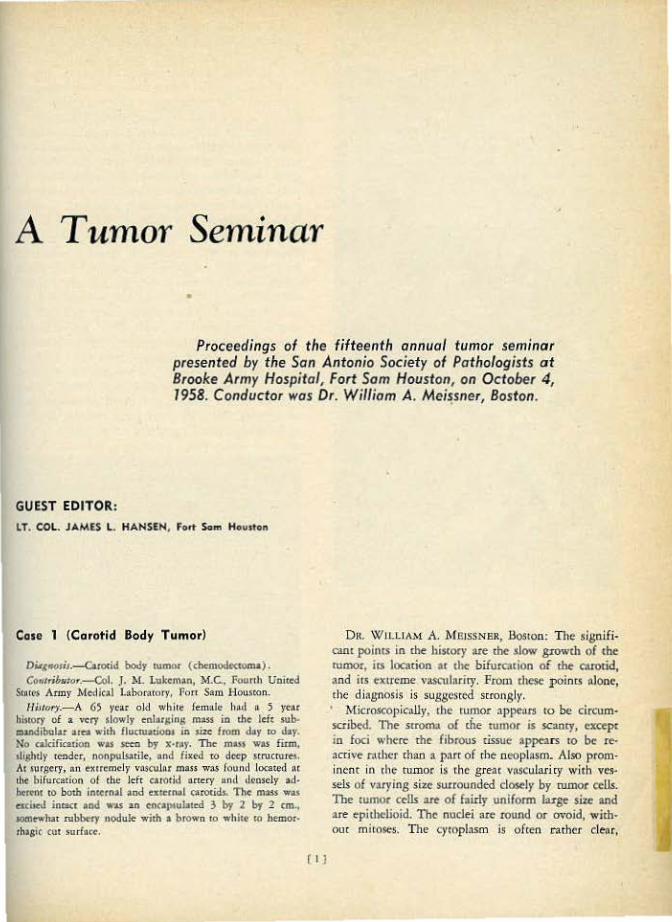
D1t W II.LIAM A. M EISSNER, J.los10n: The signifi· C'.!Jll poims in the history are the •low growth or the rutOOr, i(S location ar the- bifurcacion of the atrotid, -and irs exrrc:me vascularity. From these pointS nlone. <he di•gnosis is suggested srrongly. • 1\'[icroscopiCJ.llv, the tumor appears to be circum~ scribed. The sr.Oma of the tumor is SC3my, excepr in foci whe<t: me fibrous tissue •pp<ats ro be re· active r.uhcr than " port of the nooplasm. Also prom· incnt in the rumor is the great voscularj ry wirh ves· sel$ of varying si•c surrounded dos.!ly by rumor cells. The tumor cells :ue of fairly uoifonn luge size ond are epitbcliojd, The nuclei are round or ovoid, with· om mitoses. The cytoplasm is often cath<lr cleor,
T UMOR SEM I NA R Me i .s sner c oncinuod
\',.hkh Q.)most suggests that rhere migbi be contained m:ueri..~l such as lipid or glycogen. The c.har:.tatti.s-ric feamre of rhc rumor cells is their accumul;u:ion into clusters surn>un<ltd by • fine prolongation of the tmbo<ulor fibrous tissue. TI>e tendency of the c~lls ro form clus1ers is sccn more dearly with 'he silver stain. ·rhcsc fcarures odd up to the typical appearance of a carorid body rumor. wh ich diagtlOSis c .. u lx· mt1de \\,ith liltlc n<'ed of going into a d ifferemia.L
SubuutJt'tt Dil1gnoJc.r.-rumor of c:uotid body ( chcmodc~comn ) 33; nonchromaffin paragangliomn of carotid body. ': cumor de cuerpo carotidco1 1 ~ hemangiopedcycoJna, 1.
Fig. 1. Cost 1. Cororfd body tu-mor4 Ve-ssels o re ~ur· rounded by chJst~rs of uniform cells usually ha ving o clea r cvtoplosm.
Da. MJHSSNISR: \'(/e nre i 1'l fnir ?-gre-cmeut, l sec, exc<.:pc ftw a. slight d ifferc:ncc in c<•.rm inology.
lc JTU\)' seem rh:u rhe inclusjon of a typlcJI cntotid bod~, tumor seems hardly worthwhile in a semina.r' such Js this. This one is an e~sily d iagnosed rumor. from tt>e hisrory it might be confused with neurofibroma or oc:hcr similar slou• g.ro\ving wmors. and from the >urgical findings it might be ronfus~ with a hemongioma or even an meurysm bemuse of the tendency to such grt-at vascularity. The mici'05Copic appearance, howC!"Ver, is so rypica1 that the diagnosis is not difficulr.
Ill
The rC.lSOO for mdudiog a ttunor such as lhfs. howe:v~r. is that chese rumors are really not very common and none of us sees very many in a.ny one period of rim<'. Furchennon; the pathologiSt sometimes receives specimm.s with either 'n ioaccunue, or. m<>r< likdy, nn inadc-quare clinical hiscory. If • <umo< such •• this is jUSt scm down as ""biopsy of nuss ol neck."' it is possible char, bec.~use of their infrtquom occurrence, the patholot,.'ist may forget to include cnrotid body CUII1ors in h is diffetentiol dia,snosis. l h:lVc se~n rhis h:!ppen several times where p:nholo· gL~rs ·were trying ro make -a dj:lgnosls o£ rnera$t:ttic ~ hyrold corc inoma, for example, when the nunor was qu ire rypicnll)' one oi carotid body origin. They had been misled by inadequate clinical history. Once ch• possib ility of such a diagnosis is considered, it u•uo.l· ly becomes easy.
Tite p.1thologisr is usually nskcd by the surgeon whether or nOt the caroc.id body rumor is malignant. It is rnre <ha< 11n occasional Ct<Otid body tumor mct;tSr>srus to lymph nodes. Robson and Ellioo" hst year found 10 <such ases reponed and addtod an additional one. We ba,·c seen only one such in· sr.:tnce in our series. Romansky,u'l a few ye)rs <1go, revicwM the reported m alignant carorid body romors und found 8 cases in which visceral metnstoses had occurred. He emphaslzes, hOllt'ever, dulr rhere :I.J'C no unique ch~mges in carot id bod)' tumors rhnc distiog· u ish che mernstasizing rumors fc(>m the nonmern.sm· sizins ones. 'rhc size of the cells and the pr·escncc of uunor g iant cells seem to bave oo l):uticuJor $ig· n ificnnce wirh regard ro malignant potentinl. Dy f~r chc worsr thing nbout carotid body usmors i5 rhe fnct thar they arise in an nn>tocnic l001tion rhar m>k<s rtst.-aion dangerous and difficulr.
It is surprising how similar these tuJDON look to rhe normal coro:id body which shows the S.>mt dus· ttrs of c~Us.. !lgain accenroaced by the sih·c.r srain. The currenr view regarding carotid bodies is rh21 rhey are chemoreceptor organs and nOt part of the chrnmlffin sysrem ar all. Therefore, onmes sussesr· ing rhnr rhcy >re chromaffin tllll1ors are rnislcnding. Titc term chemodectoma is s tdtable, but ic hns bctn difficulc for us ro utiJize rhis term for clinicnl cling· nostic purposes btrnusc the renn ca.rotid body 1umor is so cnrrenched in c:he minds o f clinici:~ns ~tnd p:trh· oJogisl,s olikc. T do noc cace for the.· tcrn1 nonchrom· aHin p.tmg~mglioma pitrtly b<:cause iL is n negative dingnosis. and to me somewhac confusing. and p:wly bcC\luse carotid bod)' rumors do not aet at all din· ically like rumors o f the sofr tissues which are some· rimes coli~ by the same name.
OR. SEVIiRANCE: Col. Lukcman, do you have any £ollow·up on this case?
CoL I.UKru.tAN: Eight months af"'r operation the p:llif1lr was enrirely well and there was no evidence of !'<.'CUrrence o f rumor.
Coso 2 (Glomus Jugulore Tumor)
D11gNOs1t.-Giomw jugufarc rumor (chemodectom-a ) . CowtribNior.-Msj. R. E. Kc-Utnber~,;er, M.C., William
lbumom Army Hospiral, Et Paso. UtJ/ory.-A 2 1 y~:tr oltl colored male hnd n (rcely mnv.
able, rubbery, nl,mtendl'r m:u.s i a the postuurit:ular region antc:ior to che mi\StoiJ process of 3 momhs' duration. A ma!s lying deeP ro rhe jugu lar buJb nnd p.uliolly ovc:rlying We smeroro(.~linl aspect ol the mastoid Wlll ex<:i,cd. h was cirrunucnbc:d but apparently MC: c.n'(:apsu1otttd and COO· tained m.any large vaKuliU spaces. Two )'tan before, 11:
polyp h~ bcton rcnxwcd from che eat oo chc $1Jllt side •bicb •·iiJ followtd by .kaf.ness in dur c.ar.
OA. MJ!ISSNI!R: \VIe lu~e some addiciooal informuion on chis ocse from .Maj. Kelknberger. Apparmcly the potienr had rwo operations in 1955. at lease ooe of which w.s for removal of • polyp (rom the car. Major l(ellenberger has lent me a slide of chis whid1 is composed o f granulation tissue covc,·ed on unc surface by srrarified squamous epithelium which is ulcerared. ·n1e g.ranu1atioo tiS$ue uppc:~rs ncdve, but [ found no evidence o f rumor in chis specimen. No risoue is •vailable from the S«ond opemtion, >nd it is not even known what was removed.
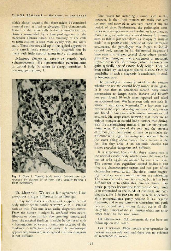
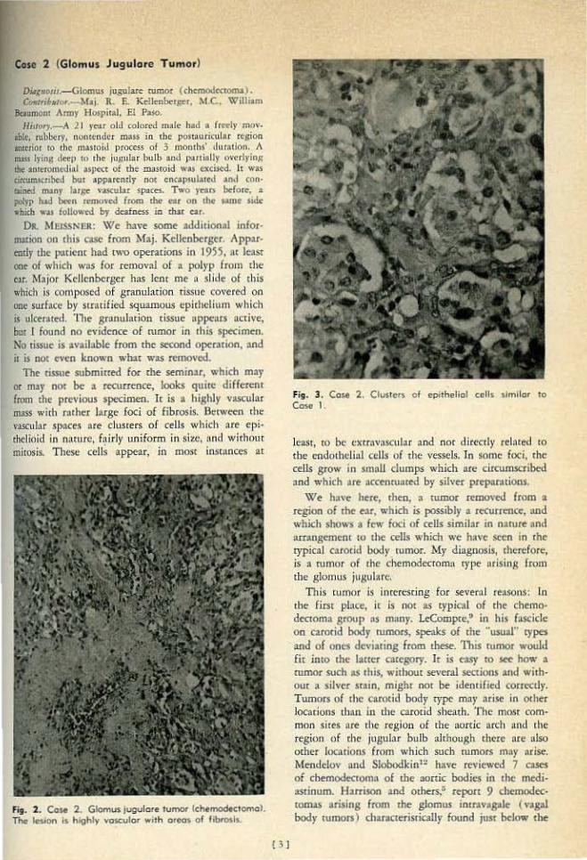
The cissue .submitred for the seminar, \Vhich m:.y or may not be a recurrence, looks quite differenr from the previous specimen. lc is a highly vascubr mass with rnther large foci of fibrosis. Bctwc."t"n che 'l:lSCulnr spaces are clusrers of celJs which nre cpi· chc.·lioid in n"turc, f,,tirly uniform in size, nnd whhom mitosi:t TheS<: cells appear, in most inscnnccs ac
fig. 2. Case 2. Glomus jugulore tumor (ch«modtctomol. The le-Sion Is hJghty vascular w•th oreo~ of ftbmsls.
O J
Fig. 3 . Cost 2. Ctusters cf cpith e.liof cells 1lmilor to Coso 1.
least, to be.: c.:xcrnvnscular and ooc directly related tO
the endothelial cells of the vessels. In some foei. rhc cells grow in small clumps which are drcunucribecl and which iUt nccennmted b)· sj!ver pzcpar.uions.
We ha\·e htre;, fhtn, a rumor removed from a <egioo of the e:~r, which is possibly a recurrence, and which shows a few foci of cells similar in nature and arrangement to the cells which we have seen in the typical carotid body rumor. My d iagnosis, therefore, is :\ rumor of the chemodecromn type nrisin,g frqm th e glomus jugulare.
This rumor i.s inrtre.sting for seveml rt:lSOnS; fn the first pl:tcc, it is not as typ ical of the chemodectoma gro<1p as many. L<Compre,• in h is f~sdcle on carotid body rumors, speaks of rbe "U$ual" types and of OOe$ deviuing from these. This n1mor would fir into the Iauer ouegory. It is easy ro ..., how a rumor .such as this, without .se'•eraJ sections and with· our a siJvcr srain, might not be ideori{ied correcd)'· Twnors of the c•.rotid body type may orise in 01her locatioos tlhut in [he carotid sheath. The mc\~r com~
moo sites are the region of the aortic arch and the region o f the jugubr bulb alchough d1C1C ore ulso ocher locarions from which such rumors mn)' arise. Menddov and Slobodkin'2 have revicw<-d 7 c>Ses of chemod<ecoma of the aortic bodies in the medilUtinurn. Harrison and other$.• ccpon 9 cbcmodec<omas arising from the glomus intr.tvagolt ( va8Jll body t\UilOI$ ) ch>racrerisrically found juot below the
TUM OR SfM·IN AR - Me j s:$ n er- co nr inue d
jugular foramen and in_ continuiry vdtl1 the vagus m.·r.ve.
Tumors arising from the glomus . jugulore often ptesenc ia.ithJly as a _polyp o f cbe ear .and ir is im· por rnnr, rbecdorc, that all such lesions be examined microscopic-all)'· Because of r11eir grear vascub ricy rhcrc is, o f course~ a- rtndCnC)' to ulcerate and bl¢ed aod it is nor $Urprising rhar these rumors may be covcrt>d by a layer of gr:\Oulation rissue and rhac their CJ:ae n.arure may .not be disclosed by ~ su_per · ficiiu biopsy. I think that is prob"bly what h.'\J>pened in this case.
From the morphologic poioc of view,. excepr for their superficial lol-adon and a n:sulting cendeoc>' tO become uJcer.{lted ot infecred, d1cse ntmors are simi!>< ro the ones arising from the carotid body. H owever, because of the locacion, they seem to run a more maJignnnt, or at least a mo~:e aggressiv~, course rhan ·d iose atising from rhc carotid. body, with direct extension and invasion imo u.djacenr bone and even into (the base o{ the brain. As a matter of face~ tumors of.·rb~ glomus jugul•re have rnrher a ]Xlor prognosis, W'inship"' rcp0rrs 35 cases, in wftich 12 died either directly or indirectly of their d isease. As with the CMOtid body rumors1 cases with mcmsrase:s have OC· cu.rred, one of which has. been reporre~:t by L1rres,s ro. ~~jsct·ra.
It is incer:esring ro ooce char glomus jugulare co· rnors show strong familial ceodencies. W inship emphasizes also rha:r frequent!}' there -are ·cono.urcn[ rLunors of tbe C;.'tiOtid body io the affecred padem or i l'l hi.s farnily. •
SttbmitJ.ed- D itr.g11t.>Je.f.- glorntiS jugularc rumor (chemodectoma) , 18; nonchrocnaffin paraganglioma of glomus jug(dare, 5; recurrent nonchrornaff in para· ganglioma of g lomus cymp:micum, 4; c:.uotid·bodylike tumor, 3; he-maogio·endocbeHoma, 7; bcmangioma ( rypc?, 1; sderosiog, 1, ca•ernous, l) , 3.
DR. MEISSI'>ER: Chemodectoma stems to be a bew:r name for rumors of 'this t)'pe, and 1 chink "glomus jub'Uiare cumor" with cbemodecroma in parentheses would be rhe \WY we would diogoose jc in .our labororoq•. l11e use of _new terms .somc:drocs can cause confusion ro tbe detriment of rhe 1)ntient
I sbould like co mendon one ime..:esring case char we -have~seen recendy. This part icular tumor projected into the larynx, and was thought by tl1e oto1aryngol· ogist ro be a laryngocde. He was surprised tO find that he could dissect it free submucosally, and he rook our qu ire a sizable vascular mass without -breaking through tht1 mucqsa. \VIe think ir l$ 9o ex-.. :tmp1e Of • chemodecroma, probably arising from l>o<f of the vagal bodies, and projecting inro the larynx. One m~t .keep in mind rhe fact that tbcse rum'Ors can occur in unusual loal:tions.
! 4 1
DR. SEVERANCE: Dr. :Kdltobergec, do you ~ave any follow-up?
.MAJ. KELLBNBJmGER: Six mombs .after removal th is man had deaf ness, and thot was all.
DR. MEISSNER: The deafness, incidenrally, atcord· lng ro an_ ear. no~e:, and throat specialist w:icb whom I tlllkcd about clliS probJem, is more likely due tO rbe evulsion of the srnpes in removing the ]Xllyp from the ear, d1an ir is ro che rumor jcsel.f. unless
--rhc wmor is very extensive.
Case 3 (Pheoch romoey.toma)
Diagnosis.:.....Ph("QChrom(l(:)' tOma wirh foci of sanslione\1· roma,.
CoJJt.ri!mJor.- Dr. Meissner. H;Jf.Q~y.-A SO }'e.tr old laborer was admicred <ompl:dn·
ing of p rogressive abdominal swcUing :lnd weakness for 3 y~ars and wcighc loSi fo r 6 months. There were -oo other S)'Jllptoms. Pby~icaJ ex-amin:tdon was oeg:1dvc; except lor :1.
nomcnder qstic abdomin-nl mass exrending_ from the pubis ro the diaphragJn. BJood pressure was LG0./92. A 3600 Gm. mulricysrk rcrrQpcriconeal m~s· filled '1\'irh red, cloudr flu id and blood d ots ·was surgicu11)· t:.'tcised.
DR. MEISSNER: We have no x-ray on this case which Shows a large abdominal nunor with distortion of the ureters, particularly the left. Thi~ was a diffkah rumor to d iagnose clinically
and its exact n<tture was not e v-en ~uspecred _priqr ro pathologic examination. Tht mass weighed '),600 Gm. It was wei! cncnpsulnred. A flnriened 'adrenal gland Ia)' at one pole. The bulk of the mass was compooe<l of mulriloclllared cysrs filler! wirh b lond d ot or red, cloudy fluid. 'fhe walls of rhc cyStic spaces were smoorh1 for C:he mosr pan 1 bur here aod chere were rollgbened by adhereor blood clot .
_Mitr:oscop·icall}~. the compressed adrena l is adjacem ro rhc (;llpsule aod '" a few points there seems to be a direct continuity be tween rhe adrenal and che r1li1SS
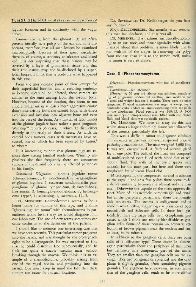
irself. Orherw.ise rhe capsule of tile l)lliSS appears dis· tinct. ~fuch of jc is nccwric, -hemor:rhagic. and t:ysik, but ar the periphery 1 parriculal'1}•, tlu~re are ide~uiftable srnlcture.S: Ute: stroma is. collagenous and .in some places fibril lar, suggesting the p resence of borh neu•·ofibrils and Schwaoa cells. In tl1ese foci, par· ticularly, tbere are large cells with cytoplasmic p ro· ce.~ses whkh l ch in..~ arc I e:.tdily ldencifiable as gan· glion cells. Some of these conrain an eccemric col· Jection of brown p igment near the nucleus and one, at lc~sc, is in mitosis.
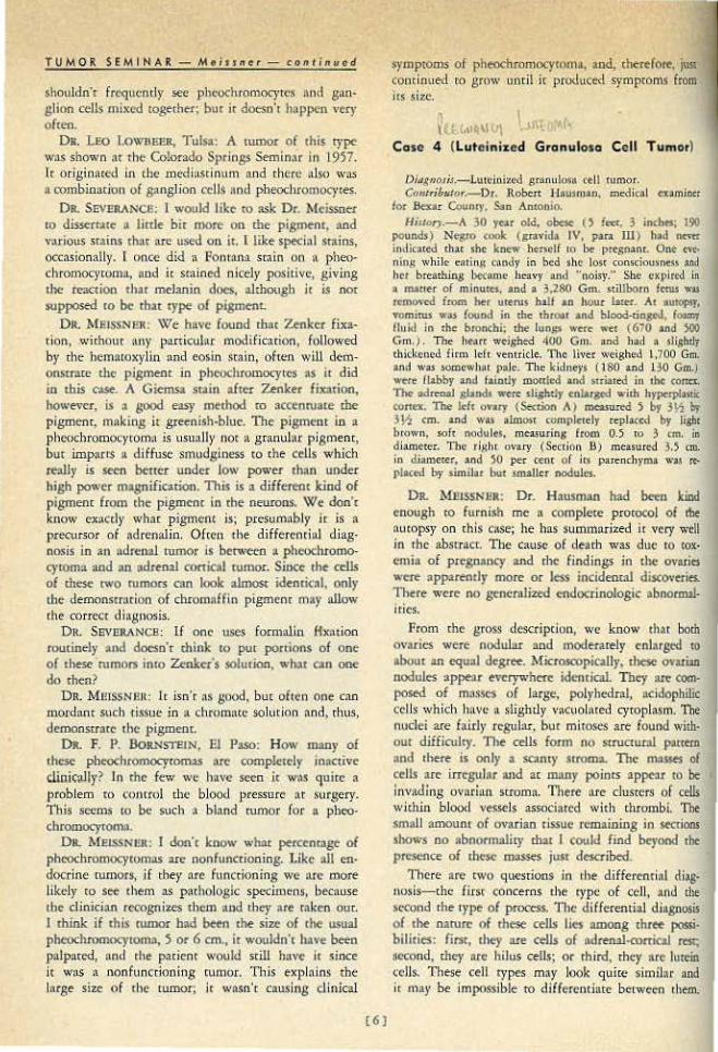
In addition ro rhe gangl.ion cells, there ar~ other ceUs of a differenr rype. These otcur in clusrers, again parricularly abour tbe periphery of the tutnOf but defiw rely as a component parr pf the rumor. They are smaller than d1e ganglion cells Oo tbcw· erage. 'rhey arc ]Xllygonal or spherica l and the Cj'roplasm is slighrly granular, o ften containing pigmcnl granules. The pigment here. howe"er, in contr~$t 10
that of rhe gang lion cells, tenc,ls to be more diffuse
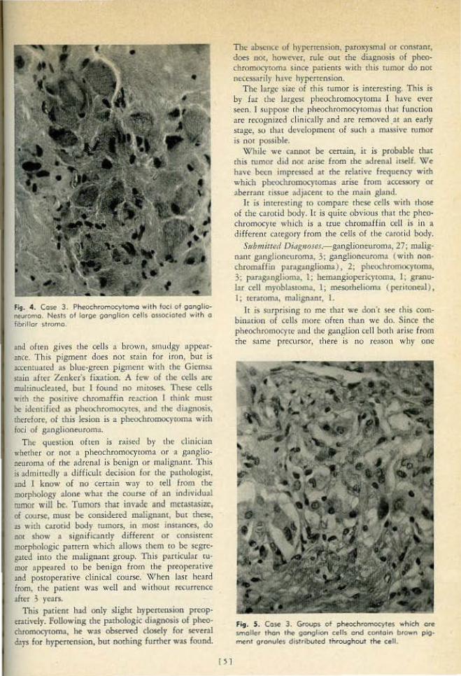
fi9. 4. Cc)e l~ Pheochromocytoma with foe• of gonglio,euromCJ. Nests of Iorge oongllon cells ossocioto-d with a Fihrilfor strorno.
nnd ofttl1 gives lhe ceiJs a brown, smu..l8)' uppcn.r· ancc. This pigmenr does nm .sra:in for iron. bur is :tccentuutt:d ns blue-greeo p ig.cnem with chc Giemsa s:t2ln Jfter Zenker's fi.xarion. A few of the cells are multinucle>~ed, bm I found no muos<S. Thrsc cells w1tb the posinve chromaffin teJ.ction I rhink musr be idemified as pheochromocytes, Jod the diagnosis, thcrdoreJ of this lesion is a pheochromocytoma wid1 foci of ganglioneuroma.
Tit!! question often is r;tlsed by 1ht: cJjnicinn wbecher 0 1' noc a pheochromocymmn or :1 s !lnslioneurom:t of rhc adrenal ls beo ign or 1nnJisnanr. T his is admincdly a difficulc decision foe the pathologlsc, :md 1 know o f no c~rmin woy tO tell from the morphology olone what me course of an individual mmor will be. Tumors that im·a<le and mttasr.t.~izc, of course, must be considered umlignant, but these, .n with carorid body tu1nors, in JOOSt instances* do ooL .show a 5ignificandy diffc.rc-nr or consisrc-m roorpbolosic p>ntrn which allows them ro be segre· gared into the malignant g roup. lbis partkuJnr ru· mor uppc,,ctd ro be benign from lhe prcoper.ui~ve and poscopcr:trive clinical Cmirse. \'\!hen lnst heard from. che p:uicnr w:ts well and wirhouc recurrence after 3 years.
This patient h•d only slight hypertension preop· auh·dy. foUowing the p:nbologic diosnosis of pheo· chromocyroma, he W:IS obsen·ed closely for several dat'$ for hypertension, bur oolhiog further was found.
OJ
The •bsence of h)•penension. paroxysm>! or consc>m, does no<, however, rule out the dillgn0$is of pheo· chromocrronu since p:ttientS with chis tumor do nor necessarily ha"c hnx:n~osion.
The Jargt :;izc of this rumor is imereMins. This is by far the Jnrgcsl pheochromocytoma 1 lu.vc ever seen. I suppose rhe pheochromocytomns thflt (\merion are recognized d inieaUr and are removed 111 nn eMly stage. so [h;n clevelopmenc of such n m'ts.sivc: rumor is not possible.
\'V'hilc we coonot be certain, ic is probable chac this rumor did no< nrise from tbe :.drenJI io~f. \VIe have been imprdSe<l at the n:lativc frequency with which pbcochromocycomas arise from accessory or aberranc tissue adj~cel'lr tO the m:'l in ghmd.
It js interesting ro compare tbc:se cells wirh 1hose of cbe carotid body. lr is quice obvious th:n the phco· dlrornocyte wh ic:h js a crue chromaffin cell is in n. diffcreoc coregory from the cells of the cnrodd body. Submiu~d D#tg1JOICS,- gang)ionturom.l, 27: malig·
nant gnnglion~urom:1, 3; ganglioncuron'l:l (with non· chromaffin parng•nglioma), 2; pheochromocyroma, 3; p.orng:onglioma, I; beoungiopericytoma, I; gr:onu· Jar ccll mrobi>Stom>. I; mesorheliolll:l ( peritooc-.tl) , I; terarom:t,. m.11ignanr. l.
Jt is surprising ro me r:bar we don'[ stt this corn· bin acion of cells more often than we do. Since the pheochromocyte nnd the ganglion cell booh arise from che same precursor, 1here is no n:nson why one
Fis . S. Cosc 3. GrOUps of pheochromocyres whkh ore $mOIIer thon the gonghon cells and contain btown pigtnent oronules di$tdbuted throughout th~ cell.
TUMOR SEMIKAR- Me;JJner - eontinued
sho~ldn'r frcquemly see pheochromocytcs and gan· glion cells mixed together; but it doesn't happen very of reo.
DR. lEO l.ownu&, Tulsa: A rumor of this 'l'P<' wns shown nt the Co!o,.do Springs Seminar in 1957. lc origioaccd in che m<-'<Jiasdnum and there ulso was a a:>mbinacion of ganglion rclls :~nd pbeochromocytes.
OR. SEV!!RANCe: I would like ro ask Or. Meissner to disse.rt'"e • little bir more on me pigment, nod \\lriO\lS srain.s th:n are used on it. r like sp«ial stains, occasionaHy. ( once did " Fonro.nn srain on !l pheo~ chromocyroma, and it srainl!d nkcl)' positive, giving the rexdoa char melanin does, although Jr is nor supposed to be chat t)'p<' of pigmenL
DR . .MEISSN<R: Wfe have found that Zenker fixa· cion, withOut any particubr modlficndon, rollowed by rhe hemomxylin and eosin stain, often wlll dem· onsua.re the pigment in pheochromocytt'S as ir did in chis esse A Giemsa stain after Zenker fix:ltioo, however, is a good easy method ro accenruate the pigment, making it greenish·bluc. The pigment in • phcochromocycorn;~ is usually llOt a granul:tr pigmenr, but impartS a diffuse smudginess to the cells which re:UJy is seen better under low po"•ec rha.n under high po"·er magnifiation. This is a different kind of pigmenr fmm the pigment in the neucons. We don' t know ex.1ctly ~vhac pigment is; prcsum:tbly it is :l
precursor of ndrenalin. Often the diffcrenrinl diag· nosis in an ndrenal ttunor is between a pheoc:hromo· cyroma and an odrenal conical rumor. Since the cells of chcse two rumors an look almost idenrical, only the demonsrr:ttion of chromaffin pigment may allow the correct di1lgnosis.
OR. SEVlli\At'ICE: If one uses formalin 11xation routinely •nd dcx:sn'r chink to put portions of one o( these rumors imo Zen.k('t's solution. wlur can ooe do then?
DR. MmSSN.CII: It isn't as good, bur often one cno mordant such tissoe in a chtotnacc solution nnd, tbus, de.monscrate the pi gruene.
OR. F. P. BoJL.'lSTEJN, EJ Paso: How m:wy of these pheochromocytomas arc compiercly inacti'\'e dinirully? II) the few we have seen it wn..~ quite a problem ·m comrol rhc blood pres.'iure ar surger)'· This seems ro be such • biMd rumor for a pbeo· duomocyrom~;.
DR. M£1SSNUR: I don'< know wb:lc p<'Ktnmgc of pheochromocytOmas are nonfuncrioning. J.ike all en· docrine rumors, if tbey nrc functioning we axe more likely to see them as p:uhologic sp<:<:imens, b«:ause the clinician recognizes rhcm and they are t3kcn out. I think if this tumor had been the size of the usWll pbeochromocy•omo, 5 or 6 em., it wouldn'r have been palpaccd, and che pac.icor would still havt it since it was ~ nonfuncdooing rumor. This ex-ph'lins the large -size of the rumor; it wa.sn'r causing clinical
(6]
symptoms of pheochcomO<:ytoma, and, dJ<:refore, juu contim.ed ro grow u.nril ir produced symptoms from its slze.
1 I IU ~· II 1 ' •
Case 4 (luteinixed Granulosa Cell Tumor)
Didgnosis.-Luu.dnizcd granulo~\1. cell tumor. Ctm,•,ibmor.- Dr:. Robert Hausman, medical cnminc:r
for .Beur Counry, San .Antonio. HiJJO:f1.-A 30 year old, obcie () feet. 3 inthts; 190
pounds) Negro <ook: (gravida IV, para 111) t»:d nc'tt indic:ued th1r she knew berselr It~ be pregnant. One- t\'t• nin,1; while eatin}l candy in bc:d vhc lost c;onsciousness an<l her brc:atbin8 be~:amc heavy nnd "noisy.'' She L'XI)ircd in • matter of minutei, tud a 3,280 Gm. stmborn feNs ~ remo,·cd from her ureru.s hs.lf an hour b.ter. A1 IUfOPST, TOmitw w.u found in rbe thro.:u .and blood·cingtd, fw.mr fluid io rhe bronchi: the lun~ were wtt (670 and 500 G m.). The hNrr weighed 400 Gm. and had a sli,ghclr thicktoed firm le£r wmuiele. 11\c Hvc:r weighed l ,700 Grn. and w.u somewh;cl paJe. The: kidneys ( 180 and 130 Cm.) wert Habby and faintly morcled and striated in 1he corea. n.c adn:nal slands were sligbtlr tnluged " 'ith hypc_rplasc.ic "'""'· The l<ft <Wuy (S«tion A) ln<'3Si1t<d 5 br 3~ b)' j Y.r em. 1Jld was almost C()mpletcly replaced by lisht brown, soft nodules. measuring: rrom O.S ro 3 tm. in diameter. The ri,.;hr m·ary (Section B) measured 3.5 tro. in diameter. and 50 per ce-nt of ils parc:nchymtL w.u reJ>Iaced h)• similar but smaUer nodules.
DR. MlliSSNSR: Dr. Hausman had been kind enough to furnish me a complete protocol of rbe nuropsy on this case; he has su.&1'l.lllnrized it very well in the abmacc. The cause of d~a<h was due ro rox· emia of pregnancy and rhc findings in the ovaries wcce opp>rendy mocc or less incidem:t.l discovtri<S. There were no 8Cfl<tali2ed endocrinologic abnomul· irics.
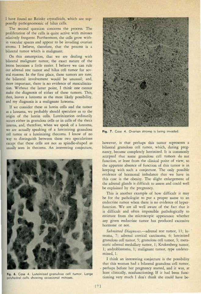
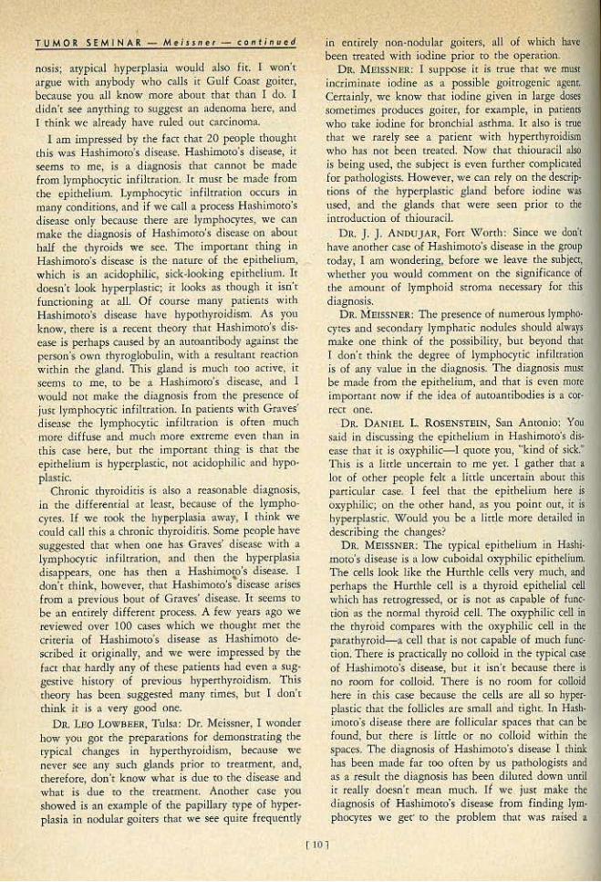
From che gross description, we know char both ovaries were nodular and modemreJ)' enlat8ed to abour an eqtlJII degree. Microscopic..Uy, these ovarilll nodules •ppenr evccywherc idenricnl They nee cocnposed of masses of large, polybcdr:U, acidophilic cells which have a slightly vacuolnred t')'ropbsm. The nudel are fairly rcgtda.r, bur mitoses are found with· out difficulty. n.e cells form no strucrucol ~»= ond rhere is onlr a SCinty Stroma. The tn:LIS<S of cells are irregular and at many pointS appear to be lnva.Jing ovaritm stromn. There nre cluscers of celLs wjdtin blood v~ssels associated with chrmnbi. Tbe small amount of ovarian tissue remaining in SKtions Jhows no abnormaliry chat I could find beyond the presence of d\C$C mosses ju.lt described.
There a_re two questions in rhe diffcr.enrial diag· uosis-ch'~ Hrst COI1cerns the rype o£ cell, nnd the >«:ond the type of process. 'Jl>e differential diagnosis of the oarure of chese cells lies among rh- possi· bilities: first, they are cells of adreJUI·ronicol =: second, they 11rc hllus cells; or third, they are lutein cells. These cell types may lnok quire similar an<l it may be impossible ro differentiate between them.
J hove found no Reinke crys~tlloid!, whicb are sup· pos.:dly pathognomonic of hilus cells.
The second question concerns rhe process. The proliferorion of <he cclls is quite acrive wi<b mitoses «btively frequenL Funhermor<:, the ceUs grow wtth· m va$CUlar spaces 2nd appt"a.I to ~ invading ovl.lritn srrOm:t, l be.lievt, rherefore, rhnc rhe process is a bilateral twnor wh i<:h is malignant.
On this assumption, that we a re dealing wich biliuml maligtWJr rumor, the cxaa nature of the lesion hccomes a linle e-asier. I believe we can rule out adren:tl <est tumor and hilus cell rumor for scv· enll rc11sons. Jn rhe fi rst placl", these tumors are rnce~
the- bihueral lovol ~o·cmenr would be unusuaJ; r.nd. more important, there is no e\'idence of IJl::l.SCuliniza· rion. W ithout the laner point, I think ooe cannO< make rhc diagnosis of cid1er of these nunors. 11>is, then, leaves a Jucoorn:t :1s rhe most likel)~ po.~sibilit)', and my <.H:tgnosis is ~L maltgnaor luteoma.
tr wr coosid~r rhcse as lurein cdls and dte rumor .s a luteoma, we probobl)' should speculate >$ to the origin of the lutein cells. I .. uu:inizarion ordinarii)• occurs either in s ranulosa cells or in cells of chc [h<.oca incerna, and1 therefore, when we sptnk of a luteoma. we nre ncrually speaking of a luceinizing granulosa ctll tumor or a lutcini2ing thecoma. I know of no w•y ro distinguish bel'l\•een these 1'1\'0 speculntions except rltac chese cells are not as spindle·shaped .s U$un.lly seeo in 1hccorna. An interestiog conjeccure,
FJg, 6. Co~ 4. Luteinized granulosa cell tvmor. Lorge polyhedral cells showing occosionol mitoses~
Fig. 7. Ccse 4. Ovorlcn strcmo is being invaded.
however, is rhar perh:.ps this tumor represents B
bihuc:.cal granuJosn cell rumor, ,.,.h jch, during l)I'Cg· nancy, became completely Juccioized. rc is commonly accepted chat some granulos;t c<:ll nunors do not fuooion, ac leas< from the clinical poinc of view, so the app:ueor t1bsenCC!' of funcuon of t:his rumor is in keeping with such n conjecture. Tlw only possible evidence of hormonal imbaJ:mcc ch:tc we bnvc in rhis Co.lc is d1e obe>ity. The sJighr enlargement of the Jdrenal glands i$ difficulc co ~~ •nd could well be explained by <he pr~gnancy.
This is another cxnmple of bow dilficulr it mny be fof the P.atholo,a;ist TO puc a proper name to on endocrine rumor wh<:n there is no evidence of hyper· funcrioo. W e are :til well aware of cbe fact thac it is difficult and ofrcn impossible p>thologicaUy ro estimrue from the microscopic tlppeamoce whethr r any given e.ndocduc rumor has produced excessive h o tn'IOIIC Or DOl.
Submiue{t D~t~guoseJ.-adrenal rr.sr rumor, ll : lu· <coo:tJ, 7; adrcn:>l cortical carcinoma, 6: lutriniud gr.t.nulosa ceJl rumor. '; granulosa ce11 rumor, 3; mem .. static ad1·en:1l rot:duHOt")' rumor. 1; Krukel'•berg tumor, 1; und,rl)bhsromn, 'I; malignant tull'lOr, rype undeu:r~
mined. I. 1 think an imores<ing conjecture is rhc possibility
that this woroon hod > bilarecnl gnnuloso ccU mmor, ~ perhnps bdore her pr<:gnancy srorted, and ir WBS, ar
le<tsc clinicnlly, nonlunccioniog. lf il' h:>d been lunc• tionins very much I don't think she could have be·
171

TUMOR SEMINA R - Meissner- continued
come prtgnanr or cotried her prcgn:mcy. Then dur· ing the C0Ur$C of the pregnancy the bibtera( grnn· uJos.t cell rumor become luteiniwd. This seemed like on intriguing idea tO me, ood 1 cried to find reference tO solid ov>rian rumors occurring in pregnaocy 10 see if 1 could find any mcnrioo of whar happens ro a gr.lnuiO$> cell rumor during pregnnncy. Solid O\'nfitln tumors :l.S a complication of pregat1ocy are uncommon, Bnd 1 here nr..: not m:tny rc:porced. Dough· erty .llld Lund" in 1950 collected 30 reponed c:•ses over a 15 yo:1r period :~nd added 6 more cases or chdr own. Thccc were 3 granulosa cell cumors; buc, uoforcun:uclr. these WCI'C nil wjchour microscopic description, so I don·c koo~v wberber rhey were Ju· 'clniz.ed or not.
DR. HAUSMAN: This is the se<:ood case thnc 1 have seen. The surgeon did :1 Owsarean sec-don on a woman, round chese rumor lDOI.SSCS in the ovaries. ancl decided 10 do a hys<cr<:<.~omy with bil1ternl oophorec· tomy. The woman live-d after tbe operatioo, and as br as 1 know w<U all right. The ovaries were enlarged and hld thtse brown tumors in tbem. That time 1 made the diagnosis of lureinized grnnul0$3 cell rumor.
DR. li1EISSNFR: Were there zny ocher eudocrino· logic :~bnormalities?
OR. HAUSMAN: There were none1 nnd T parckulatly looked for them.
DR. J. 1.. GOFORnl, Dallas: This case intrigued me very much. 1 had thought tb:1t ir was a granulosa cell nroplasm showing rhe lmeinizing effects of pceg· nancy. The problem chac worried me was whether h was benign or malignant. l leaned cowacd tbe idea that it wus malignnn<. bur kept asking myself what would hapP"n if the pregnancy were rerminuted and the neoplasm were rcmovtd. I am noc ac all sure thai
it aetu.Uy is a maligrunt process. DR. MEISSNER: I agree with thai l•don"t know
how we can pro,·c whed1er chis is malignant or 001.
It is like a lor of rumors of endocrine glands-it is difficuJt 10 say unless "'e have a follow-up, whether d1ey are benign or malignant. Our criteria for the diagnosis of malignant change in endocrine glands is more dilfit-ulc than in many ocher tissues of the body. This tumor h:tS ~ lor of miroses; ic looks like it is
~ extending into suoma. Bur, on che other hand~ if [bis is o cumor uf thecal cells or granulosa. cells, why shouldn't it excend inco i-ts own si'J'oma? It is ex· tending into its own f:.m ily, really. lr is io blood vcsscls, but ugllin 1 don't think that necessarily makes ic a nu.lignnm cumor.
DR. Leo LOWBI!Eil, TuJs.: What did this woman die of?
DR. H/.USMAN: The diagnosis of toxemia of pregn:mcy was a diagnosis of convenience. 1 have not the slightest ideo wh3t she died of.
{S]
Case 5 <Thyroid Hyperplasia)
DiJ.<HoJil.-Thyroid h;-perpl.3Si.a ( noncoxk). Cot~~ribNJo-r.-Lr. Cot James L. Harucn, M.C, Brooke
Arm)' Hospiflll, Fon Sam Hou:noo. llillo~y.-A 22 )'faX old white ftmaJe h3d an enlllr~N
chyrotd sinct sse 12 with !)'m_ptom.s of hyperrhyroidbm anJ hn,o:hy.roidism. bur 11r cirne of sur&err seemed curhJ roid. Ri~;hr lobe weighed :lO Gm., lelt I~ 18 Gm Cut >ul'f.ice v.·as slighdy nodol~r. modera1cly finn, •nd t.nYbh.
On. ~fEtSSNrut: The hisror)r in chis c..m is nn in· ccrcsting one of enlargement of the thyroid, wirh upparcnrly alternating episodes of byper- and h)•po· rhyrQiJiSill, /or perhaps 10 )"Car s. It is sign ificnnt thnt
, nc t he 1ime of surgical excision rhc p;uit•n1 w:~s ~uchyroid. \'(fe n•·• rold that the gland wM rno<lcrntel)" enlarged, the cwo lobes weighing together 48 Grn. We hove usually considered that 30 Gm. is the aver· .1gc weight for • normlll d1ycoid glund, ulrhough it is true t hJL there are great variations in the weight of the npp:orencly normal glnnd with th~ e.rrr~rnes being perhaps J< low llS 10 Gm. and as high <U '0 0!
60 Gm. Microscopicolly tbere are rwo findings of note;
one is a diffuse hyperplasio of tbc epithelium which tS presc;nt 10 about tbt same degree io all parrs of every slide. The second fe.trurc is the ptesc:nce of ' lymph(lcyric infihn tion which tends to be nodular 3nd of rcn :tssucinted wirh secondary lymplw ic nodules. The caps~<le of che gland is clistincc. There is rnoderntc v:tscuJarjcy.
Fig. 8. Cose 5. Thyroid hyperplos.io, nontoxic. The ~· thelium is hyperpl0$tic end -there is lymphocytk: lnfil· trot ion.

llle differemiaJ diagnosis here is berv.•een. thyJ:oi.doris, ~arcinol""· .and d.if£usc hyperplasia. 'rbc diagnosis of chyJ·oidicis <loes nor seem to be a sound one T his degree and . rliis type o£ lymphocytic infiltration is seen frequent!)• in a gland wim hyperpla1ia of cj1e primary or Graves' d isease cype. It is uue that if the hyperplasia d isappears, che lymphoqric infiltration Il'l::l)' remain b(;hind, and in such an evem t he diagnos·is of chronk thyroiditis ma}' appear more reasonoble. However, in the presence of dj£fusc hypecplasia, ic seems safest to condude chat rhe lymphocydc infilWltion js t~-n ~~ccompanllnent so commonly- seen rather chan a prlm~tty ptocess. Carcinoma. can be excluded by the d iffuseness of the process. Presumably, we have representative portions of both lobes and it would be unusual for a carcinoma tO be :ts diffuse throughout botb lobe.~ as is seen here. Furthermore., there is no evidence of capsuJar or vascular inv~sion.
'The best djagno:iis, therefore, would -seem co be a diffuse byperphsia of the thyroid. I should like ro coli th'is primary l>yp.erplasia of the thyroid.
ln ou.r laporafory, we ace co the )>abit of d iscing· uishing o:wo t)'Pe.l of tbyroid hyperplasia. One we call primary h)•perph~Sin and is cbe type commonly .seen associared wit'h rhe signs and symptoms of exophthalmic gpiter. The other we have C{lJied secondary hyperpl:tsia, which is rhe type thac occurs in long-standing adenom.·nous goiter. Fo.c c_(jnical ..:easons ic seems fmponanr tO distinguish_ berwecn these rwo cypes, if possible, bec•use the latter is rarely associ· aced with the exophthalmus, .lymphocyric infiltration, or sevtrc clinicill coxkity so common in primary hyperplasia. lt is a Jow·grade, smoldering type of hypcrchyroi~ism, difficult to control w ich antithyroid drugs and d1e patients are ofccn thyrocardiacs. It is quitl' a different process fJom d>e primary hyperplasia of Graves' disease.
Ir happens at rimes thor a· o:Uffuse bypet'!>lasia of the thyroid occors w ithouc evidence of c(jnlcaJ :hyperth}•roid isro or, as· the clinidans say, wicbo~.tt toxkicy. fo order tO understand whac produces h)•perplasia of the thyroid' ir is iOteresring £0 revjew the physio!om• of th)•roxin secretion. The pituitary unacr the influClnCC of the hyporhnlamus produces rbyroid-stin>u· )acing hormone. which stimulates tbe chyrojd gland <0 byperpl:osia nod ro incr..Sed production of rhy. roxin. The thyroxin, as ·it !coves cbe thyroid gland, goes pardy co rhe tissues and partly co che pituitary ancl hypothalamus where it aces as an lah.ibicor of TSH secretion. Thus, TSH and thyroxin are normally in more o r less of a balance. ln che hyperplasia of Graves' dise:tse there is 1111 unexplained hyperplasia of rhe thyroid and an excessl~e secretion of thyroxin, but the exacr poinr in rhc cycle where the difficult)' -arises is poorly tuldetsrood. Tberc arc cases, however) where the rhyrojd may be unable- to pm out either suff;icienr or funcrionally , .seful chyroxin1 _in which
[9]
case the potoutary would nor be depressed and the excc.-ssive TSH, rherefore, stimulau:s the thyroid m grearer and go:eater hyperplastic <~~:~i•ity in the presenc.:c: of clinical euthyroidism or even hypothyrqidism. Such '' smte onny be broughr about by several mecbiinisrns. For example, there are so-called goitrogenic substances, such ns thiocyanate, which inhibit.~ the ·iodine uapping mechanisms of the thyroid or thiour· acil, whicb prevents the intraglandtLlar formarion of thyroxin. Tbete are other goiuogenic substances, the mechanisms of which are poorly Wlderscood. Still, in addirion, rhere may be ~n inbom fault in thyroid rnetabolism1 for exampl~, wirh One of rhe inrrachyroid enzyme S}'Stems. so that a \!Sable thyroxin is not ade-quately formed. This is, perhaJ;>s, the explanarion of some cases .of congenital goirer.
ln any event, rhe- pathologist occasionally is con· fronted with a <.!0\Se wl1ere cbete is no evidence of clinical hypetth)'roidism, but where the thyroid gland is neverrheless d iffusely hype.cplasric. For. wMH of a becrer name we have been in the habit of calling such a gland pacli.ologically pcimar)' hyperplasia, nontoxic.
Two examples 'Yill serve co illustrate chis type of .hyperplasia. The firsr case is one- of a goiter in n 3\-2 month o ld child where cbece js a d iffuse hyper· plasia of such an e~creme degree rh.11 rhe baby ~jed from respirotory obstruction. 111e cause fo r the hyper· pfasia in chis insmnce is unknown, but the mod1er did receive deskcncd thyroid during rhe lasr crimes· rer of pregnanq, and rhe child received iron and cobalt the. first few months of life. The second ex· ample is from an adult wich recurrent enlargemenr of the thyroid and wirhouc clinical toxicity. Here again there is a diffuse hyperplasia which is quire similar to the hyperplasia found io typical Graves' disea..~e. In this caseJ agtdh, there was no obvious reason for the rhyroid enlargement
This nontoxic hyperplasia of rhe thyroid is unCOpl· mon, bur iS i.mport~Ot for the p:nhologisr to remem· bee. Jn the absence of clinical h}'perrh)'toidisrn it is Sometimes rempring to call S1.1Ch a _process carcinoma. In the case we have just discussed, 1 m iscakenl)' made a diagnosis of carcinoma on froi:en secrlon only to find on ou.r permaocnt .sections rhac the glaod waS diffusely involved wirh nnth iog bur a hyperplastic process. It is possible rhar some of che thyroid can· cers reponed in infants are also instances of an overdiagnosis of diffuse hyperplosia.
S1fb1nit;etf, diagn.oses.-duonic rhyroiditis, HashimotO, 20; chronic thyroid iUs •1onspecific, 5; Riedel's suuma, l ; hyperplasia with involution M.d l)•mphoid infi ltrotion, 3; atypical th)•roid b)·pcrplasia, 2; Hashimoto's disease ossociat<<l wid> feml adenom" of cby· roid, 1; toxic goirer (Gull Co:ost?), 1; follicular Hurthle cell -ade-o.oma, 1; adenota.rdno•na, Oj follicu~ lac :ldeoe:c:~rcinoma, 2; papillary aden.ocatdnoma, 1.
DR. 1\.fEJSSNER: Hyperplasia wicb involution and lymphoid inWrracion would b~ ·~imilar co my diag-
TU M OR SE M IN ,._ R M ei ssn et - con ti nu ed
nosis; atypical hyperplasia wo~ld :dso fit. f won't argue with anybody who calls ir Gulf CoaSt gait~<, because you :ul know more about thM d11n f do. I didn'c see aoytbirig tO suggesc 11.n adenoma here, and l think we already hnve ruled out carciooma.
l am imJ>ressed by the fact that 20 people thought rhjs was H oash.imom's dise:l-S"e. Hashimoto's disease, ir ¥"ems ro .me! is a ~ i.agnosis chat cannot: be madt ft01)1 lymphocytic infilt.rotion. Tt muse be m:~de frQm che epitheli\1-Jn. l.)'mphocytic jnfiJuario.o occurs in Jtlany conditions, and if we call a process Hashimoco's disease <.ml}' be<;a.use rhere are l)·.mphOC}'fes. we can make che diaognosis of Hasb immo'.s dise:tse-on aboU[ half the thyroids we see. The important thing in Hashimmo's disease is rhe nature of the epithelium. '"hicb is an acidophilic, sick·looking epithelitun. It doesn·c look hyperplastic; ir looks as though ir isn·t functioni_ng- ac alL Of course many patients with Hashimoto's diSease have hyporltyJoidism: As you ·know_, there is a recent rheol}.- that Hash imotO's dis· ease is perhaps caused by an am<Xmribody against the person's O\'i.'ll diyrogiobulio,. \Vich !1 result:mt reacriqn wirhin che gl~1nd. This gland is much coo acri\•t, it seems co me, to be a Hashimoco's disease, and I wOuld not nlak<: tbe dia·gnosi.s from the presence of juSt lymPhocycic infiltr\ttion. In paciems with Gr.l "<•es~ d"is<.--aS<:: Ehc l)li)1l)hocytic lnfiltmtion is ofren much mote diffuse and .much more exrremc even rh:to in chis case here. bur [be irnporcaoc thing· is that the epithelium is hyperplasric, nor acidophilic and hypo · plasric.
Chwnic thyroid itis is also a reasonable diagnosis, in the differemral m lease, because of the lympho·
. cyres. Jf we rook the hyperplasia away, I think we cou ld call this a chronic rhyroidicis. Som~ people have suggested thar when one has Graves' dise-;tse wich .a lymphocyrit irifilrradon, and eben tl1e bypcrglasia disappears, one bas then a Hashimo~o's ~ disease. I don't think, hQWC\•er, th;tt Hashimoco's disease :ujses from n prcv,ious bour of Graves' disease. Ir seems co be ail entirely different process. A few years- ago we rev'iew<:d ·o"er 100 c.1.Ses which we thonghc mer chc: Cricerin of Hashimoto's disease as Hashimoto de4
scribed i r originally, and we were impressed br the !ncr th~r hardly any of these patients had even a sug· gestive history of previous hyperthyroidism. Tbis theory has been suggested many times, but r don'r rhlnk it is a very good one.
DR. LEo LOWBEER, Tulsa: Dr. :Meissner, I wonder bow you gor the preparations foe demonsuating rhe rypical changes iu hyperthyroidism, because we never see any such glands prior co creaqncnt, :md, therefore, aon't koow whac js-due tO cbe disease .and what is" d11e ro che rrcacmeht. Anocher C.1Se you showed is an example of the papillary type of hyper· plasi:t in nodular goiters U,.ac we see quire frequently
in entirely non~no<lular goiters, al·l of which bave been tteated with iodirie prior tO the operatio~.
DR. J\.fmssN,Etc I su.ppose it is rrue rhat we muse iocrimina<e iodjne as a possi.bk goitrogenic agenr. Cena inJy, we know thac iodine _given in large 4oses sometimes prOduces goiter, lor eX71mple, i-n p:trient$ who rake Jodine for bronchial asrhma. l r al.so is crue chat ~.,e· rarely see a pacienc 'Nith hyperthyroid~n wbo has not been treated. Nqw that thiouracil also is being used, che subj<.'Ct is even further complicat~ for pathologistS. Ho-wever, we can rely on rhe descrip· tions of the hyperp lastic gland before iodine was -used, -and cbe glands chat were seen prior co d>e imroduccion of thiouracil.
DR. J. J. ANDUJAR, fort Worth:- Since we don't have anorhcr. case of Hnshimoro'.s disc.-ase in the group coday, l am wondering, before ,...,e leave che subjec.t, whedter you would commeoc on the sigoif.icance Qt tbe amounr of lymphoid suoma oecessUf)' for this diagnosis.
DR. MBISSNER : T he presence·of numerous lympho· cytcs and se~oodar.y Jymphacic nodules should a)wayj m;tke one think of the possibjliry, bur beyond rliat T don'r rhiok tbe degree of lymphocytic infilt~ation is of nny value ln the di~lgoosis. The diagnosis .mU& be mad<.: &om the epirbeliu.m-, and chat ls even mote hnportaot n6w if- rhe i~ea of autQancibodies is a cOr· recc one.
D t\. DANLBL L R OSENSTEIN, San AntOnio: You $aid in discussing the epithelium in H:tshimoro's disease that lr is oxyph ilic- ! quoce you~ ''kind of sick.• This is a lirde uncttrcain co rne )'Ct. I gache-r chat a lot of ocher people felc a little unc~rtain about this patticular c-ase. I f<..oel chat the epithe:li~tm here is oxrphllic; on tbe. ocher hand, as you poloc ouc> it }s hyperplastic. Would you be a little more deroi.lcd in describing the ch•oges?
D R. MEISSNER: The typical epithelium in HllS~i· moro's dise:.sc is :t low cuboidal oxyphilic epithelium. The cells look like the Hutthle cells very mitch, i!tld perhaps rhe Hi.trthle cell i~ ll thyroid epitbeli:i.l cell which h:u retrogressed, or is not :IS capal:tle of func· cion as the normal thyroid cell. T he m:yphilk ccll in cbe thyroid compares with the oxyphilic cell in the parathyroid-a ce) l tim is nor capable of much funt· rion. T here is ptacrica)ly no colloid in the typical me of Hitshimoro's disease, bur ir isn'r because there is no room fo< colloid. There is no room for colloid l1ere in this case beClluse the cells are all so hyper· plastic chat the folliclts ltrc small and tight. I~> Hash. !moco's disease there are follkula-r spaces rbar can he found, bm there is litrlc or no colloid witbio the spaces. The diagnosis of Hashimoto·s disease 1 rhink has been made far tOO often by us pathologistS 3nd ~~s a result ~he dillgnosis h~ been dBured down undl ir really doesn't me= much. If we just .make the diagnosis of Hashimoto's disease from fil]ding lym· ph<>C)•tes we gee· tO rhe problem chac- was raised 11
[ 101
few moouoes ago-how many l)•mphocy<es do rou need? Soventy per cc:m, or fifry per cem, Or ten l"'' cem f If one goes over ~ series of spedmens :md compares che clinicAl history and rhe follow-up, che groo findings, and the paohologic microscopic findings cogerher. on~ an arrive at a f<tirly coocr~ clinicop:uhologic cntiry which is fushimo<o's dise:l5e. But if one dia8JlOSts Hashimoto's disease just because of lrmphocytcs, ohe correl01ion with clin icnl findings is- p oor.
OJL SEVERANCP.: T nO<icc that Dr. Andujar "'M perturbed b«ause we didn't include a case which Dr. Meissner would dingoose as H tlShimmo's disease. T note char 20 oohcr people around here did chink that chis cast was H~t.llhimoto's, and we've had enough discus.<ion so we prob>bly don't need one in this seminar. This prob>bly goes b:Kk 10 cbe fact <hat • good many of us here, including myself, were led ro bdieve; or were ct'logJu somewhe-re !hat n lot of Jymphoc.yres clid givo you a gotxl t>icoure o f .1-T:tshimoro's, and maybe we wereu'r mugbl co stress (he other fearure and rhe dinical picture. \Y/t. hnvc ICOUble getting cl1e clinical informacion; it is usually no-r rt.:tdjJy a\'ailllble unless you get [he chart s<:vc.:raJ w!-<eks oc maybe a momh after tht: patic-nr has gone /rom the hosp ioal. Ma1•be that is differenr in lloston.
OR. M eJSSNI!J\: Nor vety much.
C••• 6 (Chronic T hyroidit is)
l')l(IJ!IIOiii.-Chmnic th~·roiditis, nunspec;i fic~ Ct,l#t;bu~J.-Ors. 0. A. Todd, A. M. R.ichmonJ, tln~l
S7lvi:a Jchns, Nix HO$pitlll Clinicl l:t.bo!lltOr)'• San Aoronio.
Hh:ory.- ·rbis white- fdDale. 4 t ye-Jrs o! age. !irn noticed enlttrgement or lhe neck in ftbtu:Jf')', 1957. ))hysical l"X~min~u iOJ) r(l\'Calcd a larse atlcoom:uuus growth in 1hC' zight lobe.· Pulse wo:1 82, blood p1cs~ure 140/SO. Radioacrn·t iodine uptake ~tudy with 50 mic:rocurie tnccr dose sbolol.td 17 pe.r ecru conc~nrr::uion over the thyroiJ in 24 bouu wirh pt:l(tiollr no uptak~ in the ri,gb1 lobe. A t01:al thyroidectomy was performed io .March, 19~7. The ri,g.hr, lobe W$15 !ou.od t<>- be firm, pale red. l\nd diffusely lobubrWilh no dis~rc1c nOc;IOI~. The ltft lobe, on section, J>ICSCnted " tetl lxtckgrountl wi•h firm, g.roy-whitc, indura.tcll areas.
0~. MEISSNER: The hisrory docs not help a gren deal in <he diagnosis of this inrrc<Soing cnse. \VIc arc told rhar cbe 41 year old woman had enlargement of the thyroid, especlnll)' rhe righr lobe, o f r;ubcr shorr dunuion, and thnr rhe rn.dioiodit1e uptake in che thy· roid \V!I5 racher poor. The rwo lob<.-s removed at optDrion are nondescript 11.0d more or less similar in ~ppr:tunce.
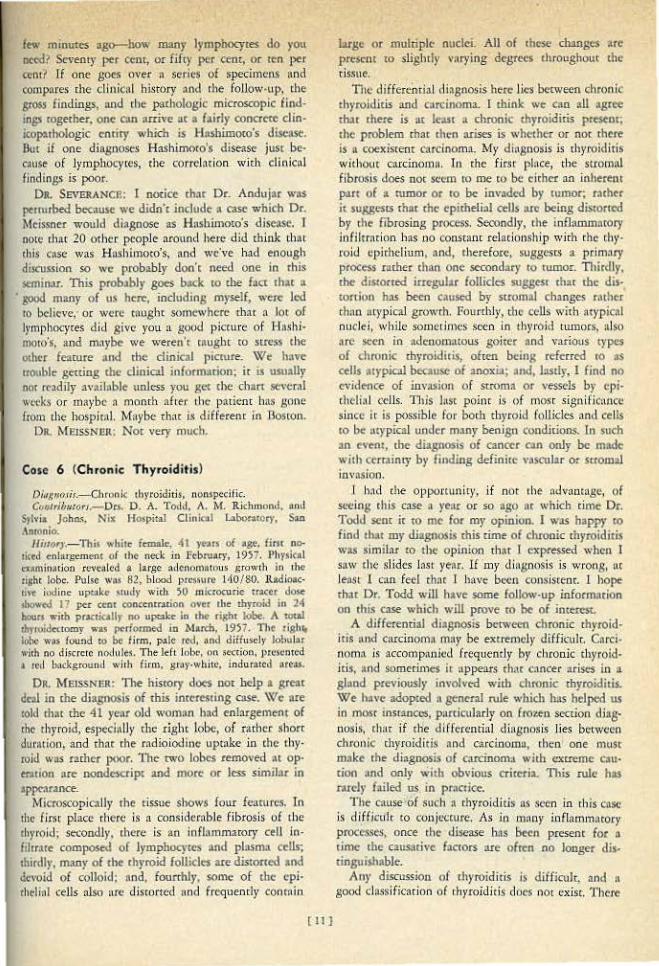
Microscopically rhe oissue shows four fcnrures. In oho first place there is a considcmble fibrosis uf rho rhyroid; Se<.'Ondly, thete is an inflammatory cell infutrllt: composed of lymphocytes and plasma a:JJs; ohirdly, many of the rhyroid follicles are disooned and dc,-oid of colloid; ond, fourrhly, some of the cpithtlinl cells also are discorted rHJd frt'C)uenrJy conroin
( 11]
large or mulnplc nuclei. AU of obese changes 2re prcsont m slig hrly varying degrees <hrougbour d1e cissue.
Tile diffetcnrial d iagnosis here lks berween chronic thyroiditis and carcinoma. 1 chink we coo all agree duo there is or l~s< a chronic thyroiditis present; the problem chat thtn n.rises is whecher or nor thtre is a coexisrent carcinom:t. 1vfy diognosis is thyroidiris without carcinoma. Jn Ute first plnce, the stromnl fibrosis does not seem ro me co be cicher an inhtrem pa.rt of a rumor or 10 be invaded by oumor; rathtr it suggesrs rhar <he cpimeliru ceUs arc being disoorred by <he fibrosing process. Secondly, rhe inflammatory iofilrr:uion has no constant relationship w ich rhe th)'roid cpichtlitun, and, rherefore, suggestS a prim:try process racber than one secondary 10 rumor. Thitdly, <hc distorted .irregul>r follicles sugges• rhu the dis<onit>O has been caused by srromal changes raoher thno :lf)'pical grow[h. Fourth!)', tbc cells wirb atypici\1 nuclei, while .somcLin"'CS seen in chyt·ojd rumocst :dso are seen in 'lcl('nomawu.s goiter r.nd v:~.ri0\1$ types of chronic chyroidiois, often being refeued 10 as cells Ol)'pical beautS<: of anoxia; and, las<iy, I find no evidence of invasion of srroma or vessels by c pichclin l cd.ls. 11\is Jusc poi1u is of rnosr siguificnncc since it is possible for bocb rbyroid follicles and cells 10 be :uypic2l under many benisn conditions. Io such an eve<~<, cbe dia8Jlosis of cana:r con only be made with ctrminty by finding definite vascuJar or stroma) invasion.
I h:t~ che o_pponunir)', if not rhe :ldvunt~tge, of seeing chis case a yenr or so ago nr which time Or. Todd sene it co roc for my opinion. I was bappr <o find ohat my diagnosis mis rime of chronic ohyroidiris was simib.r lO the opinion rbat 1 ex-pressed when J saw the slides lasL year. 1l my diagnt)Sis is wron8, :u least 1 can feel choc I h•ve bC(:n consisteor. J hope <hat Dr. Todd will have some follow-up information on this case which will prove tO be of imcr~-s<.
/1. differencial diagnosis bctw...en chronic <hyroid· iris nnd carcinoma 01:1y be e:xuemd )' diffkuk C:trci· ooma i< accomp:micd f rcquendy by chronic thyroidids, and somecimts ir appe-.us c-hac C:lncer 1triscs in n gland P""' iouslr involved wicb chronic cbyroidiois. We I»''< adop<ed o general rule which has helped us io mosr insranccs, patticularJy on r roi'.en secrion Qjag· nosis. <hac if the d ifferential diognosi< lies between chrOI'~ic chyroiJitis :tnd carcinoma, rheo one 1nusr make rhe diagposi.s of carcinoma with cxcre.me caution and oriJy witb ob\,ious criteria. This rule has rnrely f~iled us in prnetia:. Th~ cause :of such :t chyroiditis n.'\ seen i.n rhis cnse
is diffic:uJc to eonjc(.:ttire. As in mnny inflammacory processes, once the disease has been presem for n time the causative faccors are ofren no longer dis· tinguiihoble.
Any discussion of thyroidi<is is difficult, and a gond dassificuion of rhyroiditis docs •"~Ot exist. There
TUMOA· S·EMINAR- Ma l ssllor - conr;Jlued
a~e ..sever~J rensot'ls for 1his. ln the fits r place cber:e are cct.Ealn diSeases whicb have 15een included uodec che he~diog of thyroiditis which are probably noc crue inflanlmadons. l n sut'.b a catcgorr is stnun~ lymphornfl.tOSa or Hashiinoro's d isease. Perha_ps in che same caregocy comes · che lympbocycic goiter whkb is somewhat similar 'tO H ashimoto's disease~ bur without rhe extensive c_p irhdial chunges usuaJiy found in the latter. A fnrcher complkacing faccor in chc dassifiC" .• nion of thyroiditis is thar. un eciologlc dassificacion is d ifficuk Even in che cypcs of thr· roidieis wh.ich we aut recogoj:ce .re.~tsonably weU path· olog irnlly, sodl as granu lom:uous thyro iditis, we still do ·noc know the e tiologic ng~nc.
Still another cornplicacion is <h•e to ' he face chat we often see che end results of injury co r)lc glan d and for ·waur of a better n:une ktbel tbese as non· sptcifk chronic th)'IoidirisJ. evt·o though we have no idea as co what the injurious ngenc- mighr have been.
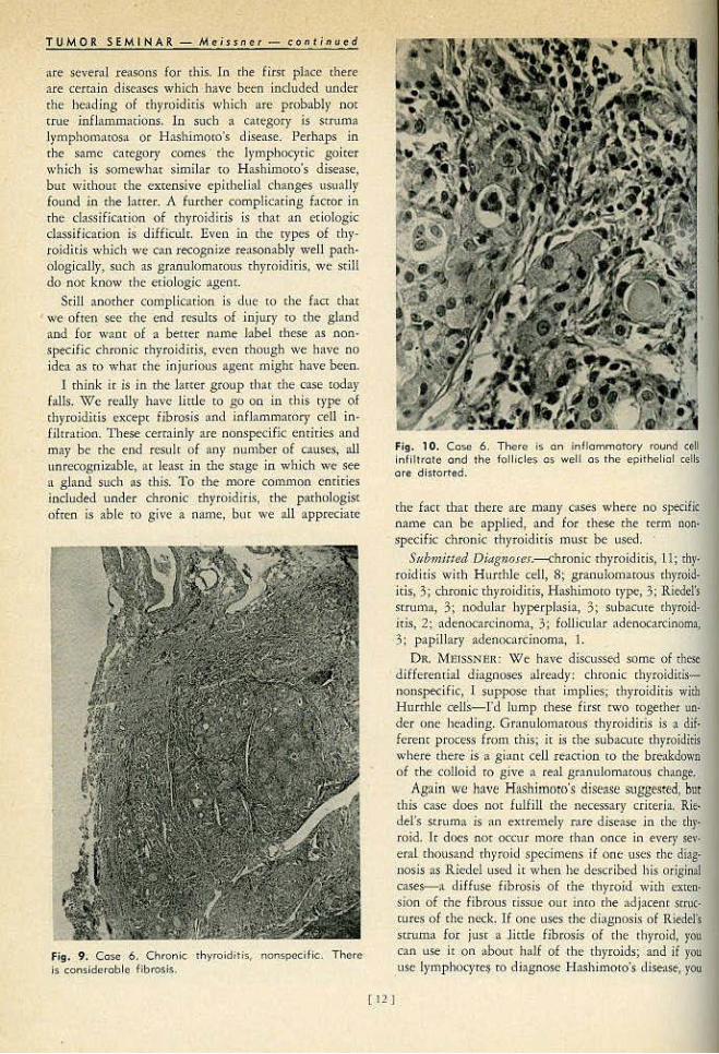
1 rhink lr is in d1e hitter grOtlp th:\t the c:nse today falls. \'\fe really have lictle to go On in chis t)•pe of c.hyroidit.is exce pr. fibrosis t~nd infktmmatorJ' cell in· filt.r1ation. The~ cerr:.linly are. oonsp<.~ic entities and mar be the end result of •n)' oumber of c~use$, all unrecogniznble, ar leasr in We smge la which we see n gl_and suc.h as th)s. To the more common cmir.ies h\cluded under chronic chyroidirjs, t he pathologist o frcn .ls able to g ive a name, bm we all apprecia[c
Fig. 9. Cose 6. Chronic. thyToiditis, nonspecific. TI~ore is considerable fibrosJs.
Fig. 10. Case 6 . There is on in flommotol'y round GeU lnflll rato a nd the follicles os well os the epithelial ceUs o re d istorted.
the facr that [here are llltWY c1ses -: ... here oo sP,ecif.ic name can be appHed1 and for 1hese che term non· !ipecific chron,ic thyroiditis mliSt be used.
Submitted DlagtJoses.~hronic ·thyroid Ids~ 11; thy· rojdi t.is wirh Hurchle ceJJ, 8~ granufomarous rhyroid· iris, 3; chronic thyroid itis, Hnshimoco type, 3; R iedel's srruma, 3; nodular hyperplnsia, 3; sdbacme rhyro.id· itis, 2; ·{ldenocardn oma, 3.; foW c.11lar adenocarcinoma; 3: papillary ~denocarcinoma, I.
O R. MlliSSNill\; We hav<: d iscussed some of rhe.~t
d ifferential dinsoose.o;- alread)r: chronic rhyro?dids•loospedfic, l supj>OSe tbat ioo.pJie-s; thyiOidiris with Hunhle ceJ.lS-Td Jump rhese first t lt'O roget.iler- un. der one heading. GranulOJtlacous thyroiditis is -a dif. fcrenc process from ch is; ic is che sub-acme chyroidiris where there js 11 giilnt cell reaction to rhe breakdown of rhe colloid ro gjve a real granulomamus change.
Agnin we have Hashimoro's dis~ose suggt>stcd, bur this c.-:asc does not fulfi1l rhe necessary cricerh1.. Riedel's st.rum~ is an extrem.ely n u c dise:tSe in che rby· roid. lr doc~s nor occur more rban once jn eve.:r se\'· eral chousaod thyroid spe9imens 1f one uses che diag· nosis os R iedel used ic when he described his origin>! cases-'lL di ffus~ fil?rosis o f the thyroid wirh exren· .${On. of chc: fibrous cisslle ou c inco the ttdjacenr stru'· cures of che neck, If ooe uses the d iagnosis of Rie<iel's
· scruma for just .a Jictle fibrosis of the thyroid, you cnn use ic on obouc h:Jif of the th yroids; and if yo~ usc {ymphOt)'te~ co diagnose H ashimQro's disease, you
[ 12 J

oould "se that Otl the other half. I wondered in this case if perhaps the pa<ienr had nor had • previous ej>isode of byperrhy•·oidism with a resulmoc lympho· eyrie infHrration·and a little fibrosis that ofren occurs with in·vohnioo1 so thu 1this rep resems the end process. This is one way chac chc nonspecific type of •·hyroidiris rhnt we see here could be explained. l wisl> l knew a good classification of thyroiditis. 1 cer.r:tinly do nor h:tve one, !U\d 1 do oot kno'v of anr ''cry good one that anybody bas.
D R. RlCHMONn : So far d1ere is no evidence of carcinoma in this case. I bad the privilege of seeing a frozen senio.n on rhis panituJar case ~ litde Over a ye;" ago. The tissue I recei,•ed from the surgeon was ~ small, encapsulated nodule dm he said was dis· cretely sepMrted from rbe thyroid aod lay alo('S the inferior borcfer. 1 cold him ac chat time if he were sure i-t was separate, ic was likely Otrcinomn. Latet I had ro ear ffi)' word'i.
OR. MEISSNER: The fact tbnr rhete was 1Ul acccssOf)' piece of ch}'roid rissue omside cbe maio gland I chink helps support th<: suggestion that th<:re was a previous hyperplasia ac one rime. Frequently in a Gr.•ves' d isease type of hyperplasia, prolongations of rhe rhyroid epirhelium push -ouc inri) lhe -adjacent sm1cnues, even into the muscle, and may remain behind after che hyperplasia i s O\'~r. It is tcmpring to call these residual foci carcinoma, especially if mere is no history of previous hyperthyroidism.
·DR. GEORGE MAN!; San Antonio: In Case 5 rhe differenrial· was carcinoma versus· hyperJ?.lnsia versus rhyroiditis, ~od in this case ic is ~gain carcinom'a \•ersuS rhyroiditis in the differentiaL In each case we had the benefit of seeing the eorice glaod. How sure can one be of rbe diagnosis when you Sl"<! only a portion of ri)e gland submitted by d1e surgeon' Also when you .n."<:cive the cmi.rc gland, how many seetions <lo yo" take before yon feel somewhat cer· min of yollf diagnosis?
DR. '1\·IEISSN ER: T hac is a good poinr. It j s worrll)' of cmphas.is <hat if one just receives a biopsy f(OUl a gland like this and sees this so.rc of epithelium wirll its atypical Cells and the .Surgeon SayS it is from OUI>
side rhe rbyroid gland, qujce a few of us mighr be taken in. Wl1en you have cbe whole gland for exam· lnadon, ir is much easier. The number of seCtions ~'"' one should mke depends on the gross oppenrance of rhe gland. 1f the proct>ss appears diffuse through· our both lobes, two, rh<ee, o' follr.blocks should be runple, but if <here is something which looks grossly suspicious of mmor, then one mighr wanr LO or 15 blocks; there is no bard and fast rule.
DR. S.t1Vi'lRANCE: In order tO save Jnyself and oihets ~ lor of worries and art:,'llmenrs, ~vould jt be pos· sible for you in a brkf fashion to run down the vadous poims of differen{ial m~croscopic diagnosis betwt¢n these forms of thyrojdiris ro ' ~onsoli&tre it in mu mfnds?
DR. MEISSNER: TI1ere are rwo 01: chree specific forms of thyroiditis thar aro relatively easy for the rpachologisr co dia,snose. One is acme, supputarive thy-roiditis, which should give no dlagno>~k trouble buc ,·vhich pa~bologiscS s.eJdom ·see because i·r is ooc treated by excision. The rype~ of thyroiditis that we see are che ones char for rhe mosc parr -are confused clh'lically with ~lrcinoma.
T he rype called gmnulomaroos cl1yroidiris, or sub· acute, is th'e one th:<t bas a history cypic:tll)' 6f 6 weeks w 2 monrlls' du.rarion. This type of rllytoidiris p roduces a bre:lkdown of colloid wid1 a resul~•nc. forcig11-body reaction to the b[Qken-down. colloid. While rhis process seems to be due to an infection, culrures are Sterile. The typical change is the forma~
cion of abscesses wirhin foUides wich che subsequent bre:1kdown of colloid, which seems ro act as a foreign materiaL The foreign body gianc celJs cause a resemblru\Ce to tuoerculosis-pseudomberculosis is a synonym. This is a rache.r specific cype of rhyroidjtis, clioically and pathologically, :md con be diagnosed as such in mosc instances. The clinician also can make the diagnosis i f he. thinks of it. Charaete.risticnlly cht-sc glands _have no iodine1.1ptake and ace assodared wi._rh a high sedimencacion vue. We httve been surprised ac th~ low incidence of preoperative diagnosis of_ chron.ic thyroiditis .. It mns abour 30 per ceot.
Anod1er tyjoe of tbyroidi tis is .Hashimoto 's disease, if one wams 10 call th is a thyroiditis. As we have -alreo:dy discussed, there is a good question whether chis sbouJd be included 115 a tme th)•roidlc-is, but there is oo ocher convenient place to put it in the thyroid classification so 1 suppose we- can ]eave it there. Ir almosr always occurs in fem~1les and most commonly around the menopausal age, which sugge.•as to me some. endocrine imb;~ Jance as a factor in irs d~velop· m<:nr.
There ls ;loOtber t)'p<: of tb}•roiditi~ rather similar: tO Hash imoto's disease whid1 doe-S not J1ttve ·a good name-as yer; ir shews ~ lyJlljJhocytic infllcracioo simi· Jar co dmt seen in H11~himoro's qise~se, in a variable degree, but ihe epithelium does nor look as · a~ido· philic. .It is seeo .io younger people as well as in older people and· cepreseors either an cadior smge of Hasb.i.llloto's disease or probably • l iightly diHec· enc proce.ss. T his is rhe lrmphoC):dc goiter w itbout the typical epithelial changes of Hashimoto's dise:15e. Tuberculosis, ~yphilis, »nd. specific inflammations of the rh)'rojd -are .rnre.
Wbeo we consider rhe t)'pes of rhyroiditi.s [1bout which we feel reasonably secure diagnostically, there 'is the gmnulo.rnntOtiS or subacute thyroiditis, HashimotO's dist'<tse, lymphocytic goiter, and Riedel's stmma . .Most of the caSL•s of chronic cl1yroiditis th." we see do nor fall into any of these. ~atcgorie.s. T11ey ate just ~ n.ouspecific chronjc [h)•roi<lit-is, like the caSe we saw J1ere, nod we do uOt~know d1e ca.use. f should like to emphasize ~b"' in r.hat fibrosis i~ i tself

TUMOR SEM f.NA: R. - Meissn e ,. - c ont;nu-od
does not. Jl)ean Riedel's mwna and lympboc)'t.es by rbemselvcs do not mean Hashimoto's disease.
DR. D. L. GALINDO, San Amonio: Do you rou_rinely do l rOzt•n SectiOL)S 0 1'\ rhe.;;e quesdon·aO!e [b}•roid lesions?
DR. MmssNEIC The question of the value of froten seccion in ch'yroid is one [hat ;llw:l)'.S comes up nmong the Surgeons. We thin.k thar h· is wor[h doing although :ldmittedly there are rimes when.) if yoo ace hooe.sr, you rnust sa)'_, ·~r do oot know." On a fro~n section if I am sure there i.s definite chronk thyroid· iris
1 1 am retic(;nc in making the 11ddirional djagnosis
of carcioomn. DR. J. H . CHll.OSRS, Galveston: Several ye:us ago
there were -a number of surgeons wl1.0 advocared partial thyroidectOmies <~nd subtotal rh)'roidcctomies in tbe t;rearment of patienrs wjth moderacely severe hypcrrhyroidJsm ~1nd ~ number of orhcr conditions. Hitve you had an opportunlly ro c._eview sequen.daJ biopsies in.. chis Jll~nne.r?
DR. MEISSNER: \Yie b:.ve. hacl ~ cbance to see some of cbese because, of course, wich :uly cbempy lor Graves' disease, \vhcrher it is radio.i9<line therapy or surgefy, there is a certain percenw.gc of cases chat will have fl. recurrence ot two ot rbree recurrences. 'fhcle glands do nor pwgress ro Hashiroow's disease. W hac they do progress ro iS • fihcotic gland-no< {lU CXt1'en1e fibrosis bur an amounr of fibrosis nbooc equh;alcnc tO tbar in the case here. ll1:is seems co be acCenn:uued ·wirb every recurrence of rhe hyperrhy~ roidism along ~vi(h s.ome lymphoqtic inliltr:ttionJ
·so rhe finuJ result is a microscopic appenrance char looks like either • nonspecific chronic thyroiditis or -n mild adenomarous goiter.
Case 7 (Follicular Carcinoma)
D/ag.(.f-OsiJ.- FoHicul-ar carcinoma. Ccm.JribuJor.-Cnpt. D. JC Surdcr, M.C, Brooke:: Army
Hospital, Fort S;un Houston. Hislor-J•.-A 50 ye!lr o ld white: male was fou.od o n pbysT
ca1 examination to b~.ve llS)'fll,Ptomntk cnJ!ugt'mcnt o f thy· roid. T he ri,sh'c lo~ contain<xl a S by 3 · by 2 em., nppar· cntly encapsulate-d, Han zn;u.:s, rbe cu r ~t.~ rfa.te of which was iorJ:iSrinccJy nochd!l( a.nd <ompo.sc:d for tbe .n\osc put of p~ok, fleshy tissue. Two foJJicnJar ;ulcoom;lS a)SQ ·were found in tht' OJlpoSi(e Jobe.
PR. lvi.EISSNER: We are cold that grossl)' this is an enc.apsulaccd nodule. ~{ictoscopicalJ)r, rhe niass is oomposed of • collection of rb)'roid cells which teod to form small follicles usuaUy without colloid. The tells individu:Uly :>re usually a lirrle larger rhan the normal thyroid 'cells, somewhar acidophilic, .and some of them bave 0\tclei considerably Jarger than the normal. An occasional cell shows a n.1:irosjs. Ar the perjpheqr of rhe f'll!lSS, there are several prolongations
[14)
Dr. William A. Meissner is o poth· ologist· at New £ngland Deaconess Hospital, Boston.
qf rhese cells [nco the aJjacem Iibrous capsule and even inro tbe adj•ceur thyroid gbod. T hese ptolonga· rions seem tO be direct invasions aubcr than j·usr lo· elusion) sio~c the cells in these foci appear to be activcl}' proliferating.
ln 1tdd ition1 there ate foci where the rumor is gro'(ving rarhcr -inrimarelr with blood vessels and lymphatics and, while I strongly suspect that borh are ilwaded by tumor, [ could not find a field good enough for a convinciog photOmicrograph. However) ~·c. can a.IL agree_ that rbis uodulc- is a. tumor and that it is ar Jensc invading through. irs capsule and is there· fore malignanr.
.l\f y d iagnosis j~ follicular cardnom;l. Subm.itted di(JgnoseJ-.-{ollicular adeoocacdnomn1
·1 2; foil icular and papill:ll)' adenocucinoma, 3;. HurthJe cell carcinoma, 2; papillary •ldenoc:t,ccinol\111, I; sm!lll cell corcinomi, 1; adenocarcinoma ( type?), 5; adenoma ( type?, 4; embryon:U, 5; Hurthle cell, 3; feral, I; foJi ictll.r, 3) !6.
PH . . Ml!ISSNER: 1 think I would need to see a little bit more io the " 'ay of papill:ll)' foci to call tbis a mixed papillary and follicular <illtciQoma. Naybe other people had more of rhis on their slides and this dingnosis, tMn, would be in or<ier. Without the capsuhtr invasion demonstrated one v.rould bave ,to leave rhis as an adenoma. benign ruther than malig· nan c.
There are .SC\'Cral in ten.ostiug points for d iscussion in a lesion such as rbi.s. ln rhe firsr p lace rhe _nomeo· d arure of these nunocs which diolcaUy and grossly seem ro b-e benign ad~nomas, hue which ·ru.rn out microscopically to have some foci of ·? rcinomn p(esenr., presems a d iffiCult problem for rhe parhblogist. They have been called by many different and coofusing names; for example, -porentiall}' malignanc-adc.· nom~, adenoma ivitb minimill ~;w~~I, ~<lenoo'~ wi~h early cancer, and adenoma with invasion • .None of these terms is wisfactory, but fro.m the clinical point of view ir seems rarher impormnt that th ese C;tSCS be placecl in a sepanue group f1·om well-devel· oped .adeno<:arcinomas. This is because such rumors irtfreqllemly metastasize and J)laoy consider rhac radj. cal surgery is unnecessary for tr<:aonenr of them. Ir is :t s iro.ilar l'roblern to iht: ooe r.hnr arises when we find a small focus of cancer in. an adcmomarous polyp. of (he recrwn-it is not always necessary w do ~ radical resect ion for s~tch . a lesion. In lieu_ of a better

term v.•e have used "adenoma wich invasion" :L$ a descripri••c one, ~cnuilly to ploce the follicular Cltci· noma in a minimal srage and1 thUS1 assist our surgical rollcogucs in deciding whether or nor radical surgery shou ld be performed. The rumor in this seminnr cose, how'ever, l be-lieve shows too much activit)' ~tnd p:.r· ticulurly too much invasion of the -c01psufe to allow it ro be considered in this minimal stoge of folliculnr arcinom:t.
It nl<o is always iate=ting ro speculate ;u ro whether thek are cancers arising from preexisting 3det~omus or whether they were cancers from the besinning. We pathologists hove been confused in our th inking about this si_oce it hns been a common habit of rhc clinicians to cnll nll nodule. of rho rll)'•
toic:l ndenomas. If such a nodule is removed and Cilm .. -er is found in it, it u.nforcun:ue!y has been :U·
sum~!'<! rh•u jt was cancer arising in an adenom:a. A few ycus ago McMaous :UJd 111 srudied • series of 500 odcuom:JS and were impressed by rhe racily of papi!IQry odenoma, in spite of the facr thar papillary carcinoma is by far the commonest single type of thyroid cancer, We were impr<:sscd further by the faet thm the nvcrage ago ar time of removal of adc· nomas :and cardnomas is nbouc the s:tme. These (aCts le.d us ro conclude that mosr of the papillary =· doom:JS do uot go through • benign rumor stogt-. Funhermore, ir is difficult for the p>rhologist to ptO\'C rh:u follirular carclnom:ls :l!ise frQm a pre· exinlng follicular adcnom:~t evc1\ though lt- muy be susaested in many c-ases. The impormm dinkal con· elusions would seem to be that a solitary nodule shou ld nor be removed pnmarily bc<:ause ic may be· come cnncer in the future; it .should be removedjust as " solietry nodule of the brensc-co see whtthtr or nor ir is cancer already.
Adenomas may be present for a long time and m'Jt become cancer.
Dr. RnthmclJ, a few years ngo, sene me t1 Cl\SC
whid1 is :m cxcellem example of a benign adeoOlnO. It is also an example of how an adenoma raay be prescnr for a long cime and sdll not rurn into cancer. This uodule wos removed from rheparient at autopsy, .00 the p.uienr wos 100 years old wbtn she died. Although tl>e rumor hod bc<:n presenr since tbe age of 20, After SO years ir wns srill 'luire benign.
OJJc other poinr of impormnce concerns rite sekoc· don of blocks by the pathologist when he examines a grossly circumscribed chyroid nodule. 1n mosr in· stances there is little value in examining the cc:otrnl portion of the nodule; not onl)• arc degenerntive chmgcs frtquenr nud confusing, bur many thyroid cana-rs, from cellular morphology alone, are indis· dnguishoble from adenomarous goiter or even from r~ltu ivcly normal thyroid. The imporr:mc thing, rhert· fore, is ro exornioe the periphery of the tumor and 10 look for in.vasion. in this aren. 11lls is where in\11Sion is most r:eadily found, either in the capsule,
lUI
in the adjacent gl•nd, or in ••ascular spaces. This is paniculorly important since many thyroid nodules can be diagnosed as cnncer only by finding invasion.
DR. J. L. GOFORTH, Pon Worth: In reference to re-rminolos)', most clinicians wiiJ rdc.:r ro any ooduk· in the thyroiU ~IS !lll adenoma. l)o )10 11 distinguish btcweeo Lhe ndt:noma which would mean, srricdy speaking, a true neoplasm, on chc one h:Lnd, and rhe nodular hyperpl.osi• on rhe orher; and il you do, do you think the one is more likclr 10 undergo rmlig· nanr changt- 1h1n the other?
DR. MEISSNER: \Ve do try to diStinguish between the rwo, although ndmirredly it is n hard rhing II)
do in m~any insrnncl".s. If che.rc arc mulriplc nodules we thiok tlu1c it is probabl}r not n n<:oplasm, bm a parr of n. nodular goicer, Or adcnom:uous goiter. There: is more eviclenct> that carcinoma :a.rises io u
Fig. 11. Cose 7. Follic\.Jio.r thyroid carcinoma. Moderately Iorge pleomorphk cells- tending to form follicles. us.uolly without colloid.
true neopl:tstic ~tdenomn d-,an jn a multinodular SOiree, and lor tlr:rt rf:rSOn , and perhAI><i 0111)' lor thnt reason, ir is r:uht•r import:tnt to distinguish between rhem.
DR. G.CORGE MANI, San i\n100io: How ofren, if ever. do you ~ follicular carcinom;u merasmsiziog as papillary a.rc1nom:tS?
DR. MErSSNI1rt: J don'c d>ink follicular carcinomas mer:.1Srasizc as papillar~' carcinom=-s unless they have :1 papillary componcm in chern somewhere. We recemly reviewed l7 c:oses of carcinom.o of rhe rbyroid with persisccnc rumor for I 0 or more years; HI in
TU MOR SE M I N AR MeiSsn e r - cont i nue.d
ocher words) we COI))j)nred rhe orlgi:p.nl bjops}..- w:jrb rumor removed again after 10 to as long as 26 years. We were impressed by cl1e lacr chac Onl)' 'o(ie of rhe 17 cwnors changed ics rype, :md <l>is one changG-d from a fol1icular cardoon1a. co a giant celJ cardnoma.
DR. LEO LO\<'DEER, Tulsa: Wich reference <Q chang ing rype. we saw recendy a carcinoma in wh ich chc p rimar)' rumor was p resumably follicular, wich occasional papillary areas,, and 3 years 1:\ter ci)e rumor ccturced RS n spindJe ceU carciiloma almosc ~nrecog· oizable as a carcinoma.
DR. J. B. HUTCHESON, Oltllns: We run .intO rrou· ble oo occasion when we usc che term adenomacous
..-_goiter, bec~use the surg~os think that this is an enlarged g land whi.ch contains multiple adenomas. Occasionally we see a goiter of chis rype which cont'ains what we consider co be actual adenomas, and as a ' onsequence we have begun using the retm nodular goicer, ~nd have avoided. using d1e tcCJ1l ade· nomacous goirer. Don't you cb.ink ir would lead to n better understanding among surgeons and parhol· og~StS- if we ~vould_ eliminate this terl.ll, nden.omarous gOic~-?
l)ll. MEISSNER: 1 ceminly agree. I don'< like the rerm adenom;~;tO\IS goiter very w'ell, bur nodl:llar goiret isn't ony !:teaer, because n lot of processes will produce a nodular goiter, induqing cluonic thyroidicis, ben ign and malignant cumo~s, and. iodioe defkiem .. y.
D.R. C. P. ScHWlNN, Houscon: We arc always having trouble witb rhe Hunhle. cell adenoma versus Hurrhl.e cell carcinoma. I wonder if you have any djagno$t'ic criteri:;~ ocher rh-an d1e usu~l tO use in -chis pa.rtk'Ular sin•arion.
D R. · M'msSNER: I don't chink there <~re any. The same criteria hold fo r che Rurchle cell mmors as for any other type of cumor. As a matter of face, I am not .sure lt is neccs~ary tO distinguish Jiunble cell tumors as a separare group. They P.tobnbly can be incorporared with follicular carcinomas and jose left like- .that lr makes ic mucb simpler, !ltld cercainly t,bey ace ':lbont rhc same as the follicub r C."ltcii'IOmas
clinically. •
Case 8 ("Atypica l;' Thyroid Carcinoma )
Vi:~goosi$.-"J\cyipical" th)•roid card nomn. ((ml-t'ib:tlor.-Col. J •. M. Lukcman1 J.tC.; Foutth Unircd
Scates Army ~1ec.lical l.aboratory_, Eort Sam HoustQn. Hi11ory.-This 31 year o ld \\'oman bf Otumsiao·lndian
descent fir-S-t nociced th~·roid cnlargc:menc Rt>proximareh• 4 ~ ye&D :.so. The thyroid gh\nd w~tS said ro ha\:e increased gradually in size, and th e _patienr stated thi!.C she aote<l in· c reasing he~'l.t inrole.ronce, nervousness, t11id. some 3.SSOCiated weWc-i~ess. )Jl Deccmlx:r, 1956, !) di:l,snosis of toxic goiter was made~ nod the p<16eoc was treated With propyJth.iotHacil. sho~ing m.1iked S}'mpronuttic impro"ement. Dudn$ rherapf the sland cnluscd rapidly. !hen progrcssi,•ely rtgr~
unti l ir wa~ defini~t~1y smaUec th:\n at the oosct of therapy. A nodule:, h6wt've,r, pers-isted in the left lobe . .Ar operation n hug~. cOmp lttely Mcapsulate<l tumor was found. Physical ext\tnio:.uioo and :r·rny 5\IZ:VC}' m thut cime teve~,tct!. nQ Cvirlcnce o f en la rged nodes of the n«k or of mewtatic tumor._ The speciJnCn received in rhe laboracory tnea..ru.ted 7 by 4.5 by 4 em. an d weig hed 35 Grn. The cut surface in Ehc f ixed swre was g-ray...t.'\0 'yirh hemorrhagic nte.u. Almost tbe entire lobe consisted o f this gn\y·isll finn tlsl-ue with a th in cap~ul~ which revealed in $e~·el'$l areas btown, g listeain8 tissue resembli.os chyrQid structures. There wcrtJjkewise are3s that cu.t with g rictiness sug~esti\'e of ctt1ci· ficntion.
D R. MmssNnR: From <be blstory of this case we find char the patient, w.i.ch ;t relarively recenr hisroff Qf hypenhycoidism concrollcd wich thiouracil, liad • 35 Gm. tucapsul'ared mass removed from on~ lobe .
Ar rhe periphcr:y of the nunor there is a C:lpSille wh.kh is weiJ defined· ~nd locftct in mo$c areas. How·
[ 16]
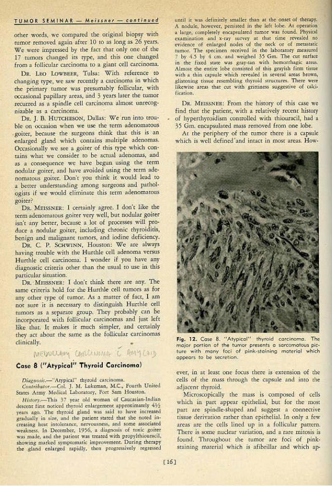
Fig . 12. Coso 8. "A typlcof" thyroid carcinoma. The major portion of the tumor p resents o sorccmo tous piclure with many fed of p ink-stol•"'ing moterio l which oppeots to be .secretion.
ever. i n a r least one focus cherc is extension of the cells of the mns.." thro11gh rhe capsu le tH'Id intO rhe adjaccm chyroid.
.Microscopically <be mass is composed of cells which in part appear <:pidJelial, bur for the most parr are SJ>iodJe-shaped •ncl suggesr a COilnective tissue derivarion racber chan epichelial. ln -only a few ar.eas are the cells lined 11p io a follicular p:meru. There i~ some nuclear variation , and a rare mitosis is found. Throvghout the nuuo.r flee Joel Qf p iok· scaining l'l'iacerial wh.ich j s afibriUar and which :ip·
peats to bt more n product of abnormal secretion th:m n true fibrosis. This ml!terbJ is iocim:uely con· nected with the cells of rhc nodule.
The problem hero is somewb>C similar co <he prob· km of rhe previous 01se. I rhink we con cooclude that this is nn invasive mmor. The problem is one of deciding on the proper name for k
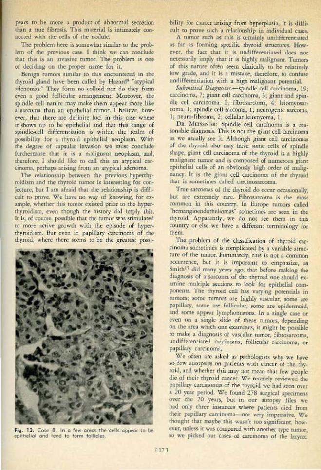
Benign rumoN simil>r ro this encountered in th• d)yroid g land have been called by Hazard' "arypical adonomas:• Tit!?)' fo rm no colloid nor do they form even a good roll iculn.r ru:rnngemenc. ~foreover, the spindle cell nAture may mnke them appear more like a sarcomn 1h:tn an epithelial tumor. 1 believe, however, thitt thcr~ urc definite foci in this case wbcre it shows up fO be eJ>ithelhll uud cbat this range of spindle-cell differcnri'fion is with in the realm o f possibility for u thyroid epithelial neoplasm. Wirh rhe degree of cnpsulor invosion we must conclude furthermore that ic: is a nt.1.lignaot neoplasm, and, <herefore, I should like co cnll chis an atypical car· cinoma, perbopo arising from ao acypical adeoorna.
The r<l11ionship btrween <he previous hypcnhy· roidism aod rhe chyroid rumor is interesting for oooje<:rure, bur I am afraid rhac chc relationship is diffi· cult ro prove. \VIe l>a,•e no way of knowing, for example:~ whether 1his 1urnor existed prior to the hyper· ll1yroidisrn, even rhough the h isrory d id im ply this. It is, of course, pc'ISSjble 1 hat rhc rumor \vas sdmuJated ro more accive growth with the episode of h>'f>er· thyroidism. Uuc cvtn in paplll:lry carcinoma of the thyroid, whCI'C there seems m be rhe gt·enre.sc possi·
Fi9. 13. Coso 8 . In o few oreos; rhe cells oppear to be epitheliol ond lend to form follide$..
[ 17]
bilicy for cancer ar ising from hyperp lasio, it is diffi. cuJr m prove such a relationship in indl\.-idual (t\SCS.
1\ rumor such ns rbis is certainly undiffcrcnri•red os far as forming spa:ific chrroid srrucrures. How· ~ver, rhe face chac ir is undifferentiated dots nO< nr:ccssarily imply <hat ic is highly malignanr. TumoN of rhis namre ofcen seem clinically ro bt rebtiv~l)' low grade, 2nd ir is a m& ake, dJcrdore, 10 confu~ undiflercnciarion wirh a high malignatu potential.
S11hmiucd DiagnoJes.-spiodle cell carcinomn, 19; carcinoma. 7; gianr cell carcinoma, 3; giant n.nd SJ>in· til<.: cell carcinoma, 1; fibrosarcoma, 4t leiomyosrtr· comn, t: spjodle cell sarcoma. 1; neurogenic so.rcorn:), 1.; ncuro·fibrom:~, 2; cellular le iomyoma, 1..
Dn. MmssNUR: Spind le cell carcinom:• is n rea· sonnblc diagnosis. l'his is noc rhe giant cell carcinomn ~~s we usually sec ir. Alchough gianc ce.ll c•tcinom:ts of the chyroid also may have some cells of spindle shnpe, gianr cell carcinoma of che chyroid is • highly mlHsn:~m rumor 'lltld is composed of nwncrotas gianr cpith~liol cells of an obviously high order of malig· nancy. It is the gianc ccU carcinoma of che rbycoid th~u is sometimes called ca.rcinosucoma.
Tnrc S.1fcomos of che chj•roid do occur occasionally, bur are extremt"Jf rare. Fibrosarcoma is lhe most common in this oouncry. in Europe rumors called ''hem;~ngioendorheliomas·· sometimes nee sc."C'o in the th)•rc)id. App,·entl)', we do not sec cbem in this counrry o r else we have • diffecenr rcrrnioolo!;Y for rhell'l.
The problem of rhe classificarioo of thyroid cor· ,cinoma sometimes is complic<lted by a variubJc srruc· cure of che cwnor. Fotntnately, d1is is noc a common cx:currcnce, but i£ is impornaor co empho.site, as Smith" did many years ago, iliac befo re making the diagnosis of n sarcoma of che ch)"Ioid one should ex· >rnint mulriple seccions ro look for ~pichelial com· ponctU$. The th)•roid cell has varying pot~ntials in rumors; .some rumors :l.rt' highly vascular, SOClle arc papill•ry, some >re follicul>r, some ace epidermoid, and some •ppear lrmphoffi3rous. In • single cas< or even on a single sUde of chese rumors, de~nding On the area which one enmines, ic m ighr be possible ro mak(; a diagnosis of vascular rurnOr1 Hbros.arcom:t, unc.liHc:rentiared carcinoma1 follicular carcinom~a, or papillnl'y carcinomn,
\Yh· of ~en na·e nsh ·d ftS jJMhologisE$ why ·we hnvc so few outopsit•s on l'atiencs with e mcee of the rh)'· roid, nnd whether this may not mean lh:lt few people die of their thyroid cancer. W e recently reviewed the papillary C•trcinomas o f che <h)'roid we had seen over n. 20 year period. We found 278 S<Ugical specimen$ over che 20 years, bur in our auropsy files we bad only three insmnces where patients died from cheir papiibry carciooma-oor very impressive. We choughr rhar maybe chis wasn'r coo significanc, how. C\'Cr. unless ir wu compared with anodlec t)'pe mmor~ so we picked out cases of carcioomo of the Luyox.

TU M OR SEM I NAR- M ~issncr conti n uo (/
While we had 32 L surgical sp<rimens of arcinom> of che larynx over 20 year$, we " 'ere :~~W~Zed to find char there wen: four insmnces in our autopsy files where patients had died of their cnrcinoma of the larynx in the s:ome 20 year period. I'm not sure what cbis proves, exccpc that st:tdstics can prove anyching.
DR. \'II A!.'l'DR KROHN, Galveston: UnfOrtlln<>tely, there is 'l mjsundersmndlog ln the rerminoiO&'Y· You h<.-simtc, Or. J..ieissner, co call chis one an uudjf!er· enriated carcinoma. Undifferenthtted, in my under· sranding. means it is ooc differentiated inro a patrem; whereas aoapla.stic refeJ:S ro rht individual cclls. Unfottunoccly, in all rexcbooks anaphurk and undifferentiated are put in che same pot. A rumor can be highly undifieremiated, being not onapl•sric; and a rumor om be well differenti:ued in[o a pattecnJ being tc.:rrifically :maplascic._
))H. MBISSNER: 1 agre-e, £com the pathologic point of view I should lil<e to call this an undifferentiated carcinoma; and yet, i£ we send rhnc dingnosis back co our surgical coUeogues, rhey '"ill immediately thinlc this is a highly malignant, wild cancer, which I don'r think ir ls. I cbooghr about this poinc last week and C\ en nored mac in Dorland's medical dictionary "unnplasia" is a synonym for "undi£feceotiarcd." lc dotsn·r necessar.ily mtan highl>• malignaor, according co the d.ictioottr)', although l am su.re mosr of us usc che recm anaplo.sia co sign ify n highly malignant tumor. COl~. LUKHM AN: Afcer our surgical reporc went:
our rhc patient was transferred co Brooke Arm)' Hos: piral where she was rco:amined with rhe object of doing a radical oeck resection. At the cime she was enmined no oodes were found, and I believe it was felt dut further surgery was nor indict ccd '" that rime.
CoL. J . .L. HANSEN, Brooke Army Hospital: No further surgery was done. To date she h!ls no fwrher evidence of disease.
Case 9 {Leiomyolipoma)
Ditttt~osiJ .-Lt.iomyolipoma. Contn'bNtor.-Dr. J. H. Childt-n., tht Universi(f of Tens
Mediffi Bnnch, Gal\"eUUn. HiJIOr).-TbU ~) rear old <Oiorcd femak noted inter
rnictent ~in in lhe leh lov--er qu:tdraftr o( d~e .lbdomc-n for 1 yeilr. A local physician told bee Sh(' had It rumor o{ the uce:rus. During the J:ut ;; months she had note!<~ ircegulur vaginal bl(ltding.. Gr05S1y, the u1erm, induding rhc: vaginal ponion o£ the ccn•ix, was 17 b)' 12.5 by 13 ern. An oval, soft, 12 by 14 <:m. lllol$S was pre$ent in the fuodu:s !lnd was SUHOUildt-d by :1 l on. .1im of 11\)'0MC'Idum. The rut surface was )'<:llow, greas)', ;~od gliJ,rt-ning. A ~econd ova), 1.7 em, rna~' was present in tht mromtllrium th:tt h3d a linn: s,ray·whh~ cut surfaoe.
DR. M UISSNilR: Presumably rhe sections for the semin>r we~ r.~ken from tbe larger ydlow myometti-
[ 18]
Fig . 14 . Coso 9 . L~iOn'lyolipotnO of t.Jterus.. The tumoris vosculor ond c:ornposed of adult fot ond smooth muscle.
al moss. Microscopically ir seems well dema.rcared from chc :u.ljnccot noJ'maL·appe-Jring uccdnt! muscle. The muss is composed mostly of adulc rnc cells which focally show only slight degeneration.
lnrerrningled wirh rhe iar cells are VOI')•iog-sized, irregular masses of cells of a dilferent type. These cells are spindle shaped with spindle shopcd nuclei and wnd to be whoded and cluStered abour blood vessels, which are numerous. The spindle shaped cell<, except for rheir whorled •rr•ngemenr. appear qu ire similar to the cell< of che adjacent normal myometrium. Thc.te is on¢_ 11iitosis, ~nd cells nnd nndci sbow linle pleomorphism. The cndorhclinl cells lining the numerous vascular space.-; in chcsc foci often are swollen and even occasionally suggest tbnc d>ey ore epirhdia) ratber than endothelial. Many of the vascul:u spaces conuio blood.
I believe rbat this is a benign rumo< composed of far and smooth muscle and cl.,~ therefore, the besr di•gn<ll'lis is a leiomyoliporna.
Submilf~cl cli"gnoser.-iipoma ( fibrolipom•, 6), 18; 1eiomyol_ipoma, 7; angiolc:iomyolipornn, 2; hamar· r·oma (fibro:lngiolipumn, I), 3; bellign mesend>ymum:l, 6~ mixed mesodermal mrnor, 1; li pos.1rcoma, I; lciomyc>sarc:oma, l: ca.ccinoma of cntlomctl'ium, l.
The mher considerable number of small vessels intermingled widt the smoorh muscle in rhis lesion suggest rho possibility of an angiomocous demenc. Lo fact, chtrc arc foci char somC\\•h>t resemble tbe
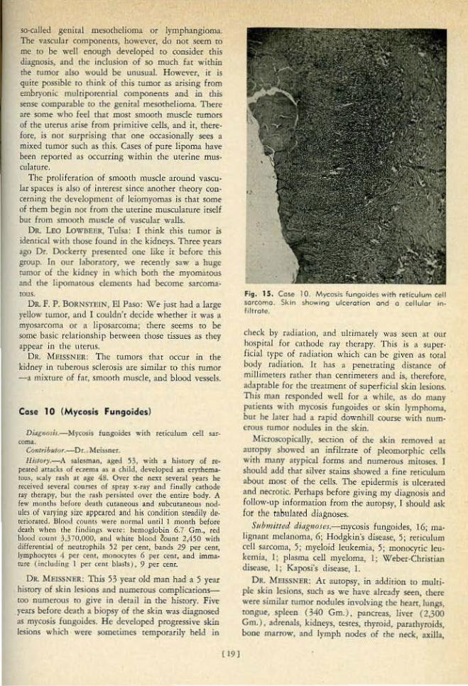
so-CliUod g<ULhll mt."Sochelioma or lymphangioma. The vasrub.r componencs~ however, do noc seem to me ro be well enough developed ro consider chis diagnosis, and rhe induslon of so much f!lt within the rumor •lso would be unusual. However, ir is quite possible ro think ol rbis rumor as arising from embryooic mulriporcnrial componenrs and in <his sense comparable co the genital mcsorhelioma. There -are some who fed th:u mosr smooth muscle rumors of the- uterus :uise from primidve tells, and it, therefore, is nor surprising rhtu one occasionally sees a mixed tumor such ns this. C'ISes of pure lipoma hove been reported ns occurring wichio rhe uredne mus .. <ulaturc.
The p roli!crnrion of smoo•h J>wscle around va.<eu· Jar spaces is nlso of inceresr since anmher cheory coo· cerning the development of Jeiomyomas is char some of them begin noc from the uterine musculnrure irself but from smooch mu•de of Vllscular walls.
DR. LEO LowoerR, Tulsa: I lhink this rumor is identical wirh tbose found in the kidneys. Three years ago Dr. Dockerry p..-orod one like ir before <his group. In our labomory, we recendy s.>w a huge rumor of me kidney in which born me myom>rous and the Hpom3tOUS l'lemenrs had become sarcom•· tous.
DR. F. P. BORNS'I'RIN, Bl Paso: W/e just bad a large rcllow tumor, !tnd I couldn't dedde whether ir w•s " myosarcomtl OJ: a li posnn.:oma~ chere seems to be some basic r<'l:nionshi1) between chose dssues a~ chey n.ppear in the urcrus.
Dlt MI1JSSNUit: The cumors tbtlr otcur in the kidney in nrberous sclerosis a.re simjhtr co this rumor -:1 mixture of fnr, smoorh muscle, and blood vessels.
Case 10 (Mycosis Fungaides)
Di.tt~Wsir .-M)'COfis fuogoidc$ .,•ilh reticulum cdl sar· COIDL
Con:ribtti<Jr.-Dr. Md.untr. H;Jiory.-A saiCJ.m:an, ased )3, with a history of re-
peated ;macks of 1:0.c:m• ~t5 a child, developed an erythema· tous. s011ly ra~h ~~ age 48. Onr the ne;rr several )'t-ars be .recejvf'd se ... cral course~ of .SI>ray x-n1y and finaUy Cllthode ray thcrnp)', but chc nuh pc:Nistcfl Qvcr the e.t1t.ire body. A {e.w monlln bcrorc dCf\lh cun~neous and .~ubcumneou.s nodules or varying sixr appe>rcd and hi< condition Slcodily ~cteriorared. Bloocl count$ were nurmal until 1 roomb before dca1h wheo d1t' (inJin,;s were: hemo8lobin 6.7 Gm., eel.! blood <ount 3,370,000, ~nd white: biO(l<l toun; 2,450 widt differendlll of ncuuuphd.s 52 per cent, bands 29 per <eor, Lymj:lbocytet 4 pe-r (tnt, monocyttS 6 per cent, and immatUn; (including I per rent bl:tSl$) , 9 per ccnr.
OR. MEISSN'ER: This 53 year old man had a 5 year bisrory of skin lesions •nd numerous complicacioRSroo numerous ro give in deroil in che hisrory. Five years before death • biopsy of rhe skin was diagnosed as mycosis fungoides. He developed progressive skin J .. ions which were sometimes temporarily held in
I 19 J
f Jg. 1 S. Case 10. Mycosis fungoides w1th reticulum eell sal'eomo. Skin show.ng ulceration and o collulor •n· lilt rote,
check b)' radiocion1 and uhUnarely was seen M our hospirnl for cathode ray therapy. This is • S\lpCt· ficinJ type uf r":tdim-ion which can be given 11.s IOtnl body radiation. Lr h35 a peneuaring disrnnce of mill imctcrs rt.uher than Ct:mimecers and is, rhcrefore, 11daprable for rhe ueacment of superficial skin Josions. This man re1ponded well for a while, as do m"ny p•ricnrs wirh mycosis fungoides or skin lymphomo, bur he brer had n r•pid downhill course with num· erous mmor noduJes in the skin.
.Microscopic:illj•, section of the skio rcmnvtd or autopsy showed an infiluare of pleomorphic ~Us wirh many atypical forms and numerous mitoses. I should add rhar silver srains showed a fine reticulum abour most of the cells. Tbe epidermis is ulcerored ond necroric. Perhnps before giving my diagnosis nnd fOIJOW•Up information from d1c autOpsy, [ ShOuld nSk for rhe rnbnlnr'ed diagnoses.
Submitted ditJg11()1l!J.-mycoois fungoides, 16; mnli8f1:tnt melanoma, 6; Hodgkin's disease, 5; n:liculu rn cd I S:'lccOm:l, 5; myeloid Jcukemi:t, 5; monocytic len· komi•. I ; plasma cell myeloma, !; Weber·ChrisdiUl clma.w, I; Kaposi's disease, I.
DR. MP.ISSNllR: Ar aurop5)', in addition ro multi· pie skin lesion., such .s we have already seen, there wtrt $imililr rumor nodules involving rhe hearr, lungs, tongue, spleen (340 Gm.), paneteilS, liver (2,300 Gm. ), adrenols, kidnC)><, resres, thyroid, pararh)•roids, bone marrow, and lymph nodes of the neck, axilla,

TUMOR SEMINAR- Me issner- co nt;nuccl
medi>Stinum, and abdomen. He died witb l"nnirud bronchopntumooia and uremia. \'11 e inrerprcted this ,. being a widespread reticulum c"ll sarcoma of a m:tlign>nt sort, which some might call Hodgkin's sarcoma.
This cnse represencs '"hat w.l$ clinically mycosis fu ngoidcs :mJ what hns ended up P"thologically as a h ighly mnlign:ult disseminaccd lymphoma, involving essendaliy all portS of rhe body.
During che past few years, beaouse of patients being referred for 01thode my chempy for the crearmenr of clinical mycosis f\lngoidl's, we hnve seen a ronsidernble number of biopsies of the skin of ruch indi\'idu.Us. The experience from rt-:~ding these bi· opsies has led us ro feel mar while mycosis fungoides tS a. reasonable clioicaJ enriry. ir may be extremely difficult co diagnose as a pathologic entity. The reason for this is thar the disease known clinically as mycosis fungoides charncceristicnlly go<:s through severn] st~l 8,C.-"$. ln the first scage~ which is known to the clinicians 35 erychemarous or prc-n"'ycotic State, :1
biopsy of rhe skin usually shows rninimal changes o i chronic, nonspecific dc:nnaritis. The round cell infiltrncc is > little dt-eper man one usually sees in ordinary derm•titis, bu< the piaure is oor diagtlOS<ic of a lymphoma aod cet~ainly nor of a specific lymphomn. As the disc::tSc progressc5 clinically ir encecs whac is called rhe plaque st:Lgc, nt which rime :1
biopsy mn)' be d iagoos•ic. In th is Sl'!l8< there is he· quencly n pleomorphic cell infilcr:He ar the corium ,,~hich ma)' reserublr: some't\•h:u a Hodgkin's disease infilcrnte, and there are often small microabscesses in the overlying epidc:nnis which arc called Paucrier abscesses. This piaure may be specific enough tO
suggesr scrongly the diognosis of cutaneous lymphoma and, more pnniculady. mycosis fungoides. The third clinical stage is known •tS che nodular or nunor <cag<:, •nd here me biof>$y shows the fullblown ch~tnges of l)•mphoma cutis, ac times re· semblin0 Hodgkin's dise;,se, tericulum ecll s.~tcoma, or Ol'hCI' lymphoma. During all of rhese Stages tbe clinicnl diagnosis may be m)'Cosis fungoiJcs1 bu[ ob,•iously tbc pathologic diagnosis is possible in only a limiced number of cases.
i\bny p<~thologists ba,,e felt d1at mycosis fungoides is only • manifCS<!ltion of rur.mcous lymphoma and, therdore, should oor be considered as a separate p>ehologic entity. This may, in p.m, be cruc from rhe p~chologic poim of view, buc it is not true from chc di1"1ic:ll poiuc of -view. To the clinician mycosis fnngoidcs is an endty usunUy di.srinc:t from ocher cypC$ of lymphoma. 1 c is mtc' th:n m~lny paticnrs with mycosis fungoides ultimately sho,-..• clissemjnared l)'mphorna such as in dte S<.:.ll"'in:lr case here; and, furcher, chis disseminated lymphoma may be similar co a lymphoma urwsociated wich mycosis fungoides.
[20]
Fig. 16. Cow 10 . The infilrtote is pl.ornorph1c:.
Gall' in a review of chc oucopsies of 129 cases of mycosis (ungoides repom that chc uhimatc lesion. whtthcr neoplastic or not, is confined to the skin in 39 ca..~es. und fwcher, rhac n.c the terl't'linacion o£ the dilcllSC there w:rs recogniznble m:rlign:cnt lympho· mot of one f<:>rm or aoocber in tW()·thirds of tbt' cnses. Our c..·urrent feeling abou[ ch<: term mycosis fungoides is that while h is -a useful one ro the dinid:uu, it is n diffjru}[ one in mosr instances for d>< pathologisc co use as a Spt<:ific di~gnosis.
DR. Cr-oRGE MANJ. S3n Anronio: lbis is mosdJ a question in cerminoJogy. In vie\\• of rhe ht:mJcologi· cnl findings terminally, why couldn't you call this some form of a cecituloendorhdiosi.s-in other words, a rcukcmi:l, h terminal leukemia, mtht:r 'h:ln l\ lytn· phosnrcomo?
DR. MccssNaR: I would ha"t no objection ro cl>ar dingnosis.. This man ar le.asr h:td a leukemoid piccure. I'm nor sure t hat he had a leukemia nlthougb the bone marrow was rnassn•ely involv«< by i11filcration with the ruwor, I c:m go •long with Custer's' views .hour che incermixrure of all of chose lymphoma;, and how they ch•nge from one rype to J.Jiotber. So co h:tve leukemic maniiesmcioas in a p.,titnt with reci<:ulum cell sarcoma seems perfectly reasonable co me, nnd I wpuld n'< objen if one w:mrc-d co cnll this leukemia in nc.klidon ro the orhN finding:;.
DR. SllVUllANCti: Were these irnm:uure cells possibl}· jusc lurnor cells circtlbring in rhe blood?
DR. MUtSSNClt: Yes, I think they may well ba.·e been jusc cumor cells, but docs that nt-eeswily mike il :t k"uktmitt? rm oor sore whether it docs or nor.

Cose 11 ("Dosmoid" Tumor)
Dittg·,J.osis~-··oe!ml>id'' tumor. Ctmlribulor.-Dr. A. 0 . Sevcr;'ln(t, Baprisc Mcmodal
HospiutJ, &.u Amonio. HiiiUT)'.-A 33 yt"o~r o ld femaJc nMiced a sligtuly p;~.inful
tolar~eo! just medid and otlet 10 the- spiDC' o( the lrfr iliac c~t. Thi} wu first noml '4 hile taking ex~rciscs after rhc: birrh o f " cluJd 2 re·us bef'orc. She h~d become con~ scious of it, but d id nor pay ~ll)' ~~uent ion to il until re· cend)•. X-ray showed ir to be u !IO!c·ri:;sue mass. not con· ne<tt:<l with bone. l!t11minadoo of the parjenc's back sho11.•cd ~ snull mass mcd~t and nexc to the spine of the left iliac cr~ 'llppearin,~: to be <ooncetcd with the fuci.a O\-'er the crftt cf thC' ilium. It v.-as )lif;.htly tender and fredr mowble-. Tite Sl>l'<imen ronsi!ted of " rouodeJ mass meas· urin8 4 by ) by 3.~ ern. lc was firm, p ink isb -gr.iy, nnd well circum$cribcd. ·n1e cut au.dacc w~•s pink ;aud gli.nening with 8rayish (ibrouJ hands inte.r'-li8iuuing in all llireccions.
OR. MEISSNER: This 4 em. circumscribed mass which had been present for '" lo•sr 2 rears was re· lTIO\'ed from 1 he subcmaneous rissue of the lower bock. Microscopically ir is CQmposed prima1·i ly of spindle shaped cells which concain spindle shaped nuclei widiOU[ mlroses.. In some foci there &s linlt ,·uiarion either in rbe ccl.ls or their nuclei, and be· cwc:t"n the cells are strands of coiiJtgenous cls.sue which :ue inrimateJ)' ns.socbted with che outlioes of the ccJis. fn Q[hcr foci che cells show- •nore variation in size :>nd shape wich sorne srella<e forms; here che nuclei art also more vari:tble with some of large siu bu[ snll wirhour miroses. 1n rhese foci the cells are in a looser arrangcmcnr, and are Jess intimately nssoci:..red with collagen fibers. 8och cbc ctw1necri ve tissue fibers nnd che cells rt:nd ro grow in nn ime.dacing, ofceo whorled parcern but without definice srruCturol for· lll:lcion. Ar rhe periphery of the = sorne of che cells wirh their collagenous fibers extend as an infiJ. muion imo the ~tdjacent srriatcd musd<:. T he S(tOI'O:l
sho'''S moderncc v~1scularity.
The problem in chis case is largely a mart<r of nomcnclarure. The spiodlc shaped cells are forming oollagen and :Itt therefore libroblnStS. ·This, then, is n lesion composed of proliferating libroblnSIS or fibrocyte'S:, which is infrltracing or iJ)V(tding :\djacenr striated muscle.
t know o( no cewt.in wa_y to detennine whether or not this lesion is even a IIue rumor. Dr. Stotn19
mak~s a particular point of thr d iff imlri~s of deter· n"'ining j f th<.·rc is such a ching as l't bcnig1"1 rumor composed of fibroblasrs.
About che only rhing of which we can be cerram here is char rhts is a mass of prolifenting fibrobl:lSIS. 11>e name lor >ttCh a lesion is 1lways n difficult problem for the parhologisc. lr ,is my feeling thnr • ltsion sudl as this shouJd ben bt' g iven a clescriprivc diognosis, such :u low-gmde proliferation of fibrous tissue. \"{le know, bow-ever, th:u in me pracdce of p•d•ology such • diagnosis is no< readily occcpcable I() our clinical colleagues They wane us 10 pur a
Fig. 17. Cose II. ''Dcsmoid tumo l'.'' Ptollferating sp)n. d lc-shoped fibroblost$ gtowing in on interlacing, o ften whorled pattern,
name to rhe process and <O tell thtm whether ir is a benign or maligr·Hmr n1mor.
To <:all chi~ an t1nqualified fibro.s:•rcoma is some· whar of an O\'ctdiagnosis in spin: o( che face rhlt the rumor is eJccending inro Jdjacem muscle. A lr:w )'C"lfS ago, ~fusgrt.wt" and McDonald wrOtt' an interesling paperJ# on cxu a·ahdominal desmoid cumors, :tnd if one ooust nppl)• n specific nnme co chis lesion [ rhiuk dc.srnoid rumor is as reasonable as noy, for SC\'Crol reasoos. firsr, che rumor has occurr~ in a female following pregnancy; <his IS a common hisrory in dt!Smoid tumors. Second, rhe rumor has s1 rinced muscle fibers ancluded wirhin ir, :tgain n common finding in dcsmoid cumo•·s. Tbe low-grade nncure o( rhe fibrous proliferacion is nlso consistent witb this diagnosis.
Mr diagnosis on rhe Jesioo, rhcn, is "dcsmoid" 111100£,
Srtbmittetl diag•uuai.-desoooid cumor, 13: fibro.1:trcorna, 7; leicunyosarcom:~, -1; rh®domyos:t.rcoma, 1: fibroma ( neuro- I; fibroooyxoma, 1), 7; neurilemmom:l, 2: fibrom.:uosis, scorifo.cm, 1; Hbromarosis, nodular, 1; "milk·line" fibroadenoma, 1; hemangiocndo! helioma, J; mallgnanr ginnr cell rumor, L.
Ot\. MEISSNCII: There is no more difficult field lor p>~hologic nomendarure <hon char of fibrous tisstlt prolilemrions of chis g~nenl rype. \VIe, as p.,hologiscs. all know how diHiculr ir may be co disdnguish, for ex:1mpJe, bctwccf'l scar tissue and a
(2 t J

TUMOR SE·M I N A R Meiss·ner contin ued
keloid) or even between star ti~ue and low-grade fibrosarcQma. Unformnatd)', jn man}' instances» the only wny the diagposis can rmly be derermined is by folJo\viog che d.inkal_ cOur$c and nocing wherher or tioc recurrences· or mec:israses occur.
It is quite possible chat r,his lesion ·reptesenrs a very low-grade fibrosarcoma. I do nor th ink so, bm on rhe ocher h~md, r have seen mecasrasizlug ,fibrosarcomas wbich were at least foc-o~Uy as weH differ-entiated as this lesion. .
Musgrove and McDonald in theil' paper made a _particular poinc of an:empting m disdngulsh berw~n e-xua-abdominal dt·Suloid rumors and rruc fibmsa.r· comas. They fou nd, as might be expc~ted, chac many fibros<rcomas could be easily distinguished from desmoid mmors by [he prcsem;e of munecous mitoses, tumor g iant cells, considen1ble pleomorphism o f cells, and. a high dtgrec of cellularity. T bey were careful to })Oin c 9uc, how<:vCr: char the diffetencial diagnosis between a fairly cellu)ar> beo.ign, fibrogc.tiic lesion nod a low-grade fibrosa.rcoma is one of the most <lifficu)L decisions a pathologist n>ust make. The only Mty tbe patboJogisr can be sure, and not !00 per ce.nr -.u rb:tt, is ro dere.rmine che final outcome in each indi\•jdual case.
It ahv:IVS has S<.'tHncxl tmforrunatt tO me thar ou.r nomencla~re of t\lmors must emphasize chc distinction becv.:(!ell benign t~.nd malignant. Tc is rrue, of course, that S\ICh a djstiuttion ofrt~n C3n be made qu.hc readjly~ buc in tissues wh,i~h are as uncomplicated as connecrive tissue, c.dteria fo r distinguishing nor only berweeo be1'1.ign and ro:tHgnnnr rumors, bur ~lso between simple str\Ctive prolifemc.ion ~nd rumor are ofren lnckiog.
DR. S B\'BRANGB: Tb is P.•cienc was last seen by the famil)' doctor nbout the middle- of August of this year (1958 ) . ln the inrecval since 1952, when chis lesion was remo:ved, chc y.1dent has had one more successful pregnancy. Tl"'ere· have bten no additional lumps or bumps of ·this type, and rhe area from which chjs was removed is sciU 'free of a ny recurrence of anything th:lt <•n be felt clinically.
OR. SMJTH, LokeJaod : 'Did you think that h istologically they got a round the nunor?
DR. SEVERANCE: 'Ibat's ~lwa)'S a goocl qoescion. J thi.ok they b"d a small amount o f far around the rumor.
OR. 1\. M. fuCHMOND, San Amoru6: Or . . Meissner, hftS ic been your observ~tio.o rhac ch is r.ype of rumor in the neck region is apr ro look much more m:~lis· oam chan it wiJl·evenrually aCt? •
DR. MEISSNER: T don't know al)out d1e rumor in rne neck 1egion specifically as looking mo.re malignant; 1 rbink all of rhesc les.ions rend to look more maligoanr than they act. They n eely metastasize -a1· though it is true chat they may recur locally, and they
m::ty recut [0 such ~ degree ch:u one;· mighr chink of them os being locally mnligon11r.
On. J. J. ANDUJAil, Fore Worth: Tbis ·is, as' D r. ~1ds,snt:r s.uys, an exercise in semantics rather tbau pathology, so [ ·would like to ·ask D rs. Sev.erance aod Mtissner wh.r tliey use rbe word excra-abdomioal desmoid rumor. 'fhcte is am]>le p re<;edem for scatil\8 cbm destnoid can occur .in numerQU$ Qdler areas. including the' nee~ so wby ~ack on excra-..a.bdominal?
Dr\ . . MEISSNER: I don't think ooe needs it. Acrually, the definition of desmoid mmor is a fibrous rumor. or fibrous pro1iferadon~ not necessarily a tumot; and rhe dictionary definition of a desmoid rumor docs not specify th:u it muse be in cbe abdomin:tl wall.
OR: SJW.ERANCE: :Don't you imagine char the first nu:enr-ion c-alled co chese tumors was tc:> chose happening tO be in the abdominal regiou following p(ognancy, and chat is the reason e\rerybodr calks 1\_bom cxtra-obdooolnnl w hen the')' occur Hsewhere?
:On. C. P. ScH\VfNN, Houston: \Y/ e had a fair munber of these rhar fi t the dassification that McDonald set ouc, and we have seen rhcm at cbe lvi. D. Anderson Hospital nnd TutQ.Or lnsdcme somctiJnes in their second, c.h ird, -o.r fourch rt>corrcm.:e. We bave found tbftt 1lc;ule oJ [bern have ever m~msrasized, bur thar rhey fxcquend)' w_ill grow· along fasci:d sheaths, nerves> and ~<> on. so rh~.c- in :~ few ·cases ampucarion has been necc:.ss:.u)'.
Case 12 {Malignant Giant Cell Tumor)
.Dirtg,H)J;.J,- M-ali.g_nam giant c:ell tu mor of soft tissue. Contrihutor;.- Drs. D. A. Todd, A. M. Richmond_, n.od
Sylvia john~. Nix Hos_pit:tJ Clinirnl J.aborarpry, San An· tonio.
NirlQry._-:Th-i.s 58. y~c o l<l (OJorcd fcma1c fjrs:: noticed a "swelling" in the Jcfr amccubhal fossa eat11 in l9S6, which W:tS said to have grawn r<tpidJy~ The l,llass was ct-moved but aJ~p;uent ly no tissu'e studies w(•rc OOJ)e. The s,cowth rapidl}' tceltrrcd, and jn M~y. 1956, s:evcr:rl circumscribed, lobuJnr , S(llid, yellowish b ro wn Li.ssut: masses were removed, the largest 13 by 25 mm. ill si~e. lo Jul)', 19).6. t.hr: patient was referre<l to a hos:pjral bc<ausc of pai.n
i n the right elbow; -.ic rh.i.s rime the antecub ital _fossa \\':lS cxp)o{t<l, and_· a noduJe was ··remove.d from rhe ulnnr nerve," The tissue for examin-ation Dovo· is thar \\1hich was removed in May. L956. Past histor)' indicateti a ''suoke .. in 195'· p¢Sith•e secoi.;:>Sf, and moderate hypertenSion.
DR. MEISSNER: !' rom the histOry On this CMe we find that rhc- cissue represenrs a ro_plcU.~, recn.rrillg growth removed from the rtgion of the e lbow. \ '(fe are nor told whctl\tr the original g(OWth involved bone or nor, but we are sure rhac rhe recuuence was removed from soft tissue.
[ 22]
¥ kroscopicall)• che tissu~ is compos<.-d primadly of v~riOus siz:ed masses of unifotm-rype structure embedded in fibrous tissue aod muscle. These masses are composed of spin<Ue, o•oid, and roond cells whicl>

scparale and surround ginnr cells. The gianc ceUs often conr-Jln mnny nuclei ~nd tend co be more numerous near blood vc"SSCIS. Some, in facr, seem co be lpng wicbin ' '..Cular SIUCCS. While the cyroplasm of £he giam ct'lls shows :m occasional vacuole, tbese are noc numerous :and no foreign nl!Ue.r-ial is found. The smiller cells with single nudd ocvathdess ha•e nuclei similar to chat St"Cn in lhe -giant cells. A.n <x:casionaJ one of 1he smalJer ceJls shows a mitosis. Abour the periphery of rbe masses of cells, there js some (ibr()us tissue and muscle wbkh is intimarcly interminglec.l with the cells jusr described. In rhe ad· jaccnc fibrous dssue 1hel'e are sn·mJJ nestS of idc:nticaJ cells present. T he str(lma shows focal deposicion of hemosiderin pigmc:nc; t·hcrc is no ne-.1t bone or <.'arti· Jnge fOI'ITIOtiOn.
This is " lesion cumposed predominantly of giam cells, and rn)· firsc decision is to co.ll rhis a giant cell rumor. To me it :tppe•rs quire compornble co the gi<Wt cell tumor o f bone. lr hos less resemblance, bemuse of itS bck of xoncbomuous stromal cb:mge, ro giam ctll rumors arising in tendon sheath. 1 think, however, chat is also a possibiliry.
The next decision pc:rrains ro whether or nor d1e rumor is m.Jig11•nt or benign. Cerninly from rhe morphology a.lont one does nor gcr lhe impression of 11 malignant mmor. 11te lesion is acti\'el)r pro· Jiferarin.g, however, nnd there arc oumerO!JS foci of rumor cells growing in connective [issue, which is
Fi51. 18. Cost I 2. Malignant gionf cetl tUmor. Sheet1 of round, ovoid ond spindle cells SUfround ond seporote numerous mult•nucleored glonl cells.
123 J
S<Ut'ICihing rh:u a benjgn gianl cell tumor is not sup· posed to do. Also. from the history we know that chis lesion has recurred prompdy and is involvins such impormnt stn.ICtuits- 3S the ulnar nerve.
I bdit\'C, therefore, that we must consid~r dUs at leaSt • locally malignant rumor of giaor cells "·hich may even have 1he opabiliry of memswis. My diag· l)()$is, therefore, is malignant giant cell tumOr of soft tissue.
St~bmirted Diag11o.res.- gianr cell rumor, rccurrcnt, I~; nngiosnrcoma, 3; s~rnovial sarcoma. 3; lipos:u· coma, 1: rhabdomyosarcoma, 1; hemangioendothcli· om:t, 6; hcrnangiomn ( w1ch giuut cdl renction, 2), 3 i neurofibroma, 2; myxofibroma, 1; pigot\(•oted villo· nodulnt S)'llOvitis, 1; malignant gaoglioncuconla, 1 ~ malignnnc rumor, 2.
D11. MUISSNUR: Giant ceU tumor, recurrent, does not indicnce whe£her ic was thought to be benign or malignant, but chat is a good point we can discuss in n tllOI'Iltnr.
"fl>ere are :ICttlilly three decisions to be nude in discussing chis case. First, is this lesion a rumor, 0<
is ir merely rt':lcrioo? \\7e know of no hiStory of traum:o, bur it is rrue thor the previous surgery may have stimulared or disseminated the process so rhar it now seems more like rumor. Thls decision is fun her complicoted by rhe fact that rhere is no ulli· vers.al opinion as tO whedter giant cell Jcsioos nrt pure ncopbsms aoyway. There are those who feel chor some of ~hcl'n :tr Lc::~sc represent merd)' rcacdons co injlll'y, or th:lt che gianr cells arc accom)':-~nimcnts of oth~r basic nonmmorous o r tlliDOfOUS proceS$eS.
The second decision deals with site of origin of a le$iOn such us chis. I believe chis tumor i• quire comparable 10 many of the giant cell mmors arising in tendon sheath. These larcer cumors, howcvtr, al· m05t alWllys occur in rhe hoods or fee<; chis would be an unusu:tl sire. Fonbcrmorc, there arc oot the exttnsive xan<hom:uous changes char one frequently finds in such lesions. Recurrences of giant ~II rumors of tendon sheath are uncommon bur· do happen. I believe rh:n a lesion such as chis mighr nrisc from Otht·r sofr tissue, perhaps synovia, muscle, and blood vessels. I ha\'<! seen seven! cases where there h~\'t bec:n such nccumuJarjons of gianc ceUs associated wirh thyroid carcinoma~ h\1t n.ot appa.rend)' fl11 inhl.!rcnr pnrt of rhe carcinoma. Rarher" believts that giant cells o f chis rype, which are variously called om'O· clos!S, fore~n body giant cells, nod so forth, may arise from the endothelial lining of vesse.ls.
The third consideration regards the question of bc:nign versus malignant tumor. Here again the d«:ision is 11 diffk'l1lt one--_rerurrence- o£ benign gi:Lnr cell mmO< is uncommon if ir has been complercly excised. In favor of • malignant process in this cnse is the hisrory of rapid growth, rapid recurrence, and in"oh·c:menc of such i.mpofiaflc StnJCt'Ult'S as the ul· nor nerve. 1l1e disseminadon of rhe lesion through·

TUMOR SEMINA R Meiss n o t co nUnuetcl
our r~ fibrous sttoma is SUS!l"'tive ro mt of malig· ru.nr narure. ~{jroses are fairly numerous in this CJ.Se.
as rhey uc in malignam v-ariants of ginnc- cell rumor; bur, og;~in, tbcy may be seen in benign rumors as well
1 nnd this cenainly is nor \'\ definitive criterion.
T he decision ns to whether a g iant cell tumor is benign or mt~lignnm is excrcmcly diff icult. nnd only the clinical course can g lve d1e true a1lswer.
11\iS p:lrticular rumor, [ believe, h:\S the po1enrials of a malignant one. 1 have seen ~ of giant cell rumor which have metastaSi2i!d which ha\'t looked no woi"S(' than this.
DR. UO loWDEER. Tulsa: Couldn't we seriously consider rhnc <his is :t rumor or mmorlike hyperplasia of ~ynovial ceHs, md nor J rom a joint, and noc froro cendon sheath, bur from che nntccubical fossa? Syn· ovial c:clls nre nmodous for acting ns l'nncmphages). storing lipid or hemosiderin, or flt~ i ng tO form giant cells. Some of the l esions in che jointS, like villooodu· lar S)'novitis, behave and Jook like tumors o.nd are nor, so perhaps tbjs is a rumor originociniJ from <he ancceut?im fossa.
DR. MmSSI<ER: I think ic is possible chat chis m•y have •cisco from syoo,<ia. I would place rbat among che first of the possibilities M <he site of origin.
DH. RICHMOND: T he surgeon in this C:LS<:: said chnt the bone \\1ll.S nor involved at Ul'l)' cime so far as chc.: x .. rn)'S were concerned. She was opcrnccd upon the lnsc cime in July of 1956, ond in December of 1957. she was aJ>pareody free of disease.
Case 13 !Undifferentia te d Sarcoma)
DuttnosiJ.-Unditletentiatcd su coau1. probably rhabdomynsarcom:t.
CrwtribNtQr.- Or. J. M. :Moore, Sctnm RO!Io'l Hospital, San Anm n10. Hili~r,_A sma11, painless mn.,\.o; wn:; nmed in the calf
of the lc~ ol :a 1 Yi ye3r ol<l male. At ellis time it wru. the or inJon ()( t hC {amJJy physicinn thllt it " '.U I cyst, l\fld he advised awo1iting further developments. The mass g radually grew !or I ~ r~rs and "~3S then excised locAlly. The twnor meuured 8 b)' S an. and w.u somewhat intgub.r, Jobul:ncd. and foitlv ., .. u citrum!Ctibed. Upon ..aioning ic ,.., found to ~ Pf'DCtit~lly $01id in coa.sinency. with • hcxnogeoeousappet.n.ncc-. Thtle "·~re also in.)".!ind nudes n:vealing rumor mcwtuis. 'l'b.c wmor w'3S thought co be embedded in mu5de anJ not attached to bone. Cobalt GO radiation is beins; ~;iven ~t 1his cime._
DR. Ml!tSSN BR : This rumoc was rcmo,•ed from ~ young child, and at th-e rime of rcmOvlll the(e nl· re-ady were mctascases. It ·apparend~, nrose in muscle.
[ dlink we can all ag ree that rh is is a malignam rumor, both from the proof provided from lymph node mewtasis ood by cbe cxronsi,•o invasion of adjnct-nt srrucnores and blood vessel$. F11r<hermore,
[ 24]
Fig. 19. Cose 13. Undtffercnt•o1cd sarcoma, pc-obcbly rhobdom)"'SSreotno. In many foc:i '"- 1umor cell.s show a tendency 10 grow in clusters.
rhe nunor does not mucruraUy suggest chc possibility of citltc.:r a Jymphom;1 or a Ci\rcioom11, so the t>robJem, rhen, is ro give this sarcoma a name.
To classify sarcoma one aut utili:ce characterjsdc srrucrur.ll 3rrnn,;emcms, r:he formation of r«egniz· able material by tl>e rumor cells, and 1he appearance of rl>e rumor cells.
One of rhe striking features of this case is the tendency of the rumor cells. in many focif to grow in clusters.. This alveol.ar arr.tngement sussests the diagnosis of alveolar soft parr snrcom:1, which is also sometimes calk><! malignant granub r cell myobl:os· coma and nonchromaffin pa.mg:tnglioma. 11ois diag· nosis is pc1·hn ps nn acceprable one since rh is is a sarcoma of soft pares and it is p:trcly growing in alveolar :1rrangcmem. These rumors characceristically have l:trgcr cells, however, often wicb gmnular cycoplasm, >tnd rhey contain lew or oo miroses. Thf.St charncterisric.s :are not present in our rumor.
In ocher loco of <he rumor <he cells 31C intimacely associated with collagen, sctoogly suggcsciog thac tbe coll11gcn is being formed by rumor cells. This turoQ[,
therefore, might be considered n v!U'b" r of ribrOS<IC· com:~..
Jn still ot hc:r foci: some of tht- rumor cells art v::tcuolared, ru1d although no mareri:tl is Qv;tilable for a far Stain, the vacuoles could be fac and che ruas· nosis of liposucoma is, therefore, o possible one.
In scill ocher loci, rhere •re rumor cells whkb hove

a deepcy acidophilic cytoplasm and in these foci chere are, at times, , JonJ>1tt<:d cells strongly suggesting srriaced muscle cells. J looked for a long cime for cross srriations but was: un~-~ble to find any.
Thjs, then~ is 11 rumOI' wjth a vnl'inbJe pa(te.ro nod perhaps a varinble cell rype, nna I Sttppose char cb~ ~sc r<.:rm js a oo.ncommiuaL one such :\S undifferenriaced sarcoma. I should like very much w c-all thls rt rhabdomyosarco ma, but in rhe absence of cross srrh rions, 1 do pot lee) quite justified iu doing so.
S11in!litJttrt Diagufr•t.- rbabdomyos:ucoma, 7 ; rhabdomyosarcoma. embr}'On.al cype, LO; soft p<l tc ~veohu san;()m9, ?; S)' novi;l,.] sarcoma, 4; myxosarcoma. 1; .reticulum cell s-<lrcoma1 L; a.ngiosnrcomaJ l : ll~S!ltcQm:l, 1; $:lKOfl'l~l (t)' pe ? ) 1 2; helttnJ.JgioendothcJio.rn:~., 3; leiomyoma: 1; 'fibroma. l ; neurilemom:\~ 1; hemJ.ngio-pe.riq ,roma. 1 ~ card ooma of sebactous gl~nds. 1.
DR. MEISSNER: Obviously there js ~._ '''ide varian ce of op in.ion. ~s to the proper name for a sarcoma such ~ts chjs,
i11e _possibility of rhabdomy,osarcoma is stiH in· crlgu.ing. D; .. H orne 1 has described f"( f'l :alveola r ar· r~ngernc:nr in some rbabdomyosar<:omas and p,erhaps che nlveolar a1·rangemenr: io chis case c.to be tx
plained by ch is diagnosiS. Prim ith't srriared musde rumors 1n;1.y have a. vtl..J'ied appe:u:Ztuce and :u- t~es
mt•)' even resemble J)'!Ophomas. Tbc neet""Ssh y for <:awful search for cypicn1 sniacioos most be empha· sized.
Fig . . 20. Case 14. lov.··Q,.ode cc rcinomo of loryngeol g la nds. Lorge epithelio1 cells fo rm olveolor st ructures·.
[15 J
The meras1:asis ro lymph nodes, by a sarcoma, is unusual in ad ult sarcomas, bur is more common in sar-comas o f ch ildre u. Io 23 C!ISes oE .r.bnbdo.lllVOS~li
coma wbk ih Horne was able ro follow adequaceiy, 15 sl1owed rneutsLases co Juogs, bm 13 showe<.l met.asmses ro l)·mph nod<os.
DR. G EORGE M ANI, San Amooio : Did )'On ar :\D)'
time. C(msidc.r <.:aUing a rumor a mescn.chymoma? DR. MEISSNER: Yes, 1 think th:n could be my best
diagnosis, a m :'lligna.nc mesenchymomn, if 1 d idn·t find any cross ~hi:tt ions. l f I had co ~all ir a .specific type, I wot.ild ·catl ic a rhabdomyosarcoma.
DR. MA~ : How many compooenrs are ne<'<led before calling a nunoc a mes.enchymoma?
D.R. M£t~~SN£~: Dt. Stoor is rbe one who i11venced chis wordt and 1 don 'r know-.maybe someone else here does-exacdy whnt h is oecess~r)' number is; I St l ppose that thrt>C or more components should be present.
DR. SEVERANCJJ: I would have said he cqnsideted cwo or mor.e, buc he Joe.sn'r include che fib rous dssue elemenL
Case 14 (Low-Grode Cordnomo of Laryngeal Glands)
Diagm>Ji s.- Low-grade C"<"" rcinom.a o( l;.ryOgeal glaods. Cn-~tltlbNiqr.--Capt. P. '«1. Kiel. M.C., DJookc .Army Hos-
pilaJ, rorr ~m .1-foos"ton. .
HiJt-MJ1.- J\ 68 }'ear old white male wirh 3 hisroty of 3
squamous cell carcinoma of the floo r of the mouth treate-d mrgi<:-aJI}· 4~ rears pre\'iousl}' -coroptai.o.ed o£ dfsphl'l8l<l. of 2 mooths' d·ltt:l_t-ion. There w·..lS- oo voice chansc Mr: "diffi· cuhy with ,.~pimtinn . ..A pedunculated lesion with a, veo· thin one--half juch stalk, Uttacbed in the regioll beneath tbe outer ~r)•rcookl , was removed. The rumor w~s bluish. fi rro, :md solllcwh11r nodular, measuring genemll}' J .6 em. Thr: cut sw.fl\ce w;ts dark red.
D R. MEfSSNER: This patient had -a hiscory of previous removal of a sqU;tmous cell -carcinoma1 grade one, from cbe floor of d1e momb. 111e lesion pte· sen ted for the seminar was pedun<11.Jarcd and acrachcd co che le£r fa lse vocal cord and ~ty<eooid. I ·do oo< believe t her e is :a rcladoqsbip bccween the squamouS celJ carcinoma of the floor of rlie mourh M1d the lesion we have .here.
lhe l<.:sio n. js composed of more or less Citcnm 4
scribed closters of cells which underlie stratified squamous epithe lium, The cypical cell type is tall, cubo id:tl, co n rnins il l:lfge nucleus~ ilnd appear.s ~cated oo a basement membrane. T be opposite si·de of dfe ce]J is d ire<:ted coward · a huneo space. The cell nucleus is large. ~iitoses occas~onaHy ~re fouod. Tbe cyropla.<m of the cell is pole or slightly acidophilic. T here. are n oc granules o r dt·oplets.
11lere ace numerous variations from rhe cypical cell apparently due ro cell disrorcion or to cell degeneratiOI'l, At cimes the cells are rounded and ac

TUMO R SEMIN AR - Me i ssne r- cortt-in u t d
other times grcatly elongated, but •II of these set'lJl
ro be -rely variancs of the bJSic cdl component. For the I11()S{ part the cells tend tO form llll alveobt strucrurc, bm ac rimes they are in more soJid mrtsses. There is uo definite ker:.uin form;ltion, no inter· cellub •· b .-idaes chac 1 could find, and no fo rmation of mucus.
The cells, where tboy arc adjacent tO the stratified squrunous t t>itheliu.n, show· a mini m:\1 invasion. The C'ells also are invading srrotna, and [here are numer· ous clustc:rs in and lining vascubr spaces.
The stroma for rhe most part is scon<y but highly vascubr. In a few aceas ir bas a hyaline appearnncc, buc rhcrc is no fo.rm:uion of carrilnge. Ac one poim near the periphery of the rumor "rc Slh'eral normal· appearing ducts. oad rhe appeamncc suggests thnt these nrc leadina from the 'epithelial surface inro the rumor.
·n,c d iffcrcnrial diagnosis in th is cnsc includes many possibili ties~ such as memstatic carcinoma, ma· lign::tnr n1elooom:;~_, hero:~1\SiOC1\dOi htlioma, and so fonh. Raoher than discuss all of rhese. I should like tO point Oat the feanues in this lesion whicb have led me to my diagnosis. In the first place, these cells look to me as tbougb they ore epithelial. 1bc tall, cuboidal nnttue and [be cendtncy ro g row in clusters wirh some alveolar at_rangemem ~ltC characrer.i.stics of iln Cl>ichelinl neoplasm. Secondly, the tumor cells. arc im•ltdjng su·on\fL, blood ves..~cls , :tnd ov<:rl)•ing squ:-~mous ephhcliwn; mereforc, this js 11 malignanr cpirhcl iol neoplasm.
11)c site or origin of d1CSC' cdls is :ln imeresting subject for conje«ure. The stroma is not very t)'p ical of mixed rumor of salh'lU}' gland, 3nd yet rhere is • suggestion here nod there of the dt11se hyaline ma· terial th., ofren is seen. The proscnco of severo] ducr suucture) \\'hich seern ro le!td inro rhc tumor suggesr.s funher that rhis is a nunor arising from nccessory mucous or serous glands of the larynx. The addo· phil io of some of rhe mmor cdls resembles somewh:u that of the so-cnJJed 01'\cocyres seen in some saJivnr)' glnnd fesions. The absence: of convincing ker3riniz.1tion 3ud of mucus is disttppointing, but neverthele$$, both of these may be absent in saJ;, •• ary &]>.nd rumors. From severn! points of evidence, then, non~ of which is in irself conclusiv~ 1 believe rhis tumor is n low grade cnrcinom01. .uising from laryngeal g lands. 1 rbink it ulso mny be con· sidcrc:d ~~ Cltciooma of accessory S."'llivary gland tissue.
Submiuc•d DMgnoJes.-Sq\19mous cell carcinoma, 13; IJ)f((ISU:Itic squrunous cell c:u·<:inom:l, S~ adeoo· carcinoma, 2; malignaor.mel:~noma, 4; hcznangioendo-rheliomn, 4~ (ldeooma, mixed bunor rype, 3; salivary gland •denoma, 2; adtnom•, 1; glomangioma, 1; bomnngioperic)'loma, 1; paragangliom>, I.
Oa. MIUSSNER: The people who CJIJed this a squa·
Fig. 2.1. Cow U. There is invos.•on of the overlying squa.tnOU$ epithelium.
mous cell cnrc-inoma app;Jrently are in rhc majority. l just could not convince m)•sclf that any of tbc pink maccritll l S•\W w~1S ken1tin. 1( [ had found keratin, however, I would just include rhe mmor in the group of s:tlivnry gland oeopla.sms where c here is an epidermoid con>ponenr as well os the g landul" com· ponenr.
Molign•m melanoma is alw>ys a po<sibili1y in a rumor that i> as difficult to c!Jssify as this. and the ~me is tn1t of hemangioendorheliom1. I musr con· ft'SS rhrtt r have nevc:r St'eJ\ any tumor C:XJCrJy like chis one before. As o ne srudies :1 serie-s o f salivary gJoml cumors, however, bt· js i•npressed by cite great diversify in possibh: morphology. This i~ cruc ooc only in 1 he mix.ed tumor group, but :t l~ in O[ber forms of salivary gland nunors, bo th benign and malignanr.
Dl\. R.. P. DERNSTEJN, E1 Paso: TI1ere is one point I definitely noticed obour this rumor, ond tlt:u is that
whil~ the cells look. vel}' much like ~p•thclial aUs. they seem to be o riented in • most pt:culiar way toward blood vessels.
On. MmssNr.R : The telacionship tO the blood vessels bo1bcred me q uire a bit, but l could not think o f nny vMt'Uiar ce.lls d>nt could look so epithelial.
On. St~l'lli\ANCE: In rhc Atlas of 01olnr)'ngic Path· olog:y by Ash nnd Raum) memion is mac.fe of cumors which look like adenomas o f the bronchus bur arc
• found in the lacynx. Maybe one does nor have ro be ron dogmatic aod say whether it is benign or malig·
[ 26 J
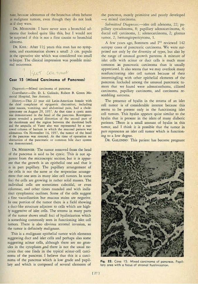
n.1n~ because adenQmis of rhe bronchus ofcea b:el:ia \re ;~s mi li,g:oam nunorst <:vco rhpugb they do oor look .!S if they were.
On. MlllSSNF.R: I h:lve never seen a bronchi•! ~d
enocil~. thar l®ked quite like this, l)ur I would not &e ~urpris~d if rhi.s is nor ·o fiist cousin. ro bronchial adenOma.
lJR. KTEr. : .Aft~r 1]1 years rbis mM h•s no symplOms.. and cxaminacion shows a small .3 cffi. papule in the operotive site which wos considered roo small n) t>iopsy. The clinical impre-ssion was possible mini:m~l r«urreacc.
! 'fx-i Ci'J,L. WM-Q~ Cose 15 (Mixed Carcinoma of Pancreas)
OiaptOJis.-MixcJ. c:.lrdooma o f p::ncrcas. €rmJribJI!Or,-Dc. D. L, Galindo. Roben B. Green Me·
mori:ll Hos_pi(a;J, San AotoQio. Ni!lu,·.- This 22 )'<:ar o!d Lmin·Amerieau !etnalt• with
tbt chief coi.'QpJaim of eJ>i8aSirk -.liscolllfon , jocludios g>tn~ n:tU$Cll, vomiting, nnd 11bdominal pain , h:.d a cbolec~ce<:romy Dn August 29, 1957. At that tin)e a c~·stic mass \\'it$ democ\Stcaced in tl1e head_ of the p<t_ncre:l$. l toentsenogmns rcvrolc-d a lxtrrial distorcion of the second part of tbe- duodenum and the rC,briOn berween the duodenal buJb :~.oJ the ampuUa oi Vater aJSo was reptc~enrcd by a nar· rowed column of b:uium in which the mucos1d panern was edematous. On NoveiUber 14, l957, tbe tumor o£ tl,\e- he>l<l of 1he pancrca.'> was resected. At the rime of opcrariQn no obstruction of the pancre(ltic or conunoo bile. duct S)'sten) was demonsa:ttc-d.
Dlt MillSSNER: 11te rumor removed from the head of the JY<UKreas is s-aid co be cystic. T his is not "-P· parenc from che m icro .. KOpic s.cccion, buc it js appar· ent that the growth is an epirbeli~L one •nd char ir is in parr papillary. The papillary arrangement of the cells is not the s:une as tb<.· serpendne arrangt·
.mcnr chac one $ees in many is1et cell m mors. in some foci tht cells are growing in rather soUd masses. 'the. jndividual cell~ are somedmes cuboidal, or even cohunn,lr, and o ther times rounded and wich indis· dnn <.-yropl:tsmic onclioes. Soine of che cells soggest s flm.· v;\cuolization but mucous stains ace negative. ln one portion of the mmor there is a field showing ~duct-like StJ:ucturC adjacent tO cells which are high· tr susses1ive of isl~c ceJis. T he sccom• in many p~ts of rhe rumor shows small foci of .hyalinizacion which is something conunonJy seen jn functioning islet cell tumors. There is also obvious stromnl inv{ISiOn1 so the mmor is definitely maHgnanr.
This is a rnalign!lll·r epithelial rurnor with elementS suggesting ducr and islet cells ana perhaps also some suggesting acinar ~ells, although dtere are no granules in chf. cytop.lasm J.tnd there is nor the usual ne· crosis that one f inds in rbc cy_pic:d acinar·cell carci· noma of rhe pancr<..oas. 1 believe chat chis is a carci. noma of the pancreas which is low grade •nd papil· Jary and which is composed of several elements of
( 27)
~he pa.ncrells, m6stly primltive and poo;fy developed - a mixed Cilrdnonla .
SubJJ}itli!d Diagno.ret.-jslet cell adeoo1Ua, 22; p:t· piUary cysrodcnom~. 8; papillary adem)C<IfCinonla, 4; duc-tal cell ~arcinoma,~ I ; adenocarcinoma, 2; glomus tumor, 2, hemang ioper k)'tOID:t, 1.
A. few ye.-trs agrtJ Sommers ttnd f h'l reviewed 142 amopsy cas.es of pancreatic carcinoma. We were Sl!r· ptised nor only by the diversity of rypos, bm also by the range o f uausuaJ gtowrh ]Xlnems. A mjxmre of islcr cells w i<h ;;scinM or ducr cells is umcb more common in pancrearic carcinoma chan i~ usually apprecitttecl .. Tr"lso seems thor we may overlook many nbofuntriorning isler cell rumors becau!ie of [heir intermingling with orher epithelial elements of the pancreas. Included among che unusual pancreack ru· mors rhat w:e found were adeooc:-uuboro:t1 cili.'lted cardnom-a1 pal'iUary carcinoma, and carcinoma re· sembling sarcom(t.
The pceseoce of h)':tlin in rbe moma of an islet cell rumor is Qf considerabJc ime resr because this seems ro be present onJ}' jo t be fuoctloniog islel ceU nunors:. This hyalin -appe-ars quire similar ro the hyalin thar is presenr- in l'he isJeL.~ of maoy <H:1beric patienr.s. Th ere is a small amount of· hralin in chis mmor, and l think it is possible rh;1t rb~ turnor in parr rcpr~senrs an islet cell rumor which is funccion· ing ro • low degree.
DR. GALh~DO: This patient has become pregnant
Fig. 12. Co$e IS. Mixed ccrcinomo of pancreas. Popll. lory area wifh a focus of stromal hyoliniz:otion.

TUMOR SEM I NA R Meiss n e r c ontinu ed
since she ldt the hospir.tl and ro date, after I year, h>S nOt hJd any more Sj'mptoms.
DR. Ll!O L0~78EER, T ulsa: 1-hwe you ever seen carcinc,ids o f the p:mcre~Ls?
Dn. M PISSN Bl!< I b• ve never $CCII o ne adslng in the pnncrcns irself. J have, howt:vcr, se<:n one in the ampulb of Varer.
DR. C!lOKCll MANJ, Snn Antonio: Could you give us :m ide-a nbout what percentage o f pure islet cell :J:drnom:.s or carcioOUl3S are nonfunetioning?
DR. MP.JSSKBR: l rhink the majority of islet cell rumoro are nonfunccioning, bur we don'r rccognh:e rhem, b<cuuse we don 't think about looking for islet cells unless the tmnor is functioning. 1f a surgeon rokes om a carcinoma of rht pancre11s1 we don't usL131l)' think of the pos.<ibiliry o f rhcrc being isler cells i1'1 thn.t tumor unless it was a functioning rumor clinically.
Dn. Mt.NI: 1 believe rbert ore some faid y specific s111ins for islet cells. Have you applied 3Jl)' hisro· chemk~ll resrs co rhcse rumors tO see if those are acruallr islet cells?
D R. MEISSNllR: Y e;, bur st.tins for isltt cells in mmors "' " Still oot complercly sarishcrorr. Unless rhe islec cell rumor is placed in fixative vecy soon ofrcr removal, ir io diff itulr ro do definitive isler cell srains.
D1<. J. 1-1. Cli i LDIJRs, Galvesron: Do you fed that rhc diHcrenrial sraining of alphn cells o ffers nny prncdcnJ itnpon:mcc as far as corrcl:uive studies of hyperpl~sia in conneccioo 't\.·ir.h ulcerat ion ()( rhe gas~
uoinrcsrinal tract? DR. ME.ISSNI!R: I baxen'r seen, in my own experi
ence, • coovincing case of ulcemrion caused by a pro· lifcr;uion of ~lpba cells.
DR. T. R. SUNBURY, Temple: In your review of pancreade tutnors, do you find rh:u they carry che ductnl .tnd isler componems :tS m<:r.tscascs?
Dn. MP.JSSN BR: Yes, rhese rumors do. T hey seerrL t'O grow with [be !iame porcm ials in the metastases as in the p rimary. ~nd I think once in :.1 while thatm2y give one a suggestion, :.t lt:.1.sc, as t() a primacy sire in the pancreas.
DR. J. J. ANDUJAR, Fon Worth: Do you rtCOgni>e <he rerm "nt'Sidioblasrom:o," •nd if so, do yott usc ic co indicate the nonfunctioningJ hjghly !LO:l·
p lnsric islet cell rumor?
Dn. M fiiSSNUR: \Y/e haven't usc:d lim term for islet cell tumors. \Y/e jusr call them islet cell rumors, benign, ma.lignam, or qucsfionablt .
DR. J. ll. WJJ.I.IAMS, Abilene: lsn'r it tn1e that often 1hese odenomas are mulriple and that some· rimt"S rhcy may occur omside of the paocrct1.s.?
DR. M EISSNER: Isler cell rumort-botb funCtioning and nonfuncrioning-are likcly to be multiple,
so th>t if only one is removc:d, there may well be anorhfr 1Im is the = of rhe persistenr bypo· glycemi:L lr is >ISO true thar sometimes rbe islet cell rumors do nOt arise from wirhin th~ pJncreu. \Y/e saw a fun«iooing adenoma recendy rhnr W<l$ behind the spJeen, eorirely separate from che pancrc-Js.
Coso 16 (Hodgkin's Disease)
Dt4R'ttOI;s.-H<>dgkin' s. dise1Se wirh m iliary 1·uberculosis. Contriblllor.-Dt. Mei.s.rocr. Hill(#') - A ) 8 )"C.":'lr old sale:snu.n .,.2.! fine admitted It
aJte ~2 btc.lute of chronic <ough. Tboracoromy with biopsy of cnlou~l mtdia$tin11 lymph node! sbow~l ''lrmpboq·· f0Cil1:• Pos1opcnac-ivd)· he receh·ed x·irr;~diation tO c..be medi· :tscinurn und over cbe- nex t ft w year5 .stver.at courses of irradi.trion en lhc neck and supr:~clav icul.tr (os:mc. He c:u • Jicd on wi1h fairly uotJual <'tcdv!ty u ntil 2 wc:ek3 before fina l udm issiuu, tlc wh icb time he dcter iornte~l rap idly with ~ d;~.ily (e-.·er of 102 F. to 104 F. :.lnd d e\•elupctl .ictenu, menin~:hmu~, autl a white blood count u ( 2.200.
DR. f\<(EISSNilR: lo reviewing (his caSt1 we can
srarr "'irh the original biopsy rokcn from the mcdi· aslinlll node wbicb shows • diffus<, r<placcmeo; of archirc:<l\uc by small cells resc:mbling lymphocytes. 1l1e di>gnosis on this was lymphocytomo or Sm:lll cell rypc of lymphosarcoma. It is inter<s<ing that cepe.ued irmliation ucaunenr p roduced good paJJia. cion, and tht: pnt ienc conrinucd tO cnrr}' our normal
Fig. 23. Case 16. Hodgkin's di.seose w11h miliary tu· befcutosls . The lymphoid e rchitectute hos be<-n reploced by pJeomorphk cells of the tymphold serfts ond multi· nudeoted cells of the Reed·Sternberg type ore present.
{281
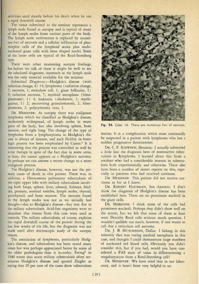
mivities wuil shortly before his de-3th ~~hen he ran • npid downhill c()tlrse.
The tissue submitttd tO the seminar represents a lymph node found or autopsy and is cypicnl of many of <he lymph nodes from various pnt!S of <he body. The lymph node arcbirecmre is repl:tced by nU<ner· OtL.Il foci of nctrosis and a ceUuJar infiltrntion of pleomorphic cells of the lymphoid scri(·$ plus multinucleated gium cells with be:tn shnped nuclei. Some of the Inner cells ore rypical of <he Reed·S<eroberg type.
There: wtre other interesting autopsy findings, but before we mlk of these ir migh< be wcll ro = tbt- rJbuloted diagnoses, inasmuch as dtc lymph node was <he only nureriol aV3ilable for <he S<:minar.
Submiued Diagnoses.-Hodg)cin's diseose (with mdillrioo cb,•nge, 4) '14; lymphoma ( rndintion change, 5; necrosis, I ; redculum cell. l; giam follicular, 1) 8; radincion necrosis) 7; m}•eloid mctnplusin (histo+ plasmosis?, I) 4; leukemia (nJeukemic, 1; myelogenous, 1) 2; necrotizing granulomarosis, 2; hisropl:tsmosis, 2; pol)'tycbcrnia ver~J I.
OR. MEISSNER: Ar auropsy rhere Mt$ malignant lymphonu which we classified 3S Hodgkin's d~, modemcly widespread, of lymph nodos in many pms of the body, bm ~lso involving spleen, bone nt.'<rOw, nnd righ< lung. The Cb>nge of rhe type oi lymphomn from n lymphocy<om• to Hodgkin's dis· e:\Sc is always of inrercsr, and such fluidicy in hiscologic p:uu:m hns been en1phasi1.cd b}' Custcr.1 It is interesting thnr <he process wM colllrollccl sQ well by x-r~•)' rhc·rap)' in sphe of £he bee Lhnt jn some a.reas, a.r le:tsr, the tumor '3ppe:.trS as :s Hodgkin·s sarcoma. So perh:J.))S we no JSSume a rc.ttnc change to a more nulign>nt process.
The Hodgkin's d~, however, was not the pri· tmry uuse of dC"Jtb in £his (Y.ttiem~ There was, in addition, n fibrocaseous pulmomuy mb<:rcuJosis of rhe righr upper lobe wjch miliary t\tbcratlosis involving both lun&S, spleen. liver, adrennl, kidne)'S, bladder, proscntc, sen.1 in:tl vesides, l)'mph nc>des, th)•roid, l"rMhyt·oid, nnd bone marrow. The necrosis found in the lymph nodes WllS no< as we initially had rhought-<lue to Hodgkin's disease-but w•s due tO
the mili>l)' ruberculosis. Acid-fasr organisms were so .>bund1nr that tissues from rhis case were used as cormols. '11>e miliatj• ruberculosis, of cou&, explains the ropid downhill cou& of the p.1ticnt during rhc lm few we<ks of his life, bur <he di•gnosis w;as not made unril after microscopic study of the Ulttopsy rissues.
The assOcitltion of lymphoma, f»<ticularly Hodgkin's disellSe, nnd cuberculosis has been noced man)' 1 imes but was perhaps appreciated better by some of the older p~uhologjscs than ir is.rod:\y. J!wjngn in 194() wrote chat acute miliary ruberculosis often ter· minorcs Hodgkin's d~ and quO!ecl Ziegler as Slying tha< 20 per cent of the cases show tuberculOtlS
[ 29]
Fig. 24. Cos~ 16. There ore '"-'tnei'OUS foci of necrosis.
lesions. lr is a complication which mu.sc coudnually be SU.SfX."Cttc.l in a patient with l)'mphomn who hns o sudden prog ressive deterior~doJ),
OR. C. 1'. S<:HWJNN, Houston: I ncrually submitted a little Jure the diagnosis here of nonrtacrive rubcrculosis in lymphoma. I learned about this from a rcsidenr who h•d .. considerable inrercs< in tubercu· losis bod> experimemaUy and O<berwisc. TI>ere also have been " number of recent report< on <htS, cspe· ci!llly in p;u iems who had rc.-ccivcd corrisone.
DR. Mlii:SSNtift.; ll1is P'.!tiem did not receive cortisone a .. ll rnr as 1 know.
Ott ROHJ.!R"t' H;\ USMAN. S:tn Anronio: 1 doD'c rhink the diagnosis of Hodgkin's disci!SC h~s been esrabl ish<:d here. There are no prominent nucleoli in 'be gi:ult cells.
OR. MUISSNEQ: I <hiok some of tile cells had prominenr nucleoli. Pt"rbaps rbey didn't show well on me screen, bur we felt that some of them ar I=< were Dorot:hy Reed cells wirbom mucb qucsrion. 1 wouldn"r quibble: too much, llO\\•ever, if one w.uns ro all rhis ~ reticulum cell s:ucoma.
Dtt. J. fl. HU1'0WSON, O.ll:ts; 1 belong in rhis minority that was vocing myeloid mcraplasin ln this Ct\set nn(l thOu8ht I cot•ld demonsmue lnrse numbers of nudente<J red blood cells. Obviously you didn't consider this, bu< if you had, would )'Ou have considered a J> AS stain of value in differeutinting a megakaryocyre Crom a Reed-Sternberg cell?
DR. M&,SSNJ!R: \Ve have cried rhar in Otlr labor· a<nry, •nd it ha.sn'r been very helpful <O us.
TUMOR. SEMINAR - Me j ssnet- continued
D R. SE\1 ERANCE: rn view o£ -So, many votes Joe hisropl~sillO$iS on rhe .screen,. were speci(l] stftins done co demonlitt'.tte ihese Organisms, or were )'oo happy when )'Ou {ound acid-fasr bacilli?
DR. l>!EJSSN.ER: When we found .cfd-fast organisms \\1C (}Ujc wbiJc we were ahead.
DR. SEVEJ\ANCE: Some time ago we sa"· n c3.1e wbich we incerprcccd as showing a lymj>hocycic type of lrmpboma associated with what '"'e thoughc was Hodgkin's disease. Do you sec char very ofren?
DR. MEISSNER: 1 don' r know bow often one s""s lymphocytic type of lymphoma curo inco Hodgkin's disease, bur we've ce.rca1nl}1 been .iroprcssed with ~~hac Custer calls the fluidity 6f these lesi0ns, cbanging back and fo rd>. It seems to me that Hodgkin's disease doesn't ofte•\ d1nngc back to somerbing else, bot .other lymphomas often cbange into Hodgkin's dis· eas·e.
DR. SEVERANCE: Whar I meant was rhac we found che rwo together, lymphosarcoma in o~e speclmen ond Hodllkin's djsease in another, botb removed at the s;t:me titnt from ;1. single parienc.
DR. MEISSNER: I agree that is unusual.
REFEI\ENCES
J,. Cu:!tu, R. P.. ;and !Xrnb;ud. W. G. : ]l)u~ud.ation~hip ol HOd~in'~ Di~!e' 2nd O thC'! l.)•mphatic Tumors, Am. ) . }.f. Sc. 216:62}·6<12 ( Occ. ) 1948.
2. Do\lA]aem'. C. M., and L1,1od. C. J.• Solid Owati11a 1\Unou O>ropll«~t.i.og Prtsnancy: CHn iOIJ.J>atholc>s iml St~,:f.ty, Am. J. Obsr. ~ Grnec. 60:26l-279 ~(Au,~t.) r9.>0.
3. !twinS. }am.<'S: Ncoplutic Disca!~, EJ! • ..1, ~hib;dHphia, W, B. S11nndeu ~ Co., " · 406, 19-10.
..1: Glll, It A.: &.i8SUM In lrmplwcnt: Retieu.Jv.m Cdl Slll'COr.u.
lind ~f~c01is E:ur)t;Oillet , Mlnne«n:c Ml!d_ 38.:()74·681, 70' { Oc:t.) 19~5.
5 . Hauisoo, £, G ., Jr.; Soule, li. H.: , and J udd, E. S.: O!mo· ~tectOJJI\lo of the Glomus lottt\"'1.g~tle { Va.gtl~body Tumor)". Cao«t L0;-(226·123{ (Nnll.-[1«.) 1957.
6. H:.utrd, J, D., a.nd KtO)'Ofl, R.: lltYJ)iC!ll /ulmottm of tlto Thymid, A.M.A. Arch, P..uh. 58:554-56~ (Dec.} 1!)54.
7. HOine, ltoba:n; Ptl'WoaJ CommunfcatioQ. $ , L:un:s, It.: N(mc;hrom.:affi.n PaNI&:~DsJiomll or ~nslioo No001'
um, <.."'aunid Body, :~od Aonic·<~.rch 6odii:s. Can~:" ~:4'67-694 (July) 19l0.
9. leComp:.e, IJ. M,: Tumc)B or tht! O u«.id 13Qd l· ~tnd ,Reh:ed S.uw:uues ( Chem<>·JtC!!"l>!Or Sy:ntm) , in Ail:t'S o! Tumo: P;1tbO!<lgy, MmN Pottt.s lost:kucc .of l"o~thh!Og_)·; [~;1sc. 16. Scttioo IV. \'(i':uhias• { (In , D, C .. 1951.
10. M~issner, \V, A .. and k/!S. 2-L A.: Pcrsi.!t~tt 1'b}·toid Ctf· d r.oml, ) . Clin, ~o.:locti..uoi. 1S~9l·98 (Jan.) 19_58.
11. Mcinne.r, \'i/, A., *nd McMJ\!1\lS, R. G.: Gc>m.l?atisoou of His· rultl-g_k PJ.tl'crn of Uenisn 110d Mallto3.nt TI)ytoid T uroots, }. Oitl. Mducrin9l. T2: l474-H79 (Nov,) 1?>2.
1'2. Mcndrlon·, H .. 11.0<.1 Slobodkin. M. : Aotdc·body 1'umor ( Chembd«Wmll) or MC<.iias-tlnuro: ReJX>n o£ Ut.se 11nd Rc~i<"'' oi Litera.ture, C-ancer 10:100S-l0t4, {Se:pt . .<XI. ) 1957.
I 3 . MusgtO\·~. J. ~.. and Mcl>-.mald, }, R.~ l~x-trll·l'llxlomit~•l -oesulo id 'fwnotl : 1'hcoir Oi!Urtn clarod Diagnol.i$ u.nd Tmrmcnt, A.M.A. Aocll. P~tb , <i):.!il3·540 ( April) 1948.
l ·i . R:tl®u, A. S., and Ellion, J. L.: Carotid Bod)' ·rumo~$ w-ith R~siouiLI L'mpb Node lm•oh-emenr, \'<.'irh ll~pon tJf i UJ~ Su~ ~2:381-385 { tr.og.) 195'1.
15. Ratbcr, L J,: Nocc on Otittin of MuJtlnucleru.cd Gitant Crils (r<.>nl Va${'UIIII Chanorh fl~ 1\uno ts: 'l'umors Ati$iO~ ill Th)'told Gl.11nd, Obnc :.nd Soft Tissu<~, A.M.A. ..Arch. Path. 52: .98·103 {July) 19,1.
HS. Rom11rt~l:i, R.: Ch;mod~roma ( Noo<htocna£fiuic Pat:cS'<Ul&Li· qma ) u( C.tumid JkKI}' with D ist:tnt M~:e:•$cases. Am. ), P-Ath. 30: J-B O:m.~F~b.) 19.54.
1 i. Sin~th, L. \V/, : Ce:uin St>o('lllled S:c.rcomas ol Th)'!Oid, A.M.A. Juo~h. P:c(h. J0:124·HO (CXt.,) 1930.
18. So:iuoffl .. S.C .. aml Mc-itt:t<-r. \'<'. A. : Uc::tu)u2) Ca.rcinon:.u oi ~ocreas, A.M.A. Atcb. Pllch . jS:LOL-111 (Aug-.) I9S4 . . 19. Stooc. A. P.: Tumou of Sof( Tissues. ia. AtJ.ts of T umot
l'uhc>.l<.>ar. Ar-m~ For«s lrmimtc of P:acholo&r, Ff!K. 5, _ScctiOA II, w':uh·ing:mn, 1), C, 195~.
20. W in,hip, T., .and IA.>u:an, J.: TuoooN' of GI()(I)US Jual.l.lar.: Net Assoti:!ted with Jugular Vein, A.M.A. /tr(.h, Ot;ola.qng. H:3.JS· ,., (On.) 19)!.
+ Dr. Meissner, New England Deaconess Hospi~l • .BO$ton. Lt. Col. Hansen, ChiefJ Anatomical PatholO,!.'Y Se-ction,
.Brooke Geocn'll HoSl>itll.f, l3roo"e- Army Medical Center, Fon Sam Houl)ton .
COPYIUGtrr 1!)60 1 '(!XA$> M!!bJ<:AL A:S!OC! ,\.'l'JON
:\USTIN, 'l'li.XkS
[ 30]