transposition ofthe great arteries associated a double...
TRANSCRIPT

British Heart Journal, 1979, 42, 483-486
Transposition of the great arteries associated witha double left ventricular outflow tract1
R. H. KINSLEY, S. E. LEVIN, AND T. G. O'DONOVAN
From the Departments of Cardio- Thoracic Surgery and Paediatrics, University of the Witwatersrand,J1ohannesburg, South Africa
SUMMARY A case is described in which, at semilunar valve level, the aorta and pulmonary artery arose
from inappropriate ventricles. Despite this, the outflow tracts to both vessels originated from the leftventricle. Embryologically, it is speculated that this anomaly is the result of normal rotation of theproximal conus, without concomitant truncal inversion, and excessive leftward shift of the proximalconus and conal septum or anterior and rightward deviation of the anterior segment of the ventricularseptum. Surgical repair using a double conduit between the right ventricle and pulmonary artery andeft ventricle and aorta, respectively, was unsuccessful.
Definition of specific outflow tract anomalies is oftencontroversial. For examplc,q transposition of thegreat arteries has variously been defined as a reversalof the anteroposterior relation of the semilunarvalves (Goor and Edwards, 1973), any alteration inthe position of the great arteries (Abbot, 1927),and the presence of mitral-aortic valve discontinuityas a result of a subaortic conus (Van Praagh et al.,1967). Recently, several authors Lave favoured amore literal definition, redefining transposition ofthe great arteries to mean that both great arteriesare 'placed across' the septum (trans-poneremeans 'placed across') reversing the connection ofthe great arteries to the morphological ventricle(Van Praagh, 1973; Shinebourne et al., 1976). Thusboth great arteries arise from morphologicallyinappropriate ventricles. By the same token, doubleoutlet ventricle is defined as a specific ventriculo-arterial connection in which more than one and ahalf great arteries arise from the same ventricular oroutlet chamber (Kirklin et al., 1973). We haveaccepted this literal definition of outflow tractanomalies.However, according to the interpretation of
embryological events by Goor and Lillehei (1975),the semilunar valves and outflow tracts of each ofthe major vessels are derived from different embryo-logical segments. It is reasonable to anticipate,therefore, that the ventricle of origin of a majorvessel (at semilunar valve level) may be at variance
'Supported by a grant from the Stella and Paul LoewensteinTrust Cardiac Fund, University of the Witwatersrand.
with that of its outflow tract. In this communicationa case will be described in which the left ventriclehas a double outflow tract (aortic and pulmonary),but the origin of the great arteries at semilunarvalve level is clearly from inappropriate morpho-logical ventricles.
Terminology
We have adopted the terminology recently proposedby Anderson et al. (1977) with regard to outflowtract anomalies. The term 'conus' (and itsderivatives) is confined to embryological events.In the postnatal, or fully developed heart, the term'infundibulum' is substituted for 'conus'. Thus thestructure interposed between the two semilunarvalves is described as the infundibular septum(parietal band of Van Praagh) (Van Praagh et al.,1975). The muscle separating semilunar from atrio-ventricular valves is termed the ventriculo-in-fundibular fold regardless of whether it exists in theright or left ventricle. The trabecula septomarginalis(septal band of Van Praagh) (Van Praagh et al.,1975) is an extensive right ventricular trabeculationwith, superiorly, anterior and posterior limbs,reinforcing the anterior and inferior borders ofmany septal defects.
Case report
A 51-year-old boy had been cyanosed since birth.Chest x-ray film showed cardiomegaly and pul-monary plethora while the electrocardiogram
483
on 14 April 2019 by guest. P
rotected by copyright.http://heart.bm
j.com/
Br H
eart J: first published as 10.1136/hrt.42.4.483 on 1 October 1979. D
ownloaded from

R. H. Kinsley, S. E. Levin, and T. G. O'Donovan
showed a mean frontal QRS axis of + 60°, withevidence of right atrial and right ventricularenglargement. Small R waves were noted in the rightchest lead. Cardiac catheterisation at age 6 daysestablished the diagnosis of transposition of thegreat arteries, mild pulmonary stenosis, and asmall ventricular septal defect. A Rashkind septo-stomy was performed with considerable improve-ment in oxygen saturation.During the next 3 years increasing polycythaemia
developed requiring multiple venesections. Cerebralthrombosis and hemiplegia occurred at age 1 2years with fairly good recovery. A Blalock-Taussigshunt was performed when the child was 4 yearsold, with subsequent improvement in oxygensaturation and fall in haematocrit. Recatheterisa-tion at 52 years confirmed the previous diagnoses. Asystolic gradient of 41 mmHg was measured acrossthe pulmonary outflow tract and the systemicoxygen saturation was 75 per cent. When the leftventricular systolic pressure was 70 mmHg, that inthe right ventricle was 80 mmHg. Elective surgerywas advised.
Using standard cardiopulmonary bypass tech-niques, a right ventriculotomy was performed and a2 to 3 mm ventricular septal defect closed with asingle horizontal mattress suture. A valved externalconduit (16 mm) was then inserted between the rightventricle and right pulmonary artery. A similarconduit (18 mm) was placed between the apex ofthe left ventricle and left lateral wall of the ascendingaorta. Initially, after discontinuing bypass, thehaemodynamics appeared satisfactory. However,just before closing the chest, ventricular fibrillationoccurred and resuscitation was not successful.Necropsy confirmed ventricular non-inversion:
the morphological right ventricle was right-sidedand the morphological left ventricle was left-sided.The aortic valve was anterior, superior, and to theright, above the right ventricle. The pulmonaryvalve was to the left of, and slightly posterior to,the aortic valve. Though the pulmonary valve wasoverriding both ventricles (similar to the aorta in thenormal heart), there was no direct communicationbetween the right ventricle and the pulmonary valveor artery. Examination of the interior of the leftventricle disclosed two outflow tracts, separatedby a muscular ridge (infundibular septum) approxi-mately in a coronal plane (Fig. 1). The aorticoutflow tract was dextro-posterior to the pulmonaryoutflow tract. The interior of the right ventricle(Fig. 2 and 3a) showed a ventricular septal defect3 mm in diameter (the surgical suture was removed),triangular in shape, and immediately subaortic. Itwas bounded by the posterior limb of the trabeculaseptomarginalis, aortic valve, and, on the left, the
Fig. 1 Photograph of left ventricle (LV) with double(aortic and pulmonary) outflow tract. The left ventriclehas been opened parallel to the left anterior descendingcoronary artery and immediately to the left of the leftventricular ascending aorta conduit. AOT, aortic outflowtract; IS, infundibular septum; MV, mitral valve;POT, pulmonary outflow tract; VSD, ventricularseptal defect.
anterior ventricular septum. The posteroinferiorrim of the defect was separated from the tricuspidvalve by a 3 to 4 mm band of muscle. A well-developed ventriculo-infundibular fold separatedthe semilunar and mitral valves though it was only2 to 3 mm wide below the aortic valve. The pul-monary outflow tract was mildly stenotic at itsorigin.
Discussion
According to the observations of Goor and Lillehei(1975) and their interpretation of previous embryo-logical descriptions, conotruncal inversion occursin two stages. Firstly, the conoventricular junction(ostium bulbi) rotates 1100 counterclockwise (look-ing downstream) during d-looping of the cardiactube. This process transfers the proximal aortic
484
on 14 April 2019 by guest. P
rotected by copyright.http://heart.bm
j.com/
Br H
eart J: first published as 10.1136/hrt.42.4.483 on 1 October 1979. D
ownloaded from
Transposition of the great arteries associated with a double left ventricular outflow tract
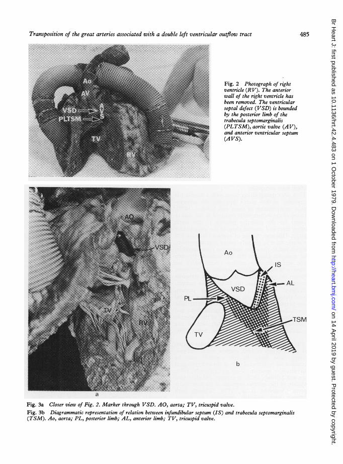
Fig. 2 Photograph of rightventricle (RV). The anteriorwall of the right ventricle hasbeen removed. The ventricularseptal defect (VSD) is boundedby the posterior limb of thetrabecula septomarginalis(PLTSM), aortic valve (A V),and anterior ventricular septum(A VS).
b
Fig. 3a Closer view of Fig. 2. Marker through VSD. AO, aorta; TV, tricuspid valve.Fig. 3b Diagrammatic representation of relation between infundibular septum (IS) and trabecula septomarginalis(TSM). Ao, aorta; PL, posterior limb; AL, anterior limb; TV, tricuspid valve.
485
on 14 April 2019 by guest. P
rotected by copyright.http://heart.bm
j.com/
Br H
eart J: first published as 10.1136/hrt.42.4.483 on 1 October 1979. D
ownloaded from

R. H. Kinsley, S. E. Lenin, and T. G. O'Donovan
conus to a posterior (but still right-sided) positionand enables it to establish contiguity with the leftventricular vestibulum. At this stage, the conotruncalridges (destined to form the conal and truncalsepta) occupy a spiral course. Secondly, but some-what later, the truncus (including the developingsemilunar valves) rotates in an identical manner. Asa result, the conotruncal ridges are uncoiled, theaortic valve is transferred to the same posteriorposition as the aortic conus, and the aorta andpulmonary artery become entwined-the situationseen in the normal heart. Failure of truncal inversionresults in dextroposition of the aortic valve (anteriorand rightward displacement) and its origin pre-dominantly, or entirely, from the right ventricle.
In the normal heart, the infundibular septumfills the Y-shaped space between the anterior andposterior limbs of the trabecula septomarginalis.Should this not occur, malalignment of the in-fundibular and ventricular septa occurs, with aresultant ventricular septa] defect and a variety ofoutflow tract anomalies.How does transposition occur with a double left
ventricular outflow tract? Applying the embryo-logical interpretation of Goor and Lillehei (1975),rotation of the ostium bulbi occurred in our case toaccount for the posterior position of the aorticoutflow tract and coronal plane of the infundibularseptum. However, failure of truncal inversion leftthe aortic valve in its early embryological position,anterior and to the right, above the right ventricle.The muscular ridge in the left ventricle betweenaortic and pulmonary outflow tracts, which webelieve to be the infundibular septum, did not insertbetween the two limbs of the Y-shaped trabeculaseptomarginalis. Rather, the infundibular septum,malaligned relative to the ventricular septum,passed through to the left side occupying a coronalplane perpendicular to the plane of the ventricularseptum. Subsequent fusion of the infundibular andventricular septa (and anterior limb of the trabeculaseptomarginalis) completed the left side of thetriangular-shaped ventricular septal defect (Fig.3b).The left-sided position ofthe infundibular septum
and hence origin of both outflow tracts and pul-monary valve from the left ventricle may beaccounted for by leftward shift of the embryologicalconventricular junction (Goor and Edwards, 1973;Goor and Lillehei, 1975) or anterior and rightwarddeviation of the anterior segment of the ventricularseptum (Anderson et al., 1974). Though bothprocesses were probably operative, we favourpredominance of the latter, because of the relativelynormal position of the pulmonary outflow tract andvalve relative to the atrioventricular valves.
Incomplete absorption ofthe subaortic ventriculo-infundibular fold occurred in our case with conse-quent mitral-aortic fibrous discontinuity. Con-comitantly, failure of truncal inversion resulted inthe aortic valve and aorta remaining dextroposedand originating completely from the right ventricle.
Although 'posterior' transposition has beenpreviously documented (Van Praagh et al., 1971;Wilkinson et al., 1975), our case is distinctive in that,at semilunar valve level, the aorta was anterior tothe pulmonary artery.
References
Abbot, M. E. (1927). Congenital cardiac disease. In Osler, W.Modern Medicine, 3rd ed., Vol. 4, p. 720. Ed. by T. McCrae.Lea and Febinger, Philadelphia; Kimpton, London.
Anderson, R. H., Becker, A. E., and van Mierop, L. H. S.(1977). What should we call the 'crista'? British HeartJ'ournal, 39, 856-859.
Anderson, R. H., Wilkinson, J. L., Arnold, R., Becker, A. E.,and Lubkiewicz, K. (1974). Morphogenesis of bulboventri-cular malformations. II Observations on malformed hearts.British HeartyJournal, 36, 948-970.
Goor, D. A., and Edwards, J. E. (1973). The spectrum oftransposition of the great arteries, with specific referenceto developmental anatomy of the conus. Circulation, 48,406-415.
Goor, D. A., and Lillehei, L. W. (1975). The embryology ofthe heart. In Congenital Malformations of the Heart, p. 38.Grune and Stratton, New York.
Kirklin, J. W., Pacifico, A. D., Bargeron, L. M., and Soto, B.(1973). Cardiac repair in anatomically corrected mal-position of the great arteries. Circulation, 48, 153-159.
Shinebourne, E. A., Macartney, F. J., and Anderson, R. H.(1976). Sequential chamber localization-logical approachto diagnosis in congenital heart disease. British HeartJ7ournal, 38, 327-340.
Van Praagh, R. (1973). Do side-by-side great arteries merit aspecial name? American Journal of Cardiology, 32, 874-876.
Van Praagh, R., Perez-Trevino, C., Lopez-Cuellar, M., Baker,F. W., Zuberbuhler, J. R., Quero, M., Perez, V. M.,Moreno, F., and Van Praagh, S. (1971). Transposition ofthe great arteries, with posterior aorta, anterior pulmonaryartery, sub-pulmonary conus and fibrous continuitybetween aortic and atrioventricular valves. AmericanJournal of Cardiology, 28, 621-631.
Van Praagh, R., Perez-Trevino, C., Reynolds, J. L., Moes,L. A. F., Keith, J. D., Roy, D. L., Belcourt, C., Weinberg,P. M., and Parisi, L. F. (1975). Double outlet right ventricle(S, D, L) with subaortic ventricular septal defect andpulmonary stenosis. Report of six cases. American Journalof Cardiology, 35, 42-53.
Van Praagh, R., Vlad, P., and Keith, J. D. (1967). Completetransposition of the great arteries. In Heart Disease inInfancy and Childhood, 2nd ed., pp. 682-744. Ed. by J. D.Keith, R. D. Rowe, and P. Vlad. Macmillan, New York.
Wilkinson, J. L., Arnold, R., Anderson, R. H., and Acerete,F. (1975). 'Posterior' transposition reconsidered. BritishHeart Journal, 37, 757-766.
Requests for reprints to Professor R. H. Kinsley,Department ofCardio-Thoracic Surgery, Universityof the Witwatersrand, Hospital Hill, Johannesburg2001, South Africa.
486
on 14 April 2019 by guest. P
rotected by copyright.http://heart.bm
j.com/
Br H
eart J: first published as 10.1136/hrt.42.4.483 on 1 October 1979. D
ownloaded from