nikaidoh procedure for situs inversus and transposition of ... · transposition of great arteries...
TRANSCRIPT
Nikaidoh Procedure for Situs Inversus and
Transposition of Great Arteries with Ventricular
Septal Defect and Pulmonary Stenosis
Heng-Wen Chou, Hsiao-En Tsai and Chung-I Chang
We present the case of a patient who underwent a Nikaidoh procedure for total correction of transposition of the
great arteries (TGA), ventricular septal defect (VSD), and pulmonary stenosis (PS). A young girl, 2 years and 4
months of age, weighing 3400 g at birth, was diagnosed with TGA, VSD, patent ductus arteriosus (PDA), and left
ventricular outflow tract obstruction (LVOTO), with valvular and subvalvular PS. Because of frequent cyanosis, she
had received a right-sided modified Blalock-Taussig (mBT) shunt when she was 8 months old. The Nikaidoh
procedure was used for total correction (aortic root translocation, LVOT enlargement, VSD patch repair, neoaorta
and coronary button reimplantation, and neopulmonary artery reconstruction by monocusp valved autologous
pericardial patch with the Lecompte maneuver). The patient’s postoperative course was uneventful, and she was
discharged from the surgical intensive care unit 10 days after the operation.
Key Words: Nikaidoh procedure � Pulmonary stenosis � Transposition of great arteries � Ventricular
septal defect
INTRODUCTION
In our institution, the in-hospital survival rate after
surgical correction of the transposition of great arteries
(TGA) is improving.1 However, in the field of cardiovas-
cular surgery, surgical management of patients with TGA,
ventricular septal defect (VSD), left ventricular outflow
tract obstruction (LVOTO), and pulmonary stenosis (PS)
continues to be a challenge. The unusual anatomical fea-
tures of patients with situs inversus and dextrocardia
make total correction even more difficult. Three methods
have been developed for correcting TGA, VSD, and PS:
the Rastelli, réparation á l'etage ventriculaire (REV), and
Nikaidoh procedures. We report a case in which situs
inversus and dextrocardia with TGA, VSD, and PS were
corrected using the Nikaidoh procedure.
CASE REPORT
A female patient was diagnosed with situs inversus
and dextrocardia. She had atrioventricular concordance
and ventriculoarterial discordance and TGA with peri-
membranous inlet-type VSD and valvular and sub-
valvular PS at birth. A modified Blalock-Taussig (mBT)
shunt was created when she was 8 months old due to in-
termittent desaturation. She adapted well to the shunt
and showed no obvious growth retardation. When she
was 2 years and 4 months old and her body weight was
11 kg, she was admitted to our hospital and scheduled to
undergo the next step of surgical intervention.
A grade 4/6 systolic murmur was audible at the right
sternal border and oxygen saturation was approximately
75-85%. Chest radiography revealed signs typical of si-
337 Acta Cardiol Sin 2012;28:337�340
Nikaidoh Procedure for TGA, VSD and PSCase Report Acta Cardiol Sin 2012;28:337�340
Received: June 21, 2011 Accepted: January 19, 2012
Division of Cardiovascular Surgery, Department of Surgery, National
Taiwan University Hospital, Taipei, Taiwan.
Address correspondence and reprint requests to: Dr. Chung-I Chang,
Department of Cardiovascular Surgery, National Taiwan University
Hospital, No. 7, Chung-Shan S. Rd., Taipei 100, Taiwan. Tel:
886-2-2312-3456 ext. 65977; Fax: 886-2-2393-4358

tus inversus with dextrocardia and a right-sided gastric
bubble. The electrocardiogram also showed typical find-
ings such as inverted P and T waves on lead 1 and dimi-
nution of R wave from V1-V6. The ejection fraction cal-
culated via follow-up echocardiography was 81.2%. The
size of the VSD was 9.4 mm. The pulmonary valve was
bicuspid with valvular and subvalvular PS. The size of
the pulmonary annulus was 7.2 mm and that of the
aorta was 17 mm. A Rastelli-type A defect with tri-
cuspid valve straddling was also observed. From the
chest computed tomography and 3-dimensional recon-
struction, the anatomy of situs inversus, TGA, LVOTO,
and valvular and subvalvular PS could be clearly ob-
served. The size and morphology of the pulmonary ar-
teries were normal. The hemodynamic data obtained
from cardiac catheterization showed a high-pressure
gradient (65 mmHg) across the LVOT and pulmonary
valve. No significant pressure gradient was detected
across the aortic valve and right ventricle. The coronary
arteries of the patient followed the usual pattern and
courses with the morphological left coronary artery
(LCA), which supplies branches to the left anterior de-
scending (LAD) and left circumflex artery (LCX), and
the morphological right coronary artery (RCA) arising
from the aortic root. The aim of the intervention was to
restore normal circulation (from the morphological left
ventricle (LV) to the aorta and from the morphological
right ventricle (RV) to the main pulmonary artery), to
repair the VSD, and to release the LVOTO. Thus, the
Nikaidoh procedure was chosen as a method for total
correction.
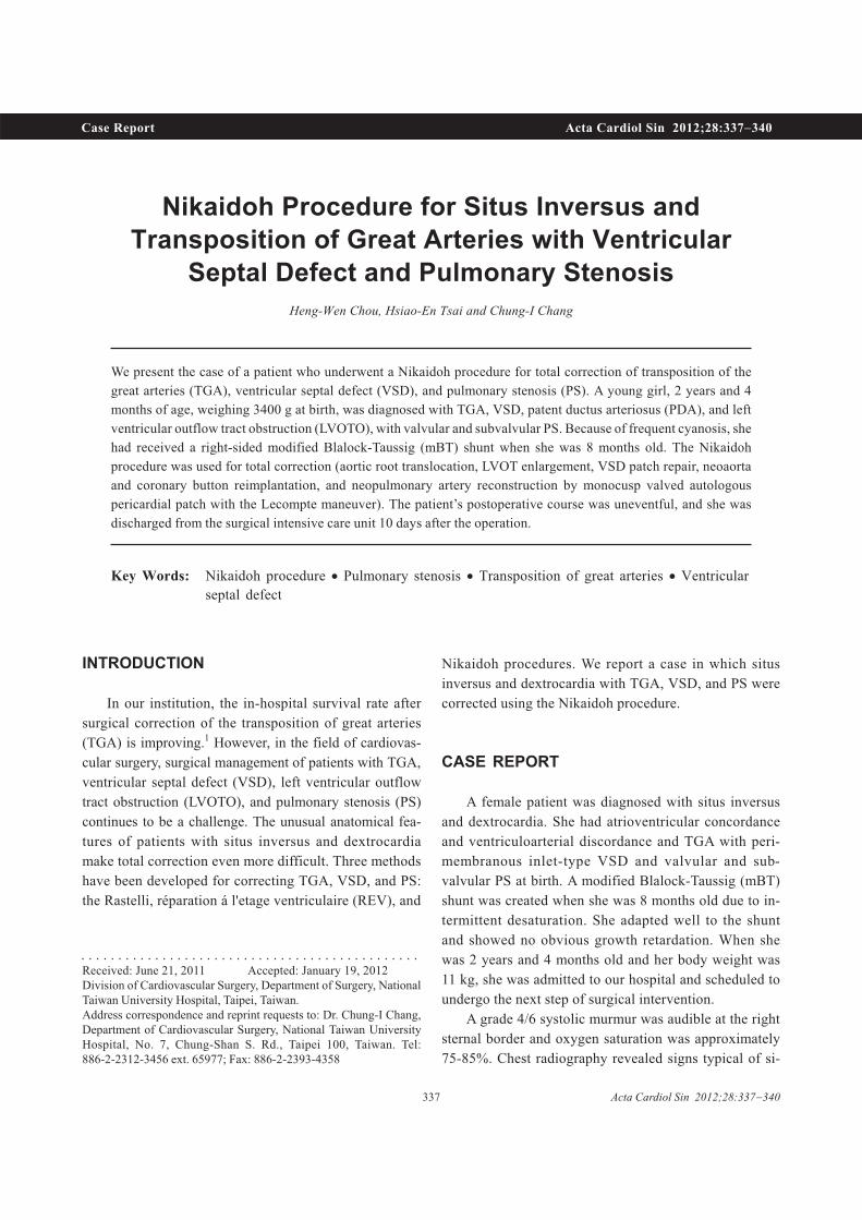
By performing a standard midline full sternotomy, a
cardiopulmonary bypass was established with the aorta,
and bicaval cannulation and systemic hypothermia (28
�C) were achieved. The mBT shunt was then divided.
After extensive mobilization of both the pulmonary ar-
teries, the main pulmonary artery (MPA) was transected
proximally from the left ventricle. After transection of
the ascending aorta, the RCA button was harvested be-
fore the aortic root was completely separated from the
RV, and the Lecompte maneuver was performed to avoid
excess tension and kinking (Figure 1B). The conal sep-
tum was severed into the VSD, and thus, the VSD and
LVOT were enlarged (Figure 1C). The VSD was re-
paired using a Hemashield prosthetic patch (Hemashield®
VantageTM; Maquet Cardiovascular, San Jose, CA, USA)
(Figure 1D; Figure 2). The aortic root was translocated
posteriorly by suturing the posterior two-thirds of the
root to the pulmonary annulus and the anterior one-third
to upper border of the Hemashield patch (Hemashield®
VantageTM) (Figure 1D). The RCA button was reim-
planted into the anterior surface of the aortic root (Fig-
ure 1E). Finally, the MPA was directly sutured to the RV
opening with autologous pericardium patch augmenta-
tion and using a monocuspid valve made of Gore-Tex
surgical membrane patch (GORE-TEX® Surgical Mem-
brane, W.L. Gore and Assoc, Flagstaff, AZ, USA) (Fig-
ure 1F).
Two days after the surgery, the sternum was approx-
imated and the wound closed. The patient was extubated
1 week later and discharged from the intensive care unit
2 weeks after the operation. Postoperative echocar-
diography showed moderate pulmonary regurgitation but
no neoaortic stenosis.
DISCUSSION
For patients with TGA, VSD, and PS, surgical cor-
rection can be performed using 3 methods: Rastelli,
REV, and Nikaidoh procedures.
Rastelli and his colleagues proposed the Rastelli
procedure in 1969.2 In patients with VSD, an intra-
cardiac baffle is placed to direct LV blood to the aorta,
and an artificial extracardiac valved conduit is con-
structed to connect the RV and the pulmonary artery.
This was the first procedure that restored the role of the
morphological LV to support systemic circulation. How-
ever, this procedure has some limitations. Unfavorable
intracardiac anatomy precluded the adoption of this
technique. Fixed size, sternal compression, and obstruc-
tion of the extracardiac conduit and recurring LVOTO
increase the rate of reoperation. A low freedom from
conduit reoperation rate after Rastelli procedure was re-
ported by Kreutzer et al.: 56%, 25%, and 21% after 5,
10, and 15 years, respectively.3 Another drawback of
this procedure was poor long-term survival. Dearani et
al.4 followed up 160 hospital survivors for at least 10
years, and reported an actuarial survival rate of 74% at
10 years and 59% at 20 years after the correctional
surgery.
In 1982, Lecompte described the REV procedure.5
Acta Cardiol Sin 2012;28:337�340 338
Heng-Wen Chou et al.
There were 2 major differences between the REV and
Rastelli procedures. First, the muscular outlet septum
was partially resected, providing a better pathway for
LVOT. Second, the pulmonary trunk was brought an-
teriorly with the Lecompte maneuver and was re-im-
planted to the RV. The REV procedure has several ad-
vantages over the conventional Rastelli procedure: the
REV operation allows complete repair during patient in-
fancy, is feasible in patients with anatomic contraindica-
tions to the Rastelli procedure, and reduces the need for
reoperation because of conduit obstruction and residual
right ventricular outflow tract obstruction (RVOTO) and
LVOTO.6,7 However, despite having better results than
the Rastelli procedure, the REV procedure has been re-
ported to result in RV-PA obstruction in 26% of patients.8
The Nikaidoh procedure, introduced by Nikaidoh in
1984,9 aimed to restore the normal anatomical orga-
339 Acta Cardiol Sin 2012;28:337�340
Nikaidoh Procedure for TGA, VSD and PS
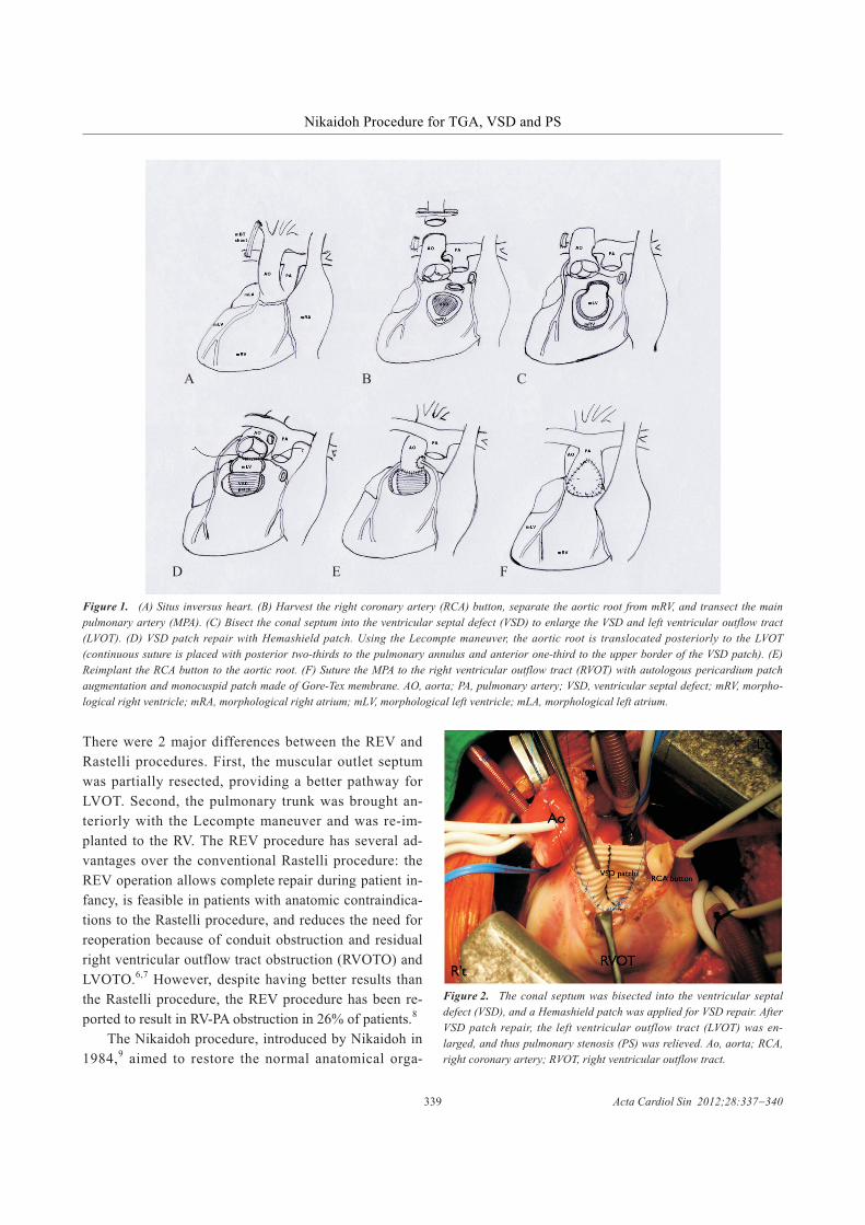
Figure 2. The conal septum was bisected into the ventricular septal
defect (VSD), and a Hemashield patch was applied for VSD repair. After
VSD patch repair, the left ventricular outflow tract (LVOT) was en-
larged, and thus pulmonary stenosis (PS) was relieved. Ao, aorta; RCA,
right coronary artery; RVOT, right ventricular outflow tract.
Figure 1. (A) Situs inversus heart. (B) Harvest the right coronary artery (RCA) button, separate the aortic root from mRV, and transect the main
pulmonary artery (MPA). (C) Bisect the conal septum into the ventricular septal defect (VSD) to enlarge the VSD and left ventricular outflow tract
(LVOT). (D) VSD patch repair with Hemashield patch. Using the Lecompte maneuver, the aortic root is translocated posteriorly to the LVOT
(continuous suture is placed with posterior two-thirds to the pulmonary annulus and anterior one-third to the upper border of the VSD patch). (E)
Reimplant the RCA button to the aortic root. (F) Suture the MPA to the right ventricular outflow tract (RVOT) with autologous pericardium patch
augmentation and monocuspid patch made of Gore-Tex membrane. AO, aorta; PA, pulmonary artery; VSD, ventricular septal defect; mRV, morpho-
logical right ventricle; mRA, morphological right atrium; mLV, morphological left ventricle; mLA, morphological left atrium.
A B C
D E F

nization by which the LVOT and RVOT can be better
aligned. It involves harvesting the aortic root from the
RV, bisecting the muscular outlet septum, and patch re-
pairing the VSD, thus relieving LVOTO, bringing for-
ward the pulmonary trunk using the Lecompte maneu-
ver, and reimplanting the aortic root to the LVOT and the
pulmonary trunk to the RV with or without MPA plasty.
In cases of TGA and LVOTO with inlet or small VSD
and small RV or straddling atrioventricular valve, which
were not amenable to the Rastelli and REV procedures,
the Nikaidoh procedure was the only way to achieve
biventricular repair.10 The limitation of the Nikaidoh
procedure becomes apparent in the presence of anoma-
lous coronary anatomy. The improper course of a major
coronary artery would make harvesting the aortic root
dangerous if not impossible. Compared to the conven-
tional Rastelli procedure for TGA, VSD, and LVOTO,
the technically challenging Nikaidoh procedure was su-
perior in terms of reintervention rate for RVOT com-
pression, LVOTO, aortic insufficiency, and midterm
survival.11
For our patient who had sinus inversus, TGA, VSD,
and LVOTO, any of the 3 procedures could be chosen
for definite correction. However, the first and difficult
part in choosing the procedure to be followed was the
unusual anatomy of the situs inversus that was totally
opposite to what the surgeon was used to managing.
Therefore, complete preoperative imaging studies for
comprehensive understanding of the anatomy were cru-
cial. The second difficulty was in the intracardiac rerout-
ing of LVOT to the aorta because of the small VSD and
Rastelli type A defect with straddling of the tricuspid
valve, as shown by echocardiography. Thus, the Rastelli
and REV procedures were precluded. The patterns of
coronary arteries of the patient were normal, but there
was a communication branch of the LCA and RCA
crossing the RVOT. Considering the anatomy, reinter-
vention rate, and long-term survival potential, we chose
the Nikaidoh procedure as the designated procedure for
total correction. We paid special attention while the
aortic root was being harvested to avoid injuring the
coronary arteries.
We used the Nikaidoh procedure for total surgical
correction in a patient with dextrocardia, situs inversus,
TGA, VSD, and PS. Compared to the Rastelli and REV
procedures, the Nikaidoh procedure can provide im-
proved mid-term survival benefits11 and lower reinter-
vention rate. To our knowledge, this is the first report of
this condition in Taiwan.
REFERENCES
1. Lu BY, Wu HD, Wang CC, et al. The impact of length of post-
operative ventilator support on outcome of the arterial switch op-
eration: report from a single institute. Acta Cardiol Sin 2010;26:
173-8.
2. Rastelli GC, McGoon DC, Wallace RB. Anatomic correction of
transposition of great arteries with ventricular septal defect and
subpulmonary stenosis. J Thorac Cardiovasc Surg 1969;58:
545-52.
3. Kreutzer C, De Vive J, Oppido G, et al. Twenty-five-year experi-
ence with Rastelli repair for transposition of the great arteries. J
Thorac Cardiovasc Surg 2000;120:211-23.
4. Dearani JA, Danielson GK, Puga FJ, et al. Late results of the
Rastelli operation for transposition of the great arteries. Semin
Thorac Cardiovasc Surg Pediatr Card Surg Annu 2001;4:3-15.
5. Lecompte Y, Neveux JY, Leca F, et al. Reconstruction of the
pulmonary outflow tract without prosthetic conduit. J Thorac
Cardiovasc Surg 1982;84:727-33.
6. Vouhe´ PR, Tamisier D, Leca F, et al. Transposition of the great
arteries, ventricular septal defect, and pulmonary outflow tract
obstruction. Rastelli or Lecompte procedure? J Thorac Car-
diovasc Surg 1992;103:428-36.
7. Lee JR, Lim HG, Kim YJ, et al. Repair of transposition of the
great arteries, ventricular septal defect and left ventricular out-
flow tract obstruction. Eur J Cardiothorac Surg 2004;25:735-41.
8. Kim YJ, Song H, Lee JR, et al. Lecompte procedure for complete
transposition of the great arteries with ventricular septal defect
and pulmonary stenosis. Ann Thorac Surg 1994;57:876-9.
9. Nikaidoh H. Aortic translocation and biventricular outflow tract
reconstruction. A new surgical repair for transposition of the great
arteries associated with ventricular septal defect and pulmonary
stenosis. J Thorac Cardiovasc Surg 1984;88:365-72.
10. Hazekamp M, Portela F, Bartelings M. The optimal procedure for
the great arteries and left ventricular outflow tract obstruction. An
anatomical study. Eur J Cardiothorac Surg 2007;31:879-87.
11. Yeh T, Ramaciotti C, Leonard SR, et al. The aortic translocation
(Nikaidoh) procedure: midterm results superior to the Rastelli
procedure. J Thorac Cardiovasc Surg 2007;133:461-9.
Acta Cardiol Sin 2012;28:337�340 340
Heng-Wen Chou et al.