time to dig deep into the plant proteome: a hunt for low ... · pdf filetime to dig deep into...
TRANSCRIPT
OPINION ARTICLEpublished: 30 January 2015
doi: 10.3389/fpls.2015.00022
Time to dig deep into the plant proteome: a hunt forlow-abundance proteinsRavi Gupta1, Yiming Wang2, Ganesh K. Agrawal3,4, Randeep Rakwal3,4,5,6, Ick H. Jo7, Kyong H. Bang7
and Sun T. Kim1*
1 Plant Functional Genomics Laboratory, Department of Plant Bioscience, Life and Industry Convergence Research Institute, Pusan National University, Miryang,South Korea
2 Plant Proteomics Group, Max Planck Institute for Plant Breeding Research, Cologne, Germany3 Research Laboratory for Biotechnology and Biochemistry, Kathmandu, Nepal4 Global Research Arch for Developing Education (GRADE) Academy Pvt. Ltd, Birgunj, Nepal5 Organization for Educational Initiatives, University of Tsukuba, Tsukuba, Japan6 Department of Anatomy I, Showa University School of Medicine, Tokyo, Japan7 Department of Herbal Crop Research, Rural Development Administration, Eumseong, South Korea*Correspondence: [email protected]
Edited by:
Joshua L. Heazlewood, The University of Melbourne, Australia
Reviewed by:
Holger Eubel, Leibniz Universität Hannover, GermanyChristian Lindermayr, Helmholtz Zentrum München - German Research Center for Environmental Health, Germany
Keywords: low-abundance proteins, high-abundance proteins, two-dimensional gel electrophoresis, RuBisCO, post-translational modifications
INTRODUCTIONTwo-dimensional gel electrophoresis (2-DGE) has come a long way since itsintroduction around 40 years by the pio-neering work of these three researchers(Klose, 1975; O’Farrell, 1975; Scheele,1975). 2-DGE was one of the majorbreakthroughs in proteomics, enablingresearchers to detect, analyze and iden-tify the whole set of proteins of a cell ortissue, simultaneously. With the advance-ment in technology, some modifications tothis technique like development of immo-bilized pH gradient (IPG) strips wereintroduced, which undoubtedly, madethis technique more simple, rapid andautonomous (Bjellqvist et al., 1982). Afterits introduction to the present, 2-DGE hasbeen the method of choice for analyzingthe complex proteomes of plants. 2-DGEhas been used extensively to investigatethe effects of biotic and abiotic stress,role of hormones, and developmentalchanges of plants, among others (Agrawaland Rakwal, 2008). However, it wasslowly realized that identification of theplant proteins led to the repeated detec-tion of high-abundance proteins (HAPs)including ribulose-1,5-bisphosphate car-boxylase/oxygenase (RuBisCO) and otherhousekeeping proteins, which are presentat 106-105 order of magnitude (Gygiet al., 2000; Patterson and Aebersold, 2003;
Görg et al., 2004). Signaling and otherregulatory proteins are generally present100 molecules per cells. Subsequently,these proteins are difficult to identifyby either gel-based or gel-free proteomicapproaches, even with access to the lat-est mass spectrometers. RuBisCO com-prises of a large percentage in the totalproteins and thus hinders the absorptionof low-abundance proteins (LAPs) on theIPG strips, which subsequently results inthe poor detection and identification ofLAPs on 2D gels and by mass spectrome-try (MS), respectively. Therefore, the timehas come for all plant proteomers to real-ize the need to hunt for the LAPs, mov-ing one step ahead from the present. AsRuBisCO is the major HAP in plant leaves,here we recommend the incorporation ofa RuBisCO depletion/removal method inevery plant protein extraction step to lookdeeper into the plant proteome. RuBisCOdepletion will definitely improve the pro-teome coverage and will lead to the detec-tion of novel unidentified LAPs.
OVERVIEW OF THE METHODSDEVELOPED FOR ENRICHMENT OFLAPs: MERITS AND DE-MERITSRuBisCO DEPLETION METHODSA number of protein extraction meth-ods with numerous depletion steps weredeveloped in the last decade to remove
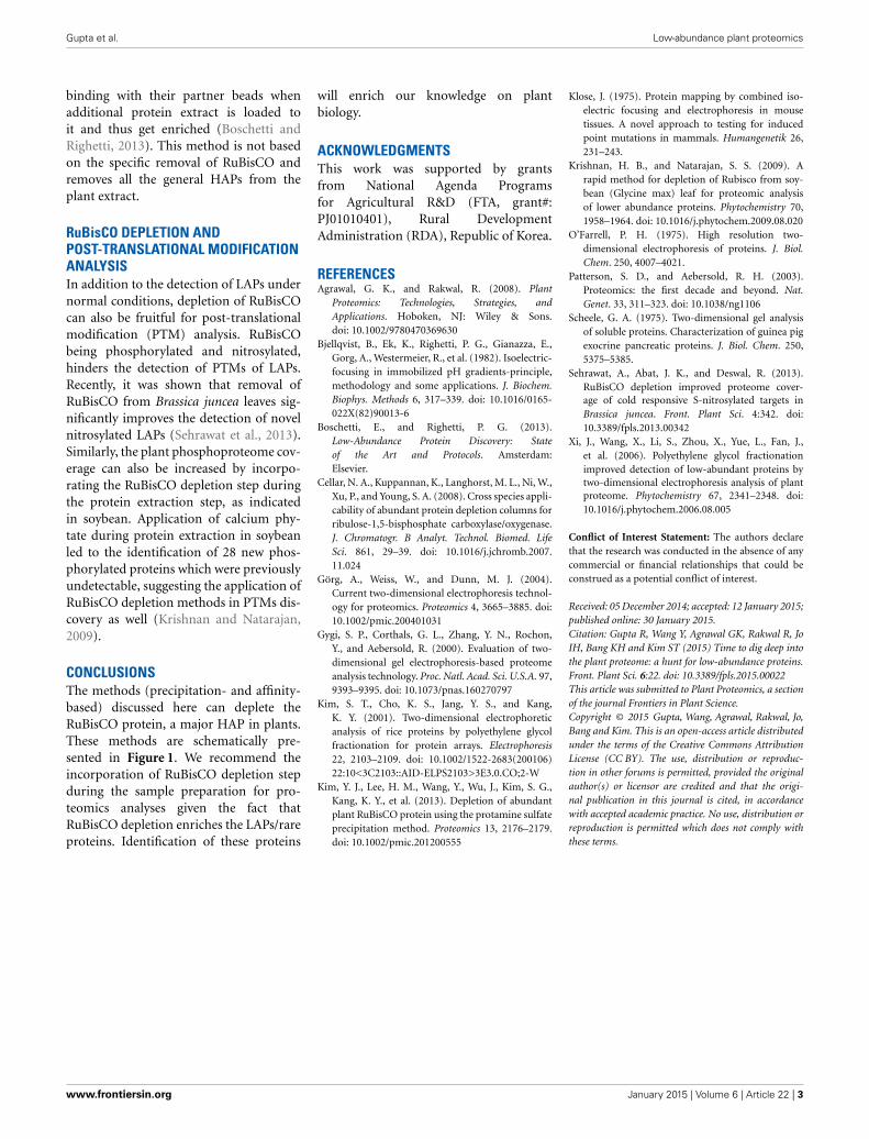
RuBisCO, which accounts for nearly halfof the total leaf protein content (summa-rized in Figure 1). Kim and co-workersinitially developed a poly-ethylene gly-col (PEG)-based method for the deple-tion of RuBisCO from rice leaves (Kimet al., 2001). Application of 20% PEG sig-nificantly precipitated the RuBisCO pro-tein (large and small subunits) in thepellet fraction, resulting in the enrich-ment of LAPs in the supernatant frac-tion. Later, it was shown that 16% PEGwas sufficient to deplete the RuBisCOfrom Arabidopsis leaves (Xi et al., 2006)indicating this method can be applied toall plants for the removal of RuBisCO.However, this PEG method is laboriousand time consuming. Following the PEGfractionation approach, a new methodusing calcium and phytate was intro-duced for the removal of RuBisCO fromleaves of soybean. Results revealed thata 10 min incubation of the leaf extractwith 10 mM calcium and 10 mM phytateat 42◦C, depleted 86% of the RuBisCOprotein in the pellet fraction (Krishnanand Natarajan, 2009). As incubation ofthe protein extract at 42◦C is abso-lutely essential for significant depletionof RuBisCO, this temperature condi-tion can lead to the denaturation ofsome heat labile proteins. Incubation atlower temperatures significantly reduces
www.frontiersin.org January 2015 | Volume 6 | Article 22 | 1

Gupta et al. Low-abundance plant proteomics
FIGURE 1 | A summary of the workflows for the enrichment of LAPs in plants using RuBisCO depletion and CPLL methods. Details of these techniquesare mentioned in the text and cited references.
the RuBisCO precipitation ability of thismethod. For example, only 44% RuBisCOdepletion was achieved at 4◦C (Krishnanand Natarajan, 2009). More recently, aprotamine sulfate-based specific RuBisCOdepletion method was introduced (Kimet al., 2013). It was shown that additionof 0.1% protamine sulfate differentiallyprecipitates the RuBisCO in the pellet frac-tion and enriches the LAPs in the super-natant fraction. Using Western blotting,no RuBisCO was detected in the super-natant fraction, suggesting this methodis able to deplete RuBisCO below thedetection limit. 2-DGE analysis showedthat application of this method in soy-bean resulted in visualization of 423 newspots in the supernatant fraction whichwere not discernible in the total frac-tion. Furthermore, in addition to soy-bean, this method was also applicableto another dicot Arabidopsis, and mono-cots rice and maize, suggesting that it
can be universally applied in plants forthe removal of RuBisCO. This protaminesulfate-based method is rapid, reliable,cost effective, and highly efficient and ismore specific than the previously pub-lished PEG and calcium-phytate basedmethods (Kim et al., 2013).
IMMUNO-AFFINITY BASED METHODSOther than the above mentioned pre-cipitation methods, an affinity-basedmethod has been developed for RuBisCOdepletion. This method utilizes theanti-RuBisCO antibodies, which arecommercially available and supplied ascolumns (IgY RuBisCO column, SigmaAldrich; Cellar et al., 2008). The beautyof this method is that it is highly spe-cific to RuBisCO. However, this methodis very expensive limiting its wide accep-tance among the scientific community,especially laboratories in the developingcountries.
In addition to the RuBisCO-depletionmethods, other techniques have also beenintroduced for the enrichment of LAPs.“Combinatorial peptide ligand library”(CPLL) technology, developed over theyears by the group of P.G. Righetti, iscommercially available under the tradename of Proteominer (BioRad) (Boschettiand Righetti, 2013). Briefly, CPLLs con-sists of several million hexapeptides (pre-pared using 16 different amino acids)that are able to recognize complemen-tary amino acid sequence in a bait proteinharvesting it from the sample matrix. Toput is simply, when the protein extractis loaded onto a CPLL column underlarge overloading conditions, beads hav-ing affinity with the abundant proteinssaturate first and therefore, the major frac-tions of these proteins are washed outdue to limited binding capacity of thebeads. However, due to low concentra-tions of LAPs, these proteins keep on
Frontiers in Plant Science | Plant Proteomics January 2015 | Volume 6 | Article 22 | 2
Gupta et al. Low-abundance plant proteomics
binding with their partner beads whenadditional protein extract is loaded toit and thus get enriched (Boschetti andRighetti, 2013). This method is not basedon the specific removal of RuBisCO andremoves all the general HAPs from theplant extract.
RuBisCO DEPLETION ANDPOST-TRANSLATIONAL MODIFICATIONANALYSISIn addition to the detection of LAPs undernormal conditions, depletion of RuBisCOcan also be fruitful for post-translationalmodification (PTM) analysis. RuBisCObeing phosphorylated and nitrosylated,hinders the detection of PTMs of LAPs.Recently, it was shown that removal ofRuBisCO from Brassica juncea leaves sig-nificantly improves the detection of novelnitrosylated LAPs (Sehrawat et al., 2013).Similarly, the plant phosphoproteome cov-erage can also be increased by incorpo-rating the RuBisCO depletion step duringthe protein extraction step, as indicatedin soybean. Application of calcium phy-tate during protein extraction in soybeanled to the identification of 28 new phos-phorylated proteins which were previouslyundetectable, suggesting the application ofRuBisCO depletion methods in PTMs dis-covery as well (Krishnan and Natarajan,2009).
CONCLUSIONSThe methods (precipitation- and affinity-based) discussed here can deplete theRuBisCO protein, a major HAP in plants.These methods are schematically pre-sented in Figure 1. We recommend theincorporation of RuBisCO depletion stepduring the sample preparation for pro-teomics analyses given the fact thatRuBisCO depletion enriches the LAPs/rareproteins. Identification of these proteins
will enrich our knowledge on plantbiology.
ACKNOWLEDGMENTSThis work was supported by grantsfrom National Agenda Programsfor Agricultural R&D (FTA, grant#:PJ01010401), Rural DevelopmentAdministration (RDA), Republic of Korea.
REFERENCESAgrawal, G. K., and Rakwal, R. (2008). Plant
Proteomics: Technologies, Strategies, andApplications. Hoboken, NJ: Wiley & Sons.doi: 10.1002/9780470369630
Bjellqvist, B., Ek, K., Righetti, P. G., Gianazza, E.,Gorg, A., Westermeier, R., et al. (1982). Isoelectric-focusing in immobilized pH gradients-principle,methodology and some applications. J. Biochem.Biophys. Methods 6, 317–339. doi: 10.1016/0165-022X(82)90013-6
Boschetti, E., and Righetti, P. G. (2013).Low-Abundance Protein Discovery: Stateof the Art and Protocols. Amsterdam:Elsevier.
Cellar, N. A., Kuppannan, K., Langhorst, M. L., Ni, W.,Xu, P., and Young, S. A. (2008). Cross species appli-cability of abundant protein depletion columns forribulose-1,5-bisphosphate carboxylase/oxygenase.J. Chromatogr. B Analyt. Technol. Biomed. LifeSci. 861, 29–39. doi: 10.1016/j.jchromb.2007.11.024
Görg, A., Weiss, W., and Dunn, M. J. (2004).Current two-dimensional electrophoresis technol-ogy for proteomics. Proteomics 4, 3665–3885. doi:10.1002/pmic.200401031
Gygi, S. P., Corthals, G. L., Zhang, Y. N., Rochon,Y., and Aebersold, R. (2000). Evaluation of two-dimensional gel electrophoresis-based proteomeanalysis technology. Proc. Natl. Acad. Sci. U.S.A. 97,9393–9395. doi: 10.1073/pnas.160270797
Kim, S. T., Cho, K. S., Jang, Y. S., and Kang,K. Y. (2001). Two-dimensional electrophoreticanalysis of rice proteins by polyethylene glycolfractionation for protein arrays. Electrophoresis22, 2103–2109. doi: 10.1002/1522-2683(200106)22:10<3C2103::AID-ELPS2103>3E3.0.CO;2-W
Kim, Y. J., Lee, H. M., Wang, Y., Wu, J., Kim, S. G.,Kang, K. Y., et al. (2013). Depletion of abundantplant RuBisCO protein using the protamine sulfateprecipitation method. Proteomics 13, 2176–2179.doi: 10.1002/pmic.201200555
Klose, J. (1975). Protein mapping by combined iso-electric focusing and electrophoresis in mousetissues. A novel approach to testing for inducedpoint mutations in mammals. Humangenetik 26,231–243.
Krishnan, H. B., and Natarajan, S. S. (2009). Arapid method for depletion of Rubisco from soy-bean (Glycine max) leaf for proteomic analysisof lower abundance proteins. Phytochemistry 70,1958–1964. doi: 10.1016/j.phytochem.2009.08.020
O’Farrell, P. H. (1975). High resolution two-dimensional electrophoresis of proteins. J. Biol.Chem. 250, 4007–4021.
Patterson, S. D., and Aebersold, R. H. (2003).Proteomics: the first decade and beyond. Nat.Genet. 33, 311–323. doi: 10.1038/ng1106
Scheele, G. A. (1975). Two-dimensional gel analysisof soluble proteins. Characterization of guinea pigexocrine pancreatic proteins. J. Biol. Chem. 250,5375–5385.
Sehrawat, A., Abat, J. K., and Deswal, R. (2013).RuBisCO depletion improved proteome cover-age of cold responsive S-nitrosylated targets inBrassica juncea. Front. Plant Sci. 4:342. doi:10.3389/fpls.2013.00342
Xi, J., Wang, X., Li, S., Zhou, X., Yue, L., Fan, J.,et al. (2006). Polyethylene glycol fractionationimproved detection of low-abundant proteins bytwo-dimensional electrophoresis analysis of plantproteome. Phytochemistry 67, 2341–2348. doi:10.1016/j.phytochem.2006.08.005
Conflict of Interest Statement: The authors declarethat the research was conducted in the absence of anycommercial or financial relationships that could beconstrued as a potential conflict of interest.
Received: 05 December 2014; accepted: 12 January 2015;published online: 30 January 2015.Citation: Gupta R, Wang Y, Agrawal GK, Rakwal R, JoIH, Bang KH and Kim ST (2015) Time to dig deep intothe plant proteome: a hunt for low-abundance proteins.Front. Plant Sci. 6:22. doi: 10.3389/fpls.2015.00022This article was submitted to Plant Proteomics, a sectionof the journal Frontiers in Plant Science.Copyright © 2015 Gupta, Wang, Agrawal, Rakwal, Jo,Bang and Kim. This is an open-access article distributedunder the terms of the Creative Commons AttributionLicense (CC BY). The use, distribution or reproduc-tion in other forums is permitted, provided the originalauthor(s) or licensor are credited and that the origi-nal publication in this journal is cited, in accordancewith accepted academic practice. No use, distribution orreproduction is permitted which does not comply withthese terms.
www.frontiersin.org January 2015 | Volume 6 | Article 22 | 3