proteome profiler tm array
TRANSCRIPT

Human Soluble Receptor Array Kit Non-Hematopoietic Panel
Proteome ProfilerTM Array
This package insert must be read in its entirety before using this product. For research use only. Not for use in diagnostic procedures.
Catalog Number ARY012
For the parallel determination of the relative levels of soluble receptors and related proteins in non-hematopoietic cells.

MANUFACTURED AND DISTRIBUTED BY:
USA & Canada | R&D Systems, Inc. 614 McKinley Place NE, Minneapolis, MN 55413, USATEL: (800) 343-7475 (612) 379-2956 FAX: (612) 656-4400E-MAIL: [email protected]
DISTRIBUTED BY:
UK & Europe | R&D Systems Europe, Ltd.19 Barton Lane, Abingdon Science Park, Abingdon OX14 3NB, UKTEL: +44 (0)1235 529449 FAX: +44 (0)1235 533420E-MAIL: [email protected]
China | R&D Systems China Co., Ltd.24A1 Hua Min Empire Plaza, 726 West Yan An Road, Shanghai PRC 200050TEL: +86 (21) 52380373 FAX: +86 (21) 52371001E-MAIL: [email protected]
TABLE OF CONTENTS
SECTION PAGE
INTRODUCTION .....................................................................................................................................................................1PRINCIPLE OF THE ASSAY ...................................................................................................................................................1TECHNICAL HINTS .................................................................................................................................................................1MATERIALS PROVIDED & STORAGE CONDITIONS ...................................................................................................2OTHER SUPPLIES REQUIRED .............................................................................................................................................3PRECAUTIONS .........................................................................................................................................................................3SAMPLE COLLECTION & STORAGE .................................................................................................................................4REAGENT PREPARATION .....................................................................................................................................................5ARRAY PROCEDURE .............................................................................................................................................................6DATA ANALYSIS ......................................................................................................................................................................8PROFILING SOLUBLE RECEPTORS IN CELL CULTURE SUPERNATES ..................................................................9PROFILING SOLUBLE RECEPTORS IN CELL LYSATES ............................................................................................. 11PROFILING SOLUBLE RECEPTORS IN SERUM .......................................................................................................... 13APPENDIX .............................................................................................................................................................................. 14

www.RnDSystems.com 1
INTRODUCTIONAnalyzing the expression profiles of soluble receptors expressed and released by non-hematopoietic cells is essential for understanding the roles these molecules play in activation of endothelial cells. The Human Soluble Receptor Array Kit is a rapid, sensitive, and economical tool to simultaneously detect changes in proteins between samples. The relative expression levels of 119 soluble receptors and related proteins can be determined without performing numerous immunoprecipitations or Western blots. Each capture and detection antibody, directed to the extracellular domain, was carefully selected using both natural and recombinant proteins.
PRINCIPLE OF THE ASSAYCapture and control antibodies have been spotted in duplicate on nitrocellulose membranes. Cell culture supernate, serum, or cell lysate samples are diluted and incubated overnight with each array part of the Non-Hematopoietic Panel. This panel is comprised of the Non-Hematopoietic Array and the Common Analytes Array, which is also used in the Hematopoietic Array Panel (R&D Systems, Catalog # ARY011). The membranes are washed to remove unbound material followed by incubation with their specific cocktail of biotinylated detection antibodies. Streptavidin-HRP and chemiluminescent detection reagents are applied, and a signal is produced at each capture spot corresponding to the amount of protein bound. Refer to the Appendix for the coordinates of analytes and controls.
TECHNICAL HINTS• FOR RESEARCH USE ONLY. NOT FOR USE IN DIAGNOSTIC PROCEDURES.• This kit should not be used beyond the expiration date on the kit label.• Do not mix or substitute reagents with those from other lots or sources. Substitution of
some high intensity chemiluminescent reagents for Chemi Reagents 1 and 2 may cause either increased background or diminished signal depending on the reagent.
• Any variation in sample handling, buffers, operator, pipetting technique, washing technique, and incubation time or temperature can alter the performance of the kit.
• The Human Soluble Receptor Array membranes are validated for single use only.• Always use gloved hands and flat-tipped tweezers to handle the membranes.• Pick up the membranes from the edge on the side with the identification number avoiding
the area with the printed antibodies.• A thorough and consistent wash technique is essential for proper assay performance.
Individual arrays should be washed in separate containers to minimize background. Wash Buffer should be removed completely from the membranes before proceeding to the next step.
• Do not allow the membranes to dry out. This will cause high background.• Avoid microbial contamination of reagents and buffers.
• For a procedure demonstration video, please visit: www.RnDSystems.com/ProteomeProfilerVideo.

For research use only. Not for use in diagnostic procedures.2
MATERIALS PROVIDED & STORAGE CONDITIONSStore the unopened kit at 2-8 °C. Do not use past kit expiration date.
PART PART # DESCRIPTIONSTORAGE OF OPENED/ RECONSTITUTED MATERIAL
Human Soluble Receptor Array Non-Hematopoietic Panel
893367 4 nitrocellulose membranes: 2 Part N each containing 62 different antibodies; and 2 Part C each containing 57 different antibodies printed in duplicate.
Return unused membranes to the foil pouch containing the desiccant pack. Reseal along entire edge of the zip-seal. May be stored for up to 3 months at 2-8 °C.*
Lysis Buffer 17 895943 21 mL of a non-denaturing buffered solution Prepare fresh for each use.Array Buffer 1 895477 2 vials (21 mL/vial) of a buffered protein base
with preservatives.
May be stored for up to 3 months at 2-8 °C.*
Array Buffer 8 895050 21 mL of a buffered protein base with preservatives.
Wash Buffer Concentrate 895003 2 vials (21 mL/vial) of a 25-fold concentrated solution of buffered surfactant with preservative. May turn yellow over time.
Detection Antibody Cocktail N 893366 1 vial of a biotinylated antibody cocktail; lyophilized; red cap.
Detection Antibody Cocktail C 893369 1 vial of a biotinylated antibody cocktail; lyophilized; blue cap.
Streptavidin-HRP 893019 200 μL of streptavidin conjugated to horseradish-peroxidase.
Chemi Reagent 1 894287 2.5 mL of stabilized hydrogen peroxide with preservative.
Chemi Reagent 2 894288 2.5 mL of stabilized luminol with preservative.4-Well Rectangular Multi-dish 607544 Clear 4-well rectangular multi-dish.
Store at room temperature.Transparency Overlay Template 607677 1 transparency overlay template for coordinate reference.
* Provided this is within the expiration date of the kit.

For research use only. Not for use in diagnostic procedures.3
OTHER SUPPLIES REQUIRED• Aprotinin (Sigma, Catalog # A6279)
• Leupeptin (Sigma, Catalog # L8511)
• Pepstatin (Sigma, Catalog # P4265)
• Pipettes and pipette tips
• Gloves
• Deionized or distilled water
• Rocking platform shaker
• Microcentrifuge
• A plastic container with the capacity to hold 50 mL (for washing the arrays)
• Plastic transparent sheet protector (trimmed to 10 cm x 12 cm and open on three sides)
• Plastic wrap
• Paper towels
• Absorbent lab wipes (KimWipes® or equivalent)
• Autoradiography cassette
• Film developer
• X-ray film (Kodak® BioMax™ Light-1, Catalog # 1788207) or equivalent
• Flat-tipped tweezers
• Flatbed scanner with transparency adapter capable of transmission mode
• Computer capable of running image analysis software and Microsoft® Excel®
PRECAUTIONSHigh levels of some array analytes are found in saliva. It is recommended that a mask and gloves be used to protect kit reagents from contamination.
Chemi Reagents 1 and 2 contain Boric Acid which is suspected of damaging fertility or the unborn child. Do not handle until all safety precautions in the MSDS have been read and understood.
Some components in this kit contain ProClin® which may cause an allergic skin reaction. Avoid breathing mist.
Wear protective gloves, clothing, eye, and face protection. Wash hands thoroughly after handling. Please refer to the MSDS on our website prior to use.

www.RnDSystems.com 4
SAMPLE COLLECTION & STORAGEThe sample collection and storage conditions listed below are intended as general guidelines. Sample stability has not been evaluated.
Since the Human Soluble Receptor Array detects relative expression levels of individual analytes, it is important to include appropriate control samples.
Note: Sample amount may be empirically adjusted to attain optimal sensitivity with minimal background. Suggested starting ranges to use for each array part are: 200-500 μL for cell culture supernates, 100-300 μg for cell lysates, and 10-50 μL for serum samples.
Cell Culture Supernates - Remove particulates by centrifugation. Assay immediately or aliquot and store samples at ≤ -20 °C. Avoid repeated freeze-thaw cycles.
Cell Lysates - Rinse cells with PBS, making sure to remove any remaining PBS before adding lysis buffer. Solubilize cells at 1 x 10⁷ cells/mL in Lysis Buffer 17 (prepared as described in the Reagent Preparation section). Pipette up and down to resuspend and rock the lysates gently at 2-8 °C for 30 minutes. Microcentrifuge at 14,000 x g for 5 minutes, and transfer the supernate into a clean test tube. Quantitation of sample protein concentrations using a total protein assay is recommended. Assay immediately or aliquot and store at ≤ -70 °C. Avoid repeated freeze-thaw cycles.
Serum - Allow blood samples to clot for 30 minutes at room temperature before centrifuging for 15 minutes at 1000 x g. Remove serum and assay immediately or aliquot and store samples at ≤ -20 °C. Avoid repeated freeze-thaw cycles.

www.RnDSystems.com 5
REAGENT PREPARATIONBring all reagents to room temperature before use.
Note: High levels of some proteins are found in saliva. It is recommended that a mask and gloves be used to protect kit reagents from contamination.
Human Soluble Receptor Array: Non-Hematopoietic Panel - Four nitrocellulose membranes; the Non-Hematopoietic Arrays (Part N; array numbers begin with N) contain 62 antibodies printed in duplicate. The Common Analyte Arrays (Part C; array numbers begin with C) contain 57 antibodies printed in duplicate. Both arrays contain four sample control antibodies. The two array parts should be used in parallel to generate a complete profile in one experiment. Handle membranes only with gloved hands and flat-tipped tweezers.
Detection Antibody Cocktail N (red cap) - Before use, reconstitute Detection Antibody Cocktail N in 100 μL of deionized or distilled water.
Detection Antibody Cocktail C (blue cap) - Before use, reconstitute Detection Antibody Cocktail C in 100 μL of deionized or distilled water.
1X Array Buffer 8/1 - Dilute 1 mL of Array Buffer 8 into 9 mL of Array Buffer 1.
Lysis Buffer 17 - Add 10 μg/mL Aprotinin, 10 μg/mL Leupeptin, and 10 μg/mL Pepstatin to the volume of lysis buffer required for cell lysate preparation.
1X Wash Buffer - If crystals have formed in the concentrate, warm the bottles to room temperature and mix gently until the crystals have completely dissolved. Add 40 mL of Wash Buffer Concentrate to 960 mL of deionized or distilled water to prepare 1000 mL of 1X Wash Buffer.
Chemi Reagent Mix - Chemi Reagents 1 and 2 should be mixed in equal volumes within 15 minutes of use. Protect from light. 1 mL of the resultant mixture is required for each membrane.

For research use only. Not for use in diagnostic procedures.6
ARRAY PROCEDURE Bring all reagents to room temperature before use. Keep samples on ice. To avoid contamination, wear gloves while performing the procedures.
Note: High levels of some proteins are found in saliva. It is recommended that a mask and gloves be used to protect kit reagents from contamination.
1. Prepare all reagents and samples as directed in the previous sections.
2. The Human Soluble Receptor Array: Non-Hematopoietic Panel is divided into two parts (N and C). For best results, incubate Parts N and C in aliquots of the same sample but in separate wells of the 4-Well Multi-dish.
3. Pipette 2.0 mL of 1X Array Buffer 8/1 into each well of the 4-Well Multi-dish to be used. 1X Array Buffer 8/1 serves as a block buffer.
4. Using flat-tip tweezers, remove each membrane to be used from between the protective sheets and place in a well of the 4-Well Multi-dish. The number on the membrane should be facing upward.
Note: Upon contact with 1X Array Buffer 8/1, the blue dye from the spots will disappear, but the capture antibodies are retained in their specific locations.
5. Incubate for one hour on a rocking platform shaker. Orient the tray so that each membrane rocks end to end in its well.
6. While arrays are blocking, prepare samples for both parts of the array (N and C) by adding the desired quantity of sample (up to 500 μL for cell lysates or 1 mL for all other sample types) to 300 μL of Array Buffer 8. Adjust to a final volume of 3 mL with Array Buffer 1.
7. Aspirate 1X Array Buffer 8/1 from the wells of the 4-Well Multi-dish and add 1.5 mL of the prepared sample to the Part N array and 1.5 mL to the Part C array. Place the lid on the 4-Well Multi-dish.
8. Incubate overnight at 2-8 °C on a rocking platform shaker.
Note: A shorter incubation time may be used if optimal sensitivity is not required.
9. Carefully remove each array and place into separate plastic containers with 20 mL of 1X Wash Buffer. The recommended container size for washing is approximately 11 x 8 x 2 cm (L x W x H). Rinse the 4-Well Multi-dish with deionized or distilled water and dry thoroughly.
10. Wash each array with 1X Wash Buffer for 10 minutes on a rocking platform shaker. Repeat two times for a total of three washes.
11. For each Part N array, dilute 30 μL of reconstituted Detection Antibody Cocktail N (red cap) to 1.5 mL with 1X Array Buffer 8/1. Pipette 1.5 mL per well of diluted Detection Antibody Cocktail N into the 4-Well Multi-dish.
12. Carefully remove each Part N array from its wash container. Allow excess Wash Buffer to drain from the array. Return the array to the 4-Well Multi-dish containing the diluted Detection Antibody Cocktail N.
13. For each Part C array, dilute 30 μL of reconstituted Detection Antibody Cocktail C (blue cap) to 1.5 mL with 1X Array Buffer 8/1. Pipette 1.5 mL per well of diluted Detection Antibody Cocktail C into the 4-Well Multi-dish.

www.RnDSystems.com 7
ARRAY PROCEDURE CONTINUED14. Carefully remove each Part C array from its wash container. Allow excess Wash Buffer
to drain from the array. Return the array to the 4-Well Multi-dish containing the diluted Detection Antibody Cocktail C, and cover with the lid.
15. Incubate for 2 hours at room temperature on a rocking platform shaker.
16. Wash the array as described in steps 9 and 10.
17. Dilute the Streptavidin-HRP in 1X Array Buffer 8/1 using the dilution factor on the vial label. Pipette 2.0 mL of diluted Streptavidin-HRP into each well of the 4-Well Multi-dish.
18. Carefully remove each membrane from its wash container. Allow excess Wash Buffer to drain from the membrane. Return the membrane to the 4-Well Multi-dish containing the diluted Streptavidin-HRP. Cover the wells with the lid.
19. Incubate for 30 minutes at room temperature on a rocking platform shaker.
20. Wash each array as described in steps 9 and 10.
Note: Complete the remaining steps without interruption.
21. Carefully remove each membrane from its wash container. Allow excess Wash Buffer to drain from the membrane by blotting the lower edge onto paper towels. Place each membrane on the bottom sheet of the plastic sheet protector with the identification number facing up.
22. Pipette 1 mL of the prepared Chemi Reagent Mix evenly onto each set of membranes.
Note: Using less than 1 mL of Chemi Reagent Mix per membrane may result in incomplete membrane coverage.
23. Carefully cover with the top sheet of the plastic sheet protector. Gently smooth out any air bubbles and ensure Chemi Reagent Mix is spread evenly to all corners of each membrane. Incubate for 1 minute.
24. Position paper towels on the top and sides of the plastic sheet protector containing the membranes and carefully squeeze out excess Chemi Reagent Mix.
25. Remove the top plastic sheet protector and carefully lay an absorbent lab wipe on top of the membranes to blot off any remaining Chemi Reagent Mix.
26. Leaving membranes on the bottom plastic sheet protector, cover the membranes with plastic wrap taking care to gently smooth out any air bubbles. Wrap the excess plastic wrap around the back of the sheet protector so that the membranes and sheet protector are completely wrapped.
27. Place the membranes with the identification numbers facing up in an autoradiography film cassette.
Note: Use an autoradiography cassette that is not used with radioactive isotope detection.
28. Expose membranes to X-ray film for 1-10 minutes. Multiple exposure times are recommended.

For research use only. Not for use in diagnostic procedures.8
DATA ANALYSISThe positive signals seen on developed film can be quickly identified by placing the transparency overlay template on the array image and aligning it with the pairs of reference spots in the corners of each array. The stamped identification number on the array should be placed on the left hand side. The location of controls and capture antibodies is listed in the Appendix.
Note: Reference spots are included to align the transparency overlay template and to demonstrate that the array has been incubated with Streptavidin-HRP during the assay procedure.
Pixel densities on developed X-ray film can be collected and analyzed using a transmission-mode scanner and image analysis software.
1. Create a template to analyze pixel density in each spot of the array.2. Export signal values to a spreadsheet file for manipulation in a program such as Microsoft
Excel.3. Determine the average signal (pixel density) of the pair of duplicate spots representing
each protein.4. Subtract an averaged background signal from each spot. Use a signal from a clear area of
the array or negative control spots as a background value.5. Compare corresponding signals on different arrays to determine the relative change in
protein levels between samples.
ABCDEF
G
1 2 3 4 5 6 7 8 9 10 11 12 13 14 15 16 17 18 19 20 21 22 23 24
Human Non-Hematopoietic Array Coordinates
These images are not to scale. They are for coordinate reference only. Please use the transparency overlay for analyte identification.
ABCDEF
G
1 2 3 4 5 6 7 8 9 10 11 12 13 14 15 16 17 18 19 20 21 22 23 24
Human Common Analytes Array Coordinates

www.RnDSystems.com 9
PROFILING SOLUBLE RECEPTORS IN CELL CULTURE SUPERNATES
Non-Hematopoietic Array
Common Analytes Array
Untreated
TNF-α Treated
TNF-α Treated
Untreated
2
14
4
5
6
1 3
6
7
7
8
89
9 10
10
11
1112
1213
13
14
14
Mea
n Pi
xel D
ensit
y
0
10000
20000
30000UntreatedTNF-α Treated
Non-Hematopoietic Array
βIG-
H3
Cath
epsin
D
Cluste
rin
ESAM
VCAM
-1
Mea
n Pi
xel D
ensit
y
0
10000
20000
30000
40000 UntreatedTNF-α Treated
Common Analytes ArrayC1
q R1/
CD93
CD31
/PEC
AM-1
CD99
CXCL
8/IL-
8
EMM
PRIN
/CD1
47
Thro
mbo
spon
din
TIMP-
1
TIMP-
2
MM
P-2
1
2
3
4
5
6
7 8
9
1011
12
1314
1A
Figure 1A: The Human Soluble Receptor Array detects multiple analytes in cell culture supernates. HUVEC human umbilical vein endothelial cells were untreated or treated with 100 μg/mL of recombinant human TNF-α (R&D Systems, Catalog # 210-TA) for 24 hours. 500 μL of cell culture supernate was run on each array. Data shown are from a 10 minute exposure to X-ray film.

For research use only. Not for use in diagnostic procedures.10
PROFILING SOLUBLE RECEPTORS IN CELL CULTURE SUPERNATES CONTINUED
Untreated
PMA Treated
Untreated
PMA Treated
Non-Hematopoietic Array
Common Analytes Array
1
13
3
24
45
5 6
6
7
7
10
8
9
9
11
11 12
12 13
13
14
14
1516
16 17
17 18
18
Mea
n Pi
xel D
ensit
y
0
10000
20000
30000UntreatedPMA Treated
Mea
n Pi
xel D
ensit
y
0
10000
20000
30000
40000
50000
60000UntreatedPMA Treated
Non-Hematopoietic Array
Common Analytes ArrayΒI
G-H3
Cath
epsin
D
Cluste
rin
Endo
glyca
n
Necti
n-2/
CD11
2
ECM
-1
Galec
tin-3
BP/M
AC-2
BP
Galec
tin-3
CXCL
8/IL-
8
EMM
PRIN
/CD1
47
Inte
grin
b1/C
D29
TIMP-
1
TIMP-
2
Thro
mbo
spon
din
N-Ca
dher
in
CREL
D2
LRP-
6
Synd
ecan
-4
1B1
2
3
4
5
6
7
8 9 10
11
12
13
14
15
16
1718
Figure 1B: HeLa human cervical epithelial carcinoma cells were untreated or treated with 0.5 mM PMA for 18 hours. 500 μL of cell culture supernate was run on each array. Data shown are from a 10 minute exposure to X-ray film.

www.RnDSystems.com 11
Figure 2A: The Human Soluble Receptor Array detects multiple analytes in cell lysates. HepG2 human hepatocellular carcinoma cells were untreated or treated with 80 nM of PMA for 24 hours. 200 μg of cell lysate was run on each array. Data shown are from a 10 minute exposure to X-ray film
Mea
n Pi
xel D
ensit
y
0
10000
20000
30000
40000 UntreatedPMA Treated
Mea
n Pi
xel D
ensit
y
0
10000
20000
30000
40000 UntreatedPMA Treated
Non-Hematopoietic Array
Common Analytes ArrayβI
G-H3
Cath
epsin
D
CREL
D2
ESAM
ALCA
M/C
D166
BCAM
Amph
iregu
lin
CXCL
8/IL-
8EM
MPR
IN/C
D147
Galec
tin-3
BP/M
AC2B
P
TIMP-
1
JAM
-A
EpCA
M/T
ROP-
1
Synd
ecan
-4
VAP-
1/AO
C3
Necti
n-2/
CD11
2
ADAM
9
Galec
tin-3
Inte
grin
β1/C
D29
Inte
grin
β5
NCAM
-L1
Non-Hematopoietic Array
Common Analytes Array
Untreated
Untreated
PMA Treated
PMA Treated
2A
10
1
1 2
23
3 4
4 5
56
6 7
7 8
8
9
9 10 11
1112
1213
13 14
14 15
1516
16 17
17 18
18
19
19
20
2021
21
1
2
34
5
6
7 8
910
11
12
13 14
15
16
17 18
1920 21
PROFILING SOLUBLE RECEPTORS IN CELL LYSATES
For research use only. Not for use in diagnostic procedures.12
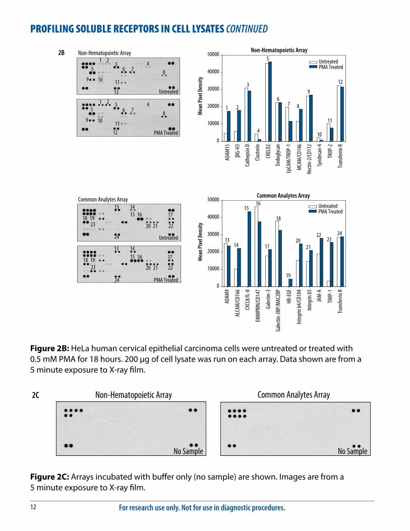
Figure 2B: HeLa human cervical epithelial carcinoma cells were untreated or treated with 0.5 mM PMA for 18 hours. 200 μg of cell lysate was run on each array. Data shown are from a 5 minute exposure to X-ray film.
Non-Hematopoietic Array
Mea
n Pi
xel D
ensit
y
0
10000
20000
30000
40000
50000UntreatedPMA Treated
Mea
n Pi
xel D
ensit
y
0
10000
20000
30000
40000
50000UntreatedPMA Treated
Common Analytes Array
βIG-
H3
Cath
epsin
D
CREL
D2
Cluste
rin
EpCA
M/T
ROP-
1
MCA
M/C
D146
ADAM
15
Necti
n-2/
CD11
2
ALCA
M/C
D166
CXCL
8/IL-
8
EMM
PRIN
/CD1
47
Galec
tin-3
BP/M
AC2B
P
TIMP-
1
JAM
-A
ADAM
9
Galec
tin-3
Inte
grin
b4/C
D104
Inte
grin
b5
Endo
glyca
n
Tran
sferri
n R
TROP
-2
Synd
ecan
-4
Tran
sferri
n R
HB-E
GF
Common Analytes Array
Non-Hematopoietic Array
Untreated
PMA Treated
Untreated
PMA Treated
2B1
1 2
2
3
3 4
45
5 6
6 7
7 8
89
9
12
10
1112
13
13 14
1415
15
16
16
17
1718
18 19
1920
20 21
21 22
2223
23
24
24
10
11
21
3
4
5
67 8
9
10
11
12
1314
1516
17
18
19
2021
2223
24
PROFILING SOLUBLE RECEPTORS IN CELL LYSATES CONTINUED
Common Analytes Array Non-Hematopoietic Array
No Sample No Sample
2C
Figure 2C: Arrays incubated with buffer only (no sample) are shown. Images are from a 5 minute exposure to X-ray film.

www.RnDSystems.com 13
Figure 3: The Human Soluble Receptor Array detects multiple analytes in serum. 25 μL of serum from a normal donor was run on each array. Data shown are from a 1 minute exposure to X-ray film.
Mea
n Pi
xel D
ensit
y
0
10000
20000
30000
40000
50000
Mea
n Pi
xel D
ensit
y
0
10000
20000
30000
40000
50000
IgM
α2-M
acro
globu
lin
C1q R
1/CD
93
Thro
mbo
spon
din
CD40
Liga
nd/T
NFSF
5
Lipoc
alin-
2/NG
AL
Tran
sferri
n R
Gal-3
BP/M
AC2B
P
RECK
NCAM
-L1
Common Analytes Array
Non-Hematopoietic Array
βIG-
H3
Cluste
rin
ECM
-1
ESAM
VCAM
-1
HPRG
VAP-
1/AO
C3 IgM
α2-M
acro
globu
lin
Tran
sferri
n R
Non-Hematopoietic Array
Common Analytes Array 11 12
1314 15 1617
18 19 20
12 3
4
56
7
8
9
10
1 23 4
5
6 78 9 10
11
12
13
14 15 16
17
18
19
20
PROFILING SOLUBLE RECEPTORS IN SERUM

For research use only. Not for use in diagnostic procedures.14
APPENDIXRefer to the table below for the Human Non-Hematopoietic Array (Part N) coordinates.
Coordinate Analyte/Control Alternate Nomenclature Entrez Gene ID
A1, A2, A3, A4 Reference Spots ___ ___
A5, A6 ADAM15 ___ 8751A7, A8 βIG-H3 ___ 7045A9, A10 BMPR-IB/ALK-6 CDw293 658A11, A12 Cadherin-4/R-Cadherin CAD4 1002A13, A14 Cadherin-11 CAD11 1009A15, A16 Cadherin-13 CDHH 1012A17, A18 E-Cadherin CD324, ECAD 999A19, A20 N-Cadherin CD325, NCAD 1000A23, A24 Positive Control ___ ___
B1, B2 Positive Control ___ ___
B5, B6 P-Cadherin PCAD 1001B7, B8 VE-Cadherin CD144, CDH5 1003B9, B10 Cathepsin D ___ 1509B11, B12 CD40/TNFRSF5 ___ 958B13, B14 CEACAM-5/CD66e CEA 1048B15, B16 CHL-1/L1CAM-2 CALL 10752B17, B18 Clusterin ___ 1191B19, B20 Coagulation Factor II/Thrombin ___ 2147C1, C2 COMP/Thrombospondin-5 ___ 1311C3, C4 CRELD2 ___ 79174C5, C6 Desmoglein 2 ___ 1829C7, C8 ECM-1 ___ 1893C9, C10 EGF R/ErbB1 HER1 1956C11, C12 Endoglycan PODXL2 50512C13, C14 EpCAM/TROP-1 ___ 4072C15, C16 ErbB2/HER2 ___ 2064C17, C18 ErbB3/HER3 ___ 2065C19, C20 ErbB4/HER4 ___ 2066C21, C22 ESAM ___ 90952C23, C24 Galectin-2 ___ 3957D1, D2 HPRG ___ 3273D3, D4 Integrin α3/CD49c ITGA3 3675D5, D6 Integrin α5/CD49e ITGA5 3678D7, D8 Integrin α6/CD49f ITGA6 3655D9, D10 Integrin α9 ITGA9 3680D11, D12 Integrin αV/CD51 ITGAV 3685continued on next page....

www.RnDSystems.com 15
APPENDIX CONTINUEDCoordinate Analyte/Control Alternate Nomenclature Entrez Gene ID
D13, D14 Jagged 1 JAG1, CD339 182D15, D16 JAM-B/VE-JAM CD322, JAM2 58494D17, D18 JAM-C JAM3 83700D19, D20 LRP-6 ___ 4040D21, D22 MCAM/CD146 MUC18 4162D23, D24 MEPE ___ 56955E1, E2 MUCDHL ___ 53841E3, E4 Nectin-2/CD112 PVRR2, PVRL2 5819E5, E6 Nectin-4 PVRL4 81607E7, E8 Neurotrimin IGLON2 50863E9, E10 Notch-1 ___ 4851E11, E12 NrCAM ___ 4897E13, E14 Periostin/OSF-2 ___ 10631E15, E16 Podocalyxin PODXL 5420E17, E18 E-Selectin/CD62e CD62E, ELAM 6401E19, E20 Semaphorin 3A SEMA3A 10371E21, E22 SREC-I/SR-F1 SCARF1 8578E23, E24 SREC-II SCARF2 91179F1, F2 Stanniocalcin 1 STC1 6781F3, F4 Syndecan-1/CD138 SDC1 6382F5, F6 Syndecan-4 SDC4 6385F7, F8 Thrombospondin-2 TSP2, THBS2 7058F9, F10 TIMP-4 ___ 7079F11, F12 TROP-2 ___ 4070F13, F14 VAP-1/AOC3 SSAO 8639F15, F16 VCAM-1 CD106 7412F17, F18 VEGF R1/Flt-1 ___ 2321F19, F20 VEGF R2/KDR/Flk-1 CD309 3791G1, G2 Reference Spots ___ ___
G5, G6 IgM (Sample Control)* ___ 3507G7, G8 α2-Macroglobulin (Sample Control)* A2M 2G9, G10 Transferrin R (Sample Control)* CD71 7037G11, G12 Vimentin (Sample Control)* VIM 7431G13, G14 PBS (Negative Control) ___ ___
G23, G24 Reference Spots ___ ___
*Sample controls are included to allow for the detection of proteins commonly present in cell culture supernates, cell lysates, and serum. If these endogenous proteins are present in a particular sample, positive signals indicate that the sample has been incubated with the array and the assay procedure has been performed correctly.
For research use only. Not for use in diagnostic procedures.16
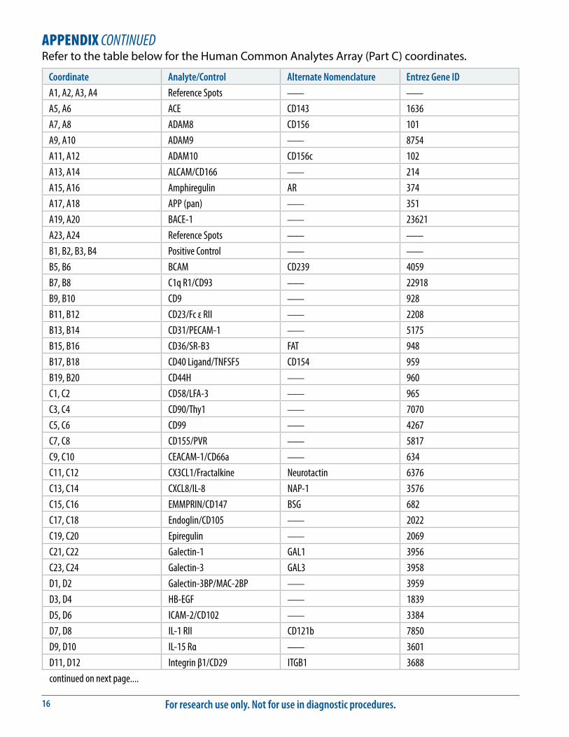
APPENDIX CONTINUEDRefer to the table below for the Human Common Analytes Array (Part C) coordinates.
Coordinate Analyte/Control Alternate Nomenclature Entrez Gene ID
A1, A2, A3, A4 Reference Spots ___ ___
A5, A6 ACE CD143 1636A7, A8 ADAM8 CD156 101A9, A10 ADAM9 ___ 8754A11, A12 ADAM10 CD156c 102A13, A14 ALCAM/CD166 ___ 214A15, A16 Amphiregulin AR 374A17, A18 APP (pan) ___ 351A19, A20 BACE-1 ___ 23621A23, A24 Reference Spots ___ ___
B1, B2, B3, B4 Positive Control ___ ___
B5, B6 BCAM CD239 4059B7, B8 C1q R1/CD93 ___ 22918B9, B10 CD9 ___ 928B11, B12 CD23/Fc ε RII ___ 2208B13, B14 CD31/PECAM-1 ___ 5175B15, B16 CD36/SR-B3 FAT 948B17, B18 CD40 Ligand/TNFSF5 CD154 959B19, B20 CD44H ___ 960C1, C2 CD58/LFA-3 ___ 965C3, C4 CD90/Thy1 ___ 7070C5, C6 CD99 ___ 4267C7, C8 CD155/PVR ___ 5817C9, C10 CEACAM-1/CD66a ___ 634C11, C12 CX3CL1/Fractalkine Neurotactin 6376C13, C14 CXCL8/IL-8 NAP-1 3576C15, C16 EMMPRIN/CD147 BSG 682C17, C18 Endoglin/CD105 ___ 2022C19, C20 Epiregulin ___ 2069C21, C22 Galectin-1 GAL1 3956C23, C24 Galectin-3 GAL3 3958D1, D2 Galectin-3BP/MAC-2BP ___ 3959D3, D4 HB-EGF ___ 1839D5, D6 ICAM-2/CD102 ___ 3384D7, D8 IL-1 RII CD121b 7850D9, D10 IL-15 Rα ___ 3601D11, D12 Integrin β1/CD29 ITGB1 3688continued on next page....

www.RnDSystems.com 17
APPENDIX CONTINUEDCoordinate Analyte/Control Alternate Nomenclature Entrez Gene ID
D13, D14 Integrin β2/CD18 ITGB2 3689D15, D16 Integrin β3/CD61 ITGB3 3690D17, D18 Integrin β4/CD104 ITGB4 3691D19, D20 Integrin β5 ITGB5 3693D21, D22 Integrin β6 ITGB6 3694D23, D24 JAM-A CD321 50848E1, E2 Lipocalin-2/NGAL ___ 3934E3, E4 LOX-1/SR-E1 CLEC8A 4973E5, E6 MD-1 LY86 9450E7, E8 MMP-2 (total) ___ 4313E9, E10 NCAM-1/CD56 ___ 4684E11, E12 NCAM-L1 L1CAM, CD171 3897E13, E14 Osteopontin OPN 6696E15, E16 PAR1 ___ 2149E17, E18 Pref-1/DLK-1/FA1 ___ 8878E19, E20 RECK ___ 8434E21, E22 Stabilin-1 CLEVER-1, FEEL-1 23166E23, E24 TACE/ADAM17 CD156b 6868F1, F2 Thrombospondin THBS, TSP 7057F3, F4 TIMP-1 ___ 7076F5, F6 TIMP-2 ___ 7077F7, F8 TIMP-3 ___ 7078F9, F10 TNF RII/TNFRSF1B CD120b 7133G1, G2 Reference Spots ___ ___
G5, G6 IgM (Sample Control) ___ 3507G7, G8 α2-Macroglobulin (Sample Control)* A2M 2G9, G10 Transferrin R (Sample Control)* CD71 7037G11, G12 Vimentin (Sample Control)* VIM 7431G13, G14 PBS (Negative Control) ___ ___
G23, G24 Reference Spots ___ ___
*Sample controls are included to allow for the detection of proteins commonly present in cell culture supernates, cell lysates, and serum. If these endogenous proteins are present in a particular sample, positive signals indicate that the sample has been incubated with the array and the assay procedure has been performed correctly.

For research use only. Not for use in diagnostic procedures.18
NOTES
09.09 752010.5 4/14
©2014 R&D Systems, Inc.
All trademarks and registered trademarks are property of their respective owners.