the studies on lymphatic venous anastomosis in lvmphedema
TRANSCRIPT

Nagoya J. med. Sci. 32: 1-21, 1969.
THE STUDIES ON LYMPHATIC VENOUS ANASTOMOSIS IN LVMPHEDEMA*
YuKro YAMADA
1st Department of Surgery, Nagoya University School of Medicine (Director: Prof Yoshio Hashimoto)
ABSTRACT
Although a considerable number of publications about the treatment of lym
phedema has been appeared in recent years, an effective method for surgical treatment has not yet been established.
In legs of adult mongrel dogs the author made a lymphatic venous end-to-end
anastomosis with 4 to 6 evenly-spaced, interrupted sutures of No. 10-0 black
tetron monofflament. All branches of the sapheneus vein were ligated and cnt
off to prevent venous blood inflow. Consequently the saphenous vein was trans
formed into a lymphatic channels. At the site of the anastomosis, the author amputated the lymphatic venous
anastomosed leg and the control leg except their bones, arteries, veins and nerves
and relsutured them respectively for formation of lymphedema. The author examined the relationship between lymphatic and venous pressure at rest or in
the manual pumping procedures. In the present study, lymphpressure ascended abruptly during manual pumping
more than venous pressure. In the amputated leg having an end-to-end anasto
mosis, the author succeeded to demonstrate the passage of a contrast medium
through the anastomosis by indirect lymphangiography, and circumferences of
the lymphatic venous anastomosed leg were less than those of the control leg.
The main cause of failure of anastomosis was presence of excess granulation
tissue at the anastomotic site and anastomotic disruption. By using No. 10-0 black tetron monofilament, however, the granulation was not prominent histolo
gically. Therefore, the author applied this method to a patient with lymphedema
clinically, and obtained considerable effects.
INTRODUCTION
Hitherto considerable number of studies on treatment of lymphedema have been reported, but an effective surgical method has not yet been established. During the past several years, the understanding of the role played by the lymphatic system has been increased, and lymphedema, chyluria and certain forms of protein-losing enteropathies are now considered by most observers to be forms of lympnatic insufficiency 1>2>3>•>. Our knowledge of lymphcirculation
[li fB 1'1' ~ Received for publication January 6, 1968. * An outline of the present paper was read at the 8th Congress of Japanese Angiological
Society in 1967.
1

2 Y. YAMADA
however remains incomplete, and mechanisms that determine lymph transport, flow and pressure are still obscure and subject to debate.
It is evident that lymphatic venous communications in the periphery may play a part as alternative pathways for lymph flow 5>6>7>8l, but any flow of lymph into the venous system which may occur at the node is not great enough to meet the needs of the part 9>.
Lymph flow is hindered by a mechanical factor. The mechanical factors may be of anatomical, organic nature, e.g. the occlusion of the lymphatics by obstructive lymphangitis, thrombosis of the lymphatics, filariasis, etc 10>. When obstruction of the normal channels prevents lymph from reaching the venous circulation through the large trunks at the base of the neck, alternative paths become important.
Clinical and experimental observations place most of these communications at the level of the lymph nodes 5> 11>. In man, though these channels may exist, they are apparently unable to compensate for the loss of the major lymphatic
trunks in the extremities. In dog, the subcutaneous lymphatics may be able to compensate for the loss of the deeper trunks, but if they are also destroyed, then edema will result.
For the formation of alternative paths, the author considered that an artificial lymphatic venous anastomosis would be effective in lymphatic obstruction. The experience with direct surgery using the lymphatics and the saphenous vein forms the basis of this study.
Masai has published a paper on the direct lymphatic venous implantation, but it had a tendency to occlusion after a long term 12> (Fig. 1). Since the author has taken a different approach toward the problem of creation of
Lymphatic venous implantation I. End-to-end implantation II. End-to-side implantation
III. End-to-end implantation with ligation of blanches of vein
IV. End-to-side implantation with ligntion of blanches of vein
FIG. 1. Method of lymphatic venous implantation

THE STUDIES ON LYMPHEDEMA 3
a lymphatic venous shunt. The present study was undertaken in the hope that the technical precision
afforded by the microsurgical techniques and the trial of various anastomotic methods might be successful in creating a long term diversion of lymph into the saphenous vein 13l 14l.
Lymphatic vessels are too fragile for formal suturing, but have a regenerative capacity which renders a precise approximation unnecessary 15l.
Low concentration of coagulation proteins in peripheral lymph makes the likelihood of intraluminal clotting remote, even under circumstances of surgical manipulation 16l.
Peripheral lymph flow is very slow and under low pressure, so that local extravasation of lymph from a loose approximation in anastomosis is not a limiting problem 17l.
From these considerations, it appeared that it would be possible to create a lymphatic venous anastomosis without the need of water-tight suture by ligating and cutting off of all branches of the saphenous vein.
To this end the following experiments were performed on 4 groups of 80 adult mongrel dogs.
MATERIALS AND METHODS
Adult mongrel dogs weighing 8 to 15 kg were used. Patent Blue dye in 10% solution was injected into the palmar surface of the paw to aid visualization of the lymphatics. After 10 to 15 minutes a skin incision was made along the saphenous vein of the hind leg in an anesthetized dog. One of two lymphatics accompanying the saphenous vein was carefully isolated from the surrounding connective tissue for a distance of about 2 em. One small polyethylene tube was inserted into the lymphatic vessel through a single incision in the wall of the lymphatics, and directed peripherally as a splint, and the lymphatic vessel was cut off at the site of incision (Fig. 2 and 3).
The saphenous vein was carefully isolated from the surrounding connective tissue for all distance, and all branches were ligated and then blood flow of the saphenous vein was intercepted.
Experiment A: End-to-end anastomosis The isolated saphenous vein was cut off adjacent to the site of cannulation
in the lymphatic, and the free end of a splint was inserted into the saphenous vein centrally. A end-to-end anastomosis was carried out between the saphenous vein and the lymphatic. The nearby lymphatics were blocked to increase lymphatic pressure.
No. 10- 0 black tetron monofilament was used for suture of the anastomosis with an operating microscope. After suturing, the splint was removed through the wall of the vein (Fig. 2). Consequently, the saphenous vein was transformed

4 Y. YAMADA
Lymphatic venous end-to-end anastomosis with ligation of all blanches of the vein
"' ~
~=> ~
FIG. 2. Method of lymphatic venous end-to-end anastomos1
Lymphatic venous end-to-side anastomosis with ligation of all blanches of the vein
FIG. 3. Method of lymphatic venous end-to-side anastomosis.
into a lymphatic channel. Long term behaviour of the anastomosis was
followed up by direct lymphangiography and histological examination. Direct
lymphangiography was done by intralymphatic injection of about 2 cc of 60%
Urografin. Histologically the author examined the specimens by Hematoxylin
Eosin staining and Elastica-Van Gieson staining.
Experiment B: End-to-side anastomosis A small incision on the wall of the saphenous vein was made adjacent to

THE STUDIES ON LYMPHEDEMA 5
the site of cannulation into the lymphatic, and the free end of a splint was
inserted into the vein through the incision (Fig. 3). A end-to-end anastomosis was carried out with 4 to 6 evenly-spaced in
terrupted sutures of No. 10-0 black tetron monofilament (Fig. 3). After com
pleting the anastomosis, the site of the anastomosis was covered with subcutaneous tissue and skin, and left for 1 week to 6 months.
Experiment C: Formation of lymphedema by amputation For alling to lymphedema clinically the author made lymphedema in the
legs of dogs. In order to test the functional capacity of such anastomosis, blockage of the normal lymphatic drainage of the legs was performed after
the manner described by Danese and Howard 6'. The author amputated the hind legs of dogs by a circumferencial incision,
except their bones, arteries, veins and nerves, and resutured the muscles and skin (Fig. 4). In one of the two hind legs the author created a lymphatic venous end-to-end anastomosis, and the opposite hind leg was observed as control (Fig. 4). One to two weeks after the operation indirect lymphangiography
was performed by intrasubcutaneous injection of 5 cc of 60% Urografin in the palmar surface of the paw.
Experiment D: Relationship between lymphatic and venous pressure Two small polyethylene tubes were inserted through a single incision in
the wall of lymphatic. One tube was directed centrally past the next valve and the other was directed peripherally. The free ends of the cannulae were connected to the two arms of a polyethylene T-tube (Fig. 5). The entire
system had previously been filled with physiological saline solution containing 1 mg heparin per ml.
The saphenous vein was cannulated with a polyethylene tube adjacent to the site of cannulation of the lymphatic. Both lymphatic and venous pressures
Formation of lymphedema in leg by amputation
Lymphatic venous anastomosed leg Control
Fig. 4. Method of formation of lymphedema by amputation.

6 Y. YAMADA
Apparatus for measurement of lymphatic pressure
FIG. 5. Cannulation method of measurement of lymphatic pressure.
were read directly in the polyethylene tube which served as a water manometer. After series of normal readings had been taken, manual pumping procedures were attempted by squeezing the paw, moving a joint or tapping the paw. In the same manner lymphatic pressure in lymphedema was measured.
EXPERIMENTAL RESULTS
Experiment A: In the legs having an end-to-end anas
tomosis of the lymphatic and vein, the author succeeded to demonstrate passage of a contrast medium roentgenographically through the anastomosis (Figs. 6, 7, 8, 9, and 10). Few early failures to demonstrate were due to surgical technical failure in suturing and in intralymphatic injection of a contrast medinm. Following nearby lymphatic blockage, all legs became edematous a little in spite of the lymphatic venous anastomosis. But 1 week after the operation light edema of the leg subsided, and the author was encouraged to think that this might be due to the function of the anastomosis.
Direct lymphangiography done 1 week after the operation demonstrated the anastomosis, the saphenous vein and femoral
FIG. 6. Direct lymphangiography showing passage of a contrast medium through the anastomosis taken 1 week following (white arrow).
THE STUDIES ON LYMPHEDEMA
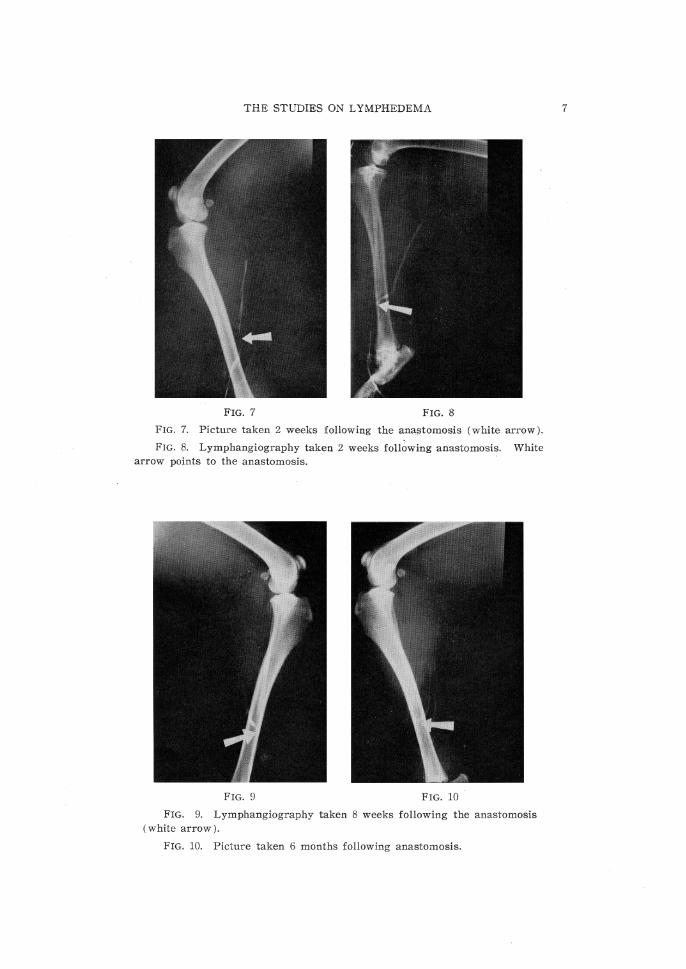
FIG. 7 FIG. 8
FIG. 7. Picture taken 2 weeks following the anastomosis (white arrow).
FIG. 8. Lymphangiography taken 2 weeks following anastomosis. White arrow points to the anastomosis.
FIG. 9 FIG. 10
FIG. 9. Lymphangiography taken 8 weeks following the anastomosis (white arrow).
FIG. 10. Picture taken 6 months following anastomosis.
7

8 Y. YAMADA
vein, patent fully (Fig. 6), and when repeated 2 weeks later, the saphenous vein was demonstrated fully by a contrast medium (Fig. 7 and 8). When repeated 8 weeks later, the vein showed narrowing of the lumen (Fig. 9). About 6 months after the operation direct lymphangiography demonstrated the lymphatic channel transformed by the saphenous vein, but ll,flrrowing of the lumen at the site of the anastomosis and at the vein was observed relatively much (Fig. 10). External and internal appearances of the anastomosis were showed in Fig. 11 and 12. The patency rate in this group was obtained 1 week, 8 weeks and about 6 months after the operation (Table I a). Histologically the author examined the area of anastomosis, and obtained that the lumen had been kept fully 2 weeks later the anastomosis (Fig. 13): When
FIG. 11. Picture shows the external appearance of lymphatic venous anastomosis.
FIG. 12. Picture shows the internal appearance of lymphatic venous anastomosis.
THE STUDIES ON LYMPHEDEMA
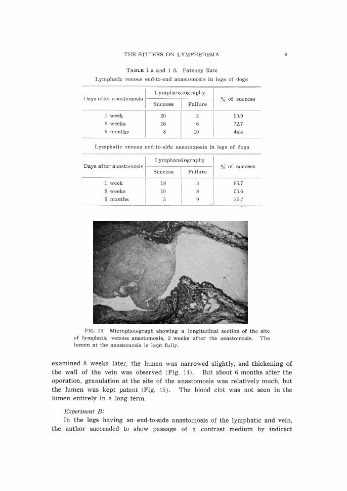
TABLE 1 a and 1 d. Patency Rate
Lymphatic venous end-to-end anastomosis in legs of dogs .;~--==::--::::-.
Days after anastomosis I Lymphangiography
I ··~ %of success
Success Failure - ·-
1 week 20 2 90.9
8 weeks 16 6 72.7
6 months 8 10 44.4
Lymphatic venous end-to-side anastomosis in legs of dogs
I Lymphansiography Days after anastomosis [---
I Success Failure % of success
1 week 1 18 3 85.7
8 weeks I 10 8 55.6
6 months ______ 5 _ _____ 9_ -'.'-""-"-; ___ 3_5~.7=
FIG. 13. Microphotograph showing a long itudinal section of the site of lympha tic venous anastomosis, 2 weeks after the anastomosis. The lumen at the anastomosis is kept fully .
9
examined 8 weeks later, the lumen was narrowed slightly, and thickening of the wall of the vein was observed (Fig. 14). But about 6 months after the oporation, granulation at the site of the anastomosis was relatively much, but the lumen was kept patent (Fig. 15). The blood clot was not seen in the lumen entirely in a long term.
Experinent B: In the legs having an end-to-side anastomosis of the lymphatic and vein,
the author succeeded to show passage of a contrast medium by indirect

10 Y. YAMADA
FIG. 14. Microphotograph of the cross section of the vein transformed into a lymphatic channel, showing thickening of the wall.
FIG. 15. Microphotograph of the longi-tudinal section of the site of lymphatic venous anastomosis 6 months after anastomosis, showing narrow
ing of the lumen. Whit~ arrow points to No. 10-0 black tetron monofilament.
yymphangiography through the anastomosis (Fig. 16). The surgical technique was more difficult than that in Experiment A, and so technical failure became frequent (Fig. 17). The patency rate in this group was poorer than that of Experiment A (Table 1 b). In others, inflammatory changes due to anastomotic disruption were noted frequently in this group.
Experiment C: In the hind legs of dogs the lymphedema appeared 3 to 7 days after the
amputation. The circumferences of the lymphatic venous ranastomosed leg
were about 1 to 3 em less than those of the opposite leg used as control. The
THE STUDIES ON LYMPHEDEMA
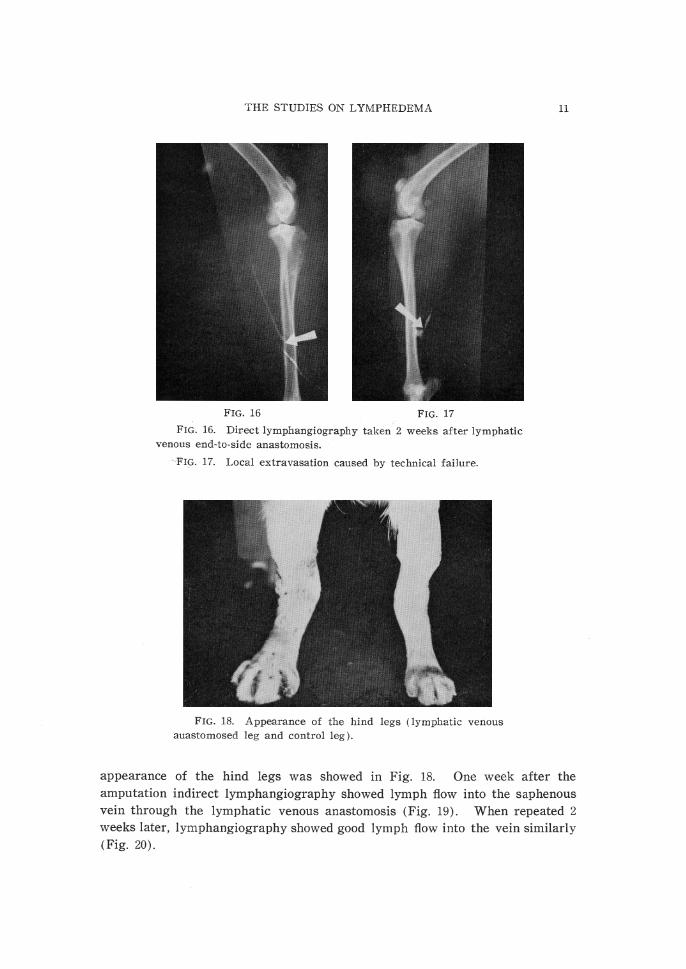
FIG. 16 FIG. 17
FIG. 16. Direct lymphangiography taken 2 weeks after lymphatic venous end-to-side anastomosis.
· FIG. 17. Local extravasation caused by technical failure.
FIG. 18. Appearance of the hind legs (lymphatic venous auastomosed leg and control leg).
11
appearance of the hind legs was showed in Fig. 18. One week after the amputation indirect lymphangiography showed lymph flow into the saphenous vein through the lymphatic venous anastomosis (Fig. 19). When repeated 2 weeks later, lymphangiography showed good lymph flow into the vein similarly (Fig. 20).

12 Y. YAMADA
Fig. 19. Indirect lymphangiography taken 1 week after amputation. Contrast medium is seen in the vein. White arrow shows the lymphatic
venous anastomosis.
FIG. 20. Indirect lymphangiography taken 2 weeks after ampntation. Contrast medium is seen in the vein. White arrow shows the lpmphatic venous anastomosis.
Exberitnent D: Immediately after cannulation both lymphatic and venous pressures were
unstable, presumably consequent on the manipulations incident to the operation.
At rest lymphatic pressure ranged from 2.0 to 8.2 em of water, while the range of venous pressure was from 8.0 to 20.6 em of water (Table 2). Pressure levels in the lymphatic and vein were generally very similar (Fig. 21). Knee joint movement increased lymphatic pressure as high as 2 or 3 times the
average control level, and caused a slight increase or slight decrease of venous pressure (Table 2). Tapping of the paw raised lymphatic pressure (Fig. 21).

THE STUDIES ON LYMPHEDEMA
T ABLE 2. Lymphatic and Venous Pressure in Legs of Dogs --- ....• --
At rest I Knee joint Tapping
I
Squeezing
I
movement
No.,J, L. P. I V . P. L. P. I V. P . L . P. I V. P. L. P. I V . P. I
~--~
I 1 3.5 lOA 9.8 10.2 13.8 14.0 21.2 18.0 2 4.8 12.0 14.0 13.0 19.0 17.4 23.8 21.2 3 3.2 8.5 9.4 10.0 12.2 13.0 20.4 18.0 4 5.0 10.3 14.8 14.0 18.8 17.5 24.4 21.6 5 8.0 12.2 16.6 17.8 20.5 20.0 26.0 24.0 6 2.5 8.0 9.0 8.0 12.8 12.0 19.2 17.0 7 7.2 20.6 20.0 18.0 23.0 22.0 27.0 26.0 8 4.8 10.0 16.0 15.4 23.0 18.7 26.2 23.0 9 5.0 11.3 15.8 15.0 18.8 19.0 26.0 24.0
10 6.0 10.8 18.0 18.0 20.6 21.0 26.8 25.3
11 i 2.0 8.0 8.0 7.5 13.6 12.0 19.8 18.0 12 i 5.0 15.0 16.0 16.0 19.5 20.0 25.8 24.0 13 5.2 13.7 15.4 12.8 18.8 16.8 24.6 20.5 14 4.3 12.0 17.0 15.2 20.4 20.0 27.4 23.0 15 4.5 12.2 15.0 15.0 19.8 18.0 26.6 23.0 16 4.6 11.8 14.8 13.6 19.6 17.0 25.2 21.0 17 4.2 10.0 15.0 13.0 20.0 18.0 25.4 22.0 18 7.4
i 17.0 18.5 :
18.0 22.5 22.0 27.0 26.0 19 8.2 18.2 17.0
I 18.0
I 20.8 23.0 27.6 27.0
20 4.0 12.0 15.8 14.4 19.0 I 18.0 26.8 23.0 I
----~-
Average I 4.9 12.2 1 14.8 1 14.1 1 18.7 1 18.0 1 2~__:::_
tH IILO
d
10
II
,, •• 11
..
Rela tionship between lymphatic and venous pressure in legs ofdogs (average)
----- Lp .
- - V.p
tlt nst ""'""J•'"" tarri•~ s,w,ezi,.g ""~"'~t,
FIG. 21. Relationship betweea lymphatic and venous pressures.
13
Reppetitive squeezing of the paw caused lymphatic pressure to rise up as high as 3 or 7 times the control level (Fig. 21). In the present experiment lymph pressure rose up about 5.0 to 14.0 em of water at rest in the legs with lymphedema.

14 Y. YAMADA
CASE REPORT
Case 1: A woman, aged 37, underwent an operation for the right hip joint tuber
culosis about 10 years before. This patient remarked swelling of the right
leg 5 years before. Physical activity was markedly and increasingly limited,
FGI. 22 FIG. 23
FIG. 22. Secondary lymphedema of the righy leg. showing preoperative
appearance.
FIG. 23. Lymphangiography showing the varicose, hyperplastic lym
phatics with some dermal backfiow.
FIG. 24. Saphenous vein with anastomosed lymphatics.

THE STUDIES ON LYMPHEDEMA 15
but the leg was painless (Fig. 22). She pressed for operative treatment for this reason. Lymphangiogrtphy disclosed hy
perplasia with varicosity of the subcutaneous lymph trunks (Fig. 23). At operation
we made 4 lymphatic venous anastomoses and 1 lymphatic venous implantation in the leg (Fig. 24) . The saphenous vein was
used, and 3 months after the operation the drcumferences of the leg markedly decreased by about 2 to 8 em (Fig. 25). As
showed in Fig. 25, the appearance of the leg became better, and the author believes
that it is due to functional artificial lymphatic venous shunts.
DISCUSSION
FIG. 25. Right lower extremity, showing appearance 3 months after operation.
The desirability to create a new pathway for lymph flow by passing the area of lymphatic obstruction is obvious to all physicians. Since the lymphatics embryologically originate from the developing venous network, lymphatic venous communications may be expected 18' . It is well documented that lymphatic tissue regenerates rapidly with restoration of normal function after manipulation, partial excision and other trauma 19>20>21>.
On the other hand, it has been both experimentally and clinically demonstrated that the thoracic duct can be sucessfully implanted into a vein with maintenance of lymph flow 22' 23' 24>. Therefore, it is clear that there is no fundamental incompatibility of anastomosis between the lymphatic and venous systems. There is some evidence that lymphatic venbus :Communications in the area of the lymph nodes normally exist in the periphery, but significance of these connections has not been firmly established 5' 6' 7' 8'.
Although there is disagreement among investigators as to intralymphatic pressures and gradients across any lymphatic venous communications 1' 25' 26',
several have reported intermittent high pressure within the lymphatics in the extremities 26' 4' . Yoffey and Courtice 18' presented ample evidence of the presence of lymphatic venous commumcations which shunt lymph from efferent channels directly into the veins prior to reaching the larger lymphatic ducts. Glenn and associates 27' have demonstrated that, when a peripheral lymphatic channel is injected with dye, the latter will appear in blood even if the thorecic

16 Y. YAMADA
duct is tied. It is considered that this occurs by way of accessory lymphatic venous communications.
Drinker 28J has similarly described the presence of accessory channels between the thoracic duct and the right lymphatic duct of doge. When the main channels are obstructed or lymph flow and lymph pressure are greatly increased, the accessory channels become enlarged and activated. Similarly, the experimental demonstration of lymphatic venous communication under conditions of lymphatic obstruction has been successfully achieved in rats 29J.
Rusznyak 10l concludes tnat, although lymphatic venous communications can be demonstrated in anatomic preparations, they are functionally negligible under normal conditions. Malek 28l concluded that damage to a lymph node by infection or trauma may lead to the formation of local lymphatic venous communications. In addition, actual demonstration of flow across these communications has been made under experimental conditions by isotope dilution methods 29l.
It would appear justifiable to conclude that lymphatic venous communications may well exist in all mammals but function only in the presence of increased lymphatic pressure or volume resulting from lymphatic obstruction.
Higher pressure on the venous side of the communications, as compared to the lymphatic side, could be a major factor interfering with Jym.ph flow. Available imformation points the existence of lymphatic venous lymph flow in certain areas despite an apparent differential pressure gradient, and this gradient could be overcome with muscular contraction.
On Starling's 30l hypothesis, the extracellular fluid originates from the blood stream by filtration across the capillary membranes, and is largely the result of the balance achieved between blood pressure which tends to force the fluid outwards and osmotic pressure of plasma proteins which tends to retain it.
Since blood pressure is higher at the arterial end of the capillary and plasma protein osmotic pressure constant throughout, movement of electrolyte across the capillary membrane is towards the tissues at the arterial end and into the lumen at the venous end of the capillary. Peripheral edema may result from disturbance of flow of these fluids.
Muscular activity is a very important factor in lymph flow. Funaoka 311
observed that radio"J'aque material, when injected into a lymphatic, remained immobile for a considerable lapse of time if the animal was at rest, but was quickly propelled on as soon as active or passive movement of the organ concerned was provoked. Kubik's 23l experiments may be regarded as an important contribution towards elucidating the question of how lymph flow is affected by passive movement. He found that there are ampullar dilatations all over the lymphatic system. The ampullae are compressed when the muscles contract, while their walls are drawn apart when the muscles relax, so that ampullae play the part of a sort of pressure pumps.

THE STUDIES ON L YMPHEDEMA 17
It is not merely in the thoracic duct and the large lymph trunks that lymph flow is influenced by arterial pulsation. To prove the correctness of this statement was the object of the experiments performed by Clark and Clark 33l , Henry 34l , McMaster and Parsons 35l . Making their experiments on the ear of rabbits, Clark and Clc.rk found that lymph in the lymphatics moved to and fro in synchronism with arterial pulsation. Heller 36l gave evidence for the fact that the mesenteric lymphatics of guinea pigs perform about ten rhythmical contractions per minute. But Folorey 37l denied the existence of lymphatic pulsation.
Horstman 38l described that the contraction of valvular segment propels lymph to the next segment. When the segment is filled, its muscles contract
and force its contents to move in the proximal direction. The role of pulsatile arterial flow in promoting lymphatic flow has been
the subject of discussions for many years, and is considered by some observers to be one of the factors producing lymph flow 39l . The volume of lymph flow under these circumstances might be insufficient in lymphatic obstruction to keep lymph circulation normal. Therefore, by creating of artificial lymphatic
venous anastomosis the volume of lymph flow might be increased. Pressure gradient is generally against lymphatic to venous flow, and by
ligating all branches of the saphenous vein the author expected the situation to change at the site of anastomosis. Minimization of technical errors provided by the microsurgical technique has proved to be a significant factor in the long term patency of vascular anastomosis 40l .
It seemed worthwhile to apply this technical refinement to the construction of lymphatic venous anastomosis. Most probably an inflammatory action oa the vascular endothelium leads to early occlusion by granulation tissue and eventual fibrosis of the vessel itself. Toti and associates 41) have showed by Thorotrast uptake in rabbits that tnere was a dilatation the of lymphatjcs and an increase in lymph flow following the occlusion of the femoral vein. Danese and associates 24l showed that after occlusion of the greater saphenous vein by septic thrombophlebitis the popliteal lymph node enlarged up to several times its original size, and that the increase in lymph pressure was smaller in that study. Reichert 43l, using direct exposure and injection of dye, demonstrated that regeneration of the lymphatic capillaries began a few days after section. Regrneration of the larger lymphatics required more time.
Many empir ical surgical procedures have been tried for the relief of lymuhedema. The introduction of lymphangiography by Kimnonth 44l in 1955
resolved. It is of interest to consider the various operations which have been devised for the treatment of lymphedema. Lymphangioplasty was introduced by Handley 45l, Ransohoff 46l and Hogeman 47l , but no convincing evidence on the efficiency of these procedures was available. Fascia excisional procedure was
performed by Kondoleon 48l and Sistrunk 49l to make a communication between

18 Y. YAMADA
the superficial and deep lymphatic channels, but the regeneration of a fascial barrier has been repeatedly observed. Subcutaneous tissue excisional procedure was dractised by Charles 50l and Homans 51l. Thompson 52l 53l 54l performed the transposition of the superficial lymphatics into the deep subfascial compartment of the limb by means of a buried skin flap. Danese 17l succeeded to create an anastomosis by reimplantation of severed lymphatics into the lymph node. Cockett 55l performed an end-to-side anastomosis between the lumbar lymphatic vessel and spermatic vein, in chyluria, and the patient no longer had chyluria after the operation. In experimental procedures on dogs, the hind leg subcutaneous lymph trunks have with occasional success been anastomosed to the adjacent femoral vein 56l, to inguinal lymph nodes 57l, and even transplanted as an autograft to the contralateral thigh to restore continuity in a second lymphatic trunk from which a corresponding segment had been removed, with subsequent restoration of lymph flow 58l.
In human being, Nielubowiez and Olszewski 59) have claimed a single initial success in a patient with secondary lymphedema by anastomosing a transected inguinal lymph node with intact afferent superficial lymph trunk, into the lumen of the femoral vein. In the present experiment venous pressure was higher than lymphatic pressure, but the differences were not great. Since the lymphatics are believed to be highly distensible 60l, lymphatic pressure may not rise significantly until these vessels are distended. Drinker and his associates o)
found that lymph flow was increased by extrinsic factors such as massage, passive motion, muscular activity and respiration. This mechanism may facilitate lymphatic drainage of the peripheral lymphatics under some conditions.
SUMMARY AND CONCLUSION
The main causes of failure in lymphatic venous anastomosis are considered as follows:
1) Surgical technical failure, 2) Disruption of anastomosis by infectiOn, 3) Excess granulation, and 4) Occlusion by blood clot. The tissue reaction at the site of anastomosis was diminished markedly by using a fine No. 10-0 black tetron monofilament,. and so histologically the granulation decreased prominently. Consequently the patency rate increased by using an operating microscope. End-to-end anastomosis was better than end-to-side anastomosis similarly to those of the blood vessels.
Ligation of all branches of the saphenous vein prevented occlusion by blood clot and promoted lymph flow through the anastomosis.
In the leg of lymphedema by indirect lymphangiography the author succeeded to demonstrate flow of a contrast medium through the lymphatic venous anastomosis, and circumferences of the leg decreased remarkably than those Qf control. In a clini~al ~ase applied this peocedure, lymphedema in the leg

THE STUDIES ON LYMPHEDEMA 19
has been reduced for several months, and hereafter it should be applied to
secondary lymphedema and hyperplasia following primary lymphedema.
ACKNOWLEDGEMENT
The author wishes to express his deep gratitude to Prof. Y. Hashimoto
for his kind guidance throuout this study. He is indebted to Associate Prof.
K. Kamiya, Dr. T. Takao, and Dr. K. Kondo for their advice and criticism.
REFERENCES
1) Blocker, T . G., Smith, ]. R., Dunton, E. F ., Protas, ]. M., Cooley, R. M., Lewis, S. R.
and Kirby, E. ] .. Studies of ulceration and edema of the lower extremity by lymph
cannulation, Ann. Surg., 149, 884, 1959. 2) Bookstein, R. D., French, A. B. and Pollard, H. M., Protein-losing gastroenteropathy;
concepts derived from lymphangiography, Amer. f. Digesr. Dis., 10, 573, 1965.
3) Jarnum, S. and Peterson, P. V., Protein losing enteropathy, Lancet, 1, 417, 1961.
4) Kinmonth, ]. B., Primary lymphedema. Clinical and lymphangiographic studies of a
series of 107 patients in which the lower limbs were affected, Brit. f. Surg., 45, 1, 1957.
5) Pressman, J. ]., Burtz, M. V. and Shafer, L., Further observations related to direct
communications between lymph nodes and veins, Surg. Gynec. Obstet, 119, 984, 1964.
6) Pressman, ]. ]., Simon, M. B., Hand, K. and Miller, ]., Passage of fluids, cells, and
bacteria via direct communications between lymph nodes and veins, Surg. Gynec.
Obsret, 115, 207, 1962. 7) Retik, A. B., Perlmutter, A. D. and Harrison,]. H., Communications between lymphatics
and veins, involving the portal circulation, Amer. ]. Surg., 109, 201, 1965.
8) Threefoot, S. A., Kent, W. T. and Hatchett, B. T., Lymphatic venous and lymphatico
lymphatic communications demonstrated by plastic corrosion models of rats and by
postmortem lymphangiography in man, f. Lab. Clin. Med., 61, 9, 1963.
9) Danese, C. and Howard,]. M., Experimental lymphedema, Ann. Surg., 61, 441, 1965.
10) Rusznyak, I., Foldi, M. and Szabo, G., Lymphatics and Lymph Circulation, Pergamon
Press, Ltd ., Oxford, 1960, p. 563. 11) Pressman, ]. ]. and Simon, M. B., Experimental evidence of direct communications
between lymph nodes and the veins, Surg. Gynec. Obstet., 113, 537, 1961.
12) Masai, o., Experimental studies on lymphedema, f. ]ap. Coli. Angiol., 7, 74, 1967 (in
Japanese). 13) Jacobson, ]. H. and Suarez, E. L., Microsurgery in anastomosis of small vessels, Sur g.
Forum., 11, 243, 1960. 14) Kosse, K. G., Suarez, E. L., Fagan, W. T., Powel, P. R. and Jacobson, ]. H., Micro
surgery in ureteral reconstruction, ]. Urol., 87, 48, 1962·
15) Halsted, W. S., Reimplantatien of Entire Limb Without Suture of Vessels, Proc. Nat.
Acad. Sci. U.S.A. , 8, 181, 1922. 16) Brinkhaus, K. M. and Walker, S. A., Prothrombin and Fibrinogen in Lymph, Amer.
f. Physiol., 150, 746, 1947.
17) Danese, C., Bower, R. and Howard, ]. M., Experimental Anastomosis of Lymphatics,
Arch. Surg., 84, 24, 1962. 18) Yoffey, ]. M. and Courtice, F. C., Lymphatics, lymph, and lymphoid tissue, Edward
Arnold Publishers Ltd., London, 1956, p. 15.
19) Danese, C., Howard, ]. M. and Bower, R., Regeneration of lymphatic vessels; a radio
graphic study, Ann. Surg., 61, 156, 1962. 20 J Tilak, S. P. and Howard, ]. M., The influence of the duel circulation on the viability

20 Y. YAMADA
of lymph nodes following interruption of their blood supply or lymphatic supply, Surg. Gyrtec. Obstet., 119, 349, 1964.
21) Tilak, S. P. and Howard, J. M., Regeneration and autotransplantation of lymph nooes, Artrt. Surg., 161, 441, 1965.
22) Harrison, E., On the treatment of wounds of the thoracic duct, Brit.]. Surg., 4, 304, 1916.
23) Joel, D. D. and Sauter, ]. H., Preparation of a chronic thoracic duct venous shunt in calves, Proc. Soc. Exp. Biol. Med., 112, 856, 1963.
24) Lure, A. S., Implantation of an injured thoracic duct in to a vein in oncologocal operations on the neck, Vop. Onkol., 7, 9, 1961.
25) Guyton, C. A., A concept of negative interstitial pressure based on pressure in implanted performed capsules, Circ. Res., 12, 399, 1963.
26) Irisawa, A. and Rushmer, F. R., Relationship between lymphatic and venous pressure in leg of dog, Amer. ]. Physiol., 196, 495, 1959.
27) Glenn, W. W. L., Cresson, S. L., Bauer, F. X., Goldstein, F ., Hoffman, 0. and Healey, ]. E., Experimental thoracic duct fistula, Surg. G_vnec. Obstet., 89, 200, 1949.
28) Malek, P., Belan, A. and Koll, J., In vivo evidence of lymphaticovenous communications in the popliteal region, Acta Radial. ( Diagn ), 3, 344, 1965.
29) Threefoot, S. A., Kossover, M. F. and Aiken, D. W., Radioisotopic detection of lymphaticovenous communications in living animals, ]. Lab. Clin. Med., 65, 688, 1965.
ilO) Starling, E. H., On the absorption of fluids from the connective tissue spaces, ]. Physiol. (London), 19, 312, 1896.
31) Funaoka, S., Der Mechanismus der Lymphbewegung, Arb. aus der III. Abt. des Anat. lnst. d. Kaiser[. Univ. Kyoto, 1, 1, 1930.
32) Kubik, I., Die hydrodynamischen und mechanischen Faktoren in der Lymphzirkulation, Acta. Morph. Hung. 2, 95, 1952.
33) Clark, E. R. and Clark, E. L., Further observations living lymphatic vessels in the transparent chamber in the rabbit's ear their relation to tissue spaces, Amer. ] . Anat., 52, 273, 1933.
34) Henry, C. G., Studies on the lymphatic vessels and on the movement of lymph in the ear of the rabbit, Anat. Rec., 57, 263, 1933.
35) McMaster, Ph. D. and Parsons, R. ]., The effects of the pnlse on the spread of sub· stances through tissue, ]. Exp. Med., 68, 377, 1938.
36) Heller, A., Uber selbstiindige rhythmische Kontraktionen der Lymphgefiisse bei saugethieren, Zbl. Med. Wiss., 7, 545, 1896.
37 J Folorey, H., Observations on the contractility of lacteals, ]. Physiol. (London), 62, 267, 1927.
38) Horstmann, E., tiber die funktionelle Struktur des mesenterialen Lymphgefiisse, Morpho!. ]ahrb., 91, 483, 1952.
39) Web, R. and Staral, T. E., The effect of blood pressure pulsations on lymph pressure in large lymphatics, Bull. fohns Hopk. Hasp., 93, 401, 1953.
40) Jacobson, ]. H., Wallman, L. ]., Schumacher, G. A., Flanagan, M., Suarez, E. L. and Donaghy, R. M. T., Microsurgery as an aid to middle cerebral artery endarterectomy, ]. Neurosurg., 19, 108, 1962.
41) Toti, A., Fabi, M., Vita, G. snd Bagni, G., Ricerche sperimentali aul comportamento funzionale del cirolo linfatico dopo interruzione isolata e contemporanea dell'arteria e della vena femorale, Arcisped. S. Anna Ferrara, 12, 1017, 1959.
42) Danese, C., Diaz, R. and Howard, ]. M., Changes in lymphatics with experimental acute thrombophlebitis, Arch. Surg., 86, 19, 1963.
43) Reichert, F. L., The regeneration of the lymphatic, Arch. Surg., 13, 871, 1926. 44) Kinmonth, J. B., Taylor, G. W. and Harper, R. K., Lymphangiography. A technique

THE STUDIES ON LYMPHEDEMA 21
for its clinical use in the lower limbs, Brit. Med. ]., 1, 940, 1955. 45) Handley, W. S., A prospective cure for elephantiasis, Lancet, 1, 31, 1909. 46) Ransohoff, ]. L., Surgical treatment of lymphedema, Arch. S ur g., 50, 269, 1945. 47) Hogeman, K. E., Artificial subcutaneous channels in draining lymphedema, Acta Chir.
Scand. , 110, 154, 1955. 48 ) Kondoleon, E., Die operative Behandlung der elephantiastischen Oedem, Zbl. Chir., 39,
1022, 1912. 49) Sistrunk, W. E., Further experiences with the Kondo leon operation for elephantiasis,
] . A . M. A ., 71, 800, 1918. 50 ) Charles, R. H., A System of Treatment, Ed. A Latham and T . C. English, 3, 416, 1912. 51) Romans, J., The treatment of elephantiasis of the legs, New Eng. ]. Med,. 215, 1099,
1936. 52) Thompson, N., Surgical Treatment of Chronic Lymphedema of the Extremities, Surg.
Clin. N. Amer., 47, 445, 1967. 53) Thompson, N., Surgical t reatment of primary and secondary :lymphedema of the
extremities by lymphatic t ransposition, Proc. Roy. Soc. Med., 58, 1026, 1965. 54 ) Thompson, N., Surgical treatment of chronic lymphedema of the lower limb. With
preliminary report of new operation, Brit. Med. ]., 2, 1566, 1962. 55) Cookett, A. T. K. and Goodwin, W. E., Chyluria; Attempted surgical treatmen thy
lymphatic venous anastomosis, ], Urol., 88, 566, 1962. 56 ) Laine, ]. B. and Howard, ]. M., Experimental lymphaticovenpus anastomosis, Surg.
Forum, 14, 111, 1963. 57) Howard, ]. M., Danese, C. and Laine, ]. B., Experimenta l lymphatic anastomosis, ].
Cordiovasc. Surg., 5, 694, 1964. 58) Stevens, G. H., Kitagawa, S. and Longmire, W. P . Jr., Autotransplantation of lymphatic
vessels in t he dog, Surg. Forum, 15, 239, 1964. 59) Nielubowiez, J. and Olszewski, W., Surgical relief of lymphedema ( Abstr. ), World
Med., 1. 90, 1966. 60) Nishirnaru, Y. and Irisawa, H., Lymph capillaries in the frogs wed, Fed. Proc., 16, 94,
1957. 61) McCarrell, ]. D., Cervical lymph pressure in thedog, Amer. ]. Physiol., 127, 154, 19:>9.