circulatory system blood circulatory system -arterial system -capillaries -venous system lymphatic...
Post on 19-Dec-2015
255 views
TRANSCRIPT
Circulatory System
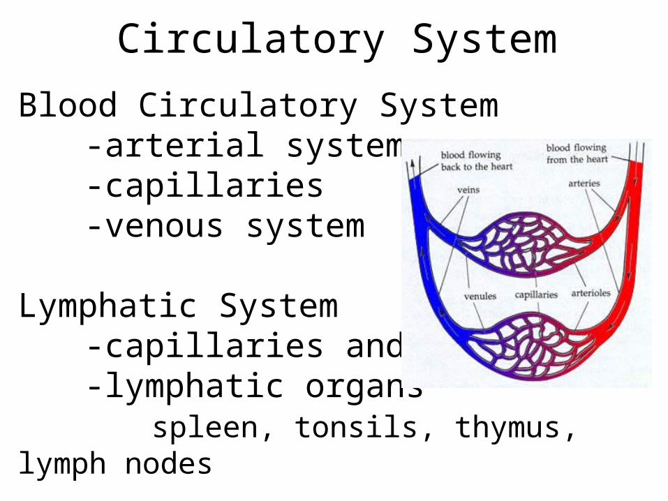
Blood Circulatory System-arterial system-capillaries-venous system
Lymphatic System-capillaries and ducts-lymphatic organs
spleen, tonsils, thymus, lymph nodes
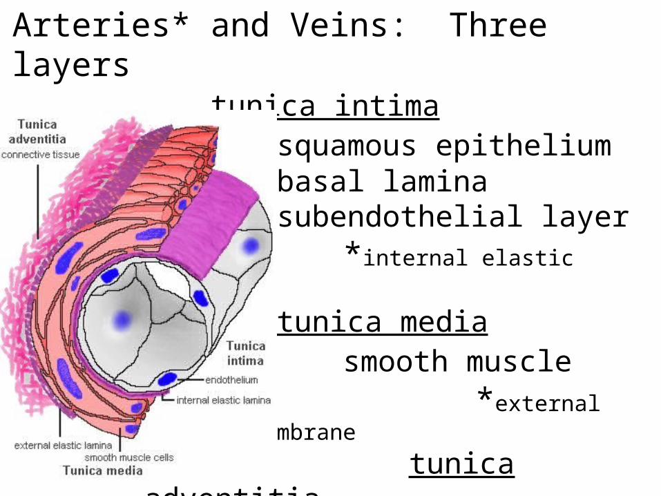
Arteries* and Veins: Three layerstunica intima
squamous epitheliumbasal laminasubendothelial layer
*internal elastic membrane
tunica mediasmooth muscle
*external elastic membrane
tunica adventitia
connective tissuevasa vasorumnervi vascularis
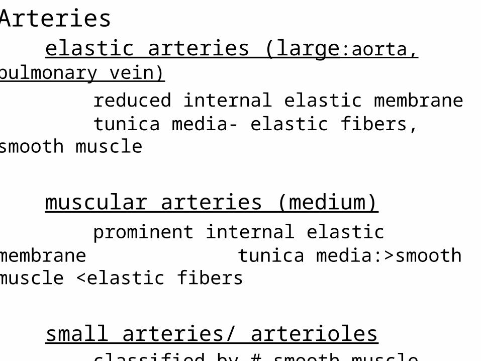
Arterieselastic arteries (large:aorta, pulmonary
vein)
reduced internal elastic membrane tunica media- elastic fibers, smooth
muscle
muscular arteries (medium)prominent internal elastic membrane tunica media:>smooth muscle
<elastic fibers
small arteries/ arteriolesclassified by # smooth muscle layers
arterioles 1-2; small artery 3-8precapillary sphincter- smooth
muscle
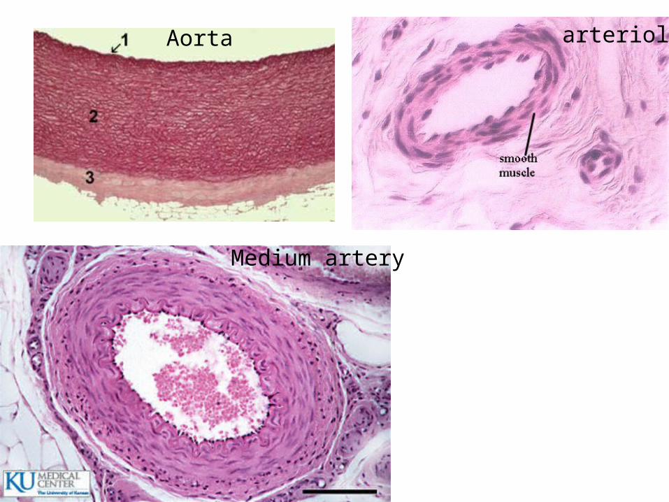
Aorta
Medium artery
arteriole
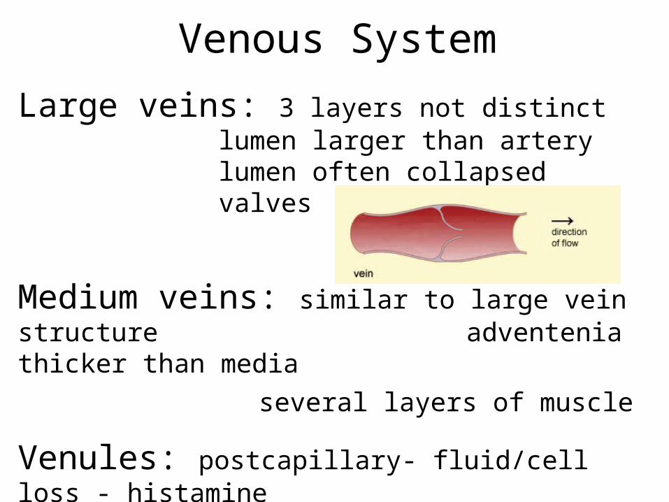
Venous System
Large veins: 3 layers not distinctlumen larger than arterylumen often collapsedvalves
Medium veins: similar to large vein structure adventenia thicker than media
several layers of muscle
Venules: postcapillary- fluid/cell loss - histamine
muscular: 1-2 layers of muscle
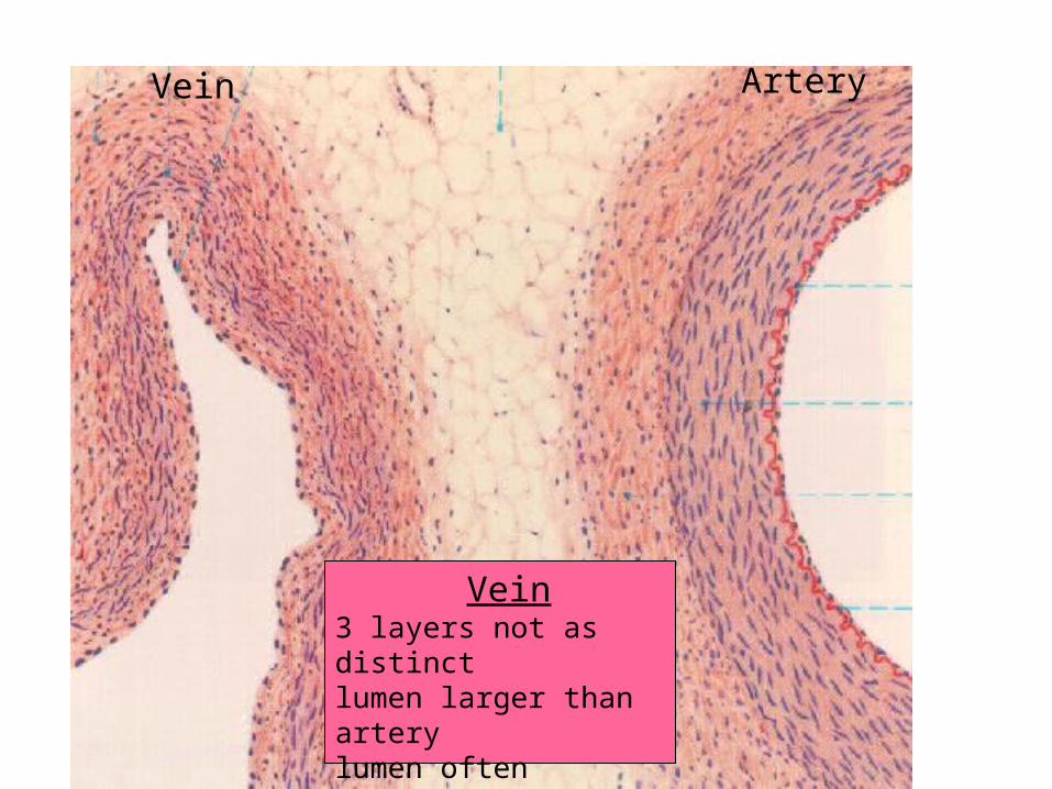
Vein Artery
Vein3 layers not as distinctlumen larger than arterylumen often collapsedvalves
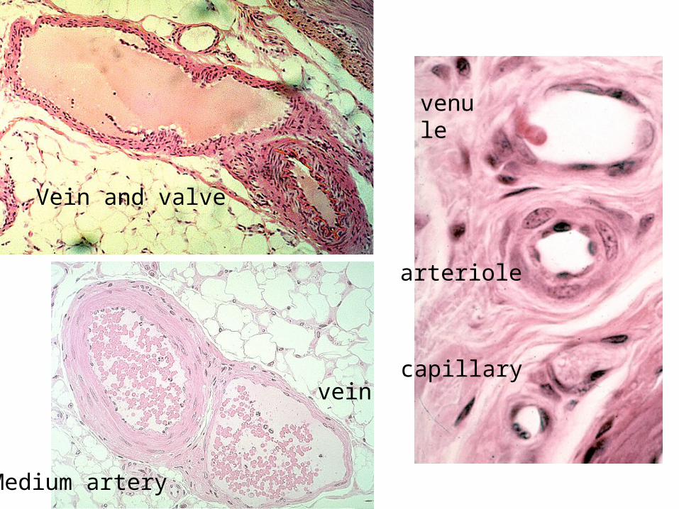
Vein and valve
venule
arteriole
capillary
Medium artery
vein
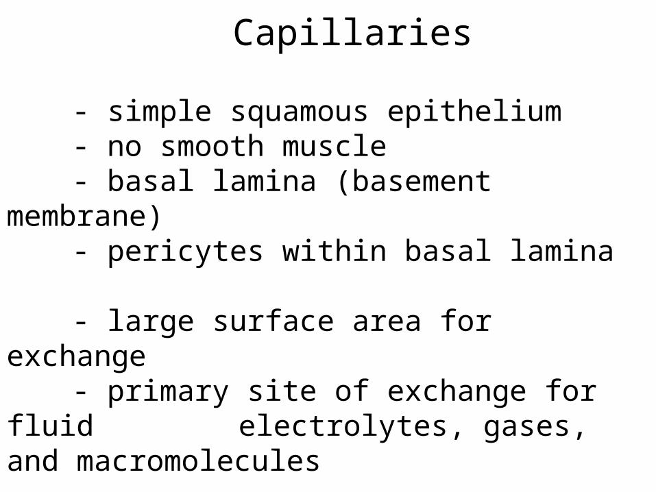
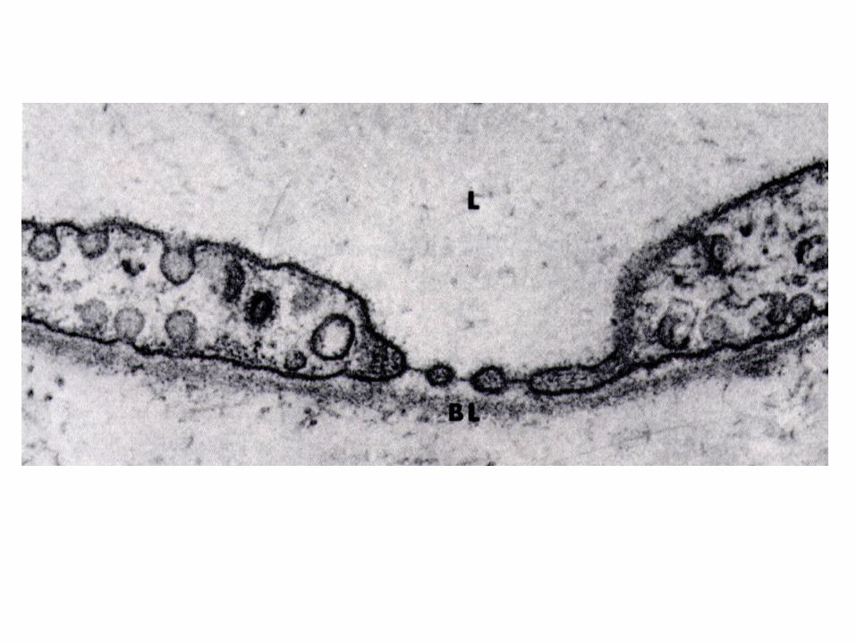
Capillaries
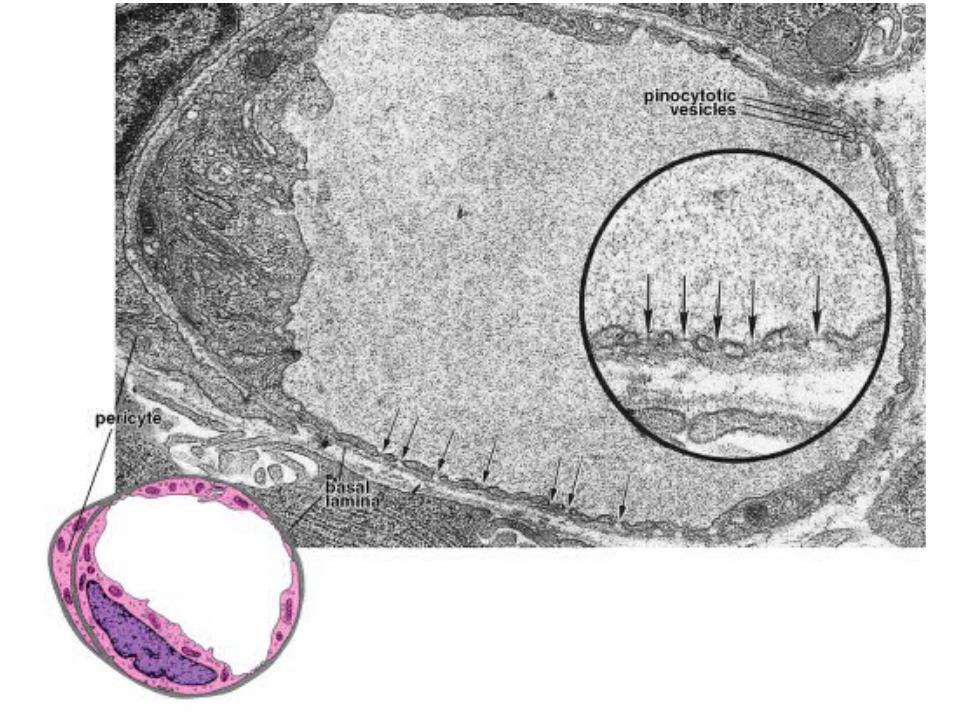
- simple squamous epithelium- no smooth muscle- basal lamina (basement membrane)- pericytes within basal lamina
- large surface area for exchange - primary site of exchange for fluid electrolytes, gases, and
macromolecules
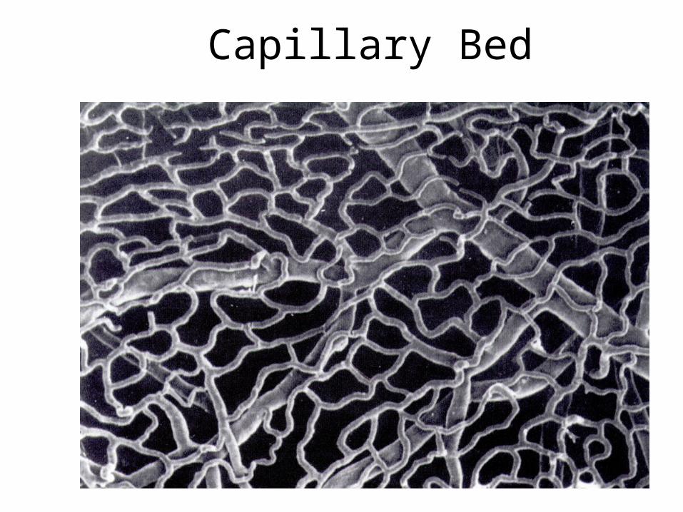
Capillary Bed

Capillaries
Types:continuousfenestrateddiscontinuous (sinusoidal)
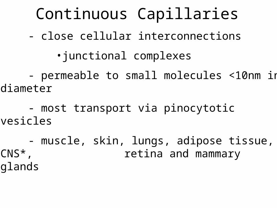
Continuous Capillaries
- close cellular interconnections
•junctional complexes
- permeable to small molecules <10nm in diameter
- most transport via pinocytotic vesicles
- muscle, skin, lungs, adipose tissue, CNS*, retina and mammary glands
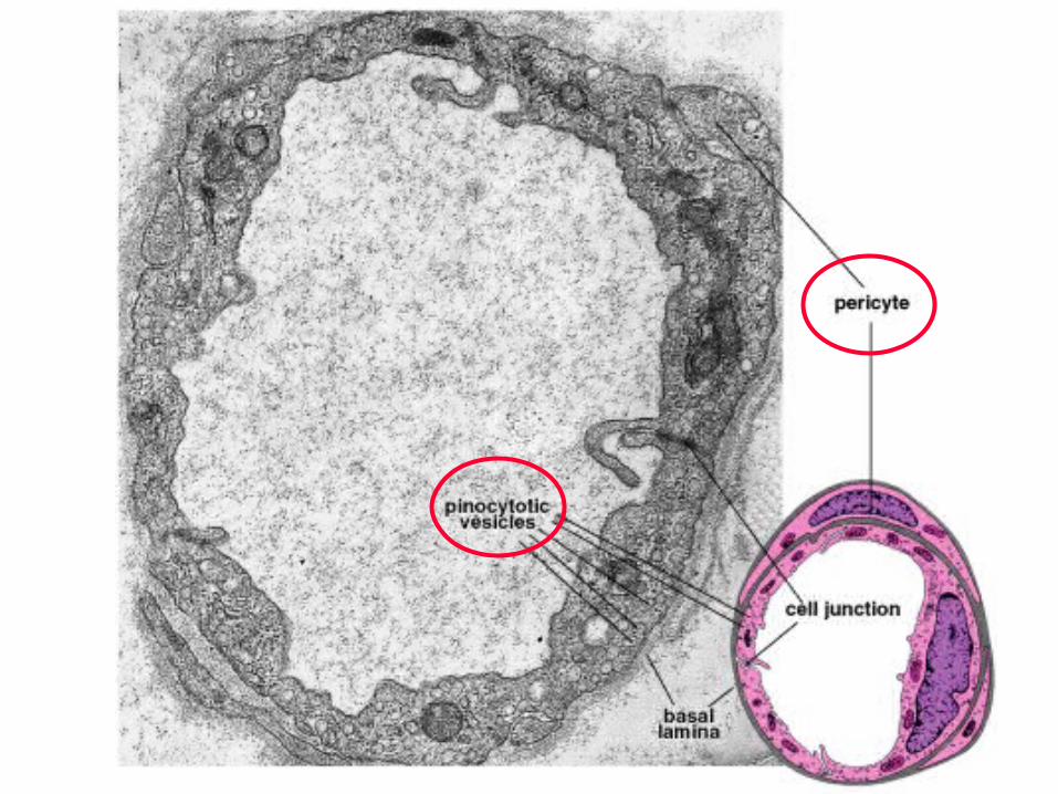
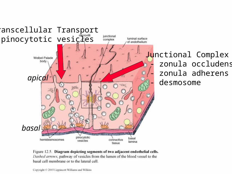
Transcellular Transport- pinocytotic vesicles
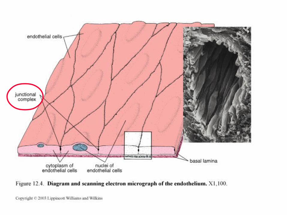
Junctional Complex - zonula occludens - zonula adherens - desmosome
apical
basal
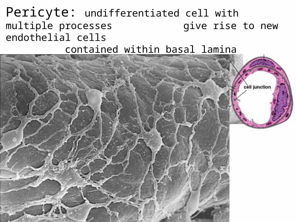
Pericyte: undifferentiated cell with multiple processes give rise to new endothelial cells
contained within basal lamina
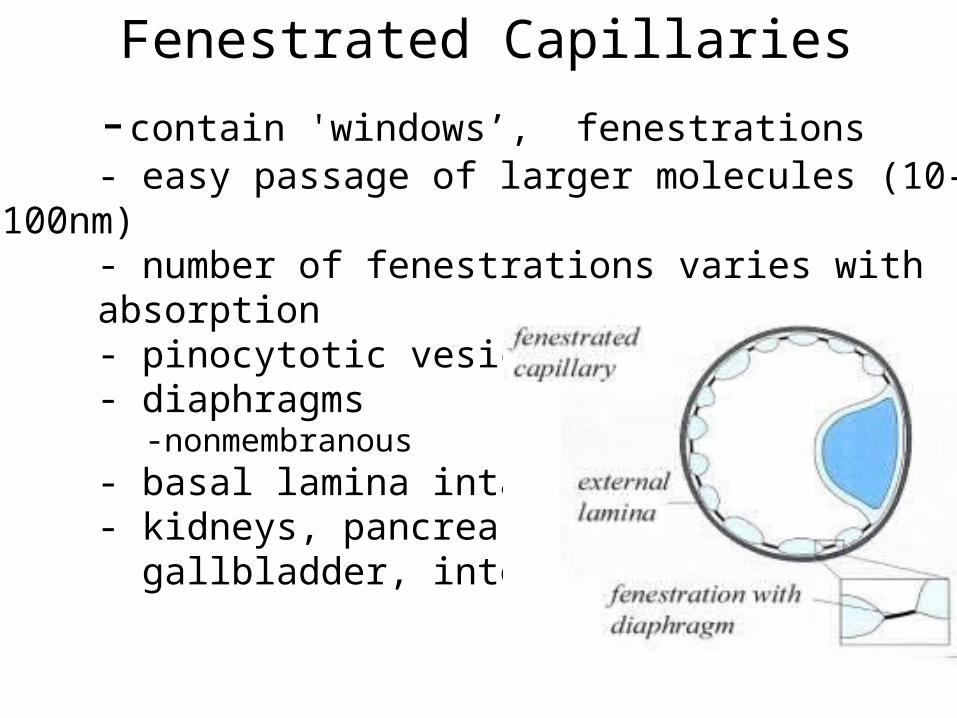
Fenestrated Capillaries
-contain 'windows’, fenestrations- easy passage of larger molecules (10-
100nm) - number of fenestrations varies with absorption - pinocytotic vesicles- diaphragms
-nonmembranous- basal lamina intact- kidneys, pancreas, gallbladder, intestine
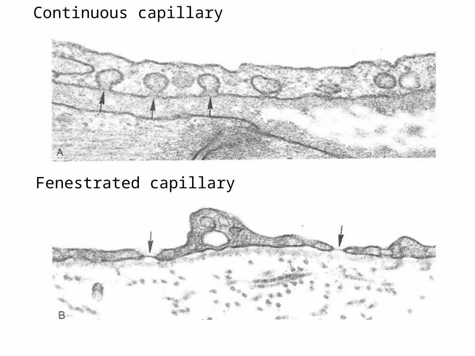
Continuous capillary
Fenestrated capillary
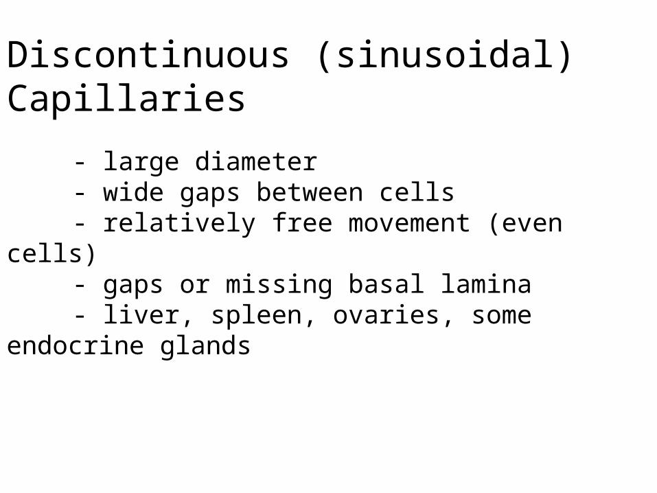
Discontinuous (sinusoidal) Capillaries
- large diameter- wide gaps between cells- relatively free movement (even cells)- gaps or missing basal lamina- liver, spleen, ovaries, some endocrine
glands
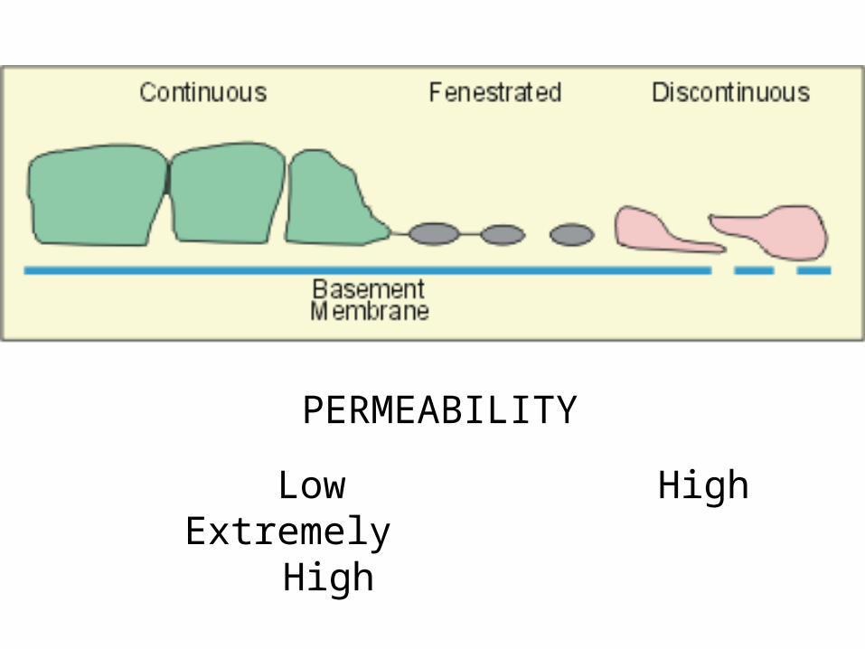
PERMEABILITY
Low High Extremely
High
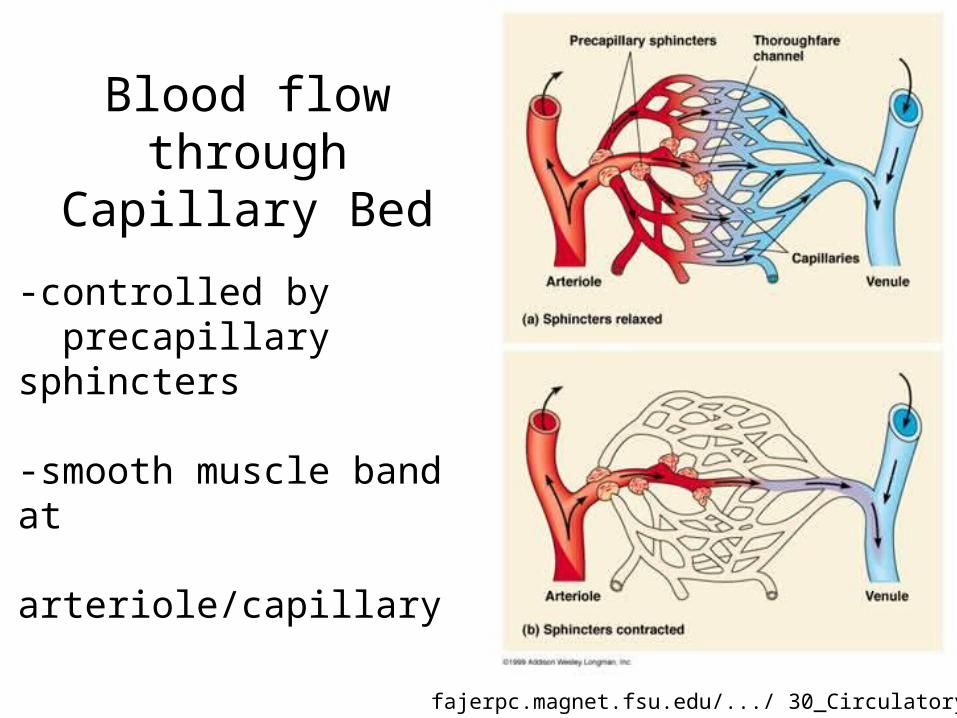
Blood flow through Capillary Bed
-controlled by precapillary sphincters
-smooth muscle band at arteriole/capillary
fajerpc.magnet.fsu.edu/.../ 30_Circulatory.htm
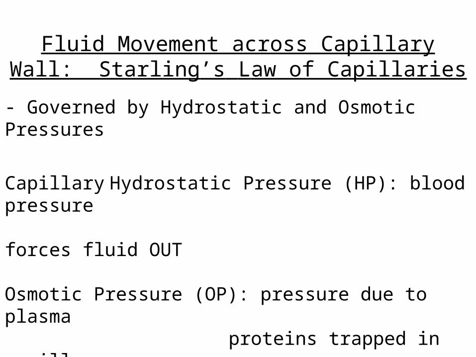
Fluid Movement across Capillary Wall: Starling’s Law of Capillaries
- Governed by Hydrostatic and Osmotic Pressures
Capillary Hydrostatic Pressure (HP): blood pressure
forces fluid OUT
Osmotic Pressure (OP): pressure due to plasma
proteins trapped in capillary
draws fluid IN
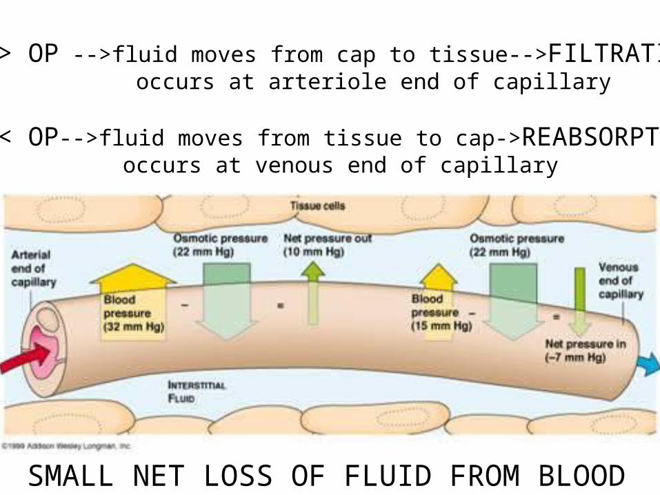
HP > OP -->fluid moves from cap to tissue-->FILTRATION occurs at arteriole end of capillary
HP < OP-->fluid moves from tissue to cap->REABSORPTION occurs at venous end of capillary
SMALL NET LOSS OF FLUID FROM BLOOD
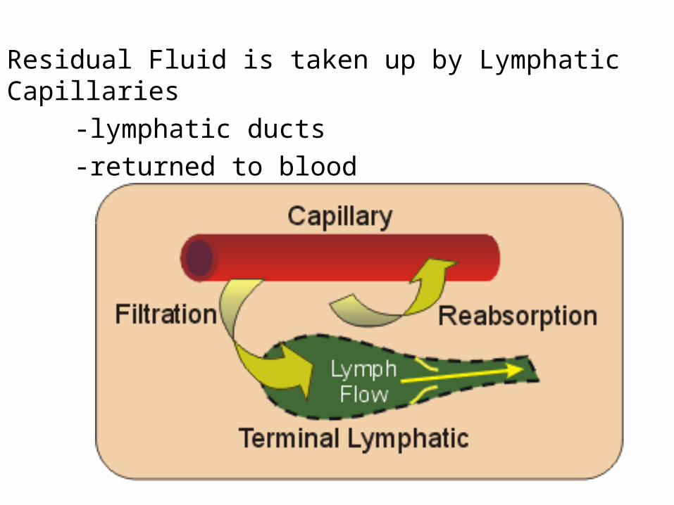
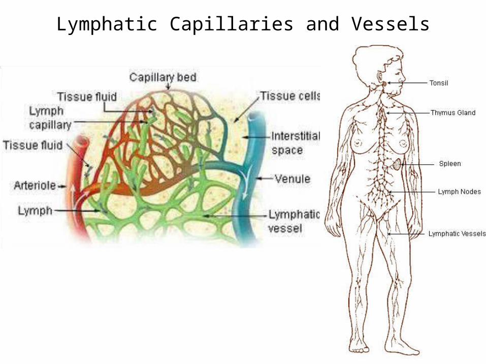
Residual Fluid is taken up by Lymphatic Capillaries
-lymphatic ducts-returned to blood
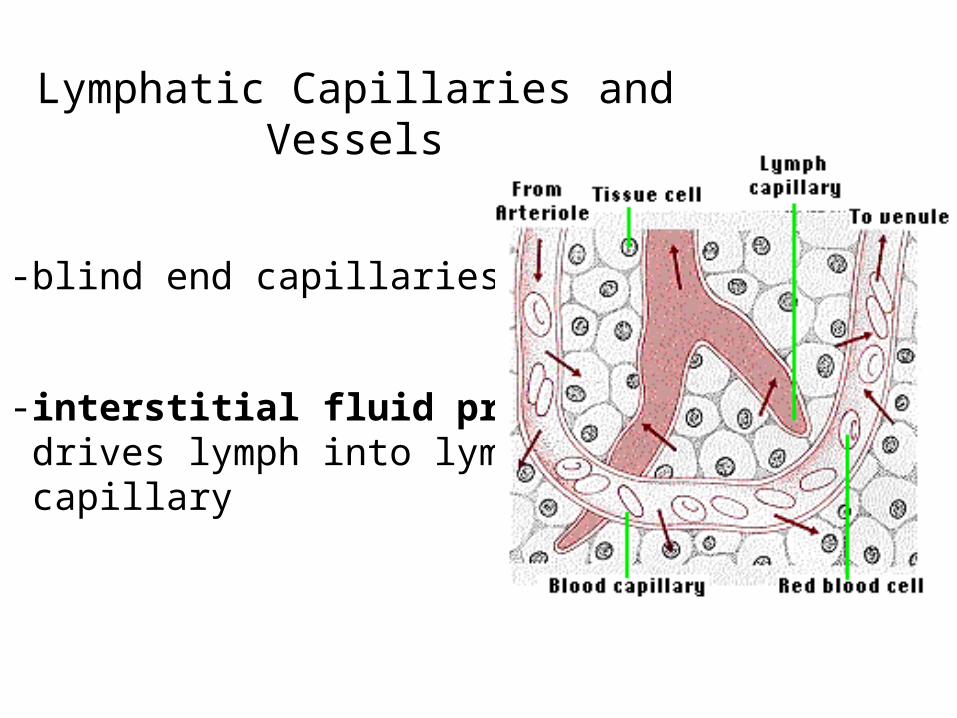
Lymphatic Capillaries and Vessels
-blind end capillaries
-interstitial fluid pressure drives lymph into lymph capillary
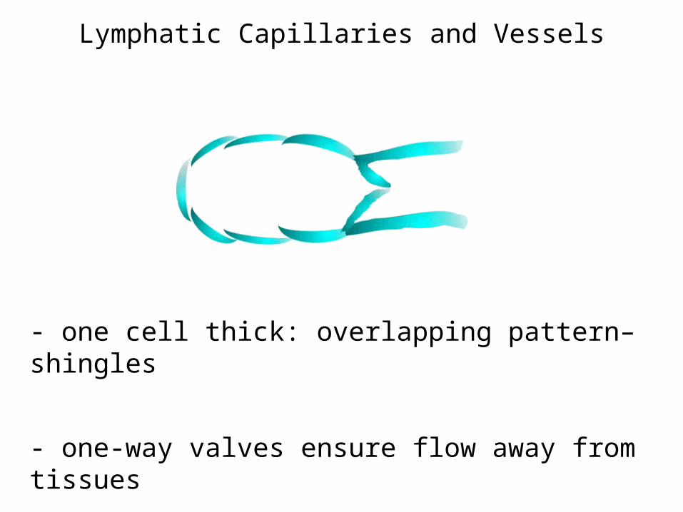
Lymphatic Capillaries and Vessels
- one cell thick: overlapping pattern– shingles
- one-way valves ensure flow away from tissues
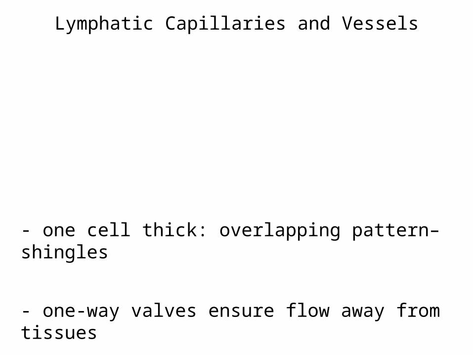
Lymphatic Capillaries and Vessels
- one cell thick: overlapping pattern– shingles
- one-way valves ensure flow away from tissues
Lymphatic Capillaries and Vessels
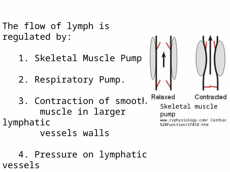
The flow of lymph is regulated by:
1. Skeletal Muscle Pump.
2. Respiratory Pump.
3. Contraction of smooth muscle in larger lymphatic vessels walls
4. Pressure on lymphatic vessels by expansion/recoil of nearby arteries
Skeletal muscle pumpwww.cvphysiology.com/ Cardiac%20Function/CF018.htm
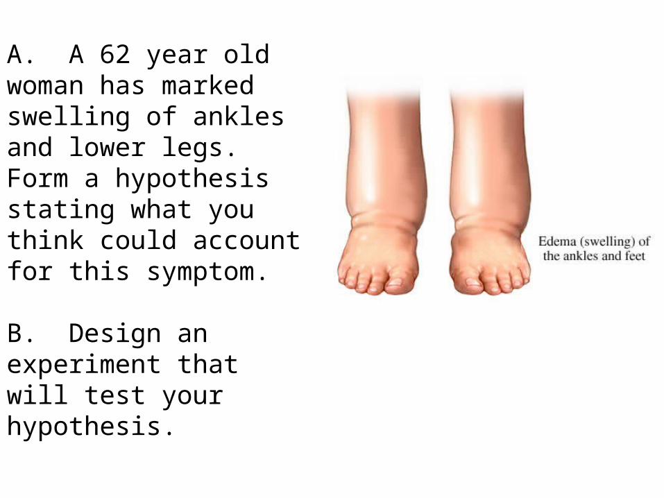
A. A 62 year old woman has marked swelling of ankles and lower legs. Form a hypothesis stating what you think could account for this symptom.
B. Design an experiment that will test your hypothesis.
Edema: accumulation of fluid in interstitial spaces
Hypotheses:
1. Increased capillary hydrostatic pressure - gravitational forces- in heart failure
2. Decreased osmotic pressure- loss of plasma proteins from kidney or liver disease
3. Increased capillary permeability- inflammatory compounds- histamine, Anaphylaxis- trauma- burns
4. Lymphatic obstruction (as occurs in filariasis)- side effect of surgery- Elephantiasis (filariasis)
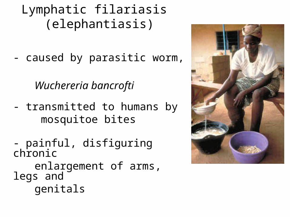
Lymphatic filariasis (elephantiasis)
- caused by parasitic worm, Wuchereria bancrofti
- transmitted to humans by mosquitoe bites
- painful, disfiguring chronic enlargement of arms, legs
and genitals
Lymphangiogram
Visualization of lymph system of legs
Inject dye between toes, visualize lymph
vessel
Inject dye directly into lymph vessel
Image dye-stained lymphatic system
Capillary PermeabilityInject tracers of variable sizes
Identify location within and outside vessels
Lymphatic System
- monitor body surfaces and internal fluids
- react to potentially harmful substances
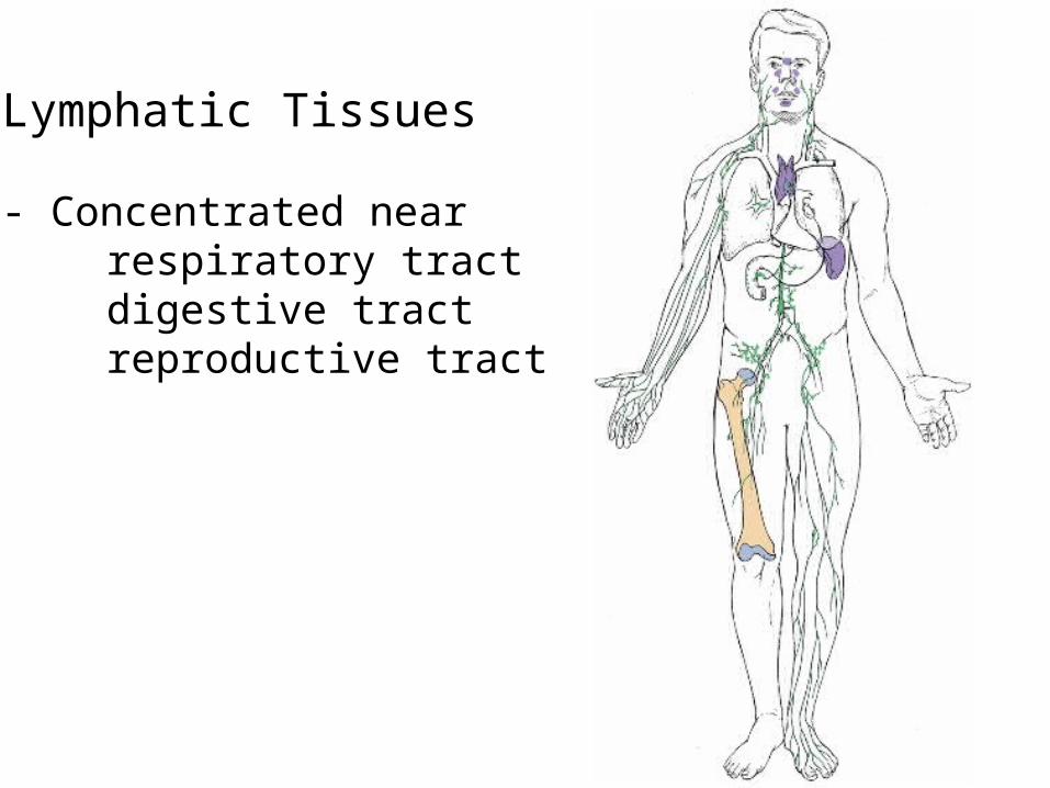
Lymphatic Tissues
- Concentrated nearrespiratory tract
digestive tractreproductive tract
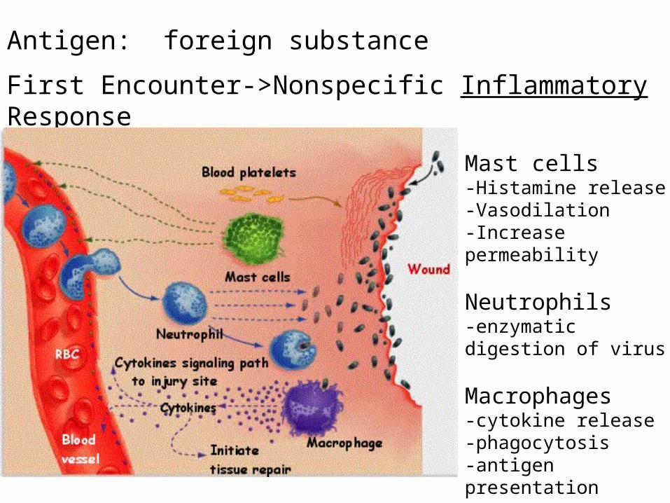
Antigen: foreign substance
First Encounter->Nonspecific Inflammatory Response
Mast cells-Histamine release-Vasodilation-Increase permeability
Neutrophils-enzymatic digestion of virus
Macrophages-cytokine release-phagocytosis-antigen presentation