the structure of the nervous system - catherine fol · the structure of the nervous system...
TRANSCRIPT
The Structure of theNervous System
INTRODUCTION
GROSS ORGANIZATION OF THE MAMMALIAN NERVOUS SYSTEMANATOMICAL REFERENCESTHE CENTRAL NERVOUS SYSTEM
The CerebrumThe CerebellumIhe Broin StemThe Spinol Cord
THE PERIPHERAL NERVOUS SYSTEMIhe Somotic PNSIhe Viscerol PNSAfferent and Efferent Axons
THE CRANIAL NERVESTHE MENINGESTHEVENTRICULAR SYSTEM
r Box 7.1 Of Special Interest: Water on the BrainIMAGING THE LIVING BRAIN
Computed TomogrophyMagnetic Resononce lmaging
r Box 7.2 Brain Food: Magnetic Resonance ImagingFunctionol Broin lmaging
r Box 7 3 Brain Food: Functional Imaging of Brain Activity: PET and fMRI
UNDERSTANDING CNS STRUCTURE THROUGH DEVELOPMENTFORMATION OF THE NEURAL TUBE
r Box 7.4 Of Special Interest: Nutrition and the Neural TLrbeTHREE PRIMARY BMIN VESICLESDIFFERENTIATION OF THE FOREBRAIN
Differentiotion of the Telencepholon ond DiencephalonForebroin Structure-F unction Relotionships
DIFFERENTIATION OF THE MIDBRAINMidbroin Structure-Function Relotionshrps
DIFFERENTIATION OF THE HINDBRAINHindbroin Structure-Function Relotionshrps
DIFFERENTIATION OFTHE SPINAL CORDSpinol Cord Structure-Function Relotionshps
PUTTING THE PIECES TOGETHERSPECIAL FEATURES OF THE HUMAN CNS
A GUIDE TO THE CEREBRAL CORTEXTYPES OF CEREBRAL CORTEXAREAS OF NEOCORTEX
Neocortico/ Evolution ond Structure-Function Relotionshrpsr Box 7.5 Path of Discovery: Evolution of My Brain, by Leah A. Krubitzer
CONCLUDING REMARKS
APPENDIX:AN ILLUSTRATED GUIDE TO HUMAN NEUROANATOMY
r68 CHAPTER 7 . THESTRUCTUREOFTHENERVOUSSYSTEM
V INTRODUCTION
In previous chapters, we saw how individual neurons function and com-municate. Now we are ready to assemble them into a nervous system thatsees, hears, feels, moves, remembers, and dreams. Just as an understand-ing of neuronal structure is necessary for understanding neuronal function,we must understand nervous system structure in order to understand brainfunction.
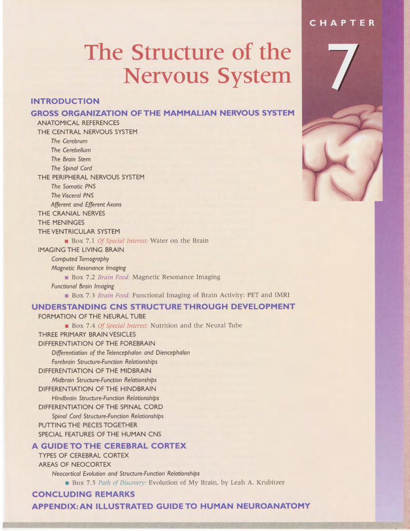
Neuroanatomy has challenged generations of students-and for goodreason: The human brain is extremely complicated. However, our brain ismerely a variation on a plan that is common to the brains of all mammals(Figure 7.1). The human brain appears complicated because it is distortedas a result of the selective growth of some parts within the confines of theskull. But once the basic mammalian plan is understood, these specializa-tions of the human brain become transparent.
We begin by introducing the genbral organization of the mammalianbrain and the terms used to describe it. Then we take a look at how thethree-dimensional structure of the brain arises during embryological andfetal development. Following the course of development makes it easier tounderstand how the parts of the adult brain fit together. Finally, we explorethe cerebral neocortex, a structure that is unique to mammals and pro-portionately the largest in humans. An Illustrated Guide to HumanNeuroanatomy follows the chapter as an appendix.
The neuroanatomy presented in this chapter provides the canvas on whichwe will paint the sensory and motor systems in Chapters 8-14. Because youwill encounter a lot of new terms, self-quizzes within the chapter providean opportunity for review.
V GROSS ORGANIZATION OFTHE MAMMALIAN NERVOUS SYSTEMThe nervous system of all mammals has two divisions: the central nervoussystem (CNS) and the peripheral nervous system (PNS). In this section, weidentify some of the important components of the CNS and the pNS. Wealso discuss the membranes that surround the brain and the fluid-filledventricles within the brain. We then explore some new methods of examin-ing the structure of the living brain. But first, we need to review someanatomical terminology.
Anatomical References
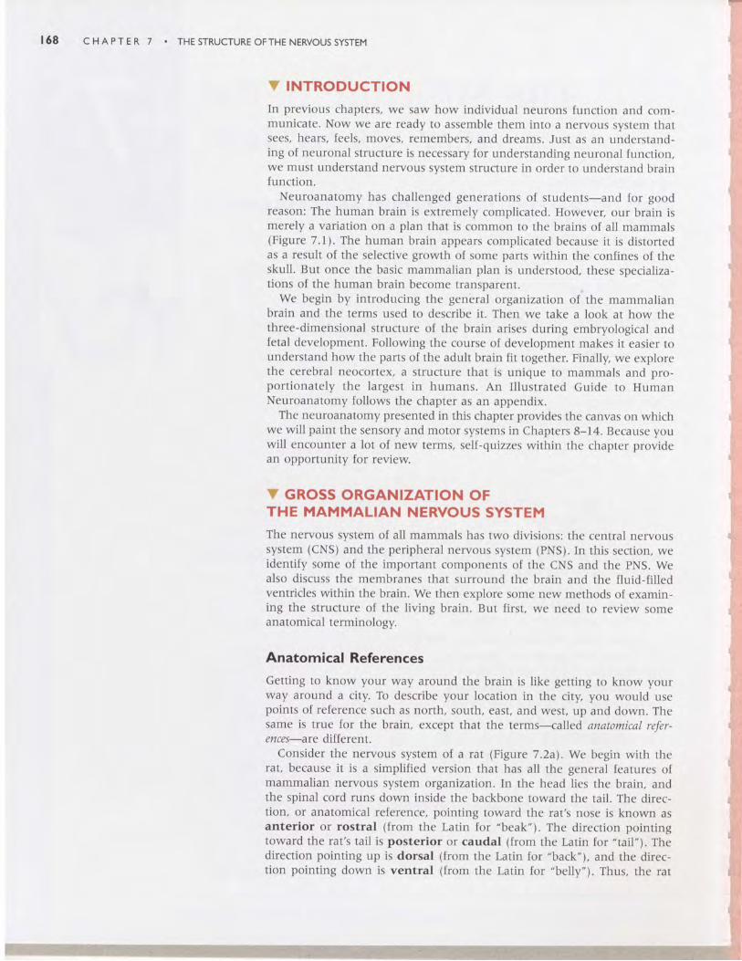
Getting to know your way around the brain is like getting to know yourway around a city. To describe your location in the city, you would usepoints of reference such as north, south, east, and west, up and down. Thesame is true for the brain, except that the terms-calle d anatomical refer-ences-are different.
Consider the nervous system of a rat (Figure 7.2a).We begin with therat, because it is a simplified version that has all the general features ofmammalian nervous system organization. In the head lies the brain, andthe spinal cord runs down inside the backbone toward the tail. The direc-tion, or anatomical reference, pointing toward the rat's nose is known asanterior or rostral (from the Latin for "beak"). The direction pointingtoward the rat's tail is posterior or caudal (from the Latin for "tail"). Thedirection pointing up is dorsal (from the Latin for "back"), and the direc-tion pointing down is ventral (from the Latin for "belly"). Thus, the rat
Y GROSS ORGANIZATION OFTHE MAMMALIAN NERVOUS SYSTEM I69
FIGURE 7 . IMammalian brains. Despite differences rn complexity, the brarns of all these species havemany features in common.The brains have been drawn to appear approximately the same stze;their relative sizes are shown in the inset on the left,
r Im.:!
Rat
Rabbit
{ffiCat
dffi,':4,.\.\i#r
Sheep
Chimpanzee
Human
/ a tf 1 ."-'\--\\
ChimDanzee
,d
t 70 CHAPTER 7 . THESTRUCTUREOFTHENERVOUSSYSTEM
Spinalcoro Dorsal
(a)
Anterioror rostral
< -
FIGURE 7,2Basic anatomical references in theneryous system oi a rat. (a) Side view.(b) Top view.
FIGURE 7.3Anatomical planes of section.
Posterioror caudal
: - - - - - - - >
spinal cord runs anterior to posterior. The top side of the spinal cord is thedorsal side, and the bottom side is the ventral side.
If we look down on the nervous system, we see that it may be dividedinto two equal halves (Figure 7.2b). The right side of the brain and spinalcord is the mirror image of the left side. This characteristic is known asbilateral symmetry. with just a few exceptions, most structures within thenervous system come in pairs, one on the right side and the other on theleft. The invisible line running down the middle of the nervous system iscalled the midline, and this gives us another way to describe anatomicalreferences. structures closer to the midline are medial; structures fartheraway from the midline are lateral. In other words, the nose is medial tothe eyes, the eyes are medial to the ears, and so on. In addition, rwo struc-tures that are on the same side are said to be ipsilateral to each other; forexample, the right ear is ipsilateral to the right eye. If the structures are onopposite sides of the midline, they are said to be contralateral to eachother; the right ear is contralateral to the left ear.
To view the internal structure of the brain, it is usually necessary to sliceit up. In the language of anatomists, a slice is called a section; to slice is /osection. Although one could imagine an infinite number of ways we mightcut into the brain, the standard approach is to make cuts parallel to one ofthe three anatomical planes of section. The plane of the section resulting fromsplitting the brain into equal right and left halves is called the midsagittalplane (Figure 7.3a). sections parallel to the midsagittal plane are in thesagittal plane.
The two other anatomical planes are perpendicular to the sagittal plane andto one another. The horizontal plane is parallel to the ground (Figure73b]'. A single section in this plane could pass through both the eyes andthe ears. Thus, horizontal sections split the brain into dorsal and ventralparts. The coronal plane is perpendicular to the ground and to the sagit-tal plane (Figure 7.3c). A single section in this plane could pass throughboth eyes or both ears, but not through all four at the same time. Thus, thecoronal plane splits the brain into anterior and posterior parts.
v SELF-QUtZTake a few moments right now and be sure you understand the meaningof these terms:
anterior
rostralposterior
caudal
dorsal
ventral
midl ine
medial
lateral
ipsilateral
contralateral
midsagittal plane
sagittal plane
horizontal plane
coronal plane
Ventral
V GROSS ORGANIZATION OFTHE MAMMALIAN NERVOUS SYSTEM
The Central Nervous System
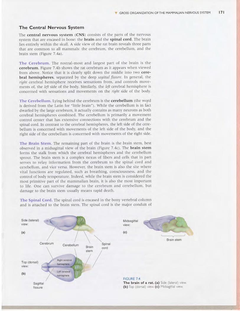
The central nervous system (CNS) consists of the parts of the nervoussystem that are encased in bone: the brain and the spinal cord. The brainlies entirely within the skull. A side view of the rat brain reveals three partsthat are common to all mammals: the cerebrum, the cerebellum, and thebrain stem (Figure 7.4a).
The Cerebrum. The rostral-most and largest part of the brain is thecerebrum. Figure 7.4b shows the rat cerebrum as it appears when viewedfrom above. Notice that it is clearly split down the middle into two cere-bral hemispheres, separated by the deep sagittal fissure.In general, theright cerebral hemisphere receives sensations from, and controls move-ments of, the left side of the body. Similarly, the left cerebral hemisphere isconcerned with sensations and movements on the right side of the body.
The Cerebellum. Lying behind the cerebrum is the cerebellum (the wogdis derived from the Latin for "little brain"). While the cerebellum is in factdwarfed by the large cerebrum, it actually contains as many neurons as bothcerebral hemispheres combined. The cerebellum is primarily a movementcontrol center that has extensive connections with the cerebrum and thespinal cord. In contrast to the cerebral hemispheres, the left side of the cere-bellum is concerned with movements of the left side of the body, and theright side of the cerebellum is concerned with movements of the right side.
The Brain Stem. The remaining part of the brain is the brain stem, bestobserved in a midsagittal view of the brain (Figure7.4c\. The brain stemforms the stalk from which the cerebral hemispheres and the cerebellumsprout. The brain stem is a complex nexus of fibers and cells that in partserves to relay information from the cerebrum to the spinal cord andcerebellum, and vice versa. However, the brain stem is also the site wherevital functions are regulated, such as breathing, consciousness, and thecontrol of body temperature. Indeed, while the brain stem is considered themost primitive part of the mammalian brain, it is also the most importantto life. One can survive damage to the cerebrum and cerebellum, butdamage to the brain stem usually means rapid death.
The Spinal Cord. The spinal cord is encased in the bony vertebral columnand is attached to the brain stem. The spinal cord is the major conduit of
Side (lateral)view:
(a)
Midsagittalview:
(c)
Cerebellum
Top (dorsal)view:
(b)
FIGURE 7.4The brain of a rat. (a) Side (lateral) view.(b) Top (dorsal) view. (c) Midsagittal view.
t 7 l
S:',
ffi{,3\{isisrs,sSid
a:{3i*{!f*
$r{'aari
l'$F:I.-*l
-qs6!3qiIS,rlSTri\il
ffiEH6\t
$i&l
ffiB{F{i
, 7 2 CHAPTER 7 . THESTRUCTUREOFTHENERVOUSSYSTEM
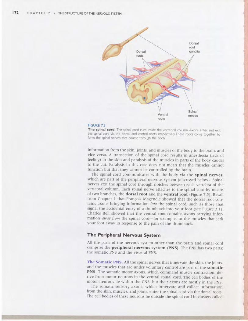
Spinalnerves
FIGURE 7.5The spinal cord. The spinal cord runs inside the vertebral column. Axons enter anc ex;1the spinal cord via the dorsal and ventral roots, respectively.These roots come tosether toform the spinal nerves that course through the body
information from the skin, joints, and muscles of the body to the brain, andvice versa. A transection of the spinal cord results in anesthesia (lack offeeling) in the skin and paralysis of the muscles in parts of the body caudalto the cut. Paralysis in this case does not mean that the muscles cannotfunction but that they cannot be controlled by the brain.
The spinal cord communicates with the body via the spinal nerves,which are part of the peripheral nervous system (discussed below). Spinalnerves exit the spinal cord through notches between each vertebra of thevertebral column. Each spinal nerve attaches to the spinal cord by meansof two branches, the dorsal root and the ventral root (Figure 7.5). Recallfrom chapter I that Frangois Magendie showed that the dorsal root con-tains axons bringing information into the spinal cord, such as those thatsignal the accidental entry of a thumbtack into your foot (see Figure 3.1).charles Bell showed that the ventral root contains axons carrying infor-mation away from the spinal cord-for example, to the musclei that jerkyour foot away in response to the pain of the thumbtack.
The Peripheral Nervous SystemAll the parts of the nervous system other than the brain and spinal cordcomprise the peripheral nervous system (pNS). The pNS has two parts:the somatic PNS and the visceral PNS.
The somatic PNS. All the spinal nerves that innervate the skin, the joints,and the muscles that are under voluntary control are part of the somaticPNS. The somatic motor axons, which command muscle contraction, de-rive from motor neurons in the ventral spinal cord. The cell bodies of themotor neurons lie within the cNS, but their axons are mostly in the pNS.
The somatic sensory axons, which innervate and collect informationfrom the skin, muscles, and joints, enter the spinal cord via the dorsal roots.The cell bodies of these neurons lie outside the spinal cord in clusters called
Ventralroots
V GROSS ORGANIZATION OFTHE MAMMALIAN NERVOUS SYSTEM I73
dorsal root ganglia. There is a dorsal root ganglion for each spinal nerve(see Figure 7.5).
The Visceral PNS. The visceral PNS, also called the involuntary, vegeta-tive, or autonomic nervous system (ANS), consists of the neurons thatinnervate the internal organs, blood vessels, and glands. Visceral sensoryaxons bring information about visceral function to the CNS, such as thepressure and oxygen content of the blood in the arteries. Visceral motorfibers command the contraction and relaxation of muscles that form thewalls of the intestines and the blood vessels (called smooth muscles), therate of cardiac muscle contraction, and the secretory function of variousglands. For example, the visceral PNS controls blood pressure by regulatingthe heart rate and the diameter of the blood vessels.
We will return to the structure and function of the ANS in Chapter 15.For now, remember that when one speaks of an emotional reaction that is.beyond voluntary control-like "butterflies in the stomach" or blushing-it usually is mediated by the visceral PNS (the ANS).
Afferent and Efferent Axons. Our discussion of the PNS is a good placeto introduce two terms that are used to describe axons in the nervous system.Derived from the Latin, afferent ("carry to") and efferent ("carry from")indicate whether the axons are transporting information toward or away
from a particular point. Consider the axons in the PNS relative to a pointof reference in the CNS. The somatic or visceral sensory axons bringinginformation into the CNS are afferents. The axons that emerge /roz the CNSto innervate the muscles and glands are efferents.
The Cranial Nerves
In addition to the nerves that arise from the spinal cord and innervate thebody, there are 12 pairs of cranial nerves that arise from the brain stemand innervate (mostly) the head. Each cranial nerve has a name and anumber associated with it (originally numbered by Galen, about 1800 yearsago, from anterior to posterior). Some of the cranial nerves are part of theCNS, others are part of the somatic PNS, and still others are part of the vis-ceral PNS. Many cranial nerves contain a complex mixture of axons thatperform different functions. The cranial nerves and their various functionsare summarized in the chapter appendix.
The Meninges
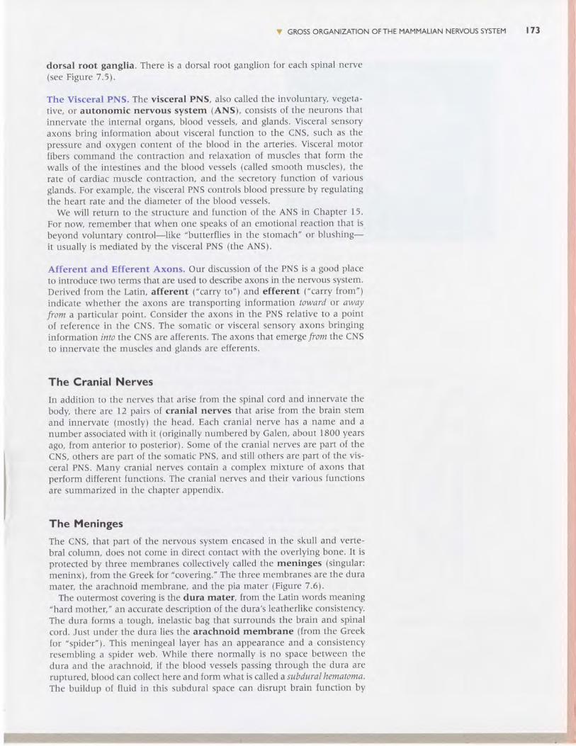
The CNS, that part of the nervous system encased in the skull and verte-bral column, does not come in direct contact with the overlying bone. It isprotected by three membranes collectively called the meninges (singular:meninx), from the Greek for "covering." The three membranes are the duramater, the arachnoid membrane, and the pia mater (Figure 7.6).
The outermost covering is the dura mater, from the Latin words meaning"hard mother," an accurate description of the dura's leatherlike consistency.The dura forms a tough, inelastic bag that surrounds the brain and spinalcord. Just under the dura lies the arachnoid membrane (from the Greekfor "spider"). This meningeal layer has an appearance and a consistencyresembling a spider web. While there normally is no space between thedura and the arachnoid, if the blood vessels passing through the dura areruptured, blood can collect here and form what is called a subdural hematoma.The buildup of fluid in this subdural space can disrupt brain function by
1 7 4 C H A P T E R 7 . T H E S T R U C T U R E O F T H E N E R V O U S S Y S T E M
Choroidplexus
Subarachnoidspace
Ventriclesin brain
Dura mater
Subduralspace
Arachnoidmembrane
Subarachnoidspace
Pla mater
Artery
Brain
(b)
FIGURE 7.6The meninges.(a)The skul l has been removed to showthe tough outer meninsealmembrane, the dura mater: (Source: Gluhbegoric and Wil l iams, 1980.) (b) l l lustratei in crosssection, the three meningeal layers protecting the brain and spinal cord are the dura mater:the arachnoid membrane, and the pia mater:
compressing parts of the cNS. The disorder is treated by drilling a hole inthe skull and draining the blood.
The pia mater, the "gentle mother," is a thin membrane that adheresclosely to the surface of the brain. Along the pia run many blood vesselsthat ultimately dive into the substance of the underlying brain. The pia isseparated from the arachnoid by a fluid-filled space. This subarachnoid spaceis filled with salty clear liquid called cerebrospinal fluid (csF). Thus, ina sense, the brain floats inside the head in this thin layer of CSF.
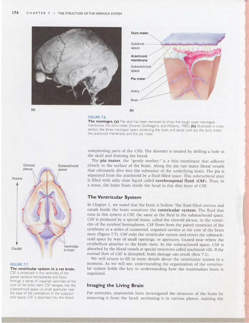
The Ventricular SystemIn chapter l, we noted rhat the brain is hollow. The fluid-filled caverns andcanals inside the brain constitute the ventricular system. The fluid thatruns in this system is cSF, the same as the fluid in the subarachnoid space.cSF is produced by a special tissue, called the choroid plexus, in the venrri-cles of the cerebral hemispheres. cSF flows from the paired ventricles of thecerebrum to a series of connected, unpaired cavities at the core of the brainstem (Figure 7.7). csF exits the ventricular system and enters the subarach-noid space by way of small openings, or apertures, located near where thecerebellum attaches to the brain stem. In the subarachnoid space, cSF isabsorbed by the blood vessels at special structures called arachnoid villi. If thenormal flow of CSF is disrupted, brain damage can result (Box 7.1).
we will return to fill in some details about the ventricular system in amoment. As we will see, understanding the organization of the ventricu-Iar system holds the key to understanding how the mammalian brain isorganized.
lmaging the Living BrainFor centuries, anatomists have investigated the structure of the brain byremoving it from the head, sectioning it in various planes, staining the
FIGURE 7 .7The ventricular system in a rat brain.CSF is produced in the ventricles of thepaired cerebral hemispheres and flowsthrough a series of unpaired ventricles at thecore of the brain stem. CSF escapes into thesubarachnoid space via small apertures nearthe base ofthe cerebellum. In the subarach-noid space, CSF is absorbed into the blood,
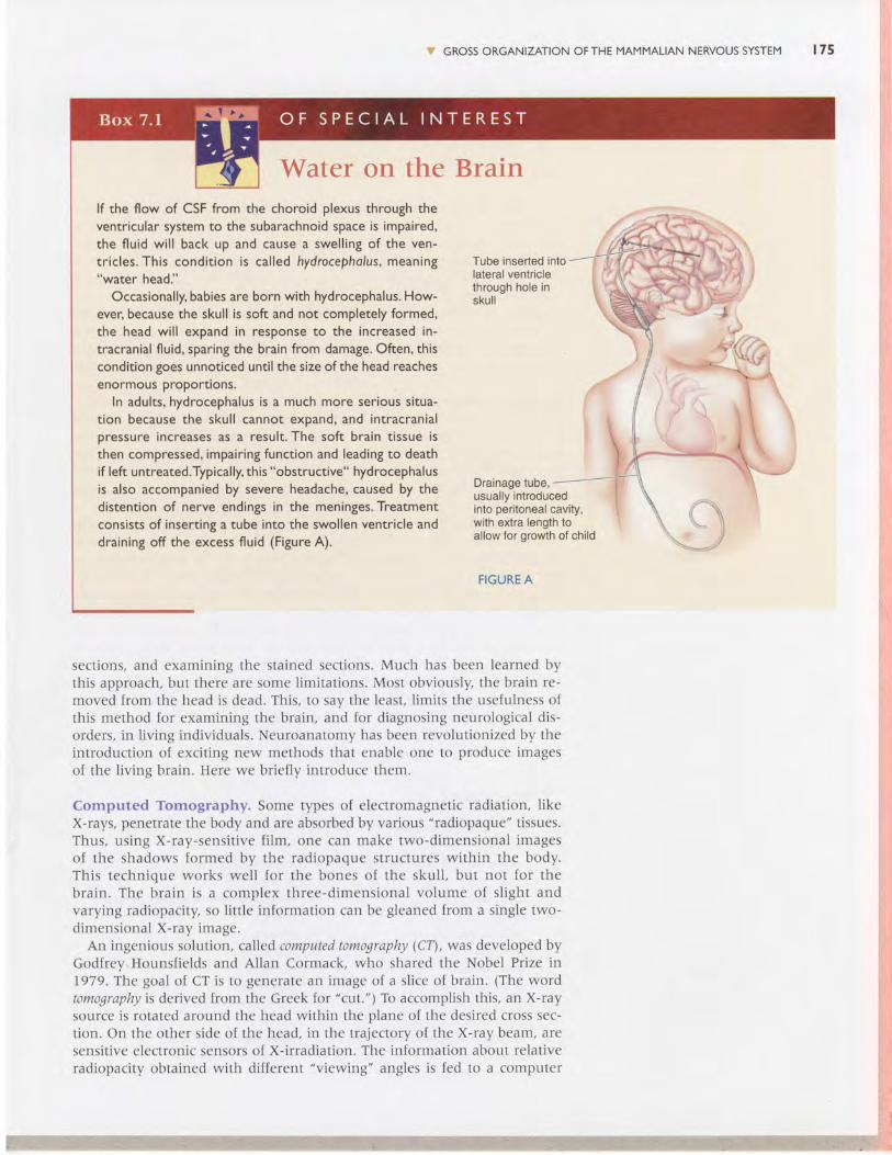
Water on the Brainlf the flow of CSF from the choroid plexus through theventricular system to the subarachnoid space is impaired,the fluid wil l back up and cause a swell ing of the ven-t r ic les. This condi t ion is ca l led hydrocepholus, meaning"water head."
Occasionally, babies are born with hydrocephalus. How-ever, because the skull is soft and not completely formed,the head wil l expand in response to the increased in-tracranial fluid, sparing the brain from damage. Often, thiscondition goes unnoticed unti l the size ofthe head reachesenormous ProPortions.
In adults, hydrocephalus is a much more serious situa-tion because the skull cannot expand, and intracranialpressure increases as a result. The soft brain tissue isthen compressed, impairing function and leading to deathif left untreated.Typically, this "obstructive" hydrocephalusis also accompanied by severe headache, caused by thedistention of nerve endings in the meninges. Treatmentconsists of inserting a tube into the swollen ventricle anddraining off the excess fluid (Figure A).
Y GROSS ORGANIZATION OFTHE MAMMALIAN NERVOUS SYSTEM t 7 5
Tube inserted intolateral ventriclethrough hole inskul l
Drainage tube,usually introducedinto peritoneal cavity,with extra length toallow for growth of child
-+4tr J ,
evt(ft.'n|\$l
FIGURE A
sections, and examining the stained sections. Much has been learned bythis approach, but there are some limitations. Most obviously, the brain re-moved from the head is dead. This, to say the least, l imits the usefulness ofthis method for examining the brain, and for diagnosing neurological dis-orders, in l iving individuals. Neuroanatomy has been revolutionized by theintroduction of exciting new methods that enable one to produce imagesof the l iving brain. Here we briefly introduce them.
Computed Tomography. Some types of electromagnetic radiation, l ikeX-rays, penetrate the body and are absorbed by various "radiopaque" tissues.Thus, us ing X-ray-sensi t ive f i lm, one can make two-dimensional imagesof the shadows formed by the radiopaque st ructures wi th in the body.This technique works wel l for the bones of the skul l , but not for thebrain. The bra in is a complex three-dimensional vo lume of s l ight andvarying radiopacity, so l itt le information can be gleaned from a single two-dimensional X-ray image.
An ingenious solution, called computed tomography (CTl, was developed byGodfrey.Hounsfields and Allan Cormack, who shared the Nobel Prize in1979. The goal of CT is to generate an image of a slice of brain. (The wordtomography is derived from the Greek for "cut.") To accomplish this, an X-raysource is rotated around the head within the plane of the desired cross sec-tion. On the other side of the head, in the trajectory of the X-ray beam, aresensitive electronic sensors of X-irradiation. The information about relativeradiopacity obtained with different "viewing" angles is fed to a computer
i:i 'r, ;r'1;.f ', '1: ' ';...:l
476 C H A P T E R 7 THE STRUCTURE OFTHE NERVOUS SYSTEM
that executes a mathematical algorithm on the data. The end result is a dig-ital reconstruction of the position and amount of radiopaque materialwithin the plane of the slice. cr scans noninvasively revealed, for the firsttime, the gross organization of gray and white matter, and the position ofthe ventricles, in the living brain.
Magnetic Resonance Imaging. While still used widely, CT is graduallybeing replaced by a newer imaging method, called magnetic resonance imag-ing (MRI), The advantages of MRI are that it yields a much more detailedmap of the brain than CT it does not require X-irradiation, and images ofbrain slices can be made in any plane desired. MRI uses information abouthow hydrogen atoms in the brain respond to perturbations of a strongmagnetic field (Box 7.21. Tl:e electromagnetic signals emitted by the atomsare detected by an array of sensors around the head and fed to a powerfulcomputer that constructs a map of the brain. The information from an MRIscan can be used to build a strikingly detailed image of the whole brain.
Functional Brain Imaging. CT and MRI are extremely valuable for de-tecting structural changes in the living brain, such as brain swelling after ahead injury and brain tumors. Nonetheless, much of what goes on in thebrain-healthy or diseased-is chemical and electrical in nature, and notobservable by simple inspection of the brain's anatomy. Amazingly, how-ever, even these secrets are beginning to yield to the newest imagingtechniques.
The two "functional imaging" techniques now in widespread use arepositron emission tomography (PET) and functional magnetic resonance imaging(fMRIl.While the technical details differ, both methods detecr changes inregional blood flow and metabolism within the brain (Box 7.3). The basicprinciple is simple. Neurons that are active demand more glucose andoxygen. The brain vasculature responds to neural activity by directing moreblood to the active regions. Thus, by detecting changes in blood flow, pETand fMRI reveal the regions of brain that are most active under differentcircumstances.
The advent of imaging techniques has offered neuroscientists the ex-traordinary opportunity of peering into the living, thinking brain. As youcan imagine, however, even the most sophisticated brain images are use-less unless you know what you are looking at. Next, let's take a closer lookat how the brain is organized.
v SELF-QU|ZTlake a few moments right now and be sure you understand the meaningof these terms:
central nervous system (CNS) dorsal root gangliabrain visceral PNSspinal cord autonomic nervous system (ANS)cerebrum afferentcerebral hemispheres efferentcerebellum cranial nervebrain stem meningesspinal nerve dura materdorsal root arachnoid membraneventral root pia materperipheral nervous system (PNS) cerebrospinal fluid (CSF)somatic PNS ventricular system
i, GROSS ORGANIZATION OFTHE MAMMALIAN NERVOUS SYSTEM 177
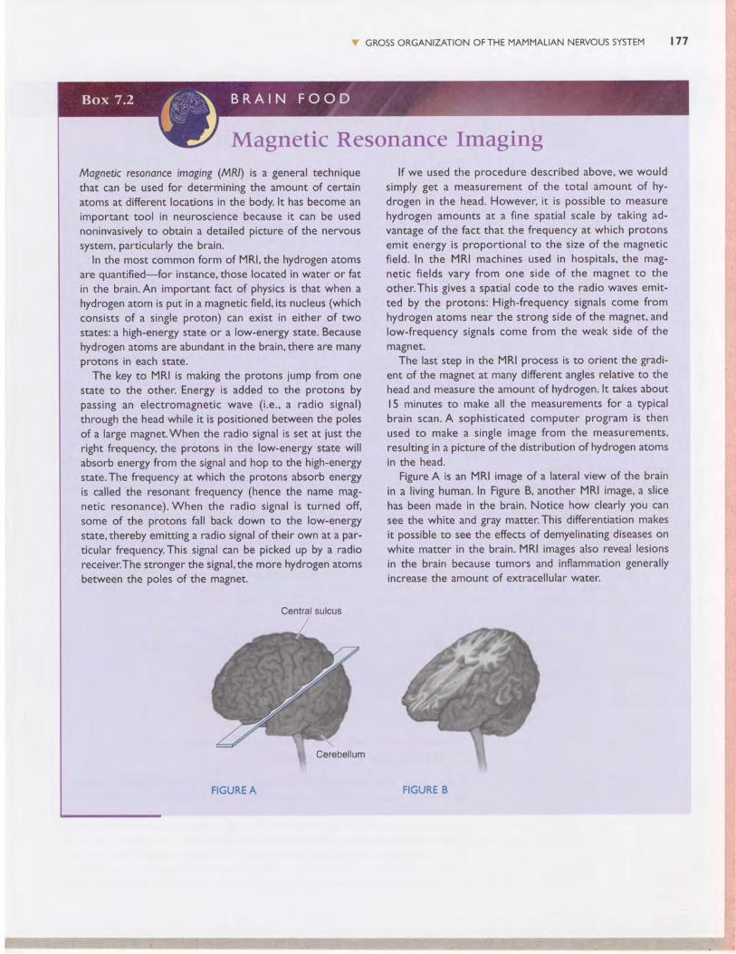
Magnetic Resonance ImagingMagnetic resononce imaging (MRl) is a general techniquethat can be used for determining the amount of certainatoms at different locations in the body. lt has become animportant tool in neuroscience because it can be usednoninvasively to obtain a detailed picture of the nervoussystem, particularly the brain.
In the most common form of MRl,the hydrogen atomsare quantified-for instance, those located in water or fatin the brain. An important fact of physics is that when ahydrogen atom is put in a magnetic field, its nucleus (whichconsists of a single proton) can exist in either of twostates: a high-energy state or a low-energy state. Becausehydrogen atoms are abundant in the brain,there are manyprotons in each state.
The key to MRI is making the protons jump from onestate to the other. Energy is added to the protons bypassing an electromagnetic wave (i.e., a radio signal)through the head while it is positioned between the polesof a large magnet.When the radio signal is set at iust theright frequency, the protons in the low-energy state willabsorb energy from the signal and hop to the high-energystate.The frequency at which the protons absorb energyis called the resonant frequency (hence the name mag-netic resonance). When the radio signal is turned off,some of the protons fall back down to the low-energystate, thereby emitting a radio signal of their own at a par-ticular frequency.This signal can be picked up by a radioreceiver.The stronger the signal,the more hydrogen atomsbetween the poles of the magnet.
lf we used the procedure described above, we wouldsimply get a measurement of the total amount of hy-drogen in the head. However, it is possible to measurehydrogen amounts at a fine spatial scale by taking ad-vantage of the fact that the frequency at which protonsemit energy is proportional to the size of the magneticfield. In the MRI machines used in hospitals, the mag-netic f ields vary from one side of the magnet to theother.This gives a spatial code to the radio waves emit-ted by the protons: High-frequency signals come fromhydrogen atoms near the strong side of the magnet, andlow-frequency signals come from the weak side of themagneE.
The last step in the MRI process is to orient the gradi-ent of the magnet at many different angles relative to thehead and measure the amount of hydrogen. lt takes aboutl5 minutes to make all the measurements for a typicalbrain scan. A sophisticated computer program is thenused to make a single image from the measurements,resulting in a picture of the distribution of hydrogen atomsin the head.
Figure A is an MRI image of a lateral view of the brainin a l iving human. In Figure B, another MRI image, a slicehas been made in the brain. Notice how clearly you cansee the white and gray matter.This differentiation makesit possible to see the effects of demyelinating diseases onwhite matter in the brain. MRI images also reveal lesionsin the brain because tumors and inflammation generallyincrease the amount of extracellular water.
Central sulcus
FIGURE A FIGURE B
t 7 8 C H A P T E R 7 THE STRUCTURE OFTHE NERVOUS SYSTEM
Functional Imaging of Brain Activity:PET and fMRr
Until recently,"mind reading" has been beyond the reach ofscience. However, with the introduction of positron emissiontomogrophy (PEI) and funaionol mognetic resononce imaging(fMRl), it is now possible to observe and measure changesin brain activity associated with the planning and executionof specific tasks,
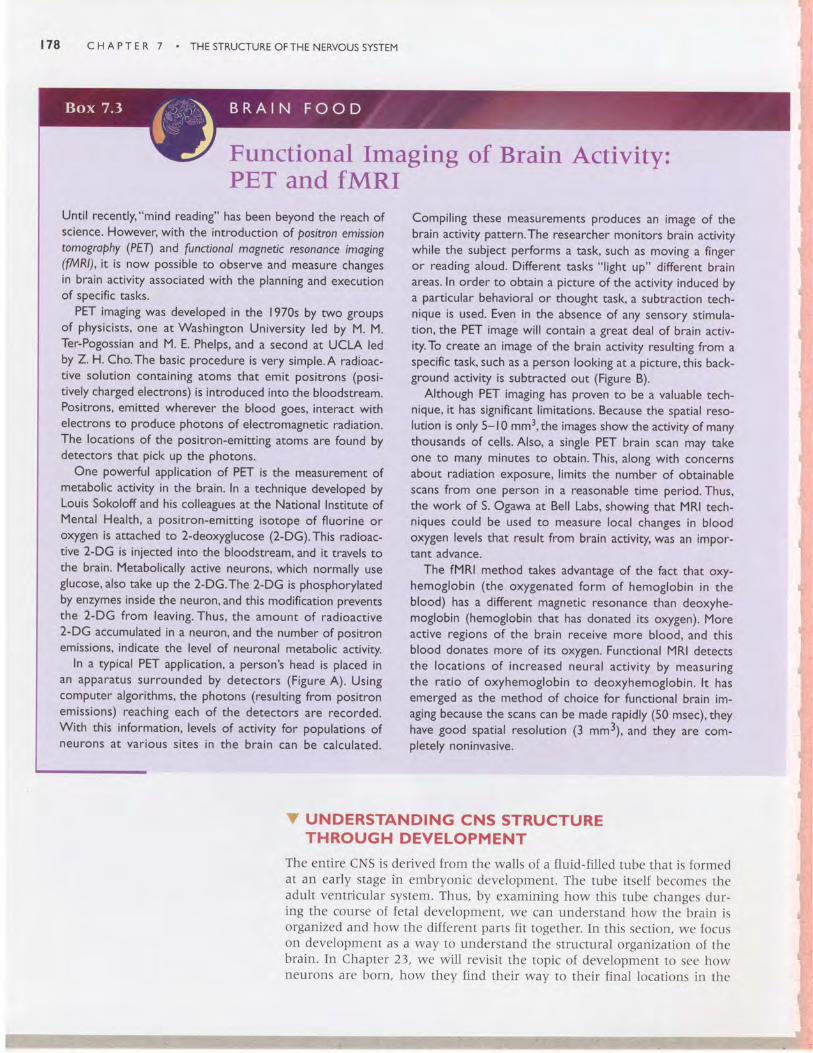
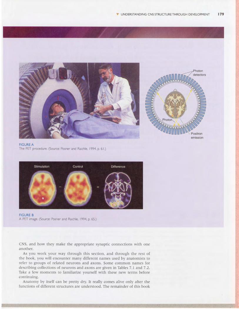
PET imaging was developed in the 1970s by two groupsof physicists, one at Washington University led by M. M.Ter-Pogossian and M. E. Phelps, and a second at UCLA ledby Z.H. Cho.The basic procedure is very simple.A radioac-tive solution containing atoms that emit positrons (posi-tively charged electrons) is introduced into the bloodstream.Positrons, emitted wherever the blood goes, interact withelectrons to produce photons of electromagnetic radiation.The locations of the positron-emitting atoms are found bydetectors that pick up the photons.
One powerful application of PET is the measurement ofmetabolic activity in the brain. In a technique developed byLouis Sokoloff and his colleagues at the National Institute ofMental Health, a posirron-emitting isotope of f luorine oroxygen is attached to 2-deoxyglucose (2-DG).This radioac-tive 2-DG is injected into the bloodstream, and it travels tothe brain. Metabolically active neurons, which normally useglucose, also take up the 2-DG.The 2-DG is phosphorylatedby enzymes inside the neuron, and this modification preventsthe 2-DG from leaving. Thus, the amount of radioactive2-DG accumulated in a neuron, and the number of positronemissions, indicate the level of neuronal metabolic activity.
In a typical PET application, a person's head is placed inan apparatus surrounded by detectors (Figure A). Usingcomputer algorithms, the photons (resulting from positronemissions) reaching each of the detectors are recorded.With this information, levels of activity for populations ofneurons at var ious s i tes in the bra in can be calculated.
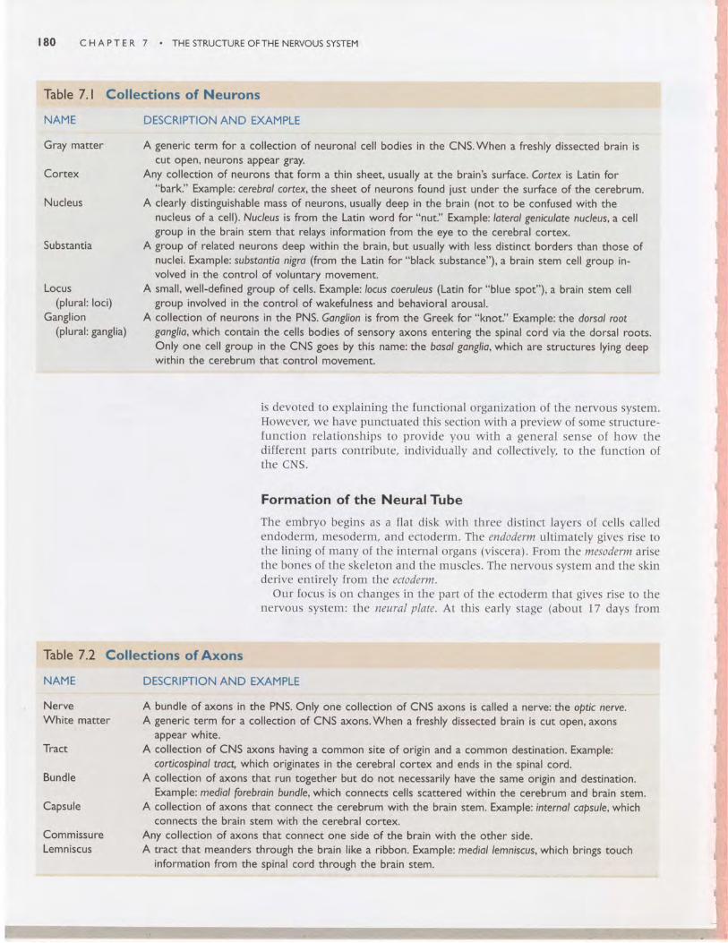
Compiling these measurements produces an image of thebrain activity pattern.The researcher monitors brain activitywhile the subject performs a task, such as moving a fingeror reading aloud. Different tasks "l ight up" different brainareas. In order to obtain a picture of the activity induced bya particular behavioral or thought task, a subtraction tech-nique is used. Even in the absence of any sensory stimula-tion, the PET image will contain a great deal of brain activ-ity.To create an image of the brain activity resulting from aspecific task, such as a person looking at a picture, this back-ground activity is subtracted out (Figure B).
Although PET imaging has proven to be a valuable tech-nique, it has significant l imitations. Because the spatial reso-lution is only 5- l0 mm3, the images show the activity of manythousands of cells. Also, a single PET brain scan may takeone to many minutes to obtain. This, along with concernsabout radiation exposure, l imits the number of obtainablescans from one person in a reasonable time period. Thus,the work of S. Ogawa at Bell Labs, showing that MRI tech-niques could be used to measure local changes in bloodoxygen levels that result from brain activity, was an impor-tant advance.
The fMRl method takes advantage of the fact that oxy-hemoglobin (the oxygenated form of hemoglobin in theblood) has a different magnetic resonance than deoxyhe-moglobin (hemoglobin that has donated its oxygen). Moreactive regions of the brain receive more blood, and thisblood donates more of its oxygen. Functional MRI detectsthe locat ions of increased neural act iv i ty by measur ingthe ratio of oxyhemoglobin to deoxyhemoglobin. lt hasemerged as the method of choice for functional brain im-aging because the scans can be made rapidly (50 msec), theyhave good spatial resolution (3 mm3), and they are com-pletely noninvasive.
C UNDERSTANDING CNS STRUCTURETHROUGH DEVELOPMENT
The ent i re cNS is der ived f rom the wal ls of a f lu id- f i l led tube that is formedat an early stage in embryonic development. The tube itself becomes theadul t ventr icu lar system. Thus, by examining how th is tube changes dur-ing the course of fe ta l development , we can understand how the bra in isorganized and how the different parts fit together. In this section, we focr,rson development as a way 1o understand the st ructura l r l rganizat ion of thebrain. In Chapter 23, we wi l l rev is i t the topic of development to see howneurons are born, how they find their way to their f inal krcations in the
UNDERSTANDING CNS STRUCTURETHROUGH DEVELOPMENT 179
i,;4- t*asdffiP
FIGURE AThe PET procedure. (Source: Posner and Raichle, 1994, p. 6 l . )
FIGURE BA PET mage. (Source: Posner and Rarch e, 199a, p.65.)
CNS, and I ' tow they lnake the appropr iate synapt ic c( ) l l tcct i ( )ns wi th oncano lhc r .
As yu r - r wo rk yo l l r way th rough th i s sec t i on , and th rough the res t o fthe book, yor- r wi l l encol ln ter many d i f ferent narnes used by anatr )n l is ts tore lcr 1t t groups of re lated neurons and axons. Sonte cornmon l - ]amcs fordesc r i b i ng co l l ec t i ons o [ neu rons and axons a re g i ven i n Tab les 7 .1 and7 .2 .Take a lew rnoments to fami l iar ize yoursel f wi th these new terms belorecon t i nu ing .
Anatorny by i tse l f can be pre11y dry. I t real ly comes a l ive only a l ter r l refunct ions of d i f ferent s l r l lc tures are understood. The remainder of t l " r is book
' ffi
ffi
I 80 cHAprER 7 . THEsrRUcruREoFTHENERVoussys rEM
Table 7. I Collections of Neurons
NAME DESCRIPTION AND EXAMPLE
Gray matter A generic term for a collection of neuronal cell bodies in the CNS.When a freshly dissected brain iscut oPen, neurons aPPear gray.
Cortex Any collection of neurons that form a thin sheet, usually at the brain's surface. Cortex is Latin for;'bark." Example: cerebral cortex, the sheet of neurons found just under the surface of the cerebrum.
Nucleus A clearly distinguishable mass of neurons, usually deep in the brain (not to be confused with thenucleus of a cell). Nucleus is from the Latin word for "nut." Example; loterol geniculote nucleus, a cellgroup in the brain stem that relays information from the eye to the cerebral cortex.Subs'can'ca ^ :::i3 ?Til[:ffiffiffi:ti*#jffI',::il::l'l#:1"::iTffi"::*::i:^;L:l?:'volved in the control of voluntary movement.
Locus A small, well-defined group of cells. Example:locus coeruleus (Latin for "blue spot"), a brain stem cell(plural: loci) group involved in the control of wakefulness and behavioral arousal.
Ganglion A collection of neurons in the PNS. Gonglion is from the Greek for "knot." Example: the dorsol root(plural: ganglia) gonglio, which contain the cells bodies of sensory axons entering the spinal cord via the dorsal roots.
Only one cell group in the CNS goes by this name: the bosol gonglio, which are structures lying deepwithin the cerebrum that control movement.
is devotcd to expla in ing the funct ional organizat ion o l the nerv<lus systent .However, we have punctuated th is sect ion wi t l ' r a prev icw of sorre st ru lc t r l rc-l u r t c t i on re la t i onsh ips t< l p rov ide you w i th a gene ra l sensc o [ l ' r ow thcdi f ferent par ts contr ibute, ind iv idual ly ancl co l lect ive ly , to 1hc f r rnct ion oft he CNS.
Formation of the NeuralTube
The embryo begir - rs as a f la t d isk wi th three 'c l is t inct layers o l ce l ls ca l lec lendoderm, mesoden-n, and ectoderm. The endoderm ul t imate ly g ives r isc tothe l ining of n-rany of the internal urgans (visccra). Fror.n the mesldertn ariscthe bones of the skeleton and the n-ruscles. The nervor . ls system ancl thc sk inderive entirelv from the ectoderm.
Our focus is on changes in the par t of the ectodernt that g ives r ise t i l thenervous system: rhe neural p late. At th is ear ly s tage (abotr t l7 days f ront
Table 7.2 Collections of Axons
NAME DESCRIPTION AND EXAMPLE
Nerve A bundle of axons in the PNS. Only one collection of CNS axons is called a nerve: the optic nerve.White matter A generic term for a collection of CNS axons.When a freshly dissected brain is cut open, axons
appear white.Tract A collection of CNS axons having a common site of origin and a common destination. Example:
corticospinol troct, which originates in the cerebral cortex and ends in the spinal cord.Bundle A collection of axons that run toSether but do not necessarily have the same origin and destination.
Example: mediol forebroin bundle, which connects cells scattered within the cerebrum and brain stem.Capsule A collection of axons that connect the cerebrum with the brain stem. Example: internol copsule, which
connects the brain stem with the cerebral cortex.Commissure Any collection of axons that connect one side of the brain with the other side.Lemniscus A tract that meanders through the brain like a ribbon. Example: mediol lemniscus, which brings touch
information from the spinal cord through the brain stem.
Rostral
Caudal
Neuralfold
Neuraltube
FIGURE 7.8Formation of the neural tube and neural crest.These schematic illustrations followthe early development of the nervous system in the embryo.The drawings above are dorsalviews of the embryo; those below are cross sections. (a) The primitive embryonic CNSbegins as a thin sheet of ectoderm. (b)The first important step in the development of thenervous system is the formation of the neural groove. (c) The walls of the groove, calledneural folds, come together and fuse, forming the neural tube. (d) The bits of neural ecto-derm that are pinched off when the tube rolls up is called the neural crest, from which thePNS will develop.The somites are mesoderm that will sive rise to much of the skeletalsystem and the muscles.
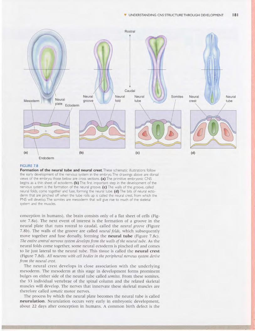
conception in humans), the brain consists only of a flat sheet of cells (Fig-ure 7.8a). The next event of interest is the formation of a groove in theneural plate that runs rostral to caudal, called the neural groove (Figure7.8b). The walls of the groove are called neural folds, which subsequentlymove together and fuse dorsally, forming the neural tube (Figure 7.8c).The entire central nervous system develops from the walls of the neural tube. As theneural folds come together, some neural ectoderm is pinched off and comesto lie just lateral to the neural tube. This tissue is called the neural crest(Figure 7 .8d). All neurons with cell bodies in the peripheral nervous system derivefrom the neural crest.
The neural crest develops in close association with the underlyingmesoderm. The mesoderm at this stage in development forms prominentbulges on either side of the neural tube called somites. From these somites,the 33 individual vertebrae of the spinal column and the related skeletalmuscles will develop. The nerves that innervate these skeletal muscles aretherefore called somatic motor nerves.
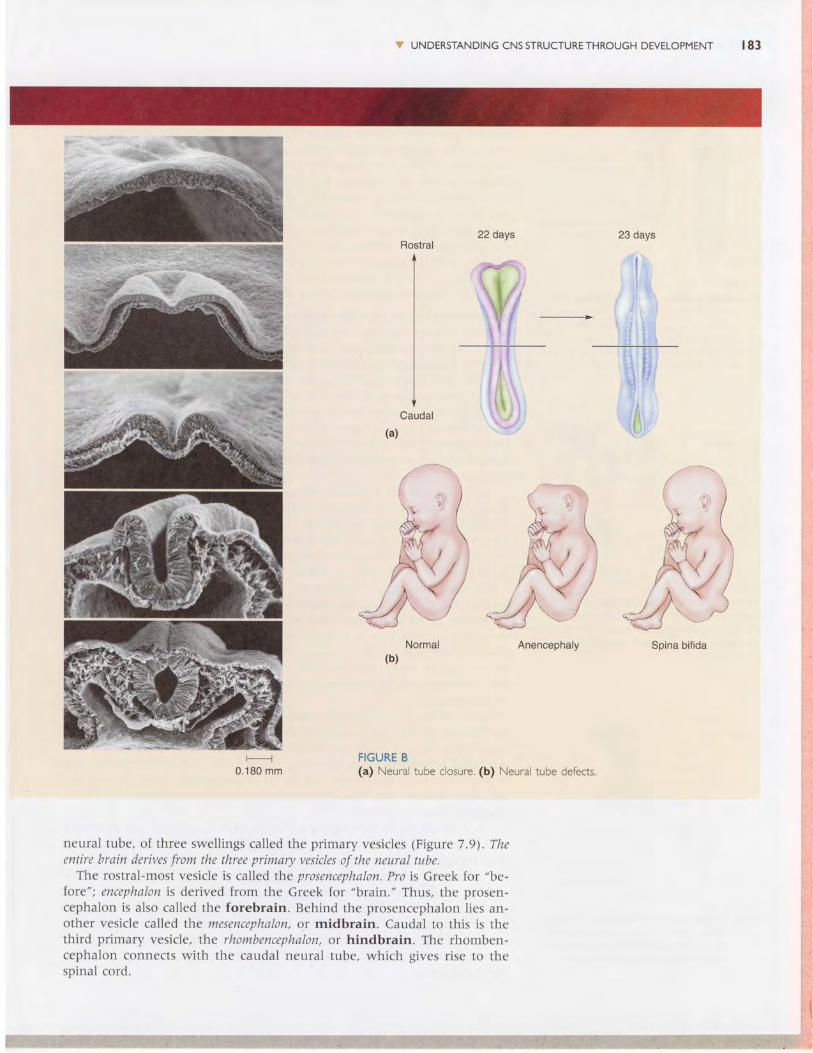
The process by which the neural plate becomes the neural tube is calledneurulation. Neurulation occurs very early in embryonic development,abott 22 days after conception in humans. A common birth defect is the
v uNDERsrANDrNGcNssrRUcruRETHRoucH DEVELopMENT l8l
Somites Neuralcrest
Neuraltube
Endoderm
I 8 2 C H A P T E R 7 THE STRUCTURE OFTHE NERVOUS SYSTEM
Nutrition andthe Neural Ti.rbe
Neural tube formation is a crucial event in the developmentof the nervous system. lt occurs early-only 3 weeks afterconception-when the mother may be unaware she is preg-nant . Fai lure of the neural tube to c lose correct ly is acommon birth defect, occurring in approximately I out ofevery 500 live births. A recent discovery of enormous pub-lic health importance is that many neural tube defects canbe traced to a deficiency of the vitamin folic ocid (or Blote)in the maternal diet during the weeks immediately afterconception. lt has been estimated that dietary supplemen-tation of folic acid during this period could reduce the inci-dence of neural tube defects by 90%.
Format ion of the neural tube is a complex process(Figure A). lt depends on a precise sequence of changes inthe three-dimensional shape of individual cells, as well as onchanges in the adhesion of each cell to its neighbors. Thetiming of neurulation also must be coordinated with simul-taneous changes in non-neural ectoderm and the meso-derm. At the molecular level, successful neurulation de-pends on specific sequences of gene expression that arecontrolled, in part, by the position and local chemical envi-ronment of the cell. l t is not surprising that this process ishighly sensitive to chemicals, or chemical deficiencies, in thematernal circulation.
The fusion of the neural folds to form the neural tubeoccurs first in the middle, then anteriorly and posteriorly(Figure B). Failure of the anterior neural tube to close re-
sults in onencephaly, a condition characterized by degenera-tion of the forebrain and skull that is always fatal. Failureof the posterior neural tube to close results in a conditioncalled spino bifida. ln its most severe form, spina bifida ischaracterized by the failure of the posterior spinal cord toform from the neural plate (bifdo is from the Latin wordmeaning "cleft in two parts"). Less severe forms are char-acterized by defects in the meninges and vertebrae overly-ing the posterior spinal cord. Spina bifida, while usually notfatal, does require extensive and costly medical care.
Folic acid plays an essential role in a number of metabolicpathways, including the biosynthesis of DNA, which natu-ra l ly must occur dur ing development as cel ls d iv ide. Al -though we do not precisely understand why folic acid defi-ciency increases the incidence of neural tube defects, onecan easily imagine how it could alter the complex choreog-raphy of neurulation. The name is derived from the Latinword for "leaf," reflecting the fact that folic acid was firstisolated from spinach leaves. Besides green leafy vegetables,good dietary sources of folic acid are liver, yeast, eggs, beans,and oranges. Many breakfast cereals are now fortified withfolic acid. Nonetheless, the folic acid intake of the averageAmerican is only half of what is recommended to preventbirth defects (0.4 mg/day). The U.S. Centers for DiseaseControl and Prevention recommends that women take mul-tivitamins containing 0.4 mg of folic acid before planning
PreSnancy.
FIGURE A >Scan^ing e ectron mic'og.3p|5 of ^eu'- lat or.(Sou'ce: Smith anc Schoe.rwoif, 1997.;
failure of appropriate closure of the neural tube. Fortunately, recent researchsuggests that most cases of neural tube defects can be avoided by ensuringproper maternal nutrit ion during this period (Box 7.4).
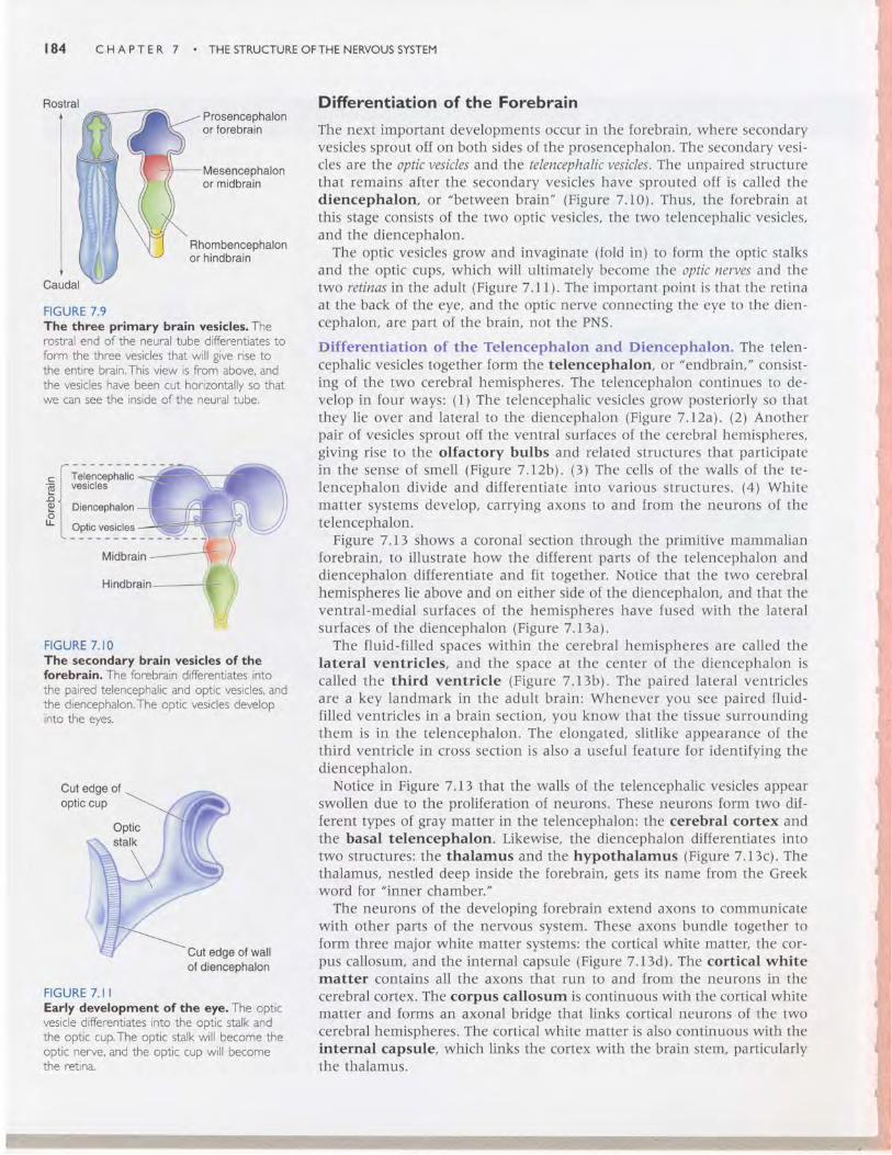
Three Primary Brain Vesicles
The process by which structures become more complex and functionallyspecialized during development is called differentiation. The first step inthe differentiation of the brain is the develoDment. ar rhe rostral end of the
I I UNDERSTANDING CNS STRUCTURE THROUGH DEVELOPMENT r 8 3
22 days 23 days
It
Rostral
Caudal
, , i ,\t'0", \ I
(a)
ritn rtkS''i
, )I ) : I
,S.r \ht,'i
4Vu- 4 )$v,Anencephaly
FIGURE B(a) Neural tube closure. (b) Neural tube defects,
Spina bif ida
0 . 1 8 0 m m
neural tube, of three swel l ings cal led the pr imary vesic les (F igure 7.91 . Theentire brain derives from the three primary vesicles of the neural tube.
The rostral-most vesicle is called the prosencephalon. Pro is Greek for "be-
tore"; encephalon is derived from the Greek for "brain." Thus, the prosen-cephakrn is also called the forebrain. Behind the prosencephalon l ies an-other vesicle called the mesencephalon, or midbrain. Caudal to this is thethird primary vesicle, the rhombencephalon, or hindbrain. The rhomben-cephalon connects wi th the caudal neural tube, which g ives r ise to thespinal cord.
t 8 4
Rostral
FIGURE 7.9The three primary brain vesicles. Therostral end of the neural tube differentiates toform the three vesicles that will give rise tothe entire brain.This view is from above, andthe vesicles have been cut horizontallv so thatwe can see the inside ofthe neural tube.
C H A P T E R 7 THE STRUCTURE OFTHE NERVOUS SYSTEM
Prosencephalonor forebrain
Mesencephalonor midbrain
Rhombencephalonor hindbrain
Difrerentiation of the Forebrain
The next important developments occur in the forebrain, where secondaryvesicles sprout off on both sides of the prosencephalon. The secondary vesi-cles are the optic vesicles and the telencephalic vesicles. The unpaired structurethat remains after the secondary vesicles have sprouted off is called thediencephalon, or "between brain" (Figure 7.10). Thus, the forebrain atthis stage consists of the two optic vesicles, the two telencephalic vesicles,and the diencephalon.
The optic vesicles grow and invaginate (fold in) to form the optic stalksand the optic cups, which will ultimately become the optic nerves and th.etwo retinas in the adult (Figure 7.1 I ). The important point is that the retinaat the back of the eye, and the optic nerve connecting the eye to the dien-cephalon, are part of the brain, not the PNS.
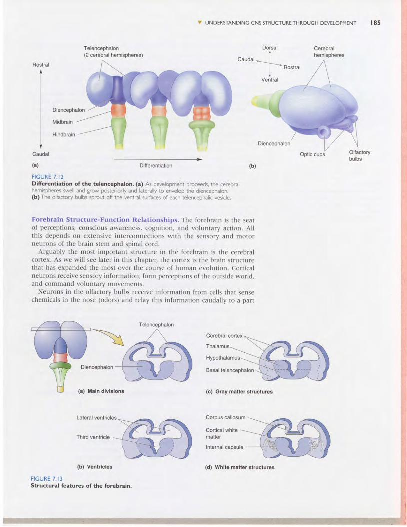
Differentiation of the Telencephalon and Diencephalon. The telen-cephalic vesicles together form the telencephalon, or "endbrain," consist-ing of the two cerebral hemispheres. The telencephalon continues to de-velop in four ways: (l) The telencephalic vesicles grow posteriorly so thatthey lie over and lateral to the diencephalon (Figure 7.l2al. (2) Anotherpair of vesicles sprout off the ventral surfaces of the cerebral hemispheres,giving rise to the olfactory bulbs and related structures that participatein the sense of smell (Figure 7.12b\. (3) The cells of the walls of the te-lencephalon divide and differentiate into various structures. (4) Whitematter systems develop, carrying axons to and from the neurons of thetelencephalon.
Figure 7.I3 shows a coronal section through the primitive mammalianforebrain, to illustrate how the different parts of the telencephalon anddiencephalon differentiate and fit together. Notice that the two cerebralhemispheres lie above and on either side of the diencephalon, and that theventral-medial surfaces of the hemispheres have fused with the lateralsurfaces of the diencephalon (Figure 7.I3al.
The fluid-filled spaces within the cerebral hemispheres are called thelateral ventricles, and the space at the center of the diencephalon iscalled the third ventricle (Figure 7.13b). The paired lateral ventriclesare a key landmark in the adult brain: Whenever you see paired fluid-filled ventricles in a brain section, you know that the tissue surroundingthem is in the telencephalon. The elongated, slitl ike appearance of thethird ventricle in cross section is also a useful feature for identifying thediencephalon.
Notice in Figure 7.LJ that the walls of the telencephalic vesicles appearswollen due to the proliferation of neurons. These neurons form two dif-ferent types of gray matter in the telencephalon: the cerebral cortex andthe basal telencephalon. Likewise, the diencephalon differentiates intotwo structures: the thalamus and the hypothalamus (Figure 7.13c). Thethalamus, nestled deep inside the forebrain, gets its name from the Greekword for "inner chamber."
The neurons of the developing forebrain extend axons to communicatewith other parts of the nervous system. These axons bundle together toform three major white matter systems: the cortical white matter, the cor-pus callosum, and the internal capsule (Figure 7.13d). The cortical whitematter contains all the axons that run to and from the neurons in thecerebral cortex. The corpus callosum is continuous with the cortical whitematter and forms an axonal bridge that links cortical neurons of the twocerebral hemispheres. The cortical white matter is also continuous with theinternal capsule, which links the cortex with the brain stem, particularlythe thalamus.
. [t*.;il*
El ;"::;",".* L _ory_":.1''::Midbrain
FIGURE 7. IOThe secondary brain vesicles of theforebrain. The forebrain differentiates intothe paired telencephalic and optic vesicles, andthe diencephalon.The optic vesicles developinto the eyes.
Cut edge ofoptic cup
Cut edge of wallof diencephalon
FIGURE 7 . I IEarly development of the eye. The opticvesicle differentiates into the ootic stalk andthe optic cup.The optic stalk will become theoptic nerve, and the optic cup will becomethe retrna.
Telencephalon(2 cerebral hemispheres)
V UNDERSTANDING CNS STRUCTURE THROUGH DEVELOPMENT r85
Cerebralhemispheres
Optic cups
Rostral
Caudal
(a)
Diencephalon
Midbrain
Hindbrain
Differentiation (b)
FIGURE 7.I2Difierentiation of the telencephalon. (a) As development proceeds, the cerebralhemispheres swell and grow posteriorly and laterally to envelop the diencephalon.(b) The olfactory bulbs sprout off the ventral surfaces of each telencephalic vesicle.
Forebrain Structure-Function Relationships. The forebrain is the seatof perceptions, conscious awareness, cognition, and voluntary action. Allthis depends on extensive interconnections with the sensory and motorneurons of the brain stem and spinal cord.
Arguably the most important structure in the forebrain is the cerebralcortex. As we will see later in this chapter, the cortex is the brain structurethat has expanded the most over the course of human evolution. Corticalneurons receive sensory information, form perceptions of the outside world,and command voluntary movements.
Neurons in the olfactory bulbs receive information from cells that sensechemicals in the nose (odors) and relay this information caudally to a part
Cerebral cortex
Thalamus
Hypothalamus
Diencephalon Basal telencephalon
Maln dlvlslons (c) Gray matter structures
(b) Ventrlcles
FIGURE 7. I3Structural features of the forebrain.
;.::"'ffi*ffllffi
Dorsal
Telencephalon
(d) Whlte matter structures
Cerebral
t 8 6 CHAPTER 7 . THESTRUCTUREOFTHENERVOUSSYSTEM
Eye Ear Skin
FIGURE 7. I4The thalamus: gateway to the cerebralcortex. The sensory pathways from the eye,ear: and skin all relay in the thalamus beforeterminating in the cerebral cortex.The arrowsindicate the direction of information flow.
of the cerebral cortex for further analysis. Information from the eyes, ears,and skin is also brought to the cerebral cortex for analysis. However, eachof the sensory pathways serving vision, audition (hearing), and somaticsensation relays (i.e., synapses upon neurons) in the thalamus en route tothe cortex. Thus, the thalamus is often referred to as the gateway to thecerebral cortex (Figure 7.I4).
Thalamic neurons send axons to the cortex via the internal capsule. Asa general rule, the axons of each internal capsule carry information to thecortex about the contralateral side of the body. Therefore, if a thumbtackentered the right foot, it would be relayed to the left cortex by the left thal-amus via axons in the left internal capsule. But how does the right footknow what the lelt foot is doing? One important way is by communicationbetween the hemispheres via the axons in the corpus callosum.
Cortical neurons also send axons through the internal capsule, back tothe brain stem. Some cortical axons course all the way to the spinal cord,forming the corticospinal tract. This is one important way cortex cancommand voluntary movement. Another way is by communicating withneurons in the basal ganglia, a collection of cells in the basal telencephalon.The term basal is used to describe structures deep in the brain, and the basalganglia Iie deep within the cerebrum. The functions of the basal ganglia arepoorly understood, but it is known that damage to these structures disruptsthe ability to initiate voluntary movement. Other structures, contributingto other brain functions, are also present in the basal telencephalon. Forexample, in Chapter 18 we'll discuss a structure called the amygdala thatis involved in fear and emotion.
Although the hypothalamus lies just under the thalamus, functionally itis more closely related to certain telencephalic structures, like the amyg-dala. The hypothalamus performs many primitive functions and thereforehas not changed much over the course of mammalian evolution. "Primitive"
does not mean unimportant or uninteresting, however. The hypothalamuscontrols the visceral (autonomic) nervous system, which regulates bodilyfunctions in response to the needs of the organism. For example, when youare faced with a threatening situation, the hypothalamus orchestrates thebody's visceral fight-or-flight response. Hypothalamic commands to theANS will lead to (among other things) an increase in the heart rate, in-creased blood flow to the muscles for escape, and even the standing of yourhair on end. Conversely, when you're relaxing after Sunday brunch, the
v SELF-QUIZListed below are derivatives of the forebrain that we have discussed. Besure vou know what each of these terms means.
PRIMAtrVESICLE SECONDAWVESICLEForebrain Optic vesicle
(prosencephalon)Thalamus
(diencephalon)
Telencephalon
SOME ADULT DERIVATIVESRetinaOptic nerveDorsal thalamusHypothalamusThird ventricleOlfactory bulbCerebral cortexBasal telencephalonCorpus callosumCortical white matterInternal capsule
V UN DERSTANDING CNS STRUCTURE THROUGH DEVELOPMENT
hypothalamus ensures that the brain is well-nourished via commands tothe ANS, which will increase peristalsis (movement of material throughthe gastrointestinal tract) and redirect blood to your digestive system. Thehypothalamus also plays a key role in motivating animals to find food,drink, and sex in response to their needs. Aside from its connections to theANS, the hypothalamus also directs bodily responses via connections with thepituitary gland located below the diencephalon. This gland communicatesto many parts of the body by releasing hormones into the bloodstream.
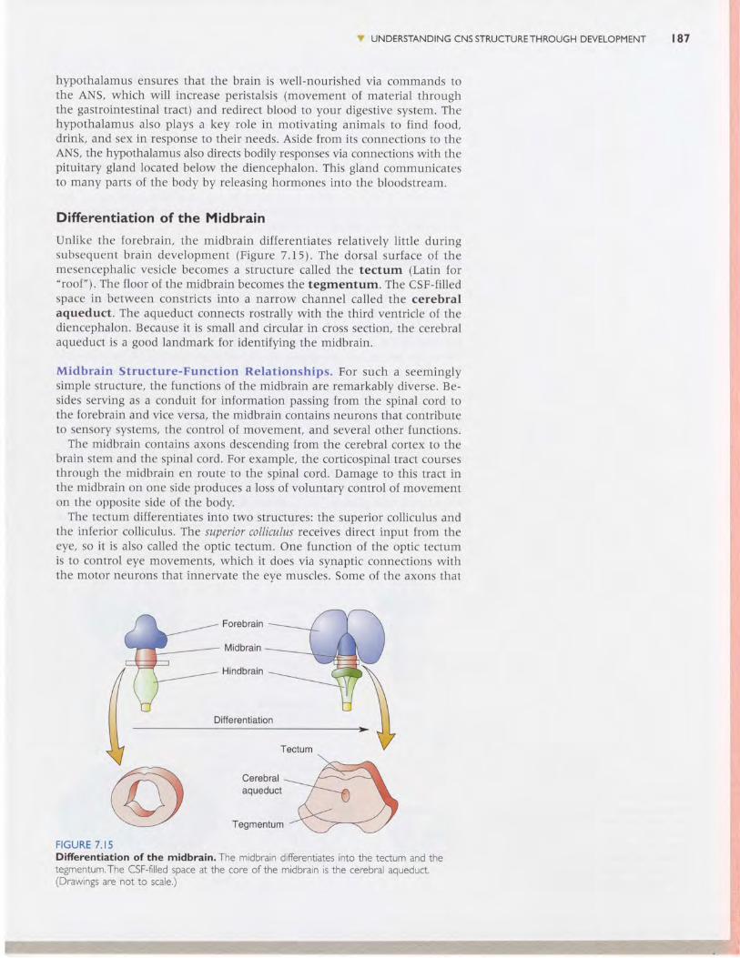
Difrerentiation of the Midbrain
Unlike the forebrain, the midbrain differentiates relatively little duringsubsequent brain development (Figure 7.I5.1. The dorsal surface of themesencephalic vesicle becomes a structure called the tectum (Latin for"roof"). The floor of the midbrain becomes the tegmentum. The CSF-filledspace in between constricts into a narrow channel called the cerebralaqueduct. The aqueduct connects rostrally with the third ventricle of thediencephalon. Because it is small and circular in cross section, the cerebralaqueduct is a good landmark for identifying the midbrain.
Midbrain Structure-Function Relationships. For such a seeminglysimple structure, the functions of the midbrain are remarkably diverse. Be-sides serving as a conduit for information passing from the spinal cord tothe forebrain and vice versa, the midbrain contains neurons that contributeto sensory systems, the control of movement, and several other functions.
The midbrain contains axons descending from the cerebral cortex to thebrain stem and the spinal cord. For example, the corticospinal tract coursesthrough the midbrain en route to the spinal cord. Damage to this tract inthe midbrain on one side produces a loss of voluntary control of movementon the opposite side of the body.
The tectum differentiates into two structures: the superior colliculus andthe inferior colliculus. The superior colliculus receives direct input from theeye, so it is also called the optic tectum. One function of the optic tectumis to control eye movements, which it does via synaptic connections withthe motor neurons that innervate the eve muscles. Some of the axons that
Ditferentiation
Cerebralaqueduct
Tegmentum
FIGURE 7. I5Difrerentiation of the midbrain. The midbrain differentiates into the tectum and thetegmentum.The CSF-filled space at the core of the midbrain is the cerebral aqueduct.(Drawings are not to scale.)
r 8 7
r88 CHAPTER 7 . THESTRUCTUREOFTHENERVOUSSYSTEM
*1stffiSi\3iifi.Y.'{
SrI$rr.f$:iitsrr,{TJ${iHSs 4iri
"*i*rU:i$.1
*;,twr.: ri
g':'j
e-iHBJ$$il;'l*':iiqtK:l{$itilH \ !
supply the eye muscles originate in the midbrain, bundling together toform cranial nerves III and IV (see the chapter appendix).
The inferior colliculus also receives sensory information, but from the earinstead of the eye. The inferior colliculus serves as an important relaystation for auditory information en route to the thalamus.
The tegmentum is one of the most colorful regions of the brain becauseit contains both the substantia nigra (the black substance) and the rednucleus. These two cell groups are involved in the control of voluntarymovement. Other cell groups scattered in the midbrain have axons thatproject widely throughout much of the CNS and function to regulate con-sciousness, mood, pleasure, and pain.
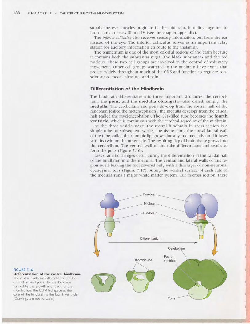
Difrerentiation of the Hindbrain
The hindbrain differentiates into three important structures: the cerebel-lum, the pons, and the medulla oblongata-also called, simply, themedulla. The cerebellum and pons develop from the rostral half of thehindbrain (called the metencephalon); the medulla develops from the caudalhalf (called the myelencephalon). The CSF-filled tube becomes the fourthventricle, which is continuous with the cerebral aqueduct of the midbrain.
At the three-vesicle stage, the rostral hindbrain in cross section is asimple tube. In subsequent weeks, the tissue along the dorsal-lateral wallof the tube, called the rhombic lip, grows dorsally and medially until it fuseswith its twin on the other side. The resulting flap of brain tissue grows intothe cerebellum. The ventral wall of the tube differentiates and swells toform the pons (Figure 7.161.
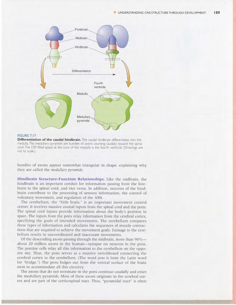
Less dramatic changes occur during the differentiation of the caudal halfof the hindbrain into the medulla. The ventral and lateral walls of this re-gion swell, leaving the roof covered only with a thin layer of non-neuronalependymal cells (Figure 7.I7). Along the ventral surface of each side ofthe medulla runs a major white matter system. Cut in cross section, these
Forebrain
Midbrain
Hindbrain
Differentiation
FIGURE 7 . I6Difrerentiation of the rostral hindbrain.The rostral hindbrain diferentiates into thecerebellum and oons.The cerebellum isformed by the growth and fusion of thertrombic lips.The CSF-filled space at thecore of the hindbrain is the fourth ventricle.(Drawings are not to scale.)
Cerebellum
FourthventricleRhombic lips
!r UNDERSTANDINGCNSSTRUCTURETHROUGHDEVELOPMENT I89
Forebrain
Midbrain
Hindbrain
Differentiation
Fourthventricle
Medulla
Medullarypyramids
F IGURE 7 . I7Difierentiation of the caudal hindbrain. The caudal hindbrain differentiates into themedulla.The medullary pyramids are bundles of axons coursing caudally toward the spinalcord.The CSF-fllled space at the core of the medulla is the firurth ventricle. (Drawings arenot to scale,)
bundles of axons appear somewhat triangular in shape, explaining whythey are called the medullary pyramids.
Hindbrain Structure-Function Relationships. Like the midbrain. rhehindbrain is an important conduit for information passing from the fore-brain to the spinal cord, and vice versa. In addition, neurons of the hind-brain contribute to the processing of sensory information, the control ofvoluntary movement, and regulation of the ANS.
The cerebellum, the "l itt le brain," is an important movement controlcenter. It receives massive axonal inputs from the spinal cord and the pons.The spinal cord inputs provide information about the body's position inspace. The inputs from the pons relay information from the cerebral cortex,specifying the goals of intended movements. The cerebellum comparesthese types of information and calculates the sequences of muscle contrac-tions that are required to achieve the movement goals. Damage to the cere-bellum results in uncoordinated and inaccurare movemenrs.
Of the descending axons passing through the midbrain, more than 90%-about 20 million axons in the human-synapse on neurons in the pons.The pontine cells relay all this information to the cerebellum on the oppo-site site. Thus, the pons serves as a massive switchboard connecting thecerebral cortex to the cerebellum. (The word pons is from the Latin wordfor "bridge.") The pons bulges out from the ventral surface of the brainstem to accommodate all this circuitry.
The axons that do not terminate in the pons continue caudally and enterthe medullary pyramids. Most of these axons originate in the cerebral cor-tex and are part of the corticospinal tract. Thus, "pyramidal tract" is often
ffi.
1 9 0 c H A P T E R 7 THE STRUCTURE OFTHE NERVOUS SYSTEM
Medulla
Pyramidaldecussation
Soinal cord
FIGURE 7. I8The pyramidal decussation. The cortico-spinal tract crosses from one side to theother in the medulla.
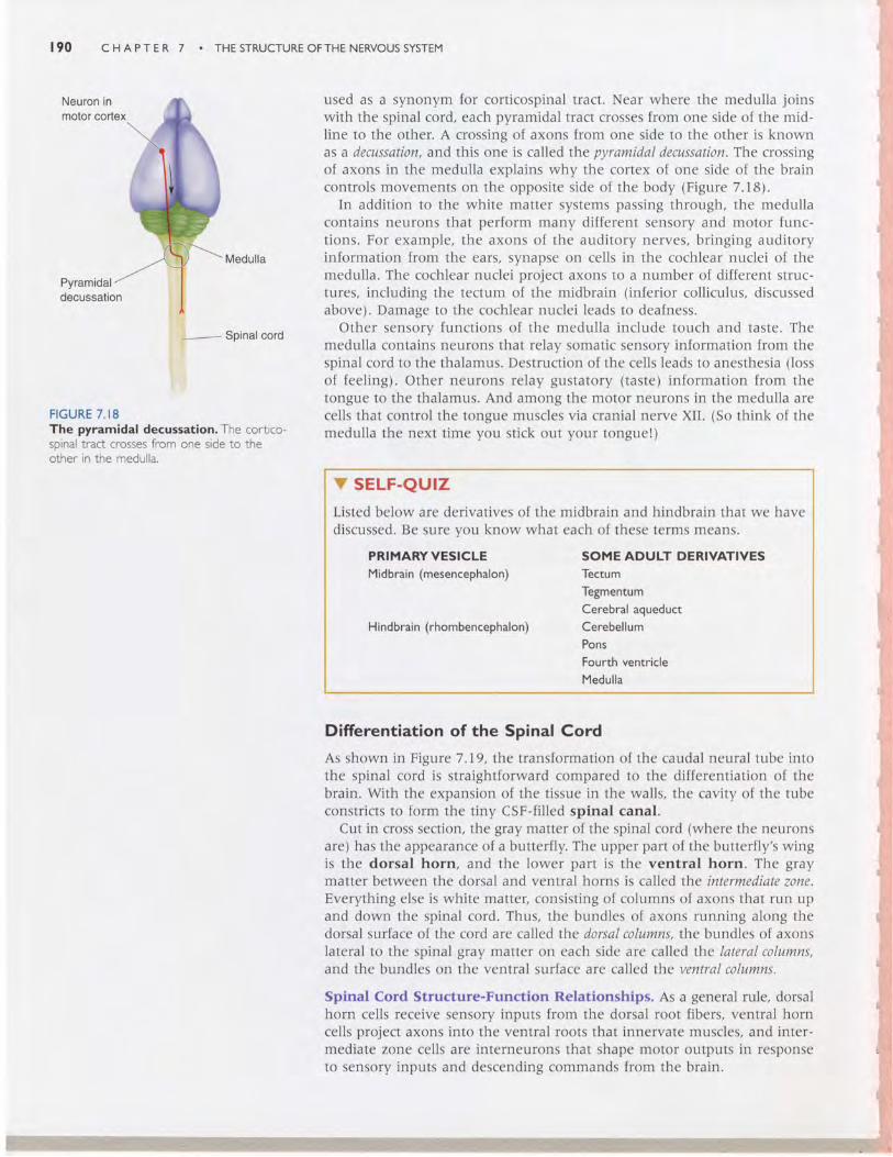
used as a synonym for corticospinal tract. Near where the medulla joinswith the spinal cord, each pyramidal tract crosses from one side of the mid-line to the other. A crossing of axons from one side to the other is knownas a decussation, and this one is called the pyramidal decussation The crossingof axons in the medulla explains why the cortex of one side of the braincontrols movements on the opposite side of the body (Figure 7.18).
In addition to the white matter systems passing through, the medullacontains neurons that perform many different sensory and motor func-tions. For example, the axons of the auditory nerves, bringing auditoryinformation from the ears, synapse on cells in the cochlear nuclei of themedulla. The cochlear nuclei project axons to a number of different struc-tures, including the tectum of the midbrain (inferior colliculus, discussedabove). Damage to the cochlear nuclei leads to deafness.
Other sensory functions of the medulla include touch and taste. Themedulla contains neurons that relay somatic sensory information from thespinal cord to the thalamus. Destruction of the cells leads to anesthesia (lossof feeling). Other neurons relay gustatory (taste) information from thetongue to the thalamus. And among the motor neurons in the medulla arecells that control the tongue muscles via cranial nerve XII. (So think of themedulla the next time you stick out your tongue!)
v SELF-QU|ZListed below are derivatives of the midbrain and hindbrain that we havediscussed. Be sure you know what each of these terms means.
PRIMARYVESICLE SOMEADULT DERIVATIVESMidbrain (mesencephalon) Tectum
TegmentumCerebral aqueduct
Hindbrain (rhombencephalon) CerebellumPonsFounh ventricleMedulla
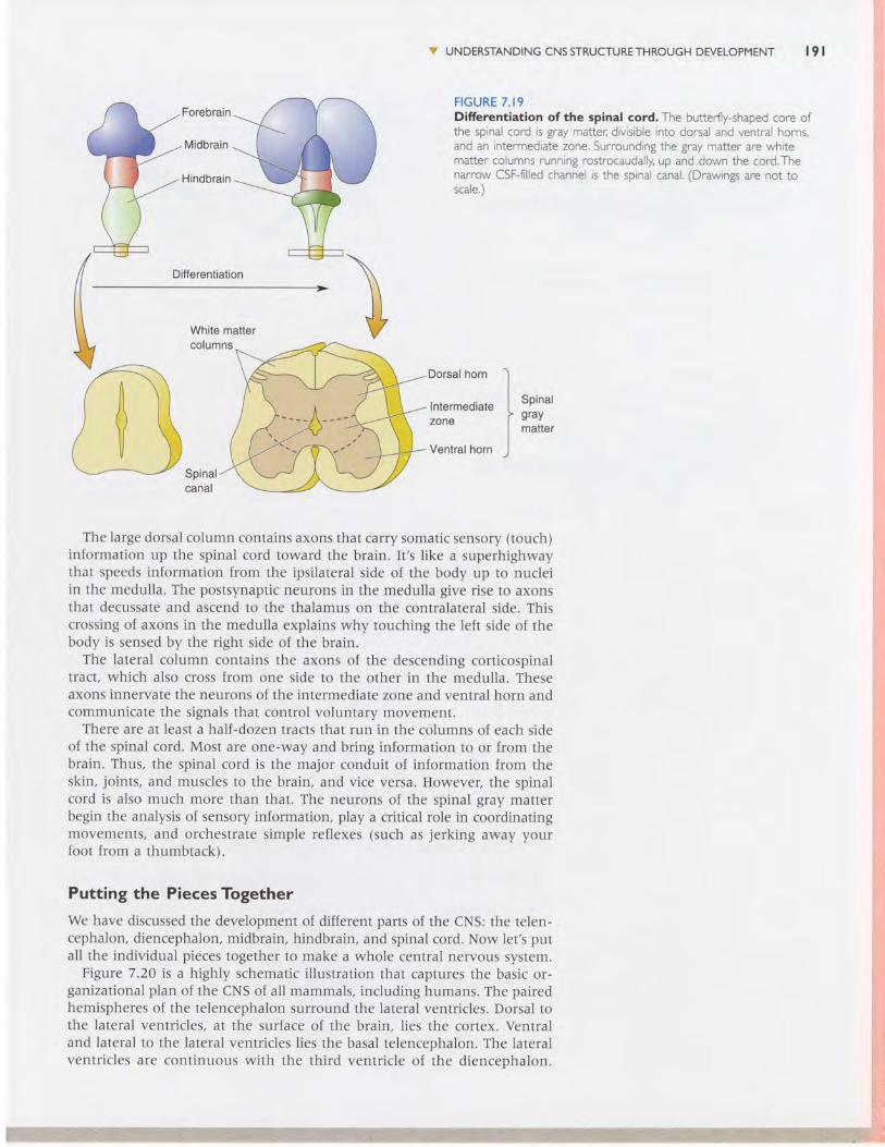
Difrerentiation of the Spinal Cord
As shown in Figure 7.19, the transformation of the caudal neural tube intothe spinal cord is straightforward compared to the differentiation of thebrain. With the expansion of the tissue in the walls, the cavity of the tubeconstricts to form the tiny CSF-filled spinal canal.
Cut in cross section, the gray matter of the spinal cord (where the neuronsare) has the appearance of a butterfly. The upper part of the butterfly's wingis the dorsal horn, and the lower part is the ventral horn. The graymatter between the dorsal and ventral horns is called the intermediate zone.Everything else is white matter, consisting of columns of axons that run upand down the spinal cord. Thus, the bundles of axons running along thedorsal surface of the cord are called the dorsal columns, the bundles of axonslateral to the spinal gray matter on each side are called the lateral columns,and the bundles on the ventral surface are called t}:.e ventral columns.
Spinal Cord Structure-Function Relationships. As a general rule, dorsalhorn cells receive sensory inputs from the dorsal root fibers, ventral horncells project axons into the ventral roots that innervate muscles, and inter-mediate zone cells are interneurons that shape motor outputs in responseto sensory inputs and descending commands from the brain.
Forebrain
Midbrain
Hindbrain
Differentiation
Dorsal horn
lntermediatezone
Ventral horn
Spinalgraymatter
The large dorsal column contains axons that carry somatic sensory (touch)information up the spinal cord toward the brain. It 's l ike a superhighwaythat speeds information from the ipsilateral side of the body up to nucleiin the medulla. The postsynaptic neurons in the medulla give rise to axonsthat decussate and ascend to the thalamus on the contralateral side. Thiscrossing of axons in the medulla explains why touching the left side of thebody is sensed by the right side of the brain.
The lateral column contains the axons of the descending corticospinaltract, which also cross {rom one side to the other in the medulla. Theseaxons innervate the neurons of the intermediate zone and ventral horn andcommunicate the signals that control voluntary movement.
There are at least a half-dozen tracts that run in the columns of each sideof the spinal cord. Most are one-way and bring information to or from thebrain. Thus, the spinal cord is the major conduit of information from theskin, joints, and muscles to the brain, and vice versa. However, the spinalcord is also much more than that. The neurons of the spinal gray matterbegin the analysis of sensory information, play a critical role in coordinatingmovements, and orchestrate simple reflexes (such as jerking away yourfoot from a thumbtack).
Putting the Pieces Together
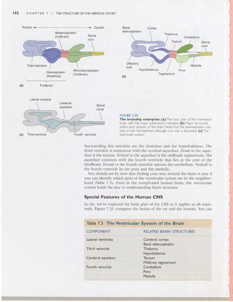
We have discussed the development of different parts of the CNS: the telen-cephalon, diencephalon, midbrain, hindbrain, and spinal cord. Now let's putall the individual pieces together to make a whole central nervous system.
Figure 7.20 is a highly schematic i l lustration that captures the basic or-ganizational plan of the CNS of all mammals. including humans. The pairedhemispheres of the telencephalon surround the lateral ventricles. Dorsal tothe lateral ventricles, at the surface of the brain, l ies the cortex. Ventraland lateral to the lateral ventricles lies the basal telencephalon. The lateralventricles are continuous with the third ventricle of the diencenhalon.
ry UNDERSTANDTNG cNs srRUcruRETHRoucH DEVELopMENT l9 |
F I G U R E 7 . I 9Difrerentiation of the spinal cord. The butter{ly-shaped core ofthe spinal cord is gray matter: drvisible nto dorsal and ventral horns,and an intermediate zone. Surrounding the gray matter are whitematter columns running rostrocaudally up and down the cord,Thenarrow CSF-f l l led channel is the spinal canal, (Drawings are not toscare.)
il:::;.::tr
r - - _ J L _ - - -
\ . / v t
' ^ 1 ' J 1 " ; ; : "
. 9 2 c H A P T E R 7 THE STRUCTURE OFTHE NERVOUS SYSTEM
Rostral Caudal
Mesencephalon(midbrain)
Basaltelencephalon
ThalamusCerebellum
Tectum
I
HypothalamusTegmentum
FIGURE 7.20The brainship enterprise. (a)The basic plan of the mammalianbrain, with the major subdivisions indicated. (b) Major structureswithin each division of the brain, Note that the telencephalon con-sists of two hemispheres, although only one is illustrated. (c)Theventricular svstem.
(b)
+Forebrain(a)
(c) Third ventricle Fourth ventricle
Surrounding this ventricle are the thalamus and the hypothalamus. Thethird ventricle is coninuous with the cerebral aqueduct. Dosal to the aque-duct is the tectum. Ventral to the aqueduct is the midbrain tegmentum. Theaqueduct connects with the fourth ventricle that lies at the core of thehindbrain. Dorsal to the fourth ventricle sprouts the cerebellum. Ventral tothe fourth ventricle lie the pons and the medulla.
You should see by now that finding your way around the brain is easy ifyou can identify which parts of the ventricular system are in the neighbor-hood (Table 7.3). Even in the complicated human brain, the ventricularsystem holds the key to understanding brain structure.
Special Features of the Human CNS
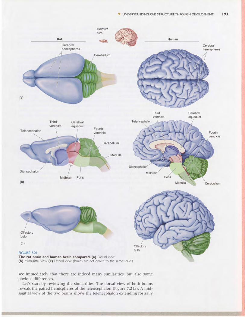
So far, we've explored the basic plan of the CNS as it applies to all mam-mals. Figure 7.21 compares the brains of the rat and the human. You can
COMPONENT RELATED BMIN STRUCTURES
Latcral Yentricles
Third ventricle
Cerobral aqueduct
Fourth ventricle
Cerebral cortexBasal telencephalonThalamusHypothalamusTectumMidbraln tegmentumCerebellumPonsMedulla
Cerebralaqueduct
V UNDERSTANDING CNS STRUCTURE THROUGH DEVELOPMENT r93
l.i
Telencephalon
Diencephalon
(b)
Cerebellum
Midbrain
l : . 1III
Pons
(c)
FIGURE 7.2IThe rat brain and human brain compared. (a) Dorsal view.(b) Midsagittal view. (c) Lateral view. (Brains are not drawn to the same scale.)
see immediately that there are indeed many similarities, but also someobvious differences.
Let's start by reviewing the similarities. The dorsal view of both brainsreveals the paired hemispheres of the telencephalon (Figure 7 .2Ia\. A mid-sagittal view ol the two brains shows the telencephalon extending rostrally
194 CHAPTER 7 . THESTRUCTUREOFTHE NERVOUSSYSTEM
from the diencephalon (Figure 7.2lbl. The diencephalon surrounds thethird ventricle, the midbrain surrounds the cerebral aqueduct, and thecerebellum, pons, and medulla surround the fourth ventricle. Notice howthe pons swells below the cerebellum, and how structurally elaborate thecerebellum is.
Now let's consider some of the structural differences between the rat andhuman brains. Figure 7.2Ia reveals a striking one: the many convolutionson the surface of the human cerebrum. The grooves in the surface of thecerebrum are called sulci (singular: sulcus), and the bumps are calledgyri (singular: gyrus). Remember, the thin sheet of neurons that lies justunder the surface of the cerebrum is the cerebral cortex. Sulci and gyriresult from the tremendous expansion of the surface area of the cerebralcortex during human fetal development. The adult human cortex, measur-ing about I100 cm2, must fold and wrinkle to fit within the confines of theskull. This increase in cortical surface area is one of the "distortions" of thehuman brain. Clinical and experimental evidence indicates that the cortexis the seat of uniquely human reasoning and cognition. Without cerebralcortex, a person would be blind, deaf, mute, and unable to initiate volun-tary movement. We will take a closer look at the structure of the cerebralcortex in a moment.
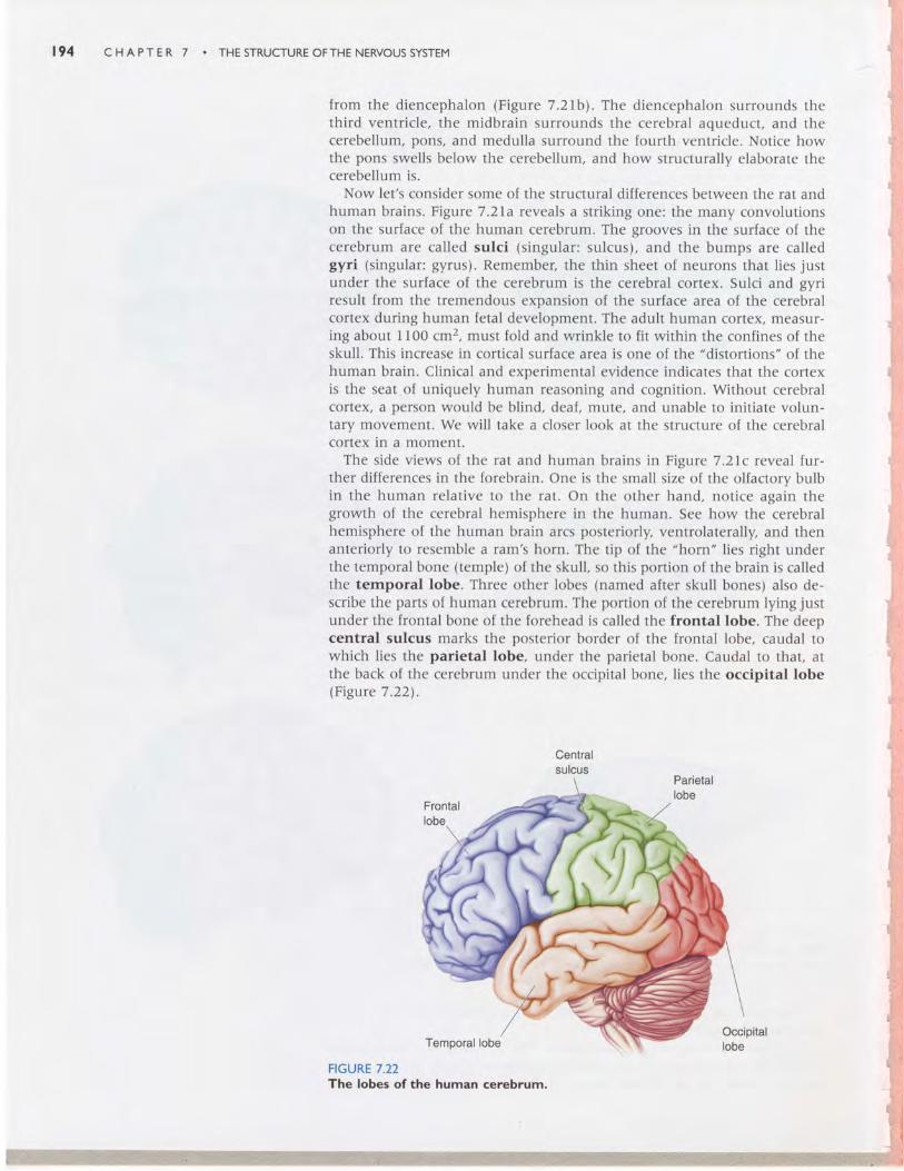
The side views of the rat and human brains in Figure 7.2 lc reveal fur-ther differences in the forebrain. One is the small size of the olfactory bulbin the human relative to the rat. On the other hand, notice again thegrowth of the cerebral hemisphere in the human. See how the cerebralhemisphere of the human brain arcs posteriorly, ventrolaterally, and thenanteriorly to resemble a ram's horn. The tip of the "horn" Iies right underthe temporal bone (temple) of the skull, so this portion of the brain is calledthe temporal lobe. Three other lobes (named after skull bones) also de-scribe the parts of human cerebrum. The portion of the cerebrum lying justunder the frontal bone of the forehead is called the frontal lobe. The deepcentral sulcus marks the posterior border of the frontal lobe, caudal towhich lies the parietal lobe, under the parietal bone. Caudal to that, atthe back of the cerebrum under the occipital bone, lies the occipital lobelFigure 7.22).
Centralsulcus
Parietallobe
Frontallobe
Temporal lobe
FIGURE 7.22The lobes of the human cerebrum.
Occipitallobe
V A GUIDETOTHE CEREBML CORTEX t 9 5
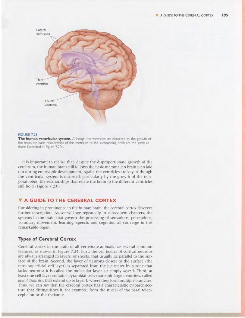
FIGURE 7.23The human ventricular system. Although the ventricles are distorted by the growth ofthe brain,the basic relationships of the ventricles to the surrounding brain are the same asthose illustrated in Figure 7.20c..
It is important to realize that, despite the disproportionate growth of thecerebrum, the human brain still follows the basic mammalian brain plan laidout during embryonic development. Again, the ventricles are key. Althoughthe ventricular system is distorted, particularly by the growth of the tem-poral lobes, the relationships that relate the brain to the different ventriclesstill hold (Figure 7.23).
V A GUIDE TO THE CEREBRAL CORTEXConsidering its prominence in the human brain, the cerebral cortex deservesfurther description. As we will see repeatedly in subsequent chapters, thesystems in the brain that govern the processing of sensations, perceptions,voluntary movement, learning, speech, and cognition all converge in thisremarkable organ.
Types of Cerebral Cortex
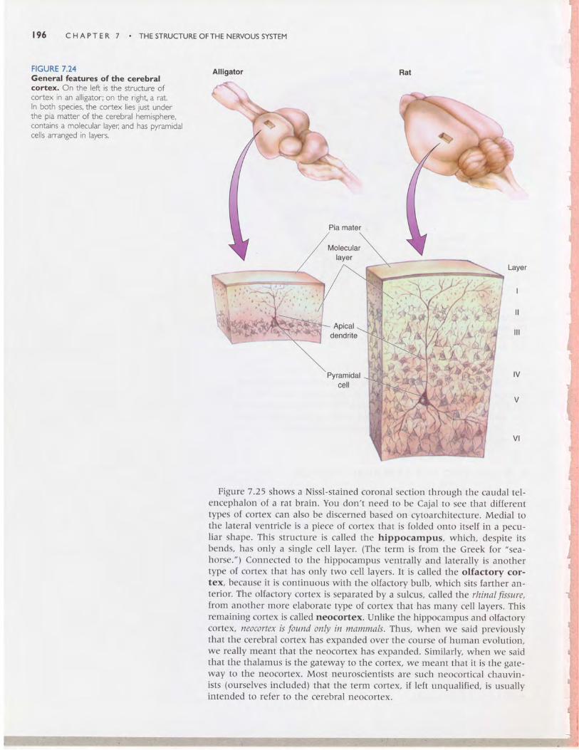
Cerebral cortex in the brain of all vertebrate animals has several commonfeatures, as shown in Figure 7.24. First, the cell bodies of cortical neuronsare always arranged in layers, or sheets, that usually lie parallel to the sur-face of the brain. Second, the layer of neurons closest to the surface (themost superficial cell layer) is separated from the pia mater by a zone thatlacks neurons; it is called the molecular layer, or simply layer I. Third, atleast one cell layer contains pyramidal cells that emit large dendrites, calledapical dendrttug that extend up to layer I, where they form multiple branches.Thus, we can say that the cerebral cortex has a characteristic cytoarchitec-ture that distinguishes it, for example, from the nuclei of the basal telen-cephalon or the thalamus.
196 cHAprER 7 . THE srRUcruREoFTHE NERVoussysrEM
FIGURE 7.24General features of the cerebrdcottex. On the left is the structure ofcortex in an alligator; on the right a rat.ln both species, the cortex lies just underthe pia matter of the cerebral hemisphere,contains a molecular layer: and has pyramidalcells arranged in layers.
Rat
Layer
Figure 7.25 shows a Nissl-stained coronal section through the caudal tel-encephalon of a rat brain. You don't need to be Cajal to see that differenttypes of cortex can also be discerned based on cytoarchitecture. Medial tothe lateral ventricle is a piece of cortex that is folded onto itself in a pecu-liar shape. This structure is called the hlppocampus, which, despite itsbends, has only a single cell layer. (The term is from the Greek for "sea-
horse.") Connected to the hippocampus ventrally and laterally is anothertype of cortex that has only two cell layers. It is called the olfactory cor-tex, because it is continuous with the olfactory bulb, which sits farther an-terior. The olfactory cortex is separated by a sulcus, called the rhinal ftssure,from another more elaborate type of cortex that has many cell layers. Thisremaining cortex is called neocortex. Unlike the hippocampus and olfactorycortex, neocortex is found only in mammals. Thus, when we said previouslythat the cerebral cortex has expanded over the course of human evolution,we really meant that the neocortex has expanded. Similarly, when we saidthat the thalamus is the gateway to the cortex, we meant that it is the gate-way to the neocortex. Most neuroscientists are such neocortical chauvin-ists (ourselves induded) that the term cortex, if left unqualified, is usuallyintended to refer to the cerebral neocortex.

A GUIDETOTHE CEREBML CORTEX 197
Rhinal fissure
Olfactorybulb
Lateralventricle
Rhinalfissure
Olfactorycortex
FIGURE 7.25Three types of cortex in a mammal. In this section of a rat brain, the lateral ventricleslie between the neocortex and the hippocampus on each side.The ventricles are not obvr-ous because they are very long and thin in this region. Below the telencephalon lies thebrain stem.What region of brain stem is this, based on the appearance of the fluid-fllledsDace at its core?
In Chapter 8, we will discuss the olfactory cortex in the context of thesense of smell. Further discussion of the hippocampus is reserved until laterin this book, when we will explore its role in the limbic system (ChapterI8) and in memory and learning (Chapters 24 and 25). The neocortex willfigure prominently in our discussions of vision, audition, somatic sensation,and the control of voluntary movement in Part II, so let's examine itsstructure in more detail.
Areas of Neocortex
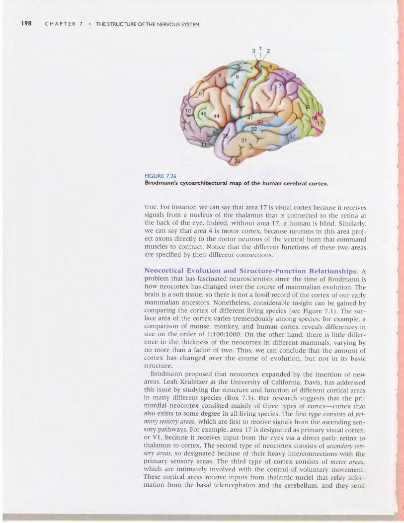
Just as cytoarchitecture can be used to distinguish the cerebral cortex fromthe basal telencephalon, and the neocortex from the olfactory cortex, it canbe used to divide the neocortex up into different zones. This is preciselywhat the famous German neuroanatomist Korbinian Brodmann did at thebeginning of the twentieth century. He constructed a cytoarchitecturalmap of the neocortex (Figure 7 .261 . ln this map, each area of cortex havinga common cytoarchitecture is given a number. Thus, we have "area 17" althe tip of the occipital lobe, "area 4" just anterior to the central sulcus inthe frontal lobe, and so on.
What Brodmann guessed, but could not show, was that cortical areas thatlook different perform different functions. We now have evidence that this is
Neocortex
t 9 8 CHAPTER 7 . THESTRUCTUREOFTHENERVOUSSYSTEM
FIGURE 7.26Brodmann's cytoarchitectural map of the human cerebral cortex.
true. For instance, we can say that arca 17 is visual cortex because it receivessignals from a nucleus of the thalamus that is connected to the retina atthe back of the eye. Indeed, without area t7, a human is blind. Similarly,we can say that area4 is motor cortex, because neurons in this area proj-ect axons directly to the motor neurons of the ventral horn that commandmuscles to contract. Notice that the different functions of these two areasare specified by their different connections.
Neocortical Evolution and Structure-Function Relationships. Aproblem that has fascinated neuroscientists since the time of Brodmann ishow neocortex has changed over the course of mammalian evolution. Thebrain is a soft tissue, so there is not a fossil record of the cortex of our earlymammalian ancestors. Nonetheless, considerable insight can be gained bycomparing the cortex of different living species (see Figure 7.1). The sur-face area of the cortex varies tremendously among species; for example, acomparison of mouse, monkey, and human cortex reveals differences insize on the order of I:100:1000. On the other hand, there is little differ-ence in the thickness of the neocortex in different mammals, varying byno more than a factor of two. Thus, we can conclude that the amount ofcortex has changed over the course of evolution, but not in its basicstructure.
Brodmann proposed that neocortex expanded by the insertion of newareas. Leah Krubitzer at the University of California, Davis, has addressedthis issue by studying the structure and function of different cortical areasin many different species (Box 7.5). Her research suggests that the pri-mordial neocortex consisted mainly of three types of cortex-cortex thatalso exists to some degree in all living species. The first type consists ol pri-mary sensory areas, w}:rich are first to receive signals from the ascending sen-sory pathways. For example, area 17 is designated as primary visual cortex,or Vl, because it receives input lrom the eyes via a direct path: retina tothalamus to cortex. The second type of neocortex consists of. secondary sen-sory areas, so designated because of their heavy interconnections with theprimary sensory areas. The third type of cortex consists of motor areas,which are intimately involved with the control of voluntary movement.These cortical areas receive inputs lrom thalamic nuclei that relay infor-mation from the basal telencephalon and the cerebellum, and they send
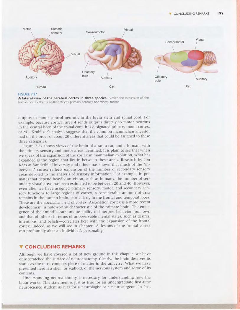
Somatic
Auditory
Human Cat
FIGURE 7.27A lateral view of the cerebral cortex in three species. Notice the expansion ofthehuman cortex that is neither strictly primary sensory nor strictly motot:
outputs to motor control neurons in the brain stem and spinal cord. Forexample, because cortical arca 4 sends outputs directly to motor neuronsin the ventral horn of the spinal cord, it is designated primary motor cortex,or Mt. Iftubitzer's analysis suggests that the common mammalian ancestorhad on the order of about 20 different areas that could be assigned to thesethree categories.
Figure 7.27 shows views of the brain of a rat, a cat, and a human, withthe primary sensory and motor areas identified. It is plain to see that whenwe speak of the expansion of the cortex in mammalian evolution, what hasexpanded is the region that lies in between these areas. Research by JonKaas at Vanderbilt University and others has shown that much of the "in-
between" cortex reflects expansion of the number of secondary sensoryareas devoted to the analysis of sensory information. For example, in pri-mates that depend heavily on vision, such as humans, the number of sec-ondary visual areas has been estimated to be between 20 and 40. However,even after we have assigned primary sensory, motor, and secondary sen-sory functions to large regions of cortex, a considerable amount of arearemains in the human brain, particularly in the frontal and temporal lobes.These are the association areas of cortex. Association cortex is a more recentdevelopment, a noteworthy characteristic of the primate brain. The emer-gence of the "mind"-our unique ability to interpret behavior (our ownand that of others) in terms of unobservable mental states, such as desires,intentions, and beliefs-correlates best with the expansion of the frontalcortex. Indeed, as we will see in Chapter 18, lesions of the frontal cortexcan profoundly alter an individual's personality.
V CONCLUDING REMARKS
Although we have covered a lot of new ground in this chapter, we haveonly scratched the surface of neuroanatomy. Clearly, the brain deserves itsstatus as the most complex piece of matter in the universe. What we havepresented here is a shell, or scaffold, of the nervous system and some of itscontents.
Understanding neuroanatomy is necessary for understanding how thebrain works. This statement is iust as true for an undergraduate first-timeneuroscience student as it is for a neurologist or a neurosurgeon. In fact,
Oltactorybulb
CONCLUDING REMARKS 199
Auditory
Rat
Visual
Visual
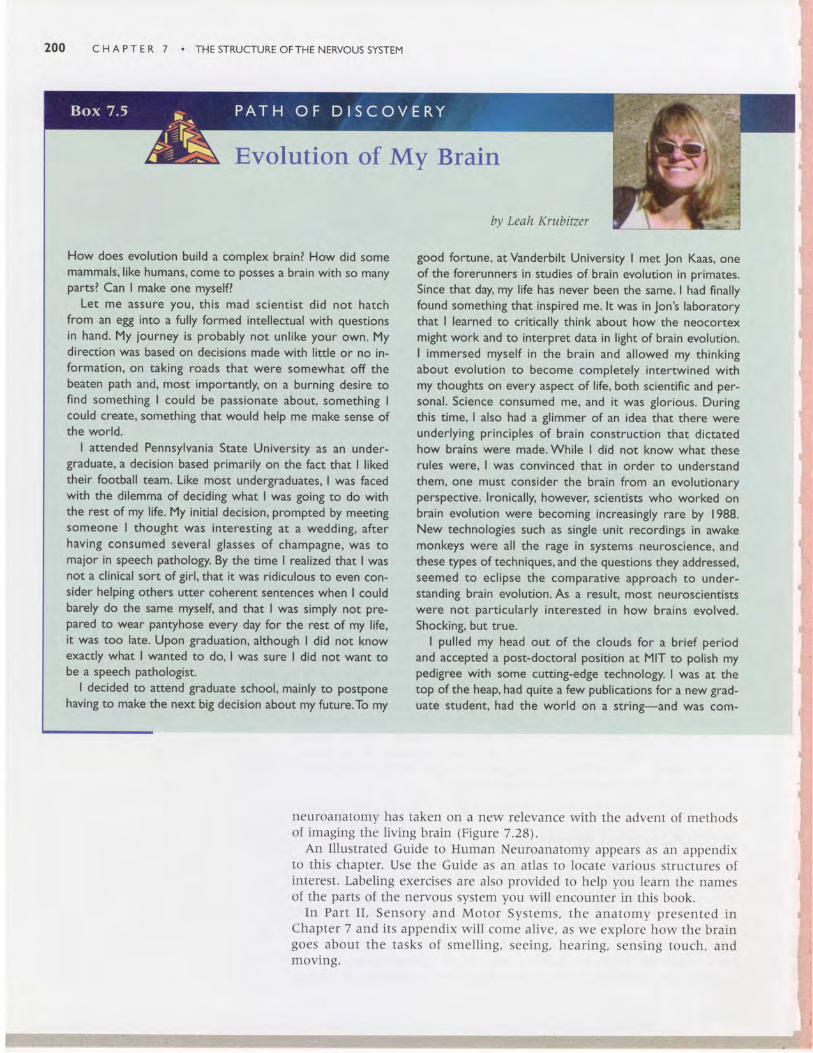
200 C H A P T E R 7 THE STRUCTURE OFTHE NERVOUS SYSTEM
Evolution of My Brain
How does evolution build a complex brain? How did somemammals, like humans, come to posses a brain with so manyparts? Can I make one myselfl
Let me assure you, th is mad sc ient is t d id not hatchfrom an egg into a fully formed intellectual with questionsin hand. My journey is probably not unlike your own. Mydirection was based on decisions made with little or no in-formation, on taking roads that were somewhat off thebeaten path and, most importantly, on a burning desire tofind something I could be passionate about, something Icould create, something that would help me make sense ofthe world.
I attended Pennsylvania State University as an under-graduate, a decision based primarily on the fact that I likedtheir football team. Like most undergraduates, I was facedwith the dilemma of deciding what I was going to do withthe rest of my life. My initial decision, prompted by meetingsomeone I thought was interesting at a wedding, afterhaving consumed several glasses of champagne, was tomajor in speech pathology. By the time I realized that I wasnot a clinical sort of girl, that it was ridiculous to even con-sider helping others utter coherent sentences when I couldbarely do the same myself, and that I was simply nor pre-pared to wear pantyhose every day for the rest of my life,it was too late. Upon graduation, although I did not knowexactly what I wanted to do, I was sure I did not want tobe a speech pathologist.
I decided to attend graduate school, mainly to postponehaving to make the next big decision about my future.To my
by Leah l(rubitzer
good fortune, at Vanderbilt University I met Jon Kaas, oneof the forerunners in studies of brain evolution in primates.Since that day, my life has never been the same. I had finallyfound something that inspired me. lt was in Jon's laboratorythat I learned to critically think about how the neocortexmight work and to interpret data in light of brain evolution.I immersed myself in the brain and allowed my thinkingabout evolution to become completely intertwined withmy thoughts on every aspect of life, both scientific and per-sonal. Science consumed me, and it was glorious. Duringthis time, I also had a glimmer of an idea that there wereunderlying principles of brain construction that dictatedhow brains were made. While I did not know what theserules were, I was convinced that in order to understandthem, one must consider the brain from an evolutionaryperspective. lronically, however, scientists who worked onbrain evolution were becoming increasingly rare by 1988.New technologies such as single unit recordings in awakemonkeys were all the rage in systems neuroscience, andthese types of techniques, and the questions they addressed,seemed to eclipse the comparative approach to under-standing brain evolution. As a result, most neuroscientistswere not particularly interested in how brains evolved.Shocking, but true.
I pulled my head out of the clouds for a brief periodand accepted a post-doctoral position at MIT to polish mypedigree with some cutting-edge technology. I was at thetop of the heap, had quite a few publications for a new grad-uate student, had the world on a string-and was com-
neuroanatomy has taken on a new re levance wi th the advent ( ) f methoclsot imaging the l iv ing bra in (F igure 7.28\ .
An I l lust rated Guide to Human Neuroanatomy appears as an appendixto th is chapter . Use the Guide as an at las to locate var ious st ructures oIinterest. Labeling exercises are also provided to help you learn the namesof the parts of the nervous system you wil l encounter in this book.
In Pa r t I I , Senso ry and Mo to r Sys tems , t he ana tomy p resen ted i nChapter 7 and i ts appendix wi l l come al ive, as we explore how the bra ingoes abou t t he t asks o f sme l l i ng , see ing , hea r i ng , sens ing touch , andmoving.
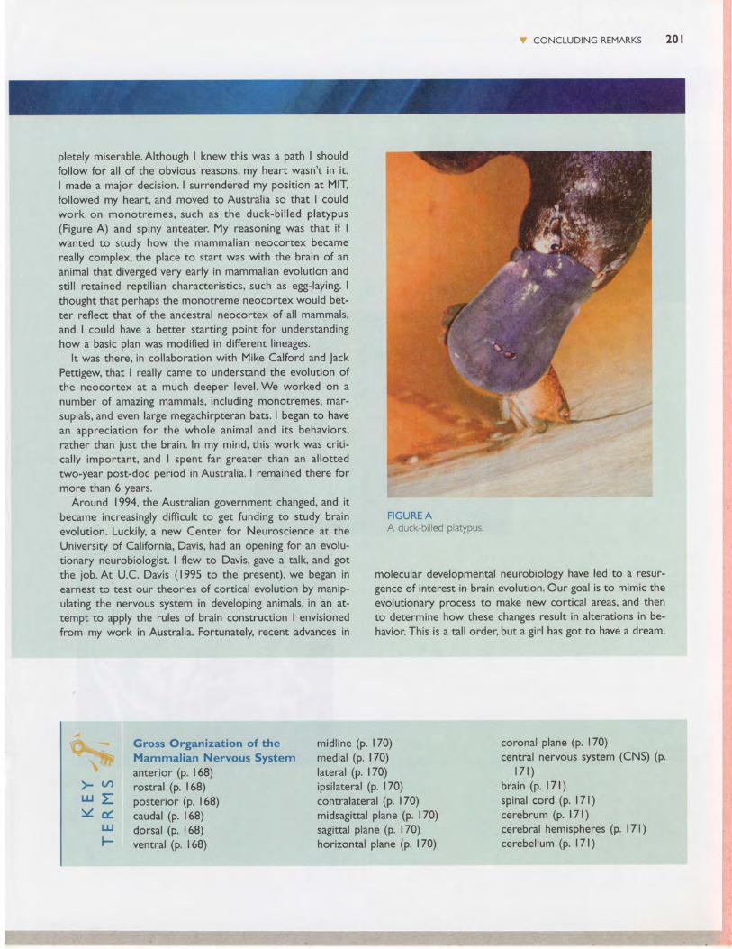
CONCLUDING REMARKS 2 0 1
pletely miserable. Although I knew this was a path I shouldfollow for all of the obvious reasons, my heart wasn't in it.I made a major decision. I surrendered my position at MIIfollowed my heart, and moved to Australia so that I couldwork on monotremes, such as the duck-b i l led p latypus(Figure A) and spiny anteater. My reasoning was that if Iwanted to study how the mammalian neocortex becamereally complex, the place to start was with the brain of ananimal that diverged very early in mammalian evolution andsti l l retained repti l ian characteristics, such as egg-laying. Ithought that perhaps the monotreme neocortex would bet-ter reflect that of the ancestral neocortex of all mammals,and I could have a better starting point for understandinghow a basic plan was modified in different lineages.
It was there, in collaboration with Mike Calford and JackPettigew, that I really came to understand the evolution ofthe neocortex at a much deeper level. We worked on anumber of amazing mammals, including monotremes, mar-supials, and even large megachirpteran bats. I began to havean appreciation for the whole animal and its behaviors,rather than just the brain. ln my mind, this work was crit i-cally important, and I spent far greater than an allottedtwo-year post-doc period in Australia. I remained there formore than 6 years.
Around 1994, the Australian government changed, and itbecame increasingly difficult to get funding to study brainevolution. Luckily, a new Center for Neuroscience at theUniversity of California, Davis, had an opening for an evolu-tionary neurobiologist. | flew to Davis, gave a talk, and gotthe job.At U.C. Davis (1995 to the present), we began inearnest to test our theories of cortical evolution by manip-ulating the nervous system in developing animals, in an at-tempt to apply the rules of brain construction I envisionedfrom my work in Australia. Fortunately, recent advances in
Gross Organization of theMammalian Nervous Systemanterior (p. 168)rostral (p. 168)posterior (p. 168)caudal (p. 168)dorsal (p. 168)ventral (p. | 68)
FIGURE AA duck-bil led platypus.
molecular developmental neurobiology have led to a resur-gence of interest in brain evolution. Our goal is to mimic theevolutionary process to make new cortical areas, and thento determine how these changes result in alterations in be-havior. This is a tall order, but a girl has got to have a dream.
> ( tU J EY d .
trJF
midl ine (p. 170)medial (p. 170)lateral (p. | 70)ipsilateral (p. 170)contralateral (p. | 70)midsagittal plane (p. 170)sagittal plane (p. 170)horizontal plane (p. 170)
coronal plane (p. | 70)central nervous system (CNS) (p.
l 7 l )bra in (p. | 7 l )sp inal cord (p. l7 l )cerebrum (p. l7 l )cerebral hemispheres (p. l7l)cerebel lum (p. l7 l )
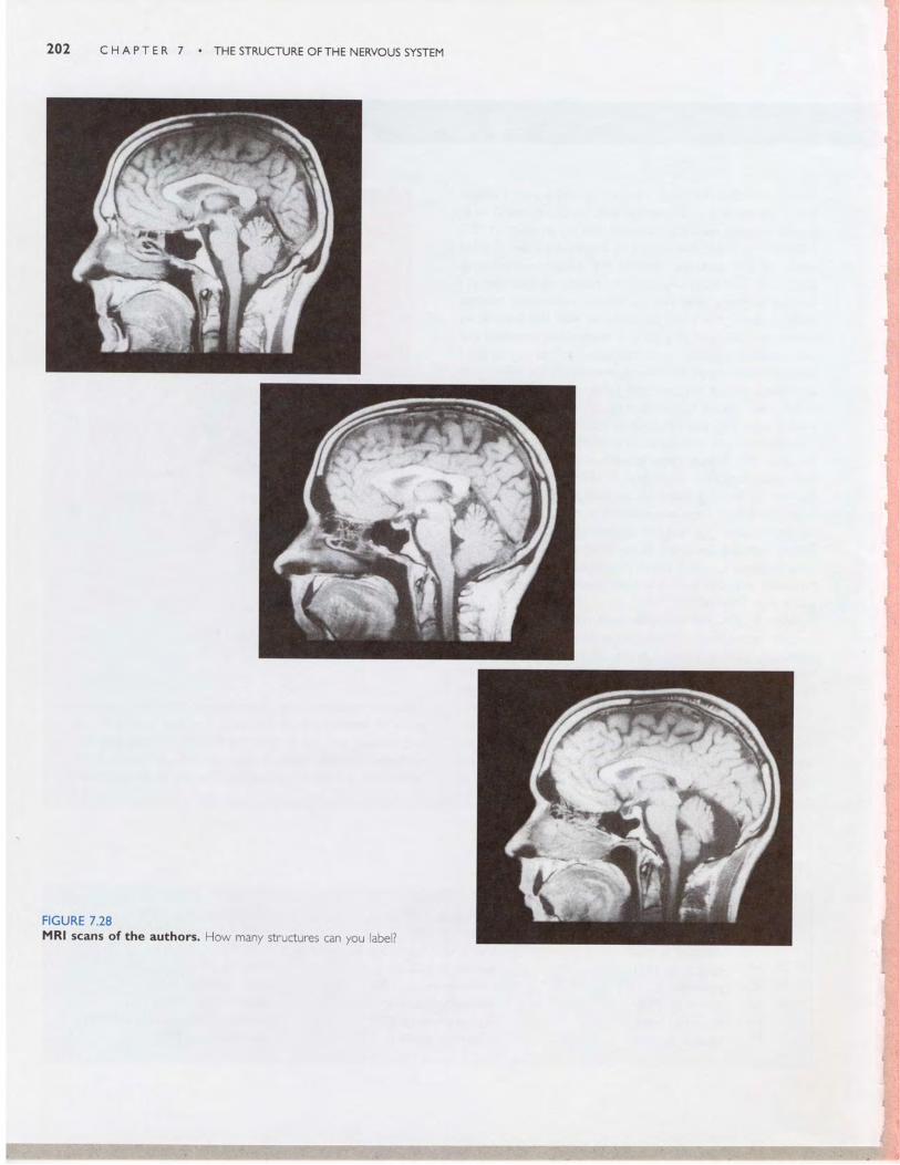
C H A P T E R 7 . T H E S T R U C T U R E O F T H E N E R V O U S S Y S T E M
FIGURE 7.28MRI scans of the authors. How many structures can you abe ?
S CONCLUDING REMARKS 203
brain stem (p. l7l)spinal nerve (p.172)dorsaf root (p.172)ventral root (p. 172)peripheral nervous system (PNS)
(p. 172)somatic PNS (p. 172)dorsal root ganglion (p. 173)visceral PNS (p. 173)autonomic nervous system (ANS)
(p. 173)afferent (p. | 73)efferent (p. | 73)cranial nerve (p. | 73)meninges (p. 173)dura mater (p. 173)arachnoid membrane (p. 173)pia mater (p. l7a)cerebrospinal fluid (CSF) (p.ventricular system (p. l7a)
Understanding CNS StructureThrough Developmentgray matter (p. 180)cortex (p. 180)nucleus (p. 180)substantia (p. 180)
locus (p. 180)ganglion (p. 180)nerve (p. 180)white mafter (p. 180)tract (p. 180)bundle (p. 180)capsule (p. 180)commissure (p. 180)lemniscus (p. 180)neural tube (p. l8 l)neural crest (p. 18l)neurulat ion (p. l8 l)differentiation (p. 182)forebrain (p. 183)midbrain (p. 183)hindbrain (p. 183)diencephalon (p. l8a)telencephalon (p. 184)
174\ olfactory bulb (p. 184)
corpus callosum (p. l8a)internal capsule (p. l8a)tectum (p. 187)tegmentum (p. 187)cerebral aqueduct (p. 187)pons (p. 188)medulla oblongata (medulla)
(p. 188)fourth ventricle (p. 188)spinal canal (p. 190)dorsal horn (p. 190)ventral horn (p. 190)sulcus (p. 194)gyrus (p. 194)temporal lobe (p. 194)frontal lobe (p. 194)central sulcus (p. 194)parietal lobe (p. 194)occipital lobe (p. 194)
A Guide to the CerebralCortexhippocampus (p. 196)olfactory cortex (p. 196)neocortex (p. 196)cytoarchitectural map (p. 197)
lateral ventricle (p. 184)third ventricle (p. 184)cerebral cortex (p. l8a)basal telencephalon (p. l8a)thalamus (p. l8a)hypothalamus (p. l8a)cortical white matter (p. l8a)
> (,/)'-&J z- oul l-& Q . t
IJJ
fO
l . Are the dorsal root ganglia in the central or peripheral nervous system?
2. ls the myelin sheath of optic nerve axons provided by Schwann cells or oligodendroglia? Why?
3. lmagine that you are a neurosurgeon, about to remove a tumor lodged deep inside the brain.The top of the
skull has been removed.What now lies between you and the brainlWhich layer(s) must be cut before you
reach the CSF?
4. What is the fate of tissue derived from the embryonic neural tube? Neural crest?
5. Name the three main parts of the hindbrain.Which of these are also part of the brain steml
6. Where is CSF produced?What path does it take before it is absorbed into the bloodstreaml Name the parts
of the CNS it will pass through in its voyage from brain to blood.
7. What are three features that characterize the structure of cerebral cortex?

CHAPTER 7 . THESTRUCTUREOFTHE NERVOUSSYSTEM