the spectrum of paediatric abdominal masses: correlation
TRANSCRIPT

THE SPECTRUM OF PAEDIATRIC ABDOMINAL MASSES: CORRELATION
OF IMAGING FINDINGS AND HISTOLOGY.
Principal investigator: Alice Adhiambo Onyango
MBchB
H58/80773/15
A DISSERTATION SUBMITTED IN PARTIAL FULFILLMENT FOR THE
REQUIREMENTS OF MASTER OF MEDICINE IN DIAGNOSTIC IMAGING
AND RADIATION MEDICINE DEGREE
COLLEGE OF HEALTH SCIENCES
UNIVERSITY OF NAIROBI
2019

ii
DECLARATION
I, Dr. Alice Adhiambo Onyango declare that this is my original work and has not been
submitted before any other university for a similar or any other degree award
Signature: ……………………… Date: …………………………….
SUPERVISORS’ APPROVAL
This research proposal is submitted for examination with our approval as University of
Nairobi supervisors.
ANGELINE AYWAK
MBChB (Nbi), M.Med (Nbi), Fellowship in Ultrasound (Thomas Jefferson University-
USA).Consultant Radiologist and Senior Lecturer, Department of Diagnostic Imaging and
Radiation Medicine, School of Medicine, College of Health Sciences, University of
Nairobi.
Signature………………………… Date………………………
WAIRIMU WAWERU
MBChB (Nbi), M.Med (Nbi),
Consultant Pathologist and Senior Lecturer, Department of Human Pathology, School of
Medicine, College of Health Sciences, University of Nairobi.
Signature………………………………… Date………………………….

iii
DEDICATION
This work is dedicated to my wonderful children Adrien and Arielle, they give me purpose.
iv
ACKNOWLEDGEMENTS
My gratitude goes to the Almighty God for giving me life, strength and ability to complete
this work.
Special gratitude goes to my chief supervisor Dr. A. Aywak for her outstanding dedication
and guidance throughout this research project. I also thank my second supervisor Dr.
Waweru from the department of pathology for her continued support.
I would also like to thank the parents and guardians of the children who participated in the
study, without whom I would not be able to realize my dream of completing this study.
I also thank my statistician, Ms. Betty Amboko for the timely analysis of the data.
Finally, I wish to thank my husband Dr. Alphonce for the unrelenting support and patience
during this study duration.

v
LIST OF ABBREVIATIONS AND ACRONYMS
CECT- Contrast Enhanced Computed Tomography
CT - Computed Tomography
FNAC - Fine Needle Aspiration Cytology
GB - Gall bladder
GIT - Gastrointestinal
HU - Hounsfield Units
KNH - Kenyatta National Hospital
MHz - Mega Hertz
MRI - Magnetic Resonance Imaging
NECT - Non – Enhanced Computed Tomography
RCC - Renal Cell Carcinoma
SD - Standard deviation
TAS - Trans Abdominal Scan
UON- University Of Nairobi
US - Ultrasound

vi
TABLE OF CONTENTS
DECLARATION ................................................................................................................ ii
SUPERVISORS’ APPROVAL .......................................................................................... ii
DEDICATION ................................................................................................................... iii
ACKNOWLEDGEMENTS ............................................................................................... iv
LIST OF ABBREVIATIONS AND ACRONYMS ............................................................v
TABLE OF CONTENTS ................................................................................................... vi
LIST OF TABLES AND FIGURES.................................................................................. ix
ABSTRACT .........................................................................................................................x
CHAPTER 1 .......................................................................................................................1
1.1 INTRODUCTION AND BACKGROUND OF THE STUDY ..................................1
1.2 LITERATURE REVIEW ...........................................................................................3
1.2.1 Abdominal Masses ............................................................................................. 3
1.2.2 Imaging Modalities ............................................................................................. 6
CHAPTER 2: .....................................................................................................................9
METHODOLOGY ..............................................................................................................9
2.1 Problem Statement .....................................................................................................9
2.2 Study Justification ......................................................................................................9
2.3 Broad Objective........................................................................................................10
2.3.1 Specific Objectives ........................................................................................... 10
2.4 Ethical Clearance and Consideration .......................................................................10
2.4.1 Ethical clearance ............................................................................................... 10
2.4.2 Ethical consideration ........................................................................................ 11

vii
2.5 Study design .............................................................................................................11
2.6 Study area .................................................................................................................11
2.7 Study population ......................................................................................................11
2.8 Sample size ...............................................................................................................11
2.9 Inclusion Criteria ......................................................................................................12
2.10 Exclusion Criteria ...................................................................................................12
2.11 Study Variables ......................................................................................................13
2.11.1 Demographics ................................................................................................. 13
2.12 Recruitment and consenting procedure ..................................................................13
2.13 Material for data collection. ...................................................................................13
2.14 Quality assurance: ..................................................................................................14
2.15 Data Validity and Reliability..................................................................................14
2.16 Data Analysis and Presentation ..............................................................................14
2.17 Procedure for Data Collection ................................................................................14
2.17.1 Ultrasound procedure ..................................................................................... 15
2.17.2 CT scan procedure .......................................................................................... 15
2.17.3 MRI Procedure ............................................................................................... 16
2.18 Conceptual Framework ..........................................................................................17
CHAPTER 3 .....................................................................................................................18
3.1 RESULTS.................................................................................................................18
3.1.1 Participants’ characteristics. ............................................................................. 18
3.1.2 Sociodemographic characteristics .................................................................... 18
3.1.3 Signs and symptoms of presentation: ............................................................... 19

viii
3.1.4 Prevalence of abdominal masses in children presenting at the radiology
department with suspected masses. ........................................................................... 20
3.1.5 Distribution of the different abdominal masses by imaging ............................. 23
3.1.6 Imaging tests conducted ................................................................................... 24
CHAPTER 4 : ...................................................................................................................32
DISCUSSION ....................................................................................................................32
4.1 Introduction ..............................................................................................................32
4.2 Socio-demographic characteristics ...........................................................................32
4.3 Description of imaging findings ...............................................................................33
4.4 Correlation of imaging findings and Histology .......................................................34
4.5 Study limitations ......................................................................................................36
4.6 Recommendations ....................................................................................................37
REFERENCES .................................................................................................................38
STUDY TIME LINES .......................................................................................................45
BUDGET ...........................................................................................................................46
APPENDICES ...................................................................................................................47
Appendix 1: Data Collection Form. ...............................................................................47
Appendix II: Consent Form............................................................................................49
Appendix III: Fomu ya Idhini ........................................................................................51
Appendix IV: Ascent Form ............................................................................................53
Appendix V: Fomu ya Idhini ya Watoto Wakubwa .......................................................54
Appendix VI: Standard of Operating Procedure in Abdominal Scans ...........................55
Appendix VII: KNH Ethical Approval Letter ................................................................57

ix
LIST OF TABLES AND FIGURES
LIST OF TABLES
Table 1: Signs and symptoms of presentation ...................................................................20
Table 2: prevalence of abdominal masses in the sampled population ...............................21
Table 3: categories of masses ............................................................................................21
Table 4: spectrum of imaging findings ..............................................................................22
Table 5: Topographical distribution on imaging ...............................................................23
Table 6: Imaging test conducted ........................................................................................24
Table 7: Comparison of imaging diagnosis and the post biopsy histological diagnosis ...31
LIST OF FIGURES
Figure 1: Conceptual Framework .....................................................................................17
Figure 2: Sociodemographic characteristics .....................................................................19
Figure 3: Sample Cases: Figure 3.1-3.6 ...........................................................................25

x
ABSTRACT
Background
Pediatric abdominal masses are a common source of morbidity and mortality more-so in
Africa where resources are limited and therefore patients tend to present late with advanced
disease. The aim of this study was to establish the local imaging spectrum and histological
correlation of abdominal masses in children at Kenyatta national hospital.
Objective
To establish the local imaging spectrum and a histological correlation of abdominal masses
in children at Kenyatta National hospital.
Materials and methods
A prospective cross-sectional study was done at Kenyatta national hospital, radiology
department between September 2018 to March 2019. Out of the 140 children who were
referred for imaging with suspicion of an abdominal mass, 123 fulfilled the inclusion
criteria and were therefore enrolled in the study. Ultrasound was done by the primary
investigator under the supervision of a consultant radiologist. CT or MRI were performed
by experienced radiographers and reported by the principal investigator under the
supervision of an experienced radiologist. Standardized reporting protocol was used in all
the cases. Description of the organ involved, size, texture, vascularity, effect of the mass
on adjacent structures and imaging diagnosis was then recorded on a data sheet. Of the 123,
only 78 patients had a histological diagnosis which was also recorded. Data was analyzed
using SPSS version 23, Microsoft Access and Excel. It was then represented on tables and
graphs. Imaging diagnosis and histological diagnosis were compared.
Results
In this study, 123 patients age range of 4 days -17 years, mean of 5.12 (SD=3.6) and a male
to female ratio is ratio was 1:1.1 were analyzed. Imaging which included Ultrasound
(94%), CT (53.7%) and MRI (1.6%) was done at the radiology. Prevalence of abdominal

xi
masses in the sampled population was 87.86%. Out of these 63.4% (n= 78) had a
histological diagnosis and these were then correlated with the imaging diagnosis which
showed an agreement in 82%. On imaging, focal tumors accounted for 51.2% (n=63) while
organomegaly accounted for 48.8% (n=60) of all the masses. On imaging, most tumors
were renal in origin accounting for 31.7% of all the focal masses and majority of these
were seen between the age of 1 -5 years. Abdominal swelling was the most common
presenting symptom (95.9%) and Ultrasound was commonly used as the first imaging
modality in 93% of the cases. Wilms tumor (41.2%) and lymphoma (29.6%) were the
commonest histological diagnosis encountered.
Conclusion
The imaging findings in pediatric abdominal masses are varied and numerous. Most
children with abdominal masses present with an abdominal swelling. Findings from this
study showed that diffuse organomegaly was commonly due to lymphoproliferative
disorders and parasitic infections while Wilms tumor/Nephroblastoma was the most
common tumor followed by lymphoma. Ultrasound was the most commonly used initial
investigation. CT was mainly used for further characterization after ultrasound. Imaging
had an overall agreement with the histology in of 82% of the cases; with a higher agreement
in diagnosing certain tumors such as Wilms tumor and Ovarian teratoma.
Key words: Abdominal masses, abdominal tumors, pediatric abdominal masses,
ultrasound, MRI, CT scan

1
CHAPTER 1
1.1 INTRODUCTION AND BACKGROUND OF THE STUDY
Pediatric abdominal masses are numerous, varied and a unique source of morbidity and
mortality. Worldwide they account for one in a thousand cancer cases (1). In Africa they
are in fact a major source of illness and mortality mostly due to late presentation, limited
resources and lack of vigilance in caregivers (2). A recent study by Carter et al (2018)
reported a major disparity between the outcome of pediatric solid tumors in low- middle
income countries and developed countries. A review of recent cancer registries in Kenya,
Zimbabwe and Malawi revealed Wilms tumor to be the most common with a 5-year
survival rate of 33% compared to >90% 5 year survival rate in developed countries (3).
Pediatric abdominal masses can present with a variety of symptoms or may also be
discovered incidentally since some of these patients may be too young to even talk and
when they do, the site of pain may also be misleading (4).
Imaging children with abdominal masses can be a challenge because the history and
physical examination are less reliable compared to adult patients. It is crucial to choose the
best first imaging modality in order to reduce the number of imaging tests done where
possible, reduce exposure to ionizing radiation and minimize expenses. The age of the
patient is an important factor to consider in choosing the imaging test because it will
determine the expected pathology and dictate patient cooperation (5, 6).
Imaging has been shown to play a major role in the diagnosis of these masses; providing a
road map to surgical intervention, guiding chemotherapy and radiotherapy. Imaging is also
important in assessing response to treatment and surveillance for recurrence of tumors.
Unfortunately, the Cross-sectional imaging modalities are not accessible to all due to
economic barriers leading to late diagnosis or lack of access to health care resulting in high
mortality rates. Proper and early use of the best imaging tools has been shown to reduce

2
cost on unnecessary investigations and surgeries and provide timely management of these
masses and therefore reducing the mortality rate (4, 6, 8).
Ultrasound is commonly used as the first imaging modality since its readily available, non-
ionizing and is able to characterize lesions quite well. Cross sectional imaging such as MRI
and CT scan are commonly used for further characterization of masses (9).
This is a prospective cross-sectional study based in a resource poor setting at Kenyatta
National hospital in Kenya. It seeks to establish the pattern of occurrence, imaging findings
of pediatric abdominal masses and compare the radiological diagnosis with the histological
diagnosis. This study will provide baseline data locally on imaging findings of pediatric
abdominal masses which will be a source of scientific knowledge both locally and
internationally. It will also provide a diagnostic imaging algorithm that will be useful in
investigating children with suspected abdominal mass.
The magnitude of the disease and economic burden caused by pediatric abdominal masses
in sub Saharan Africa warrants the Nations investment in more funds and awareness
campaigns for caregivers to be vigilant. This will ensure early diagnosis of pediatric
abdominal masses, improved outcomes and a heathy future nation (9).

3
1.1 LITERA TURE R EVIEW
1.2 LITERATURE REVIEW
1.2.1 Abdominal Masses
There are several classifications of abdominal masses (10). The masses are categorized into
congenital and acquired masses. The ones considered congenital are those that present at
the time of birth or are detected within three months after birth. However, congenital
masses are generally rare and commonly comprise: teratomas, neuroblastomas and renal
masses (11). Majority of abdominal masses in children are acquired and include masses
of traumatic origin, inflammatory, organomegaly, obstructed viscus and neoplasms (5).
1.2.1.1 Pediatric Renal Masses
Renal tumors are quite common in the pediatric population. The incidence has been
reported to be 8 per 1,000,000 children (12). Renal masses account for more than half of
the reported cases of abdominal masses in infants. Renal masses in children range from
benign ones like cystic nephroma to the aggressive malignancies such as rhabdoid tumor
(13). The most common symptoms of renal masses in children include an abdominal mass,
hematuria and flank pain. They can also present with recurrent infections of the urinary
tract and a deterioration in renal function (24). In neonates, hydronephrosis, mesoblastic
nephroma and multicystic dysplastic kidney are reported to be the most common masses
seen (11 13). Masses seen in older children include Wilms tumor, polycystic kidney
disease, rhabdoid tumor and mesoblastic nephroma (13). It is reported that Wilm’s tumor
is the most encountered form of renal masses in older pediatric patients accounting for
more than three quarters of the cases with a peak of 2-5 years. It is reported to be the third
most common childhood cancer encountered south of Sahara (23)
A Kenyan study revealed that nephroblastoma makes up to 3.4% of childhood
malignancies (3). Renal cell carcinoma is reported as the most common renal tumor seen
in adolescents and older children. Imaging has a significant role in evaluating renal tumors
and metastasis when present (16, 17).

4
1.2.1.2 Pediatric Adrenal Masses
The masses involving the adrenal glands can occur due to various congenital or acquired
causes. Pediatric adrenal masses present with symptoms which are varied in nature (18).
However, some of these masses are discovered coincidentally during medical examinations
or imaging. Adrenal tumors are not common and neuroblastoma is reported to accounts for
90% of these tumors in young children (19).Most cases of neuroblastoma are seen before
the age of 5 years and typically present with a mass in the abdomen. The differential
diagnosis of an adrenal mass should among other things include congenital lesions,
cancerous growths, infections, trauma and lesions from the supra renal fossa (20, 21).
Adrenal adenoma is reported to be the most common lesion of the adrenal gland in both
adults and older children and it is seen in about 3% of autopsies. The congenital lesions
presenting in the adrenal area include cysts, congenital adrenal hyperplasia among others.
Infective processes are rare in the adrenal area however, it can occur and lead to an adrenal
abscess (22, 23). The pediatric adrenal glands are also reported to have higher propensity
to trauma induced hemorrhage. Among neonates, trauma can result from obstructed labor/
birth asphyxia among other causes (24, 25). In older children, adrenal hemorrhage occurs
mostly due to blunt trauma. The general appearance of blood in the adrenal gland is
reported to be related to the age of the blood (26, 27).
Cystic adrenal masses are not common. These include the pure cyst, cysts arising from
infections such as hydatid and cystic tumors (28).
Adrenocortical tumors are very rare accounting for 3-6 % of all carcinomas in children.
According to a study done in south Brazil, more than half of these carcinomas are seen in
children below the age of 5 years (29). In neonates, ultrasound has been advocated as the
imaging modality of choice for evaluating the adrenal masses. However, the most
important tool for evaluating the adrenal is cross sectional studies such as CT with a
specific protocol for the adrenal gland (30, 31).

5
1.2.1.3 Gastrointestinal Masses
The second most common abdominal masses in children under the age of five years are the
gastrointestinal tract masses. The GI tract masses account for up to a quarter of all the
abdominal masses in children (32). There are several differential diagnoses of the GI tract
masses. These may include gastrointestinal lymphoma, duplication cysts, omental or
mesenteric cysts among other cysts (33, 34). In infants and children GI masses are mostly
expressed in the form of duplication, fecal mass and Meckel’s diverticulum. (10).
1.2.1.4 Hepatobiliary Masses
Hepatobiliary masses in children account for just 5% of abdominal masses in infants (44).
Hepatic tumors are categorized into primary and secondary hepatic tumors. Most liver
masses tend to be malignant and are mostly metastatic (45). Benign liver tumors account
for only 30% of all liver masses. Most described benign liver tumors in pediatrics include,
infantile hepatic hemangioendothelioma, mesenchymal harmatoma, focal nodular
hyperplasia, nodular regenerative hyperplasia and hepatic adenoma (46, 47). Other lesions
affecting the liver include, hepatic cysts, hemangioma, angiomyolipoma, lipoma and
biliary tumors (biliary cystadenoma, bile duct hamartoma or adenoma and papillary
adenoma (18).
Hepatoblastoma is the most common primary malignant hepatic tumor in children.
Malignant liver tumors account for more than 1% of pediatric malignancies with
hepatoblastoma contributing about two thirds of all malignant liver tumors (48).
Harmatomas and hemangiomas are most prevalent from 0-1 year of age. Hepatoblastoma
is common between 1-10 years and hepatocellular carcinoma common between 11-17
years (42, 49).
1.2.1.5 Pelvic Masses
The third most common differential diagnosis of abdominal masses in infants are the Pelvic
masses that extend into the abdomen. These make up to 15% of abdominal masses in
children. The two most common forms of pelvic masses in children are ovarian cysts and

6
teratomas. Other pelvic abdominal masses include sacrococcygeal teratoma and
hematocolpos (49). In neonates pelvic masses include ovarian cysts, hydrometrocolpos and
gastrointestinal duplication (50).
1.2.1.6 Other Pediatric Abdominal Masses
These include intraperitoneal, omental and mesenteric tumors. Intraperitoneal tumours are
seen less frequently in children than in adults. The localized intraperitoneal tumours
include, mesenteric fibromatosis, Castlemans disease and inflammatory myofibroblastic
tumours (51).
Primary tumors of the peritoneum in pediatrics are mostly mesenchymal in origin. Diffuse
peritoneal disease can result from non-Hodgkin lymphoma, desmoplastic round cell tumor
or rhabdomyosarcoma. Lymphomas in the abdomen accounts for 5-6% of all malignancies
and they can either be nodal or extra-nodal (11, 42).
Peritoneal tumors can present with an abdominal mass, abdominal fullness or sign of
obstruction. Peritoneal tumors when encountered are mostly from metastatic disease (52).
The other group of masses are retroperitoneal masses which are not commonly seen in
pediatric patients. They tend to have common features with other abdominal masses (53).
1.2.2 Imaging Modalities
1.2.2.1 Radiography
Plain abdominal radiography is sometimes used in children with distended abdomens (6).
This is normally done to exclude possibility of the obstruction of the gastrointestinal tract.
It can also be used to locate the source and density of the abdominal mass and identify
calcifications if present (19, 54). Presence of intra-abdominal calcifications are
encountered in; neuroblastoma, teratomas or urolithiasis. It is also useful when ruling out
the possibility of skeletal metastasis. The draw backs of this modality is that it is
nonspecific, with a low sensitivity and its ionizing (55).

7
1.2.2.2 Sonography
It is the preferred initial method of investigating children with pathology in the abdomen
since it is non ionizing and readily available (56). Principally sonography produces images
by the reflection of ultra-high sound beams at tissue interfaces. The characteristics of the
reflected wave will then be used to provide information about a mass such as size, location
and its consistency (54, 57). It can also be useful in determining the effect of the mass on
the surrounding structures (58). In previous studies done by Annuar et al, ultrasound
correctly diagnosed up to 87% of abdominal masses in children. In situations where the
presence of abdominal mass is not definite, ultrasound can relay a high degree of certainty
on whether or not the mass is associated with the certain specific organs (59). Ultrasound
was reported to be reliable in diagnosing Wilm’s tumor and neuroblastoma with an
accuracy of more than 80%. However, the diagnosis of psoas abscess, and hemorrhage in
the adrenals was reported as non-specific (60). In another study ultrasound was reported
to be more than 80% accurate in differentiating a solid mass from a cystic one and more
than 60% accurate in identifying the organ of origin of a mass (61).
1.2.2.3 Computed Tomography
CT scan is important in providing details about masses in the abdomen. It uses X-rays to
obtain cross sectional images that can be re-constructed (62).
The non-invasive nature of the CT scan and its ability to acquire images rapidly makes it
an important tool for further investigation of masses in children (63). It is also quite
invaluable in determining the extent of the abdominal mass invasion and evaluation of
presence of metastasis within the abdomen (64). According to previous studies, CT scan
proved to have a higher sensitivity of more than 90% compared to a sensitivity of less than
50% by ultrasound. The main disadvantage of CT is exposure to ionizing radiation and
therefore a special pediatric protocol aimed to minimize the dose according to ALARA
principle should be applied (65).

8
1.2.2.4 Magnetic Resonance Imaging
MRI is employed when there is need for more characterization of a mass (66). In principle,
MRI uses focused radio waves and powerful magnetic fields to relay high resolution
images with excellent tissue contrast. It provides cross-sectional images and with the use
of the various sequences it is able to provide a lot of information about a mass. In addition,
it is often the more preferable method in older children because it is non- ionizing and it
has an excellent soft tissue contrast (66, 67). It is especially helpful in determining the
respectability of hepatic tumors by assessing vascular invasion and in treatment follow-up
of several tumors (68).

9
CHAPTER 2:
METHODOLOGY
2.1 Problem Statement
Pediatric abdominal masses are a common cause of morbidity, suffering and even death in
children. Worldwide they account for one in a thousand cases (1, 2). In Sub Saharan Africa
especially where most countries are middle to low income countries, they are a major cause
of mortality due to economic barriers to health care accessibility and lack of adequate
vigilance in caregivers. Imaging plays a major role in characterizing these masses, in
providing a road map to surgical intervention, guiding chemotherapy and radiotherapy.
Imaging is also invaluable in assessing response to treatment and in surveillance for
recurrence of tumors. Unfortunately, some of the Cross-sectional imaging modalities are
too expensive for the local Kenyans.
Proper and early use of the right imaging tools has been shown to reduce cost on
unnecessary investigations, surgeries and provide timely management of these masses.
This study seeks to establish the pattern of occurrence of pediatric abdominal masses and
provide a diagnostic imaging algorithm that will be useful in investigating children with
confirmed or suspected abdominal mass with the hope of reducing mortality from pediatric
abdominal masses. It will also be a source of reference in the National data base which
might be used for future planning.
2.2 Study Justification
Pediatric abdominal masses cause a significant disease and economic burden and are a
major source of mortality. In this setting which is resource limited, most patients tend to
present with advanced disease and others are lost to follow up resulting in high mortality
rates(5,17). Timely management of these masses is necessary in order to make an impact
on the outcome of these masses. Imaging plays an important role in the initial diagnosis of
these masses, surgical planning, post treatment follow up and in surveillance for tumor
recurrence(6). It is therefore important to be able to accurately describe the spectrum of

10
these abdominal masses on imaging and provide a radiological diagnosis as majority of
these masses are evaluated on imaging. The findings in this study will set out to promote
vigilance in clinicians and parents for early detection and management of pediatric
abdominal masses.
To the best of my knowledge, there is no local data describing the spectrum of abdominal
masses on imaging. This data obtained will be fundamental in establishing a national data
base on the pattern of pediatric masses on imaging,
Findings obtained from this study will be published and therefore contribute to the overall
scientific knowledge.
2.3 Broad Objective
To determine the prevalence, pattern and the spectrum pediatric abdominal masses on
imaging and compare with histological diagnosis.
2.3.1 Specific Objectives
1. To determine the prevalence and distribution of abdominal masses in pediatric patients
in Kenyatta National Hospital.
2. Describe the imaging findings of pediatric abdominal masses in pediatric patients in
Kenyatta National Hospital.
3. Correlate imaging diagnosis and the post biopsy histological diagnosis for those who
will need biopsy.
2.4 Ethical Clearance and Consideration
2.4.1 Ethical clearance
Ethical clearance was sought from Kenyatta National Hospital/ UON ethics committee
before the study was done. Permission was also obtained from Kenyatta National hospital
Radiology department for the study to be done. The study was conducted in accordance to
Helsinki declaration (1975) revised in 2008.

11
2.4.2 Ethical consideration
Informed consent was obtained from the parents/caregivers allow their children to be
included in the study, emphasizing voluntary participation.
Privacy and confidentiality was maintained at all times and the data collection forms were
assigned numbers and not patient names to conceal the identity of the participants.
Data collected was handled securely and stored in a hard disc only accessible to the
investigator.
This study did not in any way interfere or delay the patients’ intended management and the
patients were not required to pay an extra fee for the study. The patients were only
subjected to the investigation requested by the primary doctor. Unnecessary investigations
were not conducted in any way. Additional investigations were only performed if requested
by the primary doctor.
2.5 Study design
Prospective descriptive cross-sectional study
2.6 Study area
Kenyatta National Hospital radiology department
2.7 Study population
Patients between 0-17 years referred to Kenyatta National Hospital radiology department
for imaging with the suspicion of an abdominal mass.
2.8 Sample size
A convenience sampling of 140 was done. Out of these, 123 met the criteria and were
included in the study. 17 patients had either normal findings, abdominal distention due to
intestinal obstruction with fecal loading and massive intestinal infestation.

12
The desired sample size was calculated as follows using Cochran’s formula.
A previous study by Sharma et al in a similar setting found the prevalence of abdominal
masses to be 7.2% (23)
(Cochran 1963)
n0 - Sample size for target population >10,000;
Z - Standard variate (1.96) corresponding to 95% confidence interval;
e - The desired level of precision;
p - Estimated prevalence
Therefore, n= 1.962 ×0.072× (1-0.072)/0.05×0.05. 95 % confidence
The sample size for this study was 103 children aged between 0-17 years.
20% was added into the sample size to cater for attrition giving 123 patients
2.9 Inclusion Criteria
Children from age 0 to 17 years.
Children whose parents/guardians will give consent to participate in the study.
Those who will have a request form from the doctor indicating that they have a suspected
or confirmed abdominal mass and they are to be imaged with either ultrasound, CT Scan
or MRI.
2.10 Exclusion Criteria
Persons older than 17 years.
Children in whom no mass was found on imaging.

13
2.11 Study Variables
2.11.1 Demographics
1.Age
2.Gender
Independent variable
Histological diagnosis of the abdominal masses.
Dependent variable
Imaging diagnosis/ findings of an abdominal mass.
Clinical diagnosis of the patients with abdominal masses.
2.12 Recruitment and consenting procedure
The study participants were met at the radiology department when they had already paid
and registered and were ready to undertake the specific imaging procedure which had been
requested. Details pertaining to the study and the imaging procedure were explained
adequately to those who did not already have the images. Once they were content that they
had understood the nature of the study and that it is voluntary participation, the prospective
participants were all required to sign a consent form and recruited.
The study participants filled their contacts in the data collection form. The participants
were followed up and once the histological diagnosis was obtained the investigator
obtained a copy.
2.13 Material for data collection.
Data collection form in Appendix I which is in English and Swahili and Consent forms
were used. Data was stored safely in a computer, flash disk and backed up in an external
hard disc.

14
2.14 Quality assurance:
The principal investigator observed standard stepwise procedure and protocol while
performing each ultrasound the images were saved and discussed with a consultant
radiologist with experience in ultrasound. Standard abdominal ultrasound assessment
procedure was undertaken for all patients using Standard operation of procedures
(Appendix VI) to ensure uniformity. CT and MRI images were also reported as per the
standard template used in KNH making sure that adequate description of the mass, its
vascularity and all the other abdominal organs was done in a systematic manner so as not
to miss any pathology. CT and MRI images were reported by the primary investigator and
then verified by a consultant radiologist. Controversial cases were reviewed by a third
senior consultant radiologist and if there was a disagreement the images were discussed in
a forum until a consensus was reached.
2.15 Data Validity and Reliability
The investigator observed standards of Operating procedure while doing ultrasound scans
under the supervision of a consultant radiologist. CT and MRI were reported by the primary
investigator and validated by a consultant radiologist.
2.16 Data Analysis and Presentation
The data was coded and analyzed using Microsoft Access, Excel and SPSS version 23
software
2.17 Procedure for Data Collection
Written informed consent was obtained from the patient before the procedure. Convenient
sampling of children aged 0-17 who came to the Kenyatta national hospital radiology
department for imaging with suspected abdominal masses during the study period was done
until the desired sample size was reached.

15
2.17.1 Ultrasound procedure
The ultrasound scans of the abdomen were performed by the primary investigator who is a
senior radiology resident using GE LOGIQ P7, GE LOGIQ P8 and Aplio canon 400
ultrasound machines. Curved transducer with a frequency of 3.5MHz and linear transducer
with a frequency of 7.5-11MHz was used. The active setting used was pediatric abdomen.
Prior fasting for a maximum of 4 hours was done for those who could. Patient was placed
in supine position, coupling gel was applied to the region of interest and the abdomen
scanned systematically. Any masses identified had their sizes measured, vascularity,
echotexture and relationship to adjacent organs.
2.17.2 CT scan procedure
Non-contrast and post contrast CT scans of the abdomen or abdomen and pelvis depending
on the extent of the mass, were obtained using a Siemens 128 slice CT scanner. The
scanning protocol was modified according to the clinical indication, age and weight of the
patient. In older children who could follow instructions, single breath hold technique was
used. Radiation minimization practices were observed such as: reducing the number of
scans and only scanning the region of interest.
Low osmolar non-ionic intravenous contrast 300mgI/ml was administered at a rate of 2mg
per kilogram body weight. Oral contrast was administered via nasogastric tube in infants
while in older children it was administered orally 24 h prior to or 1 h and 15 min before
the examination to some patients. Oral contrast administered was either negative or
positive. Oral contrast was not administered in all cases and the decision to give oral
contrast was determined by the clinical indication.
The slice thickness was between 3-5mm, the region of interest was scanned with 1 mm
slices. Multi-planar reformatting was done during reporting using a PACS system. The
images were reported by a senior radiology resident on a monitor with supervision of a
consultant radiologist. The main pitfall encountered was relative lack of abdominal fat in
children greatly reducing the contrast of abdominal scans.

16
2.17.3 MRI Procedure
A Siemens 1.5 tesla MRI scanner was used to acquire MRI images of the abdomen in all
the required sequences. Consent was obtained in all cases as is the standard of procedure.
Fasting up to 6 hours was done before the procedure depending on the indication.
Evaluation of renal function was done prior to the procedure.
A standard protocol was followed which was determined by the indication and section of
the body to be imaged. Coil selection was done to match the field of view. Younger children
(< 5 years) required sedation with an anesthetist with General anesthetic machines within
immediate reach.
Standardized protocols were used to obtain the images based on the specified indication.
The number of sequences depended on the indication. The sequences obtained were, Non
contrast T1 W axial and coronal sequence; T2 W spin echo sequence; diffusion weighted
sequence and the apparent diffusion coefficient (ADC map); T1 W Gradient Echo
sequence; T1 with fat suppression sequence and T1W with (contrast) Gadolinium and
dynamic imaging with T1 weighted gradient echo. Reporting of the 2 MRI images was
done by the primary investigator and an experienced consultant on a reporting monitor.
The findings were then recorded.
The following were recorded on the data collection form: age, gender, presenting symptom
and duration of symptom, clinical finding on physical examination, imaging findings,
radiological diagnosis. The imaging findings recorded include: organ involved, size of the
mass, echogenicity or attenuation pattern or signal intensity, presence of calcifications,
necrosis, vascularity and Doppler characteristics, weather the mass is well defined or not
and weather the adjacent organs are invaded.

17
2.18 Conceptual Framework
Figure 1: Conceptual Framework
Conceptual Framework Narrative
Patients who present at Kenyatta National Hospital radiology department for imaging with
suspected abdominal mass will be selected. If one consents and is between 0 – 17 years
with confirmed or suspected abdominal mass they will be followed. The imaging diagnosis
will be provided and this will be compared to the final histological diagnosis once the
patient obtains it. The patient will only be subjected to the investigation requested by the
primary doctor
MRI mass
confirmed
Ultrasound
mass confirmed
KNH Radiology
department
Age 0-17 years
Confirmed or suspected
abdominal mass
Imaging diagnosis
CT scan mass
confirmed Histological diagnosis
Consent obtained

18
CHAPTER 3
3.1 RESULTS
3.1.1 Participants’ characteristics.
This study was carried out between September 2018 and March 2019, among children aged
from 4 days to 17 years. Out of the 140 children conveniently sampled, only 123 met the
study criteria and were analyzed. Of these, focal tumors accounted for 51.2% (n=63) while
organomegaly accounted for 48.8% (n=60) of all the masses. Among these, only 63.4%
(n=78) had a histopathological diagnosis.
3.1.2 Sociodemographic characteristics
The age range was between 4 days old to 17 years with a mean age 5.12 (SD=3.6) years,
and median age 4.0 (IQR=5.0) years. The male to female ratio of ratio was 1:1.1 (females
accounted for 52% while males were 48% of the cases).
Majority of the patients, 58 (47.2%) were in the >5-year category. Within the 1-5-year
category there were 56(45.5%) patients with a female to male ratio of 1:1.15 and those
below 1 year of age were 9(7.3%) with a male to female ratio of 1:3 (Figure 2)

19
Figure 2: Sociodemographic characteristics
3.1.3 Signs and symptoms of presentation:
The clinical information in the young ones was obtained from the caregivers. The mean
age of presentation to the hospital from the time the symptoms appeared was 2.5(SD=3.9)
months with the median 2.0 (IQR=2.0)
Abdominal swelling was the most common presenting symptom (96%) followed by
abdominal pain (30%) and weight loss at (27.7 %) (Table 1). Symptoms such as vomiting,
hematuria and jaundice were relatively rare. Majority of the children presented with a
constellation of symptoms and each was analyzed independently.

20
Table 1: Signs and symptoms of presentation
Frequency Percentage
Abdominal swelling 118 95.9
Abdominal pain 37 30.1
Weight loss 34 27.6
Fever 22 17.9
Vomiting 2 1.6
Hematuria 5 4.1
Others 18 14.6
Others included: jaundice, constipation, nose bleeding, loss of appetite, fatigue, cervical
lymphadenopathy, dyspnea, neck swelling, cough and headache.
3.1.4 Prevalence of abdominal masses in children presenting at the radiology
department with suspected masses.
One hundred and forty (140) children were clinically suspected to have an abdominal mass
however on thorough imaging, 123 were found to have diffuse organomegaly or focal
masses. The remaining 17 had other findings including distended bowel (5), urinary
bladder obstruction due to posterior urethral valve (n=1), the rest (n=11) had fecal loading.
The prevalence of abdominal masses in the sampled population was 87.86% (Table 2).

21
Table 2: prevalence of abdominal masses in the sampled population
Frequency Percentage
Tumors /organomegaly on imaging 123 87.86
Other findings 17 12.14
Of the 123, tumor masses were seen in 63 (51.21%) of the cases and most (34 in number)
were renal in origin. Organomegaly was seen in 60(48.78%) of the cases (table 3).
Lymphoproliferative disorders (lymphoma) were the most common cause of
organomegally and parasitic infections such as visceral Leishmaniasis and tropical malaria
was also seen. Imaging alone was not able to make a specific diagnosis in 18 % of the cases
and these were mostly peritoneal and retroperitoneal tumors.
Table 3: categories of masses
Tumors Organomegaly
Number 63(51.21%) 60(48.78%)
On imaging, Wilms tumor was the most common tumor (24.28%) followed by lymphoma
(17.14%) and neuroblastoma (3.57%). The least seen masses were Hepatoblastoma n=2
(1.4%), rhabdomyosarcoma n=1(0.7%) and solid papillary neoplasm of the pancreas n=1
(0.7%). Among the cases of organomegaly, hepatomegaly was most common seen in n=32
(22.85%) and splenomegaly n=24 (17.14%) of the cases (Table 4).

22
Bilateral nephroblastoma was diagnosed in one case; in a 1 year old girl. The youngest
patient who was 4 days old had an imaging diagnosis of hepatoblastoma.
Table 4: spectrum of imaging findings
Radiological diagnosis Frequency Percentage
Wilms tumor 34 24.28
Lymphoma 24 17.14
Hepatomegaly 32 22.85
Splenomegaly 24 17.14
Hepatoblastoma 2 1.4
Neuroblastoma 5 3.57
Multilocular cystic nephroma 2 1.4
Caroli disease 1 0.7
Retroperitoneal teratoma 1 0.7
Ovarian teratoma 4 2.85
Primitive neuroectodermal tumor 1 0.7
Metastasis 3 2.14
Rhabdomyosarcoma 1 0.7
Intussusception 2 1.4
Tuberculoma 1 0.7
Hepatitis 1 0.7
Solid pseupapillary neoplasm of the
pancreas
1 0.7
Ovarian cystadenocarcinoma 1 0.7

23
3.1.5 Distribution of the different abdominal masses by imaging
Most of the tumors were renal in origin accounting for 31.7% (n=39) of all the masses and
majority were seen between the age of 1 -5 years (46.4%) with a P value of 0.005 which
was statistically significant. Hepatobiliary masses were the second most common (25.2%)
and these were mostly seen in children> 5 years of age.
A total of 7 pelvic masses were seen in the study (5.7 %) of the cases. Of these, 5 were
ovarian teratoma, 1 case of dysgerminoma seen in an 8-year-old girl and 1 case of ovarian
mucinous cystadenomcarcinoma in a 16-year-old girl. Most of the pelvic masses were seen
in adolescent girls. Masses arising from the gastrointestinal tract were 3 (2.4%) and only
seen in the 1-5-year age group. The rare masses encountered were
gastrointestinal/mesenteric (2.4%), pancreatic (0.8%) and peritoneal (0.8%) (Table 5)
Table 5: Topographical distribution on imaging
≤1 year
n (%)
1-5
years
n (%)
>5
years
n (%)
P-value Total Percentage
Renal 2 (22.2) 26
(46.4)
11
(19.0)
0.005 39 31.7
Hepatobiliary 5 (55.6) 13
(23.2)
13
(22.4)
0.112 31 25.2
Hepatobiliary and
splenic
0 7 (12.5) 12
(20.7)
0.243 19 15.6
Splenic 1 (11.1) 4 (7.1) 9 (15.5) 0.322 14 11.4
Pelvic 0 0 7 (12.1) 0.024 7 5.7
Retroperitoneal 0 2 (3.6) 4 (6.9) 0.799 6 4.9
Gastro-
intestinal/Mesenteric
0 3 (5.4) 0 0.200 3 2.4
Others 1 (11.1) 1 (1.8) 2 (3.5) 0.352 4 3.3
Total 9 56 58 123 100
Others include: pancreatic, peritoneal tumors

24
3.1.6 Imaging tests conducted
A majority of the patients had more than one imaging test before appropriate management
was instituted.
Ultrasound was the most commonly used initial mode of imaging, seen in 94.3% (n=116)
followed by ultrasound and CT 48.8% (n=60). Ultrasound was used as the only imaging
modality in 56 (45.5%) cases. CT scan and MRI were used in patients who already had
ultrasound, mainly for further characterization of lesions. MRI was used alone in one case
(0.8%) who was on follow up for a retroperitoneal tumor recurrence. MRI was used in
combination with CT scan in 1 case (0.8%) (Table 6).
Table 6: Imaging test conducted
Frequency Percentage
Ultrasound 56 45.5
CT Scan 6 4.9
Ultrasound & CT scan 60 48.8
MRI 1 0.8
MRI & CT 1 0.8
TOTAL 123 100
3.7 Description of imaging findings
On imaging the findings were diverse, including heterogenous and homogenous focal
lesions which were either solid, nodular or cystic. Some lesions were as a result of diffuse
organomegally with and without heterogenous texture. The characteristics of the lesions
radiologically and the organ of origin was considered in providing the radiological
diagnosis.

25
Figure 3: Sample Cases: Figure 3.1-3.6
Imaging description of sample cases
Case 1
A 4-year-old girl who presented with progressive abdominal swelling for 4 months. CT
scan done shows a left heterogeneously hypodense mass with punctate calcifications within
it and minimal heterogenous enhancement. The left kidney was displaced inferiorly by the
mass. The final histological diagnosis was neuroblastoma.
Fig 3.1 a) b) c)
Ultrasound: heterogenous left upper quadrant mass
26
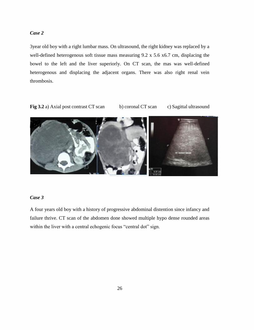
Case 2
3year old boy with a right lumbar mass. On ultrasound, the right kidney was replaced by a
well-defined heterogenous soft tissue mass measuring 9.2 x 5.6 x6.7 cm, displacing the
bowel to the left and the liver superiorly. On CT scan, the mas was well-defined
heterogenous and displacing the adjacent organs. There was also right renal vein
thrombosis.
Fig 3.2 a) Axial post contrast CT scan b) coronal CT scan c) Sagittal ultrasound
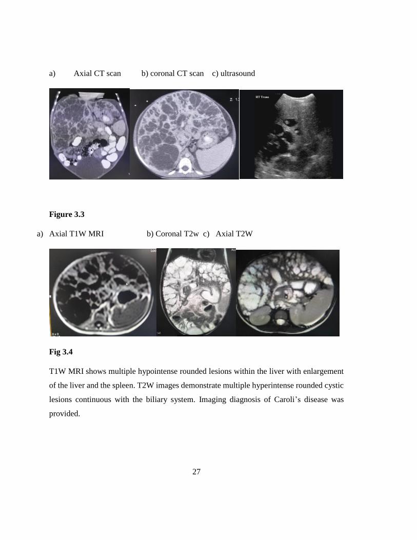
Case 3
A four years old boy with a history of progressive abdominal distention since infancy and
failure thrive. CT scan of the abdomen done showed multiple hypo dense rounded areas
within the liver with a central echogenic focus “central dot” sign.
27
a) Axial CT scan b) coronal CT scan c) ultrasound
Figure 3.3
a) Axial T1W MRI b) Coronal T2w c) Axial T2W
Fig 3.4
T1W MRI shows multiple hypointense rounded lesions within the liver with enlargement
of the liver and the spleen. T2W images demonstrate multiple hyperintense rounded cystic
lesions continuous with the biliary system. Imaging diagnosis of Caroli’s disease was
provided.

28
Case 4
A 1 year 8 months baby who presented with a progressively increasing left abdominal mass
and loss of appetite. This was a biopsy confirmed case of immature teratoma.
Fig 3.5
Ultrasound- there was a large left hypochondrial heterogeneous intra- abdominal mass crossing the midline
and extending into the pelvis. The mass was thought to be a massively enlarged spleen due to its location
sonographically.
Ct scan done 1 month later,
a) Precontrast saggital b) Post contrast coronal scan

29
Fig 3.6
Axial post contrast scans of the mass.
Non contrast abdominal CT image showed, a large homogenous soft tissue density mass (21.7 x 13.6 x 9.8cm) arising
from the left hypochondrium crossing the midline and extending into the pelvis with heterogeneous enhancement post
contrast. A normal spleen was seen displaced superiorly. CT diagnosis was rhabdomyosarcoma. Surgery was performed
and histology showed an immature retroperitoneal teratoma.
Case 5
CT scan of a 12-month-old who presented with progressive abdominal swelling over 1
month, fever and weight loss showed, a large central heterogeneously hypo dense nodular
confluent masses in pre aortic region displacing the kidneys posteriorly without contrast
enhancement. Histology confirmed a diagnosis Burkitt’s lymphoma.

30
Fig 3.7
a) Coronal CT post contrast b) axial c) sagittal
3.8 Comparison of imaging diagnosis and histological diagnosis
Out of the 123 patients, 78 underwent tissue biopsy, 30 bone marrow aspirate and 11 had
specific laboratory tests in addition to the imaging findings. In some patients, a definitive
imaging diagnosis was not provided but rather a description of the mass and histology was
then recommended.
Overall imaging diagnosis of specific lesions and histology diagnosis had an agreement of
82% Comparison of the histological diagnosis (gold standard) and imaging findings in the
78 showed the highest level of agreement in ovarian teratoma (100%, n=5), multilocular
cystic nephroma (100%, n=1) and Wilms tumor (94.1%, n=34 )( Table 7 ).

31
Table 7: Comparison of imaging diagnosis and the post biopsy histological
diagnosis
Imaging diagnosis (test) Frequency Histology
(standard)YES
Histology
NO
Agreement
(%)
Wilms tumour 34 (41.2%) 32 2 94.1
Lymphoma 24 (29.6%) 17 7 70.8
Ovarian teratoma 5 (6.17%) 5 0 100
Metastasis 3 (3.7%) 2 1 66.7
Neuroblastoma 5 (6.17%) 4 1 80
Primitive
neuroectodermal tumour
1 0 1 0
Hepatoblastoma 2 (2.5%) 1 1 50
Rhabdomyosarcoma 2(1.2) 1 1 50
Tuberculoma 1 1 0 100
Multilocular cystic
nephroma
1 1 0 1 (100)
The two cases that were thought to be Wilms tumor were confirmed histologically to be
renal rhabdoid tumor and focal renal abscess. The 7 cases that were thought to be
lymphoma were confirmed histologically to be leukemia. One case was thought to be
metastatic lesion, but it was renal abscess. The case that was thought to be a primitive
neuroendocrine tumor ended up being a rhabdomyosarcoma while one case which looked
like a hepatoblastoma radiologically was confirmed to be an infantile
hemangioendothelioma on histology.

32
CHAPTER 4:
DISCUSSION
4.1 Introduction
Pediatric abdominal masses comprise a variety of lesions which are specific to each age
group. They are a major source of morbidity and mortality. Imaging has been reported to
be important in the diagnosis since history and physical examination may be unreliable
(60)
This prospective cross-sectional study strived to determine the prevalence, spectrum of
pediatric abdominal masses on imaging and compare with the histological diagnosis. Out
of the 140 conveniently sampled children with an age range of 4 days-17 years referred for
imaging, abdominal masses were seen in 87.86%. The lesions encountered were numerous
and diversified. Overall renal tumors (Wilms tumor) and lymphoma were the most
common. Organomegaly was also frequently seen in association with lymphoproliferative
disorders and parasitic infections. Imaging had an overall agreement of 82% with
histology. A higher agreement was seen in Wilms tumor and ovarian teratoma.
4.2 Socio-demographic characteristics
In this study, the male to female ratio was 1:1.1 which is in agreement with a study done
by Nkorowo et al (2015) who had similar findings (20). However, this differs from one by
Lema et al in Northern India who found a slight male predominance of pediatric abdominal
masses at 1.1:1 (8).This difference is however not statistically significant
The prevalence of masses in the sampled population was found to be 87.86%. Sharma et
al however found a prevalence of 7.2% (60). This difference could be due to the sampling
method in which patients who were already suspected of having a mass were included in a
study. The findings in this study cannot therefore be generalized to be representative of the
population at large.

33
4.3 Description of imaging findings
The imaging findings on the abdominal masses were highly differing. Out of all the patients
who underwent imaging, 12.2% (n=17) did not have actual tumors or organomegaly on
imaging leaving 123 to be evaluated. Of the 17, 11 had constipation with bowel fecal
loading, 5 had radiological evidence of intestinal obstruction and one child had a distended
bladder due to posterior urethral valves all later confirmed. Overall tumors were 51.2%
(n=63) while organomegaly involving the liver and spleen was seen in 48.2% (n=60)
(Table 5). Densmore et al found that organomegaly accounted for 50% of all the cases.
These findings are also comparable to those reported by Adeseyun et al (2, 11). This study
found that Wilms tumor, lymphoma and neuroblastoma were the most commonly
encountered tumors. Similar findings have been shown in other studies (10, 16). Renal
tumors were common in the 1-5 year age group while ovarian tumors were all seen in
children >5 years of age category, similar findings have been reported in other studies
elsewhere (6,55, 62, 67).
On imaging, Wilm’s tumor presented as an intra-renal mass replacing the whole kidney or
partially with a ‘claw sign’ forming between the mass and adjacent normal renal tissue. On
Ultrasound Wilms tumor presented as a solid heterogeneous, well-defined mass. On CT
scan most masses were well encapsulated, solid and homogenous while others, were
heterogeneous most likely due to hemorrhage or necrosis. Invasion of the renal vein and
inferior vena cava was seen in a few cases on Doppler and CT. These findings are similar
to what has been reported in other studies done elsewhere (5, 6, 60). Lymphoma was
suggested on imaging by the presence of nodular or confluent retroperitoneal masses of
soft tissue density elevating the aorta with no enhancement. These findings have also been
reported by Manzella A et al (2013) and Adesiyun O et al (2017) (11, 61)
Organomegaly was seen in lymphoproliferative (lymphoma, lymphocytic leukemias),
myeloproliferative (myelocytic leukemia), in parasitic infections such as visceral
leishmaniasis and less commonly due to malaria infection. Previous studies have also
reported lymphoma and hematological malignancies to be a common cause of

34
organomegally in children. The features seen on imaging include; diffuse organ
enlargement, heterogenous texture and multiple hypoechoic and echogenic nodules (10,
12). Organomegally due to parasitic infections is well documented in Africa where the
burden of disease from malaria and leishmaniasis is still high (9, 11, 68). There were no
specific findings on imaging that suggested either lymphoproliferative disorder or parasitic
infection and the definitive diagnosis was made from histology or microbiological
examination. This observation has also been reported in other studies (9, 11).
In this study, the most common presenting symptom was an abdominal swelling followed
by abdominal pain. This finding is similar to those reported by several previous studies (1-
4, 12, 55). This is probably because majority of these patients cannot communicate
adequately and most masses tend to be large at the time of presentation, drawing the
attention of the caregiver (1, 2, 45). The mean duration at presentation to the hospital from
the time the symptoms were discovered was 2.5(SD=3.9) months. Soheila Z in a recent
study reported a delay of between 1-3 months between the onset of symptom and
presentation to the hospital. Other authors have also reported a similar delay in developing
countries (55, 65). The reason for this delay was however not provided. In our setting, the
delay could be due to lack of awareness and financial constraints in patients since the study
was done in a public hospital which serves mostly people in the low socio-economic group.
Ultrasound was the most commonly initial imaging modality in 116 (94.3%) of the cases.
This is because it is considered safe since it is non-ionizing, readily available and relatively
cheap however it is user dependent. This finding was also reported by Papaioannou et al
(2005) and Malkan et al (6, 47, 63). Ultrasound in expert hands and with standardized
protocol plays a vital role in evaluation of pediatric masses as was demonstrated in this
study where it was the sole diagnostic imaging tool in 56 (45.5%) of the cases(47).
4.4 Correlation of imaging findings and Histology
On correlation of imaging findings and histology a higher accuracy was seen in Wilms
tumor, teratoma and multilocular cystic nephroma which have characteristic findings on
imaging. Teratoma was easily diagnosed by the presence fat, cyst and calcification. This

35
finding has also been reported in other previous studies (67).The overall agreement
between imaging diagnosis and histology was seen in 82% of the cases. This finding is
comparable to other previous studies (60). In 18% of the cases, imaging was not accurate
in making a definitive diagnosis. Some of these cases include; ovarian cystadenoma, renal
rhabdoid tumor, retroperitoneal immature teratoma and focal renal abscess among others.
A specific case of immature teratoma of the retroperitoneum was missed on imaging due
to its large size, heterogeneous appearance on both ultrasound and CT and increased
vascularity. Since the mass was arising from the left hypochondrium crossing the midline
and extending into the pelvis, demonstrating high vascularity on ultrasound, it was thought
to be an enlarged spleen. On CT it was thought to be a rhabdomyosarcoma since the spleen
was displaced superiorly towards the thoracic cavity. Image guided biopsy was then
performed and histology revealed it was an immature teratoma. In other cases, imaging
could not provide a specific diagnosis and therefore a description of the mass was provided,
and histology recommended. One such case was in a 16-year-old girl who had a complex
large (19.2 x 15.7 x12.9cm) multi-septated left ovarian mass. The histology results in this
case was an ovarian cystadenocarcinoma.
In this study, the most common diagnosis seen was nephroblastoma at 41.2% followed by
lymphoma at 29.6%. These findings are similar to other previous studies (1, 2). However,
they differ from a study done in Nigeria (2015) which found lymphoma as the most
common diagnosis followed by nephroblastoma (16). Another similar study conducted in
Lahore by Haniff et al found that Neuroblastoma was the commonest accounting for
29.6%, Wilms tumor ;25.1% and lymphoma ;15.5% (10).

36
CONCLUSION
The spectrum of imaging findings in pediatric abdominal masses is diversified. Most
children with abdominal masses presented with abdominal swelling and pain. Neoplasms
accounted for the majority (51.2%) while organomegaly accounted for 48.8% of the masses
most of which were lymphoproliferative and parasitic infections. Ultrasound was used as
the initial investigation in majority of the cases.
Wilms tumor was the most common neoplasm and was seen in children below the age of
5 years. Imaging had an overall agreement of 82% in making specific diagnoses. Imaging
is therefore important in the diagnostic workup of pediatric abdominal masses.
It is therefore important for clinicians to have a high index of suspicion and appropriately
refer children with abdominal swelling for further early management. This study supports
other research findings already published.
4.5 Study limitations
Several limitations were encountered in this study including the cross-sectional design with
no control groups and its correlational nature limiting the directionality and validity of the
findings. Subjects were patients with suspicion of an abdominal mass, hence generalization
of the prevalence to the whole population cannot be done. Sample size was small which
also restricts generalizability of the results to a larger target group.
The MRI machine at KNH national hospital had been nonoperational for the better part the
study period therefore further imaging had to done by CT scan.

37
4.6 Recommendations
Studies focusing on specific abdominal masses with larger sample size in a multicenter are
warranted in future to build up on available data.
To the policy makers, radiological investigations should be made accessible to children to
facilitate early diagnosis and appropriate early management.
Capacity building to increase awareness on early signs of abdominal masses in both health
care givers and guardians is recommended.

38
REFERENCES
1. Willi UV. Imaging the Child with an Abdominal Mass. In: Diseases of The Abdomen
and Pelvis [Internet]. Springer, Milano; 2006 [cited 2018 Mar 2]. p. 224–31. Available
from: https://link.springer.com/chapter/10.1007/88-470-0508-6_31
2. Densmore JC, Oldham KT. Chapter 388. Abdominal Masses. In: Rudolph CD, Rudolph
AM,Lister G, et al editors. Rudolph’s Pediatrics [Internet]. 22nd ed. New York, NY:
The McGraw-Hill Companies; 2011 [cited 2018 Mar 6]. Available from:
accesspediatrics.mhmedical.com/content.aspx?aid=7035952
3. Pediatric_cancer_in_africa.pdf [Internet]. [cited 2017 Nov 14]. Available from:
http://bethanykids.org/wp-content/uploads/2016/10/pediatric_cancer_in_africa.pdf
4. Nicholas H. Carter et al:Pediatric Solid Tumors in Resource-Constrained Settings: A
Review of Available Evidence on Management, Outcomes, and Barriers to Care
5. Abdominal mass - an overview | ScienceDirect Topics [Internet]. [cited 2018 Mar 6].
Available from: https://www.sciencedirect.com/topics/medicine-and-
dentistry/abdominal-mass
6. Olukayode AA, Richard IO, Rachael AA et al. Pattern of computed tomography scan
findings in children with Wilms’ tumor in a tertiary hospital in Lagos, Nigeria. Indian
J Med Paediatr Oncol Off J Indian Soc Med Paediatr Oncol. 2014 Mar;35(1):31.
7. Malkan AD, Loh A, Bahrami A et al. An Approach to Renal Masses in Pediatrics.
Pediatrics. 2015 Jan 1;135(1):142–58.
8. Das D, Lema PC, Datta A. Ultrasound of a distended pediatric abdomen in a limited
resource setting. Critical Ultrasound J. 2011 Dec 1;3(3):163–5.
9. Maki E, Oh K, Rogers S et al: Imaging and differential diagnosis of suprarenal masses
in the fetus. Vol. 33, Journal of Ultrasound in Medicine. 2014. p. 895–904.

39
10. hp_feb06_child.pdf [Internet]. [cited 2018 Mar 8]. Available from: http://www.turner-
white.com/pdf/ hp_feb06_child.pdf
11. Adesiyun O, Adeniyi WA, Ololu-Zubair HT et al. Radiological evaluation of childhood
abdominal masses in Ilorin, Nigeria. East Afr Med J. 2017 Jan;94(6):427–32.
12. Shamberger RC. Pediatric renal tumors. Semin Surg Oncol. 1999 Mar;16(2):105–20.
13. Potisek NM, Antoon JW. Abdominal Masses. Pediatr Rev. 2017 Feb 1;38(2):101–3.
14. Lowe LH, Isuani BH, Heller RM, Stein SM, Johnson JE, Navarro OM, et al. Pediatric
Renal Masses: Wilms Tumor and Beyond. RadioGraphics. 2000 Nov 1;20(6):1585–
603.
15. Mullen EA, Weldon C, Kreidberg JA. Pediatric Renal Tumors. In: Avner E, Harmon
W, Niaudet P, Yoshikawa N, editors. Pediatric Nephrology [Internet]. Springer Berlin
Heidelberg; 2009 [cited 2017 Nov 9]. p. 1431–55. Available from:
http://link.springer.com/referenceworkentry/10.1007/978-3-540-76341-3_59
16. Siegel MJ, Chung EM. Wilms’ tumor and other pediatric renal masses. Magn Reson
Imaging Clin N Am. 2008 Aug;16(3):479–497, vi.
17. Axt J, Abdallah F, Axt M, Githanga J, Hansen E, Lessan J, et al. Wilms Tumor Survival
in Kenya. J Pediatr Surg. 2013 Jun;48(6):1254.
18. Gaillard F. Paediatric renal tumours and masses | Radiology Reference Article |
Radiopaedia.org [Internet]. Radiopaedia. [cited 2017 Oct 20]. Available from:
https://radiopaedia.org/articles/paediatric-renal-tumours-and-masses
19. Fufezan O, Asavoaie C, Blag C et al. The role of ultrasonography for diagnosis the
renal masses in children. Pictorial essay. Med Ultrasonography. 2011;13(1):59–71.
20. Hospital N. Clinicopathological characteristics of malignant abdominal tumors in
children : 10 years experience. 2016;6:142–8.

40
21. Wale DJ, Wong KK, Viglianti BL et al. Contemporary imaging of incidentally
discovered adrenal masses. Vol. 87, Biomedicine and Pharmacotherapy. 2017. p. 256–
62.
22. Bittman ME, Lee EY, Restrepo R et al. Focal Adrenal Lesions in Pediatric Patients.
Am J Roentgenol. 2013 May 23;200(6):W542–56.
23. Malayeri AA, Zaheer A, Fishman EK et al. Adrenal masses: Contemporary imaging
characterization. Computed Assisted Tomography. 2013;37(4):528–42.
24. Adrenal tumors [Internet]. [cited 2017 Nov 19]. Available from
:http://www.pediatricurologybook.com/adrenaltumors
25. Balassy C, Navarro OM, Daneman A. Adrenal masses in children. Vol. 49, Radiologic
Clinics of North America. 2011. p. 711–27.
26. Czerwińska K, Roik D, Sopyło B et al. Atypical imaging features of adrenal gland
lesions in children – report of three cases and review of literature. Pol J Radiol.
2012;77(2):73–9.
27. Gaillard F. Adrenal haemorrhage | Radiology Reference Article | Radiopaedia.org
[Internet]. Radiopaedia. [cited 2017 Nov 19]. Available from: https://radiopedia.org
28. Cystic adrenal lesions: focus on pediatric population (a review). - PubMed - NCBI
[Internet]. [cited 2017 Sep 26]. Available from:
http://login.research4life.org/tacsgr1www_ncbi_nlm_nih_gov/pubmed/28246490
29. Adrenal lesions (differential) | Radiology Reference Article | Radiopaedia.org
[Internet]. [cited 2017 Nov 8]. Available from: https://radiopedia.org
30. Ribeiro RC, Michalkiewicz EL, Figueiredo et al. Adrenocortical tumors in children.
Brazilian J Med Biol Res. 2000 Oct;33(10):1225–34.

41
31. Urrutia A, Santesmases J, Benítez RM et al. Adrenal gland abscess due to Streptococcus
pneumoniae. J Infect. 2010 Jan 1;60(1):88–9.
32. Ranganath SH, Lee EY, Eisenberg RL. Focal Cystic Abdominal Masses in Pediatric
Patients. Am J Roentgenol [Internet]. 2012 Jul 1;199(1):W1–16. Available from:
https://doi.org
33. Servaes S, Khanna G, Naranjo A et al. Comparison of diagnostic performance of CT
and MRI for abdominal staging of pediatric renal tumors: a report from the Children’s
Oncology Group. Pediatr Radiol. 2015 Feb;45(2):166–72.
34. Chung EM, Biko DM, Arzamendi AM et al. Solid Tumors of the Peritoneum,
Omentum, and Mesentery in Children: Radiologic-Pathologic Correlation: From the
Radiologic Pathology Archives. RadioGraphics. 2015 Mar 1;35(2):521–46.
35. Onur MR, Bakal U, Kocakoc E et al. Cystic abdominal masses in children: A pictorial
essay. Vol. 37, Clinical Imaging. 2013. p. 18–27.
36. Litten JB, Tomlinson GE. Liver Tumors in Children. The Oncologist. 2008 Jul
1;13(7):812–20.
37. Chiorean L, Cui X-W, Tannapfel A et al. Benign liver tumors in pediatric patients -
Review with emphasis on imaging features. World J Gastroenterol WJG. 2015 Jul
28;21(28):8541–61.
38. Kochin IN, Miloh TA, Arnon R et al. Benign liver masses and lesions in children: 53
cases over 12 years. Vol. 13, Israel Medical Association Journal. 2011. p. 542–7.
39. Dezsofi A, McLin V, Hadzic N. Hepatic neoplasms in children: A focus on differential
diagnosis. Vol. 38, Clinics and Research in Hepatology and Gastroenterology. 2014. p.
399–402.

42
40. Dawn E. Light, M.D., Frances R et al. Abdominal Mass in an Infant Case.
2015;4(1):22–5.
41. Varich L. Ultrasound of Pediatric Liver Masses. Vol. 5, Ultrasound Clinics. 2010. p.
137–52. 51.
42. Levy AD, Arnáiz J, Shaw JC et al. Primary Peritoneal Tumors: Imaging Features with
Pathologic Correlation. RadioGraphics. 2008 Mar 1;28(2):583–607.
43. Pickhardt PJ. Primary Malignant Tumors of Peritoneal and Retroperitoneal Origin.
Clinical and Imaging Features. Surg Oncol Clin N Am. 2014;23(4):821–45.
44. Scali EP, Chandler TM, Heffernan EJ et al. Primary retroperitoneal masses: what is the
differential diagnosis? Abdominal Imaging. 2015 Aug;40(6):1887–903.
45. Rahhal R., Eddine A., Bishop W.,(2006). A Child with an Abdominal Mass. Case
presentation. Hospital Physician. Pediatric Rounds. February 2006 37-42
46. Yılmaz, G. et al., (2015). The Radiologic Evaluation of Pediatric Acute Abdomen;
Results of Tertiary Referral Center. Journal of the Belgian Society of Radiology. 99(2),
pp.34–42. DOI: http://doi.org/10.5334
47. Milla S., Lee E., Buonomono C et al (2007) Ultrasound Evaluation of Pediatric
Abdominal Masses.Elsavier ultrasound clinics.Volume2, Issue 3, July 2007, pp541-559
48. Grossman H., (1976) EvaluatingCommon Intra-AbdominalMasses in Children. A
SystematicRoentgenographicApproach. CA: A Cancer Journal for Clinicians.26(4
49. Berger P., Munschauer R, and Kuhn P.(1980).Computed Tomography and Ultrasound
of Renal and Perirenal Diseases in Infants and Children.Relationship to Excretory
Urography in Renal Cystic Disease and Neoplasm. Pediatric Radiology.9,pp91-99.
50. Holm, H.(1971). Ultrasonic scanning in the diagnosis of space-occupying lesions of
the upper abdomen. Br. J. Radiol. 44:24-36

43
51. Annuar Z., Sakijan A., Annlular N et al. (2010) Ultrasound in the diagnosis of palpable
abdominal masses in children Med. J. Malaysia Vol. 45 No. 4 December 1990
( (Accessed: 19 January 2018)
52. Bhargava SK, Jain S. Role of ultrasonography in evaluation of pediatric abdominal
masses. Ultrasound Int. 2004;10(2):41–8.
53. Servaes S, Geller JI, Ehrl et al. Comparison of diagnostic performance of CT and MRI
for abdominal staging of pediatric renal tumors: a report from the Children’s Oncology
Group. Pediatr Radiol. 2015 Feb;45(2):166–72.
54. Leonidas J., Carter B., Leape L.(1987) Computer tomography in diagnosis of
abdominal masses in infancy and childhood Comparison with excretory
urography.Archives of Disease in Childhood, 53, pp120-125.
55. Soheila Z, Sezaneh H, Hamid R. et al. Original Article Iran J Ped Hematol Oncol. 2016,
Vol6.No3, 142-148? Clinicopathological characteristics of malignant abdominal
tumors in children: 10 years experience
56. Peña B., Mandl K., Kraus J.et al (1999)Ultrasonography and Limited Computed
Tomography in the Diagnosis and Management of Appendicitis in
Children. JAMA.;282(11):1041–1046. doi:10.1001/jama.282.11.104
57. Ladino-Torres F., Strouse J., (2011) Gastrointestinal tumors in children. Radiologic
Clinics of North America, Volume 49( (Accessed: 19 January 2018)
58. Elsayes K.,Mukundan G.,Narra V et al(2004). Adrenal Masses: MR Imaging Features
with Pathologic Correlation.Adrenal Imaging - Continuing Medical Education.
Radiographics. https://doi.org/10.1148/rg.24si045514
59. Hoffer F.(2005).Magnetic Resonance Imaging of Abdominal Masses in the Pediatric
Patient.Seminars in ultrasound, CT and MR.(26) 4, pp 212-223.
https://doi.org/10.1053/j.sult.2005.04.002

44
60. Sharma N, Memon A, Sharma AK et al. Correlation of radiological investigations with
clinical findings in cases of abdominal mass in the paediatric age group. Afr J Paediatr
Surg 2014;11:132-7
61. Manzella A, Borba-Filho P, D’Ippolito et al. Abdominal manifestations of lymphoma:
spectrum of imaging features. ISRN Radiol [Internet].;2013(Figure 6):483069.
Available from:http://www.ncbi.nlm.nih.gov/pubmed/24967280%5Cnhttp://www.pub
medcentral.nih.gov/articlerender.fcgi?artid=PMC4045537
62. Dingeldein MW. Pediatric abdominal masses. In: Common Surgical Diseases: An
Algorithmic Approach to Problem Solving, Third Edition. 2015. p. 283–5.
63. Papaioannou, G., & McHugh, K. (2005). Neuroblastoma in childhood: review and
radiological findings. Cancer imaging : the official publication of the International
Cancer Imaging Society, 5(1), 116–127. doi:10.1102/1470-7330.2005.0104
64. Kebede AG, Nigussie Y. Ultrasound evaluation of abdominal masses in Ethiopian child
patients. Trop Doct. 2011;41(3):157–9.
65. Nicholas H. Carter et al:Pediatric Solid Tumors in Resource-Constrained Settings: A
Review of Available Evidence on Management, Outcomes, and Barriers to Care
66. Nwokoro CC, Fatungase OM, Salami BA, Shonubi AO, Adekoya AO, Oyelekan AA.
Abdominal Masses in Children: A 10-Year Review. 1. 2015;1(2):44–7.
67. Surbhi Gupta, Deepak Meena, GL Meena ,Tumors and beyond: an Array of Abdominal
masses in children. Symbiosis, Internal journal of Pediatrics child care: Open Access
22(2): 1-9
68. Mazigo H. D et al Anaemia and organomegaly associated with parasitic infections
among schoolchildren in Sengerema District, north‐ western Tanzania H.D: Tanzania
Journal of Health Research Volume 12, Number 2, April 2010

45
STUDY TIME LINES
2017 2018 2019
ACTIIVITY Nov Dec Jan Feb Mar April May June Jul Aug Sep Oct Nov
Proposal
Writing
Proposal
Presentation
Ethical
Approval
Data
Collection
and analysis
Finalization
of Research
Project
Dissemination
of Study
Findings
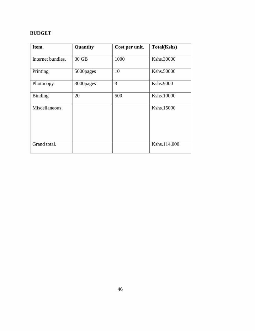
46
BUDGET
Item. Quantity Cost per unit. Total(Kshs)
Internet bundles. 30 GB 1000 Kshs.30000
Printing 5000pages 10 Kshs.50000
Photocopy 3000pages 3 Kshs.9000
Binding 20 500 Kshs.10000
Miscellaneous
Kshs.15000
Grand total. Kshs.114,000

47
APPENDICES
Appendix 1
DATA COLLECTION FORM.
Patient No ……… Age. ………YEARS
Gender: Male…… Female ………
Contact ………………….
PRESENTING SYMPTOM ( Tick appropriately)
Abdominal pain
Abdominal swelling
Fever
Hematuria
Jaundice
Weight loss
Others (Specify)
Duration of symptom
Clinical findings
Clinical diagnosis
IMAGING MODALITY REQUESTED.
IMAGING MODALITY USED TICK APPROPRIATELY
Ultrasound
CT scan
MRI

48
TOPOGRAPHIC LOCATION OF THE MASS.
LOCATION TICK APPROPRIATELY
Renal
Non renal flank
Hepatobiliary
Gastrointestinal
Pelvic
Mesenteric
Splenic
Others
Brief description of the imaging findings ………………………………………………….
……………………………………………………………………………………………..
Please indicate if there is need for further imaging and specify the imaging modality
requested.
………………………………………………………………………………………………
………………………………………………………………………………………………
RADIOLOGICAL DIAGNOSIS: ...…………………………………………………..
HISTOLOGICAL DIAGNOSIS: ………………………………………………………

49
Appendix II
CONSENT FORM
The purpose of the study.
I, Dr. Alice Adhiambo from the University of Nairobi would like to conduct a study to
determine the pattern of occurrence of pediatric abdominal masses. This is in partial
fulfillment for a degree of Master of Diagnostic imaging and radiation medicine.
How you participate.
I have provided some questions regarding you and your child. Please answer them as
accurately as possible. The information will help me to be to understand the illness that’s
affecting your child from the time you noticed there was a problem till this particular time.
Voluntary participation.
You and your child’s participation in the study is voluntary. You can withdraw from this
study at any time if for any reason you feel you don’t want to continue, without
intimidation. Please note there will be no financial benefit to you or your child by
participating in the study.
Risks of the study.
There is no risk anticipated in this study.
Benefits of the study.
The study will provide a baseline for accurate diagnosis and management of pediatric
patients with abdominal masses in future.
Confidentiality.
The information given to me will be kept in confidence and will not be released, or
published with your identity. If you are content with my explanation and are willing to
allow your child to participate in this study, kindly sign the consent form.
I…………………………………………having understood the nature of the study as
explained by Dr. Alice Adhiambo Onyango of the University of Nairobi, is willing to have
my child participate in the study.
……………………………….Signature………………..Date………..
I confirm that I explained the nature of the study to caregiver

50
Name and contacts of investigator:
Dr. Alice Adhiambo Onyango,
Department of diagnostic imaging,
School of medicine, University of Nairobi.
Cell: 0716521406
Email: [email protected]
Name and contact of the supervisors:
Dr Angeline Aywak
Department of diagnostic imaging and radiation medicine,
University of Nairobi.
DR WAIRIMU WAWERU
MBChB (Nbi), M.Med (Nbi),
Consultant Pathologist and Senior Lecturer, Department of Human Pathology, School of
Medicine, College of Health Sciences, University of Nairobi.
The secretariat,
KNH-UoN Research ethics and standards committee,
P.O BOX 20723-00202, Nairobi.

51
Appendix III
FOMU YA IDHINI
Madhumuni ya utafiti
Mimi, Daktari Alice Adhiambo, kutoka Chuo Kikuu cha Nairobi ningependa kufanya
utafiti ili kujua tukio la uvimbe kwenye tumbo ya watoto. Hii ni kutimiza nusu kwa
kiwango cha shahada ya juu katika ‘masters in diagnostic imaging and Radiation medicine’
Jinsi unaweza kushiriki
Haya ni baadhi ya maswali kuhusu ugonjwa wa mtoto wako. Tafadhali uyajibu kwa usahihi
kadri iwezekanavyo.
Ushiriki wa hiari
Wewe na mtoto wako kushiriki katika utafiti huu ni kwa hiari na unaweza kujiondoa
kutoka kwa utafiti huu katika hatua yoyote bila vitisho.
Hatari ya utafiti
Hakuna hatari katika utafiti.
Faida
Hakuna faida ya kifedha katika utafiti huu kwako. Utafiti huu utatuwezesha kufahamu
zaidi kuhusu uvimbe unaopatikana katika tumbo za watoto.
Usiri Maelezo ambayo nitapata kwako yatawekwa katika usiri kali na hayawezi kuwa huru au
kuchapishwa kwa utambulisho wako.
Kama umeridhika na maelezo yangu na uko tayari kuruhusu mtoto wako kushiriki katika
utafiti huu, tafadhali weka saini fomu za kuridhia.
Ridhaa fomu Mimi ......................................................, baada ya kuelezwa asili ya utafiti katika barua
hii kutoka kwa Dr Alice Adhiambo wa Chuo Kikuu cha Nairobi, niko tayari kushirikisha
mtoto wangu katika utafiti.
Jina la mzazi /mlezi.............................................................Sahihi....................Tarehe.........

52
Mimi nathibitisha kwamba nilieleza asili ya utafiti kwa mlezi.
Jina la mchunguzi.
Dr. Alice Adhiambo Onyango,
Chuo kikuu cha Nairobi.
Nambariya simu: 07165212406.
email: [email protected]
Jina la wasimamizi:
1. ANGELINE AYWAK,
Department of Diagnostic Imaging,
2. WAIRIMU WAWERU
MBChB (Nbi), M.Med (Nbi),

53
Appendix IV
ASCENT FORM
My name is Dr. Alice Adhiambo. I am a doctor but I am also a masters student. Right now,
I am trying to learn more about swellings or masses that grow within childrens’ abdomen.
If you agree, you will be asked to fill a form that will only take a few minutes in order to
allow us to use information regarding your illness.
You may be helping us understand better how to investigate and manage masses occurring
in childrens’ abdomen.
If you agree to help us, you should know that your friends, teachers or even classmates
won’t know what you have because we will keep the information in secret. You should
also know that if you decide to help us or if you decide to say “no,” your choice will not
affect weather people like you or not.
Please talk this over with your parents before you decide if you want to be in my study or
not.
I will also ask your parents to give their permission for you to be in this study, but even if
your parents say “yes,” you can still say “no” and decide not to be in the study.
If you don’t want to be in my study, you don’t have to be in it. Remember, being in the
study is up to you and no one will be upset if you don’t want to be in the study or if you
decide to stop after we begin, that’s okay, too.
You can ask any questions that you have about the study. If you have a question later that
you didn’t think of now, you can call me or ask your parents to call me at: TEL NO.
0716521406
I…………………………………………having understood the nature of the study as
explained by Dr. Alice Adhiambo Onyango of the University of Nairobi, is willing to
participate in the study.
Name ……………………………….Signature………………..Date………..
TEL NO: …………………….
I confirm that I explained the nature of the study to parent/guardian

54
Appendix V
FOMU YA IDHINI YA WATOTO WAKUBWA
Jina langu ni Dr Alice Adhiambo. Mimi ni daktari lakini mimi pia mwanafunzi. Sasa hivi,
mimi ninajaribu kujifunza zaidi juu ya uvimbe inayomea ndani ya tumbo ya watoto '
Kama utakubali, utaulizwa kujaza fomu ya idhini. Itachukua tu dakika chache kushiriki.
Tunakuomba uturuhusu kutumia taarifa kuhusu ugonjwa wako. Kushiriki kwako
utatuwezesha kuelewa vizuri jinsi ya kuchunguza na kutibu uvimbe unaomea kwenye
tumbo ya watoto.
Ukikubali kutusaidia, unapaswa kujua kwamba rafiki yako, walimu au hata wanafunzi
wenzako hawatajua yale iliyotuambia maana taarifa yote itawekwa siri na jina lako halisi
halitatumiwa. Lazima pia ujue kwamba ukiamua kutusaidia au ukiamua kusema "hapana,"
ni haki yako na hakuna yeyote atakasirika .
Tafadhali zungumza na wazazi wako juu ya hili jambo kabla ya kuamua kama unataka
kushiriki katika utafiti wangu au la. Mimi pia nitaomba idhini kutoka kwa wazazi, lakini
hata kama wazazi wako wamekubali bado unaweza kukataa na kuamua kutoshiriki katika
utafiti. Kumbuka, kuwa katika utafiti ni uamuzi wako na hakuna mtu atakasirika ukikataa.
Ukiamua kutoka baada ya kuanza, pia ni sawa.
Unaweza kuuliza maswali yoyote ambayo unayo kuhusu utafiti huu. Ukiwa na swali
baadaye usisite, unaweza kunipigia mimi au uwaulize wazazi wako wanipigie simu katika:
NAMBARI YA SIMU; 0716521406
Mimi ................................................ baada ya kuelewa asili ya utafiti kama ilivyoelezwa
na Dr Alice Adhiambo wa Chuo Kikuu cha Nairobi, nina nia ya kushiriki katika utafiti.
Jina .................................... .Sahihi .................. ..Tarehe......... ..
Ninathibitisha kwamba alielezea asili ya utafiti.

55
Appendix VI
STANDARD OF OPERATING PROCEDURE IN ABDOMINAL SCANS
1. Purpose 1. The purpose of this guideline is to provide staff with a specific procedure to follow. This
will ensure that every abdominal ultrasound scan, undertaken by a Sonographer, is
complete and standardized.
2. Introduction Ultrasound is regarded as a first-line examination for a vast array of abdominal symptoms,
owing to its non-invasive and to some extent accessible nature.
3. Definition
A full abdominal ultrasound survey includes assessment of the liver, pancreas, gallbladder,
biliary tree, both kidneys, spleen and abdominal aorta.
4. Examination preparation Any patient booked for an ultrasound scan of the upper abdomen should be informed to
fast four hours prior to the scan.
5. Consent The consent process is a continuum beginning with the referring health care professional
who requests the ultrasound examination and ending with the sonographer who carries it
out.
Consent for those of an intimate or invasive nature should be recorded in the ultrasound
report.
6. Performing the scan Anatomy to be examined:
• Liver: shape, contour and echotexture. Appearances of intrahepatic vessels and ducts
• Gallbladder: size, shape, contour and surrounding area ultrasound characteristics of the
wall and the nature of any contents
• Common duct: maximum diameter and contents; optimally it should be visualized to the
head of pancreas
• Pancreas: size, shape, contour and ultrasound characteristics of head, body, tail
• Spleen: size, shape, contour and ultrasound characteristics
• Abdominal Aorta: Antero-posterior (AP) diameter
• Kidneys: size, shape, position and orientation, outline and ultrasound characteristics of
cortex, medulla, collecting system
• Adrenals: not routinely viewed but any apparent abnormality of size and ultrasound
characteristics should be noted
7. Images to be Stored A series of static images should be recorded on the Radiology patient archive and
communication system (PACS). This should include:
• Liver: Longitudinal sections (LS) and Transverse sections (TS) of left and right lobes.
• RUQ to demonstrate comparative echotexture of liver and right kidney.
• Common duct measurement.
• Gallbladder.
• Right kidney: LS and TS

56
• Left kidney: LS and TS.
• Spleen: AP measurement.
• Abdominal aorta: AP diameter.
• Pancreas.
• Any pathology identified.
8. Reporting 8.1 The following should be documented in the electronic report, recorded on the radiology
information system:
• Description of the liver (size, shape, echotexture).
• Description of the common duct and gallbladder.
• Description of both kidneys.
• Description of the spleen.
• Description of the pancreas.
• Description of abdominal aorta, including AP measurement if aneurismal.
• State any reason for limited examination/suboptimal views.

57
Appendix VII: KNH Ethical Approval Letter

58

59