case report - scielo.br · complications of hc ruptures vary from constant abdominal symptoms...
TRANSCRIPT

864
Rev Soc Bras Med Trop 50(6):864-867, November-December, 2017doi: 10.1590/0037-8682-0292-2017
Case Report
Corresponding authors: Dr. Reza Shahkaram. e-mail: [email protected] 27 July 2017Accepted 10 October 2017
Traumatic rupture of liver hydatid cysts into the peritoneal cavity of an 11-year-old boy: a case report from Iran
Sadaf Sabzevari[1], Alireza Badirzadeh[2],[3], Reza Shahkaram[4], and Mohammad Seyyedin[5]
[1]. Vector-borne Diseases Research Center, North Khorasan University of Medical Sciences, Bojnurd, Iran. [2]. Department of Medical Parasitology and Mycology, School of Medicine, Shahid Beheshti University of Medical Sciences, Tehran, Iran. [3]. Department of Microbiology and Parasitology, School
of Medicine, Ardabil University of Medical Sciences, Ardabil, Iran. [4]. North Khorasan University of Medical Sciences, Bojnurd, Iran. [5]. Razi Vaccine and Serum Research Institute, Mashhad, Iran.
Abstract This is the fi rst published case report of an 11-year-old patient with a rupture of a liver hydatid cyst (HC) into the peritoneal cavity after an abdominal trauma in Iran. The disease was diagnosed using focused abdominal sonography for trauma. To date, no cases of traumatic ruptures of liver HCs in children have been reported in Iran. In the endemic regions of the world, where patients suffer from a history of trauma and constant abdominal symptoms or anaphylactic shock, early diagnosis of HC is crucial as it may disseminate to other organs. The condition needs conservative surgery and follow-up.
Keywords: Hydatid cyst. Trauma. Echinococcus granulosus.
INTRODUCTION
Hydatid cyst (HC) disease, a zoonotic parasitic infection, occurs during the larval stage of a cestode named Echinococcus granulosus (E. granulosus)1. The disease is endemic and hyper-endemic in pastoral regions of the world2,3. Although HC may develop in any organ, it generally occurs in the liver (50-75%) and lungs (18-30%), and is characterized by a cystic form of lesions4. Hydatid cysts (HCs) develop slowly, making the viscera, and especially the abdominal cavity, highly susceptible to traumatic ruptures. Complications of HC ruptures vary from constant abdominal symptoms (abdominal pain, tenderness, and vomiting) to peritonitis and shock with allergy symptoms (cutaneous rash, urticaria, and anaphylactic shock)2,5. While there have been a few reports of HC ruptures after trauma in adults in Iran5, to date, no reports have been published describing this condition in children. Here, we present the fi rst documented report from Iran of an 11-year-old male patient with traumatic rupture of liver HCs into the peritoneal cavity.
CASE REPORT
An 11-year-old boy was admitted to the emergency department of Imam Ali Hospital, North Khorasan University of Medical Science. He had intractable pain in the abdomen, nausea, and vomiting from trauma sustained after falling
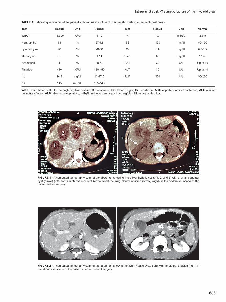
down a few steps. On physical examination, his abdomen was not distended and he was not pale. His situation was stable and normal, except for generalized rebound tenderness and involuntary guarding during abdominal palpation. He presented with fever, itching, and a generalized rash. All his preliminary laboratory parameters, such as results of a liver function test, showed normal values (Table 1). The patient had no history of trauma, surgery, or systemic disease. Owing to the trauma, focused abdominal sonography in trauma (FAST) and conventional ultrasonography were performed. These revealed free fl uid in Morison's pouch and multiple cystic masses in the right hepatic lobe. A spiral computed tomography scan (CT) showed multiple hepatic HCs with small daughter cysts in segments II, III, VI, and VII, as well as intraperitoneal free fl uid (Figure 1). A diagnosis of liver HCs with severe ruptures was made, and emergency surgery was performed. After a total cystectomy and excision, the cyst pouch was washed with hypertonic saline (5%), and the peritoneal spaces were irrigated with isotonic saline for 10 minutes. Moreover, the abdominal spaces and cyst pouches were irrigated with povidone iodine in order to kill the viable protoscoleces. The histopathology of biopsies taken from the cyst content confi rmed an infection of Echinococcus granulosus. Based on the radiological and histopathological fi ndings, hydatid disease (cysts) was diagnosed. Post-surgery, to prevent recurrence of the infection, the patient was started immediately on a course of albendazole (10mg/kg/day) for four months. On the follow-up visit, six months post infection, the physical examination, medical laboratory investigations, and CT were repeated. The patient was found to be well and normal (Figure 2). Written informed consent was provided by the patient’s father.
865
Sabzevari S et al. -Traumatic rupture of liver hydatid cysts
Test Result Unit Normal Test Result Unit Normal
WBC 14,300 103/µl 4-10 K 4.3 mEq/L 3.6-5
Neutrophils 73 % 37-72 BS 130 mg/dl 80-150
Lymphocytes 20 % 20-50 Cr 0.8 mg/dl 0.6-1.2
Monocytes 6 % 0-14 Urea 36 mg/dl 17-43
Eosinophil 1 % 0-6 AST 30 U/L Up to 40
Platelets 450 103/µl 150-450 ALT 30 U/L Up to 40
Hb 14.2 mg/dl 13-17.5 ALP 351 U/L 98-280
Na 145 mEq/L 135-146
TABLE 1: Laboratory indicators of the patient with traumatic rupture of liver hydatid cysts into the peritoneal cavity.
WBC: white blood cell; Hb: hemoglobin; Na: sodium; K: potassium; BS: blood Sugar; Cr: creatinine; AST: aspartate aminotransferase; ALT: alanine aminotransferase; ALP: alkaline phosphatase; mEq/L: milliequivalents per litre; mg/dl: milligrams per deciliter.
FIGURE 2 - A computed tomography scan of the abdomen showing no liver hydatid cysts (left) with no pleural effusion (right) in the abdominal space of the patient after successful surgery.
FIGURE 1 - A computed tomography scan of the abdomen showing three liver hydatid cysts (1, 2, and 3) with a small daughter cyst (arrow) (left) and a ruptured liver cyst (arrow head) causing pleural effusion (arrow) (right) in the abdominal space of the patient before surgery.

866
Rev Soc Bras Med Trop 50(6):864-867, November-December, 2017
DISCUSSION
Echinococcus granulosus, the causative agent of HC disease, is a small tapeworm that lives in the bowel of dogs. This tapeworm sheds it eggs in the feces of infected dogs, and these eggs can be accidentally ingested by intermediate hosts such as grazing animals (sheep, cattle) as well as by humans. Hydatidosis is an endemic disease found in many sheep- and cattle-rearing countries such as Iran. The liver and the lungs are the most common organs where hydatid disease is seen6-9.
Liver HC rupture into the peritoneum cavity is very rare (1-8%) and may result in a spontaneous rupture due to increased pressure of the cystic fl uid or trauma10. These uncommon conditions are mostly seen in young children, particularly children with large cysts and a superfi cial localization in the organs. True cysts have two layers: the pericyst and the endocyst. When both these layers are torn apart, the cyst contents spill into the peritoneal cavity. This is called a direct rupture. Here, the hydatid fl uid and sands, brood capsules, and protoscoleces are released into the spaces of the peritoneum. A long-term outcome of direct rupture into the peritoneal spaces is the implantation of protoscoleces, which leads to a metastatic form of the disease11. Direct ruptures of HCs into the peritoneal spaces take two main clinical forms with different manifestations: small fi ssures and large ruptures. The former is very common and is caused by an ordinary trauma that is unrecognized, while the latter is rare and is caused by sudden and severe blunt trauma10. HC ruptures cause severe clinical presentations that are accompanied by abdominal pain, urticarial rash, anaphylaxis, and sometimes sudden death5,11. In this situation, as there may be allergic reactions or anaphylactic shock, the rupture requires emergency medical care10.
The preoperative diagnosis of ruptured HCs must be confirmed immediately as emergency intervention is essential11. This diagnosis requires a complex workup, such as ultrasonography and a CT scan. Ultrasonography is a non-invasive technology that is very helpful in identifying intra-abdominal free fl uids and cysts with detached membranes. The CT scan is the main diagnostic tool for this condition as it has high sensitivity (100%). It is used to determine the exact site of HC ruptures and HC features such as the presence of daughter cysts in the abdominal cavity10,11. A histopathological examination is the fi nal confi rmatory diagnosis tool for the identifi cation of the causative agents of the HCs. In the present case, the causative species of HC was E. granulosus, which is an endemic species in Iran.
Although the disease is common and endemic in Iran, reports of HC ruptures after trauma in the country are rare5,12. This case is one of the fi rst-documented offi cial reports of HC rupture caused by trauma in a child in Iran. Upon initial examination, our patient suffered from abdominal pain, itching, and rash. The itching and rash, in this case, may have been a direct result of the ruptured HC. After HC was suspected, the patient was immediately sent for emergency surgery in order to prevent allergic reactions or anaphylactic shock, and, more importantly, because of potential metastasis to other parts of the abdominal cavity.
Despite developments in chemical therapy, surgery is still the best choice for treatment of this disease10. In the case of ruptured HCs, the initial focus must be on medical treatment of the allergic reaction, followed promptly by immediate emergency surgery. The key steps to effective and successful surgical management of the ruptured cysts are a complete wash of the peritoneal cavity using scolicidal agents such as hypertonic saline and thorough removal of all cystic content — especially protoscoleces of the cysts. In the current case, the patient’s peritoneal cavity was washed with hypertonic saline and isotonic saline for 10 minutes. To prevent recurrence, treatment with albendazole is initiated immediately after surgery, for a course of approximately four months10. In our case, the patient received a prolonged course of albendazole, which contributed to a good clinical follow-up evaluation and no recurrence. Albendazole is the drug of choice for HC disease and is very effective in sterilizing the cysts, reducing the risk of anaphylaxis, and decreasing the recurrence rate in infected patients.
In conclusion, although HC ruptures into the abdominal cavity are rare, especially in children, they present serious challenges for clinicians and surgeons who work in emergency wards. Early diagnosis of HC cases by using simple techniques — such as a combination of routine clinical history and a CT scan — is vital to preventing severe and deadly complications of the disease.
Acknowledgements
The authors are grateful to the clinical research development center of Imam Ali Hospital, North Khorasan University of Medical Science for their contributions and assistance.
Confl icts of interest
The authors declare that there is no confl ict of interest.
REFERENCES
1. Stewart BT, Jacob J, Finn T, Lado M, Napoleon R, Brooker S, et al. Cystic echinococcosis in Mundari tribe-members of South Sudan. Pathog Glob Health. 2013;107(6):293-8.
2. Çetinkaya ÖA, Çelik SU, Kocaay AF, Kırımker EO, Akyol C. Spontaneous rupture of a hepatic hydatid cyst with anaphylaxis: a case report. JAEMCR. 2016;7(2):31-3.
3. Valizadeh M, Haghpanah B, Badirzadeh A, Roointan E, Fallahi S, Raeghi S. Immunization of sheep against Echinococcus granulosus with protoscolex tegumental surface antigens. Vet World. 2017;10(8):854-8.
4. Salih AM, Kakamad FH, Hammood ZD, Yasin B, Ahmed DM. Abdominal wall Hydatid cyst: A review a literature with a case report. Int J Surg Case Rep. 2017;37:154-6.
5. Kalantari N, Bayani M, Abbas-zadeh M. Rupture of hydatid liver cyst into peritoneal cavity following blunt abdominal trauma: a case report. Emerg. 2015;3(1):45-7.
6. Abdelraouf A, El-Aal AAA, Shoeib EY, Attia SS, Hanafy NA, Hassani M, et al. Clinical and serological outcomes with different surgical approaches for human hepatic hydatidosis. Rev Soc Bras Med Trop. 2015;48(5):587-93.
7. Tekin R, Avci A, Tekin RC, Gem M, Cevik R. Hydatid cysts in muscles: clinical manifestations, diagnosis, and management of this atypical presentation. Rev Soc Bras Med Trop. 2015;48(5):594-8.

867
8. Abbas I. Molecular and epidemiological updates on cystic echinococcosis infecting water buffaloes from Egypt. Vet World. 2016;9(12):1355-63.
9. Tekin R, Onat S, Tekin RC. Hydatid cysts in a patient with multiple organ involvement. Rev Soc Bras Med Trop. 2016;49(4):534.
10. Dirican A, Yilmaz M, Unal B, Tatli F, Piskin T, Kayaalp C. Ruptured hydatid cysts into the peritoneum: a case series. Eur J Trauma Emerg Surg. 2010;36(4):375-9.
11. Limeme M, Yahyaoui S, Zaghouani H, Ghannouchi M, Khnissi A, Amara H, et al. Spontaneous intraperitoneal rupture of hepatic hydatid cyst: a rare cause of ascites. BMC Surg. 2014;14(1):99.
12. Hosseinian A, Mohammadzadeh A, Shahmohammadi G, Hasanpour M, Maleki N, Doustkami H, et al. Rupture of a giant cardiac hydatid cyst in the left ventricular free wall: successful surgical management of a rare entity. Am J Cardiovasc Dis. 2013;3(2):103-6.
Sabzevari S et al. -Traumatic rupture of liver hydatid cysts