the skeletal system: bone tissue chapter 6. the skeletal system: bone tissue functions of bone and...
TRANSCRIPT

The Skeletal System: Bone TissueChapter 6


The Skeletal System: Bone Tissue Functions of Bone and Skeletal System Structure of Bone Histology of Bone Tissue Blood and Nerve Supply of Bone Bone Formation Bone’s Role in Calcium Homeostasis Exercise and Bone Tissue Aging and Bone Tissue

Functions of Bone and Skeletal System Support Protection Assistance in Movement Mineral Homeostasis Blood Cell Production Triglyceride Storage

Functions of Bone and Skeletal System Support
Structural framework of the body Supports soft tissues Provides attachment points for tendons of skeletal
muscle
Protection Protects important internal organs
Cranium protects brain Vertebrae protects spinal cord Ribs protect lungs and heart

Functions of Bone and Skeletal System Assistance in Movement
Skeletal muscle attaches to bone Skeletal muscle contraction pulls on bone producing
movement
Mineral Homeostasis Bone tissue stores several minerals
Acts to serve as a reservoir of critical minerals Calcium (99% of body’s content) Phosphorus

Functions of Bone and Skeletal System Blood Cell Production
Red bone marrow produces (Hemopoiesis) Red blood cells White blood cells Platelets
Triglyceride Storage Yellow bone marrow
Triglycerides stored in adipose cells Serves as a potential chemical energy reserve

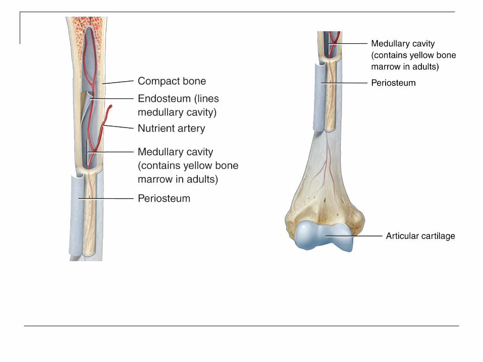
Structure of Bone
Diaphysis Epiphysis Metaphysis
Epiphyseal growth plate Articular cartilage
Perforating fibers Periosteum Medullary cavity Endosteum
Long Bone Anatomy (Humerus)

Histology of Bone Tissue Extracellular matrix surrounding widely
separated cells Matrix
15% water 30% collagen fibers 55% crystallized mineral salts
The most abundant mineral salt is calcium phosphate
Histology of Bone Tissue A process called calcification is initiated
by bone-building cells called osteoblasts
Mineral salts are deposited and crystallize in the framework formed by the collagen fibers of the extracellular matrix
Bone’s flexibility depends on collagen fibers
Histology of Bone Tissue Four types of cells are present in bone tissue Osteogenic cells
Undergo cell division (stem cells); the resulting cells develop into osteoblasts
Osteoblasts Bone-building cells Synthesize extracellular matrix of bone tissue
Osteocytes Mature bone cells Exchange nutrients and wastes with the blood

Histology of Bone Tissue Osteoclasts
Release enzymes that digest the mineral components of bone matrix (resorption)
Regulate blood calcium level

Histology of Bone Tissue Bone may be categorized as:
Compact Spongy
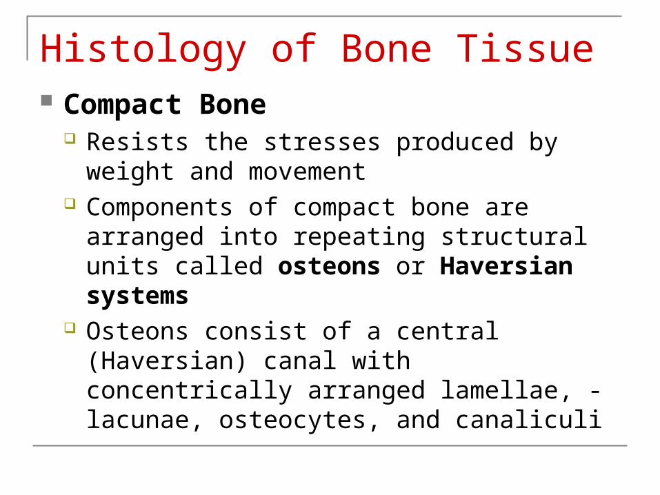
Histology of Bone Tissue Compact Bone
Resists the stresses produced by weight and movement
Components of compact bone are arranged into repeating structural units called osteons or Haversian systems
Osteons consist of a central (Haversian) canal with concentrically arranged lamellae, - lacunae, osteocytes, and canaliculi


Histology of Bone Tissue Osteon
Central canals run longitudinally through bone Around the central canals are concentric
lamellae Rings of calcified matrix (like the rings of a tree trunk)
Between the lamellae are small spaces called lacunae which contain osteocytes
Radiating in all directions from the lacunae are tiny canaliculi filled with extracellular fluid and the plasma membrane extensions of the osteocyte

Histology of Bone Tissue
Osteon Canaliculi connect
lacunae, forming a system of interconnected canals Providing routes for
nutrients and oxygen to reach the osteocytes
The organization of osteons changes in response to the physical demands placed on the skeleton

Histology of Bone Tissue Spongy Bone
Lacks osteons Lamellae are arranged in a lattice of thin
columns called trabeculae Spaces between the trabeculae make bones
lighter Trabeculae of spongy bone support and protect
the red bone marrow Hemopoiesis (blood cell production) occurs in red
bone marrow of spongy bone

Histology of Bone Tissue Spongy Bone
Within each trabecula are lacunae that contain osteocytes
Osteocytes are nourished from the blood circulating through the trabeculae within the red bone marrow.
Interior bone tissue is made up primarily of spongy bone
The trabeculae of spongy bone are oriented along lines of stress helps bones resist stresses without breaking
while keeping them light.

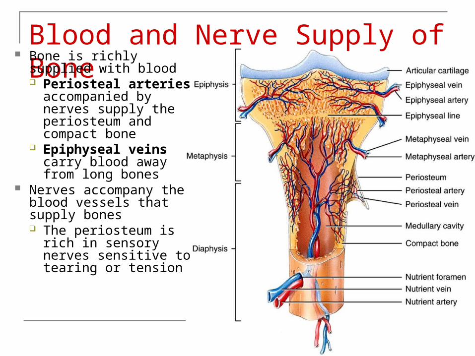
Blood and Nerve Supply of Bone
Bone is richly supplied with blood Periosteal arteries
accompanied by nerves supply the periosteum and compact bone
Epiphyseal veins carry blood away from long bones
Nerves accompany the blood vessels that supply bones The periosteum is
rich in sensory nerves sensitive to tearing or tension

Bone Formation The process by which bone forms is
called ossification Bone formation occurs in four situations:
1) Formation of bone in an embryo 2) Growth of bones until adulthood 3) Remodeling of bone 4) Repair of fractures
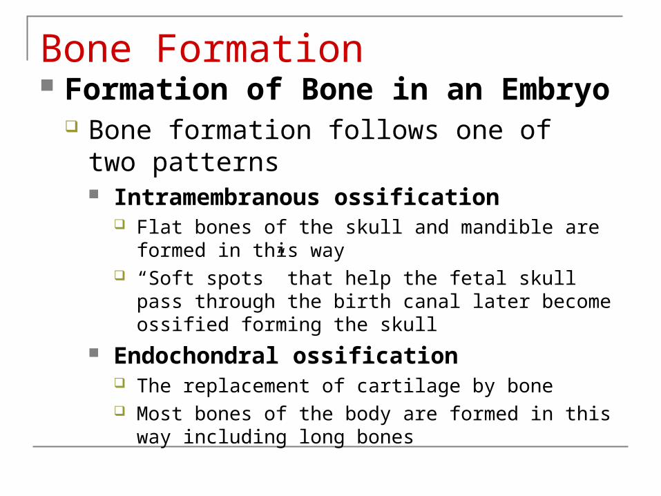
Bone Formation Formation of Bone in an Embryo
Bone formation follows one of two patterns Intramembranous ossification
Flat bones of the skull and mandible are formed in this way
“Soft spots” that help the fetal skull pass through the birth canal later become ossified forming the skull
Endochondral ossification The replacement of cartilage by bone Most bones of the body are formed in this way including
long bones

Bone Formation Formation of Bone in an Embryo
Cartilage formation and ossification occurs during the sixth week of embryonic development
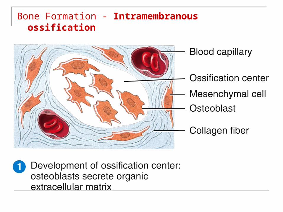
Bone Formation - Intramembranous ossification

Bone Formation - Intramembranous ossification

Bone Formation - Intramembranous ossification

Bone Formation - Intramembranous ossification

Bone Formation - Endochondral ossification
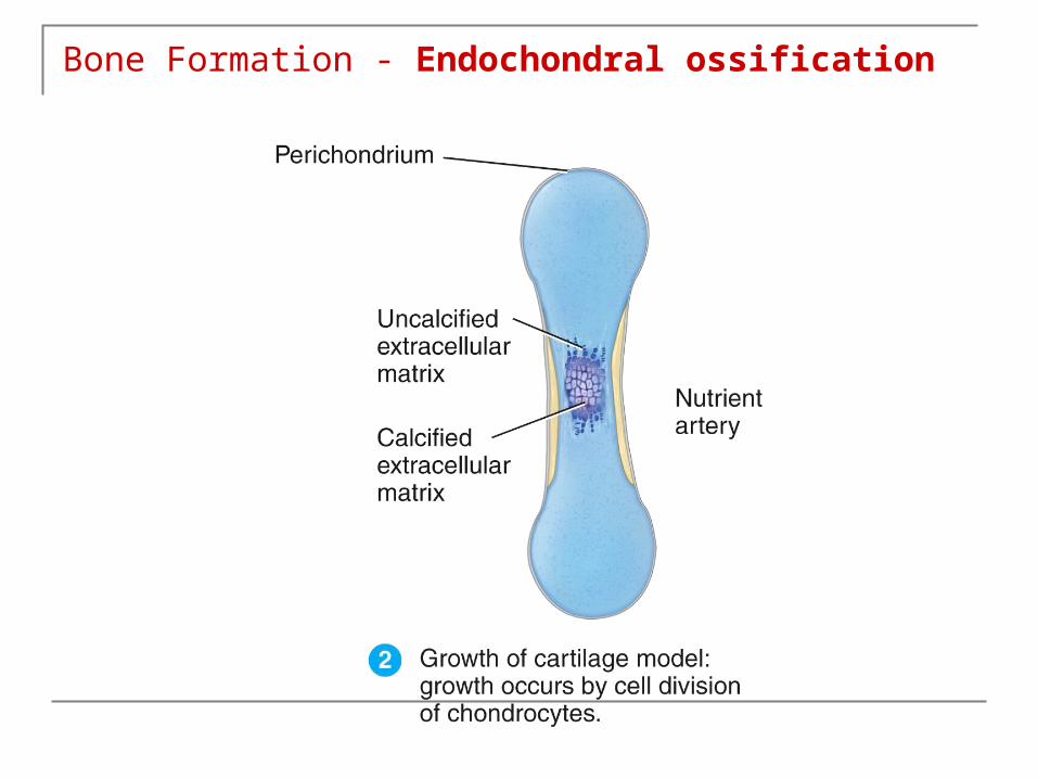
Bone Formation - Endochondral ossification

Bone Formation - Endochondral ossification

Bone Formation - Endochondral ossification
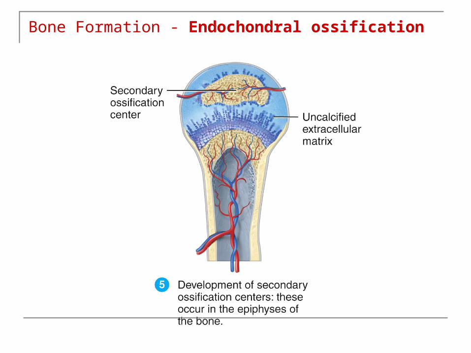
Bone Formation - Endochondral ossification

Bone Formation - Endochondral ossification


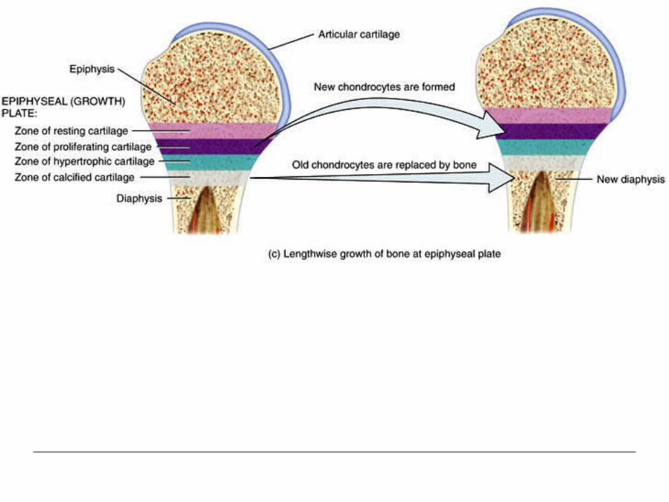
Bone Growth During Infancy, Childhood and Adolescence Growth in Length The growth in length of
long bones involves two major events: 1) Growth of cartilage
on the epiphyseal plate
2) Replacement of cartilage by bone tissue in the epiphyseal plate

Bone Growth During Infancy, Childhood and Adolescence
Osteoclasts dissolve the calcified cartilage, and osteoblasts invade the area laying down bone matrix
The activity of the epiphyseal plate is the way bone can increase in length
At adulthood, the epiphyseal plates close and bone replaces all the cartilage leaving a bony structure called the epiphyseal line

Bone Growth During Infancy, Childhood and Adolescence Growth in Thickness
Bones grow in thickness at the outer surface Remodeling of Bone
Bone forms before birth and continually renews itself
The ongoing replacement of old bone tissue by new bone tissue
Old bone is continually destroyed and new bone is formed in its place throughout an individual’s life

Bone Growth During Infancy, Childhood and Adolescence A balance must exist between the actions of
osteoclasts and osteoblasts
If too much new tissue is formed, the bones become abnormally thick and heavy
Excessive loss of calcium weakens the bones, as occurs in osteoporosis
Or they may become too flexible, as in rickets and osteomalacia
Factors Affecting Bone Growth and Bone Remodeling Normal bone metabolism depends on several factors Minerals
Large amounts of calcium and phosphorus and smaller amounts of magnesium, fluoride, and manganese are required for bone growth and remodeling
Vitamins Vitamin A stimulates activity of osteoblasts Vitamin C is needed for synthesis of collagen Vitamin D helps build bone by increasing the
absorption of calcium from foods in the gastrointestinal tract into the blood
Vitamins K and B12 are also needed for synthesis of bone proteins

Factors Affecting Bone Growth and Bone Remodeling Hormones
During childhood, the hormones most important to bone growth are growth factors (IGFs), produced by the liver and bone tissue (under influence of hGH) IGFs stimulate osteoblasts, promote cell division at the
epiphyseal plate, and enhance protein synthesis
Thyroid hormones (T3 and T4) also promote bone growth by stimulating osteoblasts
Insulin promotes bone growth by increasing the synthesis of bone proteins

Factors Affecting Bone Growth and Bone Remodeling Hormones
Estrogen and testosterone cause a dramatic effect on bone growth Cause of the sudden “growth spurt” that occurs
during the teenage year Promote changes in females, such as widening of
the pelvis Ultimately-Shut down growth at epiphyseal plates
Parathyroid hormone, calcitriol, and calcitonin are other hormones that can affect bone remodeling
Bone’s Role in Calcium Homeostasis Bone is the body’s major calcium reservoir Levels of calcium in the blood are maintained
by controlling the rates of calcium resorption from bone into blood and of calcium deposition from blood into bone Both nerve and muscle cells depend on
calcium ions (Ca2+) to function properly Blood clotting also requires Ca2+
Many enzymes require Ca2+ as a cofactor

Bone’s Role in Calcium Homeostasis Actions that help elevate blood Ca2+ level
Parathyroid hormone (PTH) regulates Ca2+ exchange between blood and bone tissue PTH increases the number and activity of
osteoclasts (i.e., resorption) PTH acts on the kidneys to decrease loss of
Ca2+ in the urine PTH stimulates formation of calcitriol (active
form of Vitamin D) a hormone that promotes absorption of calcium from foods in the gastrointestinal tract

Bone’s Role in Calcium Homeostasis

Bone’s Role in Calcium Homeostasis Actions that work to decrease blood Ca2+
level
The thyroid gland (i.e., parafollicular cells) secretes calcitonin (CT) which inhibits activity of osteoclasts
The result is that CT promotes bone formation and decreases blood Ca2+ level
See Table 6.2

Fracture and Repair of Bone Fracture Types
Open (compound) fracture The broken ends of the bone protrude through the skin
Closed (simple) fracture Does not break the skin
Comminuted fracture The bone is splintered, crushed, or broken into pieces
Greenstick fracture A partial fracture in which one side of the bone is broken and the other side
bends Impacted fracture
One end of the fractured bone is forcefully driven into another Pott’s fracture
Fracture of the fibula, with injury of the tibial articulation Colles’ fracture
A fracture of the radius in which the distal fragment is displaced Stress fracture
A series of microscopic fissures in bone

Fracture and Repair Anatomical appearance – like breaking a green twig
Greenstick

Fracture and Repair Anatomical appearance – the distal part is
shoved up into the proximal part.
Impacted

Fracture and Repair Anatomical appearance – though not seen
here, one or both bones are “open” to the outside.
Open (compound)

Fracture and Repair Eponyms – Colles’ is a fracture of the distal
radius ± ulna.
Colles’
Fracture and Repair of Bone Calcium and phosphorus needed to strengthen and
harden new bone after a fracture are deposited only gradually and may take several months
The repair of a bone fracture involves the following steps 1) Formation of fracture hematoma
Blood leaks from the torn ends of blood vessels, a clotted mass of blood forms around the site of the fracture
2) Fibrocartilaginous callus formation Fibroblasts invade the fracture site and produce collagen fibers
bridging the broken ends of the bone 3) Bony callus formation
Osteoblasts begin to produce spongy bone trabeculae joining portions of the original bone fragments
4) Bone remodeling Compact bone replaces spongy bone

Compact boneSpongy bone
Periosteum
Fracture hematoma
Fracturehematoma
BonefragmentOsteocyte
Red bloodcell
Blood vessel
Formation of fracture hematoma
Phagocyte
Osteon
1
Phagocyte
Osteoblast
Fibroblast
Fibrocartilaginouscallus
Collagen fiberChondroblastCartilage
Fibrocartilaginous callus formation2
Compact boneSpongy bone
Periosteum
Fracture hematoma
Fracturehematoma
BonefragmentOsteocyte
Red bloodcell
Blood vessel
Formation of fracture hematoma
Phagocyte
Osteon
1
Bony callus
Spongy bone
Osteoblast
Bony callus formation
Osteocyte
3
Compact boneSpongy bone
Periosteum
Fracture hematoma
Fracturehematoma
BonefragmentOsteocyte
Red bloodcell
Blood vessel
Formation of fracture hematoma
Phagocyte
Osteon
1
Phagocyte
Osteoblast
Fibroblast
Fibrocartilaginouscallus
Collagen fiberChondroblastCartilage
Fibrocartilaginous callus formation2
Spongy bone
OsteoblastOsteoclast
New compactbone
Bony callus formation Bone remodeling
Osteocyte
3 4
Compact boneSpongy bone
Periosteum
Fracture hematoma
Fracturehematoma
BonefragmentOsteocyte
Red bloodcell
Blood vessel
Formation of fracture hematoma
Phagocyte
Osteon
1
Phagocyte
Osteoblast
Fibroblast
Fibrocartilaginouscallus
Collagen fiberChondroblastCartilage
Fibrocartilaginous callus formation2
Bony callus

Once a bone is fractured, repair proceeds in a predictable pattern:
The first step, which occurs 6-8 hours after injury, is the formation of a fracture hematoma, as a result of blood vessels breaking in the periosteum and in osteons.
Fracture and Repair

The second and third steps involve the formation of a callus (takes a few weeks, to as many as six months). Phagocytes remove cellular debris and fibroblasts
deposit collagen to form a fibro-cartilaginous callus...
Fracture and Repair

Fracture and Repair ... which is followed by osteoblasts forming a bony
callus of spongy bone.
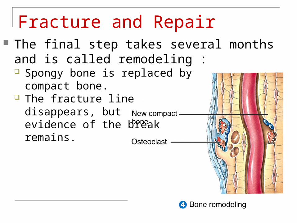
Fracture and Repair The final step takes several months and is
called remodeling : Spongy bone is replaced by
compact bone. The fracture line
disappears, but evidence of the breakremains.

Exercise and Bone Tissue Bone tissue alters its strength in response to
changes in mechanical stress Under stress, bone tissue becomes stronger through
deposition of mineral salts and production of collagen fibers by osteoblasts and fibroblasts
Unstressed bones diminishes because of the loss of bone minerals and decreased numbers of collagen fibers
The main mechanical stresses on bone are those that result from the pull of skeletal muscles and the pull of gravity
Weight-bearing activities help build and retain bone mass

Aging and Bone Tissue The level of sex hormones diminishes during
middle age, especially in women after menopause A decrease in bone mass occurs Bone resorption by osteoclasts outpaces bone
deposition by osteoblasts Female bones generally are smaller and less
massive than males Loss of bone mass in old age has a greater
adverse effect in females

Aging and Bone Tissue There are two principal effects of aging on bone tissue:
1) Loss of bone mass Results from the loss of calcium from bone matrix The loss of calcium from bones is one of the symptoms in
osteoporosis 2) Brittleness
Results from a decreased rate of protein synthesis Collagen fibers gives bone its tensile strength The loss of tensile strength causes the bones to become very brittle
and susceptible to fracture
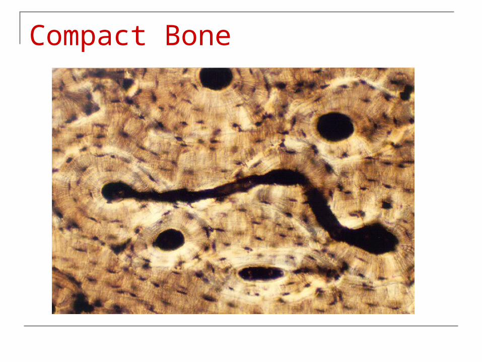
Compact Bone
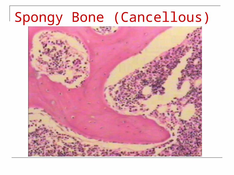
Spongy Bone (Cancellous)

Hyaline Cartilage
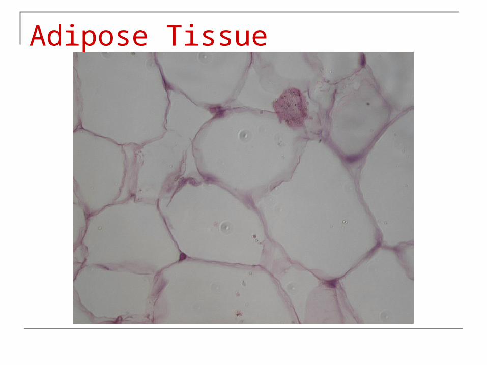
Adipose Tissue