the retrograde connections and anatomical segregation of ... · of broca, midline para-ventricular...
TRANSCRIPT

ORIGINAL RESEARCHpublished: 05 December 2016
doi: 10.3389/fnana.2016.00117
Frontiers in Neuroanatomy | www.frontiersin.org 1 December 2016 | Volume 10 | Article 117
Edited by:
Jackson Cioni Bittencourt,
University of São Paulo, Brazil
Reviewed by:
Tomas Gonzalez-Hernandez,
University of La Laguna, Spain
Hugues Duffau,
Gui de Chauliac Hospital, Montpellier
University Medical Center and
INSERM U1051, France
*Correspondence:
Anders C. Meidahl
Received: 20 September 2016
Accepted: 21 November 2016
Published: 05 December 2016
Citation:
Meidahl AC, Orlowski D,
Sørensen JCH and Bjarkam CR
(2016) The Retrograde Connections
and Anatomical Segregation of the
Göttingen Minipig Nucleus
Accumbens.
Front. Neuroanat. 10:117.
doi: 10.3389/fnana.2016.00117
The Retrograde Connections andAnatomical Segregation of theGöttingen Minipig NucleusAccumbensAnders C. Meidahl 1*, Dariusz Orlowski 1, Jens C. H. Sørensen 1 and Carsten R. Bjarkam 2
1Department of Neurosurgery, Department of Clinical Medicine, Faculty of Health, Center for Experimental Neuroscience,
Aarhus University Hospital, Aarhus University, Aarhus, Denmark, 2Department of Neurosurgery, Institute of Clinical Medicine,
Aalborg University Hospital, Aalborg, Denmark
Nucleus accumbens (NAcc) has been implicated in several psychiatric disorders such
as treatment resistant depression (TRD), and obsessive-compulsive disorder (OCD),
and has been an ongoing experimental target for deep brain stimulation (DBS) in
both rats and humans. In order to translate basic scientific results from rodents to
the human setting a large animal model is needed to thoroughly study the effect of
such therapeutic interventions. The aim of the study was, accordingly, to describe the
basic anatomy of the Göttingen minipig NAcc and its retrograde connections. Tracing
was carried out by MRI-guided stereotactic unilateral fluorogold injections in the NAcc
of Göttingen minipigs. After 2 weeks the brains were sectioned and subsequently
stained with Nissl-, autometallographic (AMG) development of myelin, and DARPP-32
and calbindin immunohistochemistry. The minipig NAcc was divided in a central core
and an outer medial, ventral and lateral shell. We confirmed the NAcc to be a large and
well-segregated structure toward its medial, ventral and lateral borders. The fluorogold
tracing revealed inputs to NAcc from the medial parts of the prefrontal cortex, BA
25 (subgenual cortex), insula bilaterally, amygdala, the CA1-region of hippocampus,
entorhinal cortex, subiculum, paraventricular and anterior parts of thalamus, dorsomedial
parts of hypothalamus, substantia nigra, ventral tegmental area (VTA), the retrorubral field
and the dorsal and median raphe nuclei. In conclusion the Göttingen minipig NAcc is a
large ventral striatal structure that can be divided into a core and shell with prominent
afferent connections from several subrhinal and infra-/prelimbic brain areas.
Keywords: fluorogold, striatum, deep brain stimulation, treatment resistant depression, sus scrofa, calbindin, OCD
INTRODUCTION
Nucleus accumbens (NAcc) has been implicated in several psychiatric disorders such as depressionand obsessive compulsive disorder (OCD) and has accordingly been an ongoing experimentaltarget for deep brain stimulation (DBS) in rats, primates and humans (Schlaepfer et al., 2007;Bewernick et al., 2010; de Koning et al., 2011; Li et al., 2013).
In humans NAcc forms the main part of the so-called ventral striatum located beneath theanterior limb of the internal capsule where the head of the caudate nucleus and the ventro-anterior part of putamen meet (Lucas-Neto et al., 2013). NAcc borders medially the lateral septal
Meidahl et al. Nucleus Accumbens of the Göttingen Minipig
nuclei and ventrally the olfactory tubercle (Basar et al., 2010).The dorsal and lateral borders of NAcc to the rest of striatumare, however, more difficult to define. In fact cytoarchitectureand immunohistochemical characteristics indicate that no sharpborders between NAcc and the rest of the striatum exist (Voornet al., 2004) and the NAcc blends in with the rest of the striatumin Nissl stained sections (Heimer et al., 1997).
NAcc was initially divided into two regions—an outer medial,ventral, and lateral shell and a more dorsal and centrally locatedcore based on differences in cytology and chemoarchitectonicsas well as a differential distribution of cholecystokinin-immunoreactivity (Záborszky et al., 1985). Today calbindinimmunohistochemistry is generally considered themost acceptedstaining for making this distinction (Meredith et al., 1996;Heimer et al., 1997; Groenewegen et al., 1999; Brauer et al.,2000).
Previous tracing studies of NAcc connectivity, primarily inrats, show afferent connections from themedial prefrontal cortex,cortical infralimbic area/Brodmann area 25, the diagonal bandof Broca, midline para-ventricular thalamus, basal amygdala,subiculum and CA1 regions of the hippocampal formation,ventral tegmental area (VTA), nucleus tractus solitarius anddorsal raphe nucleus of the mesencephalon (Groenewegen et al.,1980, 1987, 1999; Burstein and Giesler, 1989; Berendse andGroenewegen, 1990; Berendse et al., 1992; Brog et al., 1993;Wright et al., 1996; Heimer et al., 1997; Perez-Santana et al., 1997;Delfs et al., 1998; Friedman et al., 2002; French and Totterdell,2003; Van Dongen et al., 2005; Ikemoto, 2007; Thompson andSwanson, 2010).
The Göttingen minipig is increasingly replacing dogs andnon-human primates in preclinical studies (Lind et al., 2007;Sauleau et al., 2009; Ganderup et al., 2012; Suenderhaufand Parrott, 2012). To adequately study novel neurosurgicalinterventional therapies in preclinical settings large animalmodels are needed as rodents’ brains are too small and do notallow application of humanDBS equipment (Bjarkam et al., 2009,2016; Dolezalova et al., 2014). The Göttingen minipig has a largegyrated brain (6 × 5 × 4 cm) and is thus much more suitablefor conventional MRI, PET imaging and translational studiesinvolving neurosurgical techniques like cell replacement-basedtherapies and DBS than rodents (Bjarkam et al., 2004).
There are increasing ethical concerns when using non-humanprimates in experimental research. Accordingly, non-humanprimates should only be used as research animals when there areno in vivo or in vitro alternatives (Goodman and Check, 2002).The minipig provides such an in vivo alternative. Non-humanprimates are also difficult to procure, to keep and they requireexpensive housing facilities. In comparison pigs are affordableand straight forward to keep in regular animal housing. Pigscan thus represent an effective large animal model to be usedin preclinical cell replacement modeling replacing non-humanprimates in the study of neurodegenerative disorders (Dolezalovaet al., 2014).
In order to apply neurosurgical techniques to pigs in apreclinical setting it is important to study and understand thebasic anatomical and physiological properties of the pig brain asvariations between species occur.
The aim of this study was to investigate the retrogradeconnections of the NAcc in the Göttingen minipig by an MRI-guided stereotactic retrograde tracing-procedure, as well as todescribe the basic anatomy of the NAcc in the Göttingen minipigby various histological and immunohistochemical procedures.
MATERIALS AND METHODS
All experimental protocols were approved by the Danish Councilof Animal Research Ethics (DANCARE). 6 Göttingen minipigswere used in the basic anatomy study of NAcc and 5 otheranimals successfully received an MRI-guided injection of 1µL FluoroGold (FG a selectively fluorescent retrograde tracer,hydroxyl-stilbamidin methanesulfonate from Fluorochrome,Denver, CO; CAT no H22845, Molecular Probes Inc. diluted to2% in distilled water) on one side of the brain in the tracing part.
In the tracing part of the experiment the animals were sedatedusing an intra-muscular injection of 6 ml Midazolam (1 mg/ml)and 4 ml Ketamine (25 mg/ml). Intravenous access was obtainedby ear vein catherization. To allow endotracheal intubation theanimals received an intravenous injection of 3 ml Midazolam(1 mg/ml) and 2 ml Ketamine (25 mg/ml). Throughout therest of the surgical procedure the animals were anesthetized byventilation with Sevoflurane (1–2%). The pigs were then placedin anMRI compatible localizer box with applicable side-fiducials.The localizer box was fixed to the skull by pointed titaniumscrews in the os zygomaticus (Bjarkam et al., 2004). Next anMRI-scan was carried out in accordance with the protocol describedin Bjarkam et al. (2009) and the images were transferred to aLeksell SurgiPlan system from which we could localize the NAccand calculate the coordinates for the injection point and pathwayof the tracer substances (Figure 1). Afterwards, the side-fiducialswere replaced by a modified stereotaxic Leksell frame (Bjarkamet al., 2009) with an attached Hamilton micro syringe. A scalpincision was made followed by a drill hole using a Midas Rexpower drill exposing the dura mater covered surface of the brain.The dura was gently cut open with a dura knife and the syringestereotaxically guided into the NAcc target area. The injection of1 µL was carried out in accordance with (Sørensen et al., 1995)resulting in a slow stepwise retraction of the syringe. The scalpincision was sutured in one layer and the animals made sure tobreathe on their own prior to extubating.
The animals were afterwards kept, at the university largeanimal research facility, for 2 weeks in order to ensure retrogradeaxonal transportation of the tracer. At the day of sacrifice thetracing study animals and the basic anatomy study animalswere first sedated and then euthanized by an overdose (20ml) of 40% pentobarbital and transcardially perfused (Ettrupet al., 2011) with approximately 5 l of phosphate buffered 4%paraformaldehyde (pH 7.4). Afterwards the brains were removedand placed in formaldehyde for 1 week and then cut in 9 mmcoronal brain slabs using a HistOtech brain slicer (Bjarkamet al., 2001). It was then immersed in a sucrose solution (30%phosphate buffer) for 1 week before freezing and final cryostatsectioning into 40 µm coronal sections.
One series was mounted with Depex without any furtherprocessing, another followed by Nissl-staining and a third by
Frontiers in Neuroanatomy | www.frontiersin.org 2 December 2016 | Volume 10 | Article 117

Meidahl et al. Nucleus Accumbens of the Göttingen Minipig
FIGURE 1 | Injection pathway. (A–C) show the located NA and the intended injection pathways in a coronal (A), horizontal (B), and sagittal (C) cut view. In (D) the
anterior commissure is marked by a yellow arrow and the internal capsule by a red arrow.
myelin staining using an autometallographic (AMG) technique(Larsen et al., 2003). Two series were kept free floatingin a cryoprotective ethylene glycol solution (deOlmos) andimmunohistochemically stained for DARPP-32 and calbindin.
The anti DARPP-32 (Dopamine and cAMP-regulatedneuronal phosphoprotein) immunohistochemical procedureswere performed in accordance with the avidin-biotin method(Bjarkam et al., 2004, 2005). Anti-DARPP-32 was applied asprimary antibodies, whereas the secondary antibody was biotin-labeled anti-sheep IgG. The sections were rinsed in tris-bufferedsaline (TBS) + 1% Triton X-100 for 15 min and incubatedwith avidin 0.1% for 20 min, followed by another rinse withTBS for 2 min and incubation with biotin 0.01% for 20 min.After another 2 min TBS rinse the sections were pre-incubatedwith 1% TritonX-100 and 0.2% milk in TBS for 30 min. Theprimary antibodies were then added to these solutions andstored overnight at 4◦C. Next day the sections were rinsed withTBS + Triton for 3 × 15 min and incubated with the secondarybiotinylated antibody (1.400 in TBS 1% Triton + 0.2% milk)for 1 h. This was followed by a quick rinse in TBS + Tritonand a blockade of endogenous tissue peroxidase with a solutionconsisting of TBS, methanol and hydrogen peroxide for 15 min.After another 3 × 15 min. TBS + 1% Triton rinse, the avidinperoxidase (diluted 1:400 in TBS + 1% Triton and 0.2% milk)was applied for 1 h at room temperature. Another 3 × 15 minTBS + 1% Triton rinse followed, and the avidin-peroxidasecomplex was visualized by incubation for 10 min with 0.1%diaminobenzidin (DAB) solution including 0.3% hydrogenperoxide. The sections were finally to be mounted with Depex.
Other brain sections were stained free-floating withmonoclonal mouse Anti-Calbindin antibody (Abcam, ab82812)diluted 1:1000. Briefly, sections were washed in TBS, and theactivity of the endogenous peroxidase was blocked by 3% H2O2
+ 10% metanol in TBS. Target retrieval was performed byheating the sections for 30 min in DAKO retrieval buffer in 80◦Cfollowed by cooling of the sections for further 30 min. Then,sections were pre-incubated with 0.2% milk and incubated withprimary antibody overnight in 4◦C followed by incubation withbiotinylated anti-mouse IgG diluted 1:200 in TBS + triton (RT)and finally for 1 h in ABC Vectastain kit for 1 h in RT. The DABreaction was carried out for 5 min.
Myelin staining was carried out using an autometallographic(AMG) technique (Larsen et al., 2003). Sections on glass slideswere placed in jars and covered with the AMG developer. Thesections were allowed to develop for 1–2 h in a water bath at 26◦Ccovered by a light-tight lid. Then the development was stoppedby replacing the AMG developer with 5% thiosulphate for 10min. The glass slides were then rinsed several times in distilledwater. Counterstaining was performed with 0.1% toluidine bluein citrate buffer, pH 4.0. Finally the sections were rinsed twice indistilled water, dehydrated in 99% alcohol, imbibed with xylene,mounted with Depex, and coverslipped.
To study the cell sizes a number of randomly chosen cellswere measured using ImageJ program on the microphotographsusing 20x objective, taken from the five areas: the dorsal-lateral, the dorsal-medial, the middle, the ventro-lateral and theventro-medial parts of NAcc (in total 500 cells were measured).Cell density was described qualitatively based on microscopic
Frontiers in Neuroanatomy | www.frontiersin.org 3 December 2016 | Volume 10 | Article 117

Meidahl et al. Nucleus Accumbens of the Göttingen Minipig
observations of the five microscopic coronal sections fromdifferent parts of the NAcc in the anterio-posterior axis.
The identification of fluorogold traced neurons was carriedout through a thorough fluorescence-microscopic search fortracer substance in all the cut brain sections. The stainingintensity and distribution within the various areas were allocatedto either one of three descriptive categories: weak, medium andstrong tracing. Identification and naming of traced structures andneighboring areas were carried out by reference to the Göttingenminipig brain atlas (Bjarkam et al., 2016).
RESULTS
Anatomy and DichotomyNissl and anti DARPP-32 staining show how the anatomicaland topographical relations of the Göttingen minipig NAcc aresimilar to that of humans (Voorn et al., 2004; Basar et al., 2010;Lucas-Neto et al., 2013). DARPP-32 is first and foremost localizedto regions that receive dopaminergic innervation and the highestDARPP-32 levels are found in caudatoputamen, NAcc, olfactorytubercle, bed nucleus of stria terminalis, and portions of theamygdaloid complex with DARPP-32 immunoreactivity beingpresent in neuronal cell bodies and dendrites (Hemmings et al.,1984; Svenningsson et al., 2004). Thus, anti DARPP-32 stainingenables us to visualize the striatum including NAcc. As seenin Figures 2, 3 the minipig NAcc forms the main part of theventral striatum where the head of the caudate nucleus and theventroanterior part of the putamen meet beneath the anteriorlimb of the internal capsule, with AMG-myelin stained sectionsclearly visualizing the internal capsule. Ventrally NAcc is seenbordering the easily recognizable olfactory tubercle and mediallythe septum, subgenual cortex, infralimbic cortex, and the anteriorhorn of the lateral ventricle (Bjarkam et al., 2016). As in previousstudies we have not been able to demonstrate any sharp bordersbetween NAcc and the rest of the striatum in the caudal, lateral,and dorsal projections, but a dorsal border will by definition beset as ventral to the anterior commissure. The ventral borderto the olfactory tubercle and the lateral border to prepiriformcortex can, likewise, be determined by assistance of the Nissl andanti-DARPP-32 stained sections (Figure 3).
The cerebral MRI scans allowed us to clearly identify the NAccand confirm the basic anatomical findings above (Figure 1).
Antibody against Calbindin (CaB) was shown to staincells and matrix within striatum (caudate, putamen, and lessintensively globus pallidus) including NAcc (Figure 4). Fibers inthe internal capsule remained unstained. CaB staining intensitywithin NAcc (in core) is strongest in its medio-rostral part andslightly lower in the caudal part. It seemed that most of thestaining is a matrix staining, some CaB positive cell bodies couldalso be found; more positively stained cell bodies were seen in thecaudal part of the NAcc.
The observed staining pattern is a bit similar to that seen inrats (Meredith et al., 1996). The difference in staining intensitywithin the NAcc gives rise to a very clear divisional dichotomywith a strongly stained core and weakly a stained shell. Theborder between the core and shell is quite visible, especially inthe rostral part of NAcc. In this part of NAcc, the core is located
dorsally, with the shell below it, and extends from the medial tothe ventral part of the brain hemisphere. The core area is quitelarge with well-defined borders. The CaB staining within the coreis uneven and within strongly stained areas some weakly stainedpatches could be found. Meanwhile the border between the NAcccore and shell is quite visible, the outer border of the shell is insome brain sections not well defined because of the low contrastbetween NAcc shell staining and background staining. Stainingwithin the shell appears more homogeneous than in the core andgradually fades toward the outer border of the NAcc.
Toward the caudal part of the NAcc the shell startssurrounding the core. In the most caudal part NAcc consists ofa small fragment close to the midline. It can, however, still bedivided into two parts based on the staining intensity. The partswith stronger staining, lying on the dorsal and ventral part, andthe middle part with weaker staining.
We found that the average soma diameter of neurons in theNAcc was 9.5 µm and no significant changes in diameter wereobserved in the anterior-posterior axis. Average diameter of thecell bodies measured in the dorsal part of the anterior NAcc wasa bit smaller in the lateral parts than in the middle of the NAcc(8.89 ± 1.49 µm vs. 10.19 ± 1.38 µm, n.s.) and cell density inthe lateral parts was found to be lower than in remaining parts ofNAcc through qualitative assessment. In the posterior parts of theNAcc the difference in cell soma diameter between the lateral andmedial parts disappears. However, we see a slight difference in thecell density between the dorsal and ventral part of the posteriorNAcc (with lower cell density in the ventral part).
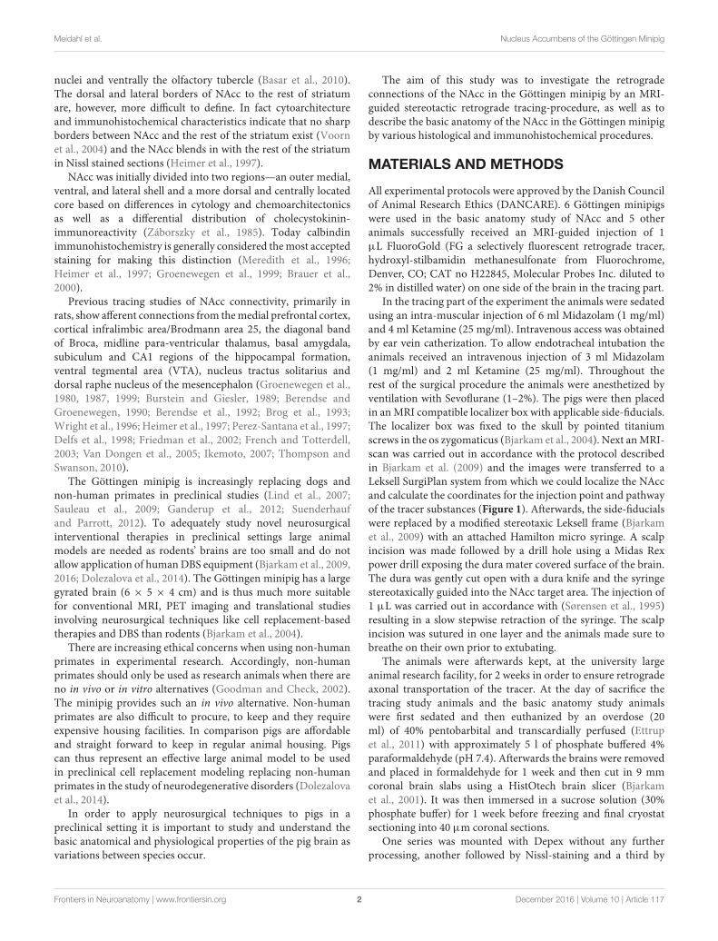
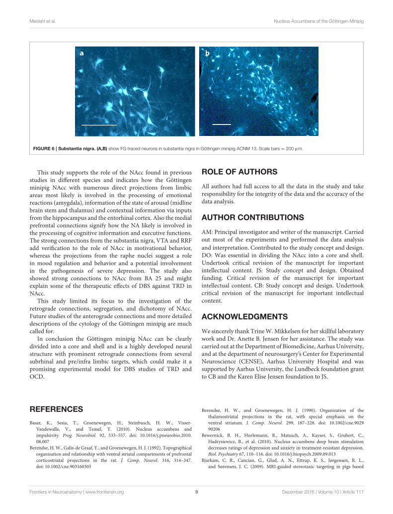
TracingAs shown in Table 1 the FG tracing technique revealed inputs toNAcc originating from medial parts of the prefrontal cortex, BA25 (subgenual cortex), the contralateral NAcc, basal amygdala,ventral CA1 region of hippocampus and entorhinal cortex,subiculum, the paraventricular and anterior parts of thalamus,dorsomedial parts of hypothalamus on the border betweenthalamus and hypothalamus, insula bilaterally, medial forebrainbundle (MFB), substantia nigra, VTA, retro rubral field (RRF),and dorsal and median raphe nuclei. FG was also found in fibersin the anterior commissure and the MFB. Figure 5 demonstratesFG-tracing to BA 25 and the medial prefrontal cortex andFigure 6 demonstrates FG-tracing to substantia nigra.
In the first pig the injection was made in the caudal partof NAcc. In the second it was more rostral in the NAcc. Theinjection in third was more ventral, medial and caudal, whereasit was more ventral in the fourth. As for the fifth pig the injectionwas in the dorsal, caudal part of the NAcc.
In mesencephalon very heavy tracing was found bilaterally inneurons in substantia nigra (A9)—Figure 6—in all pigs, but mostpronounced on the ipsilateral site of the injection. FG tracing wasalso quite pronounced in the ventral tegmental area VTA (A10)bilaterally and in the retro rubral field (A8) unilaterally. Tracedneurons were also found bilaterally in both themedian and dorsalraphe nucleus in all pigs. Fiber tracing in the MFB was found inall pigs.
In diencephalon intense FG tracing was found unilaterallyin the paraventricular parts of thalamus. FG was found in the
Frontiers in Neuroanatomy | www.frontiersin.org 4 December 2016 | Volume 10 | Article 117

Meidahl et al. Nucleus Accumbens of the Göttingen Minipig
FIGURE 2 | Nucleus accumbens Nissl and anti DARPP-32 staining. (A,C) show Nissl stained coronal sections of the nucleus accumbens (NA), the internal
capsule (IC), the putamen (Put), the prepiriform cortex (PP) and the olfactory tubercle (TO). (B,D) Corresponding anti-DARPP-32 staining. Scale bar = 5 mm.
anterior thalamic structures in pig number 3, 4, and 5, but notin number 1 and 2. Traced neurons in the ventral thalamus wereonly seen in number 4 and not in any other brains. Tracingto the ventromedial thalamus was only seen in number 2. Inhypothalamus the tracing was less pronounced, but weak tomoderate neuronal tracing was found in the dorsomedial partsof hypothalamus on the border to thalamus in all pigs.
In telencephalon heavily labeled cells were consistently seenin the medial parts of the prefrontal cortex, most pronounced onthe same site as the injection. Also prominent tracing to the insulawas persistently seen in all the pigs. In three out of five animals
the insular tracing was bilateral. The tracing was, however, muchmore moderate on the contra lateral site of the injection. Labeledneurons in the perirhinal cortex were seen in four pigs. Amygdalawas also heavily FG labeled.
Consistent and strong labeling of cells was seen in Brodmannarea 25. In one animal (number 3) more moderate contra lateraltracing was also noted.
In the ventral CA1 region of the hippocampus and subiculumweak, but evident, tracing was found in neurons in animalnumber 1, 2, 4, and 5. Whereas very strong tracing was foundin the ventral CA1 region and subiculum in number 3. Weak
Frontiers in Neuroanatomy | www.frontiersin.org 5 December 2016 | Volume 10 | Article 117

Meidahl et al. Nucleus Accumbens of the Göttingen Minipig
FIGURE 3 | Continuous coronal sections of nucleus accumbens stained with NISSL, autometallographic (AMG) myelin staining and anti-DARPP-32
staining showing nucleus accumbens (NAcc), the internal capsule, caudate nucleus (NC), putamen (Pu), the posterior part of the anterior olcaftory
nucleus (AOP), olfactory tubercle (Tu), septum (Sep), and Broadmann Area 25 (BA25).
Frontiers in Neuroanatomy | www.frontiersin.org 6 December 2016 | Volume 10 | Article 117

Meidahl et al. Nucleus Accumbens of the Göttingen Minipig
FIGURE 4 | Nucleus accumbens stained with monoclonal mouse
Anti-Calbindin antibody. The star marks the core of the NAcc and the
arrows mark the contours of the core toward the shell. The shell more
gradually fades out toward the border of the NAcc.
but evident tracing in neurons was also found in gyrus cinguliin number 4 and 5. Moderate tracing was found in the anteriorolfactory nucleus (AON) in number 2 and 5 and in the habenularnucleus in 1 and 4.
No tracing was found in any other parts of the striatumaside from the NAcc, nor in any other cortical areas than thosementioned above. Also, no tracing was found in the cerebellum.Septum was, likewise, carefully examined in all brains, but notracing was seen.
Traced fibers were also found in the anterior commissure andin the MFB in all pigs.
DISCUSSION
The afferent connections to the NAcc in the Göttingen minipigare for most parts in agreement with the connections foundin previous studies, primarily in rats. Inputs from structuressuch as VTA, substantia nigra, the paraventricular parts ofthalamus, hypothalamus, medial parts of the prefrontal cortex,insula, the dorsal raphe nucleus, hippocampus, subiculum andamygdala were all found in both the studied literature andin this experiment (Groenewegen et al., 1980, 1987, 1999;Burstein and Giesler, 1989; Berendse and Groenewegen, 1990;Berendse et al., 1992; Brog et al., 1993; Wright et al., 1996;Heimer et al., 1997; Perez-Santana et al., 1997; Delfs et al.,1998; Friedman et al., 2002; Van Dongen et al., 2005; Ikemoto,2007).
To our knowledge only one other study (Haber et al., 1995)found tracing to the BA 25. This is very interesting considering,that this is another target for DBS treatment of TRD and OCDin humans, and because BA 25 is thought to play an important
TABLE 1 | List of the anatomical structures where FG traced neurons were
found.
Traced areas/animal number 1 2 3 4 5
Prefrontal cortex medial ttt ttt+b ttt+b ttt
Area 25/subgenual cortex ttt ttt ttt+b ttt ttt
Insula ttt ttt+b tttb ttt+b tt
Prepiriform cortex t / t
Perirhinal cortex tt t / tt tt
Cingulate cortex / / t t /
Septum / / / / /
Anterior commissure t+b t+b t+b
Hippocampus CAl t tt ttt t t
Subiculum / / / / /
Regio diagonalis t t
Amygdala ttt tt ttt ttt ttt
Anterior olfactory nucleus t / tt
Entorhinal cortex t t tt tt tt
Nucleus habenularis t tt
Thalamus paraventricular/medial tt tt tt ttt ttt
Thalamus ventromedial t / /
Thalamus, anterior ttt tt ttt
Thalamus ventral ttt /
Border between thalamus and
hypothalamus
t t tt t t
Raphe nucleus, dorsalis tt+b t+b tt+b t+b t+b
Raphe nucleus median t+b t+b t+b t t
MFB–medial forbrain bundle ttt tt tt ttt ttt
VTA ttt ttt+b tt+b ttt+b ttt+b
Substantia nigra ttt+b ttt+b ttt+b ttt+b ttt+b
RRF (A8) t tt tt
ttt, strong tracing; tt, medium tracing; t, weak tracing; b, bilateral. In the anterior
commissure and MFB traced fibers and not neurons were seen.
role in the pathophysiology of the former condition (Hauptmanet al., 2008). The therapeutic effects of NAcc-DBS and BA25-DBSmight accordingly be elicited by stimulation of a limbic circuitryanchored in these two structures. Descriptions of projectionsfrom the AON, the cingulate cortex and nucleus habenularis tothe NAcc were not to our knowledge shown in previous rat orcat studies. Tracing to the AON was only found in ACNM 2and 5. Considering how close the AON is located to the NAccit seems likely to be spill-over tracing from the NAcc injectionsite. Hence this study does not provide sufficient evidence toconclude the existence of connections to the NAcc originatingfrom AON. Traced neurons in the cingulate cortex were onlyfound in animals 3 and 4. The injection in these pigs wasmade in the ventral part of NAcc, and it is possible that theventral NAcc receives different afferent connections from therest of the NAcc. However, further anterograde studies fromthe cingulate cortex in the Göttingen minipig are needed toverify if such connections exist. Traced fibers in the anteriorcommissure show that this very likely is the way by which theNAcc receives its afferents from the contra lateral side of thebrain. We cannot, however, rule out that some amount of tracersubstance might have spread to the anterior commissure. The
Frontiers in Neuroanatomy | www.frontiersin.org 7 December 2016 | Volume 10 | Article 117
Meidahl et al. Nucleus Accumbens of the Göttingen Minipig
FIGURE 5 | BA 25. (A,B,D) show coronal Nissl- staining with scale bars = 5 mm. BA 25 is marked with green in (B,D). (C) Shows fluorescence microscopy of FG
traced neurons in BA 25, scale bar = 200 µm.
injections in pigs, number 1, 2, and 5 were, however, in verydifferent areas of the NAcc, why it seems most likely to besignificant.
Tracer spread into neighboring areas of the injection site cannever entirely be ruled out despite the use of a very preciseMRI-guided stereotaxic injection method. The consistency ofthe results in all five pigs with injection sites found in variouslocations of the nucleus, as well as, the fact that the findingsalign with previous studies in non-human primates and rodents,however, makes our findings credible.
Heimer et al. described how in primates various projectionsto the NAcc are topographically organized and that variousbrain areas predominantly seem to project to certain partsof the NAcc (Heimer et al., 1997). Even though our findings
are in agreement with that of Heimer our study calls forfuture topographical projection studies in Göttingen minipigas DBS or cell replacement therapies could have differenteffects depending on what area of NAcc that is targeted ifthere same topographical pattern were to be found in theminipig as in primates. However, such studies would currentlyonly have post-mortem implications in neuro interventionalstudies with DBS and cell replacement therapies in the sensethat current MRI scanners and stereotactic devices as seenin Figure 1 would not enable one to certify more preciselywhere for example DBS electrodes had been placed within theNAcc due to its quite complicated complicated neurochemicalanatomy and the precision of stereotactic devices and MRIresolutions.
Frontiers in Neuroanatomy | www.frontiersin.org 8 December 2016 | Volume 10 | Article 117
Meidahl et al. Nucleus Accumbens of the Göttingen Minipig
FIGURE 6 | Substantia nigra. (A,B) show FG traced neurons in substantia nigra in Göttingen minipig ACNM 13. Scale bars = 200 µm.
This study supports the role of the NAcc found in previousstudies in different species and indicates how the Göttingenminipig NAcc with numerous direct projections from limbicareas most likely is involved in the processing of emotionalreactions (amygdala), information of the state of arousal (midlinebrain stem and thalamus) and contextual information via inputsfrom the hippocampus and the entorhinal cortex. Also themedialprefrontal connections signify how the NA likely is involved inthe processing of cognitive information and executive functions.The strong connections from the substantia nigra, VTA and RRFadd verification to the role of NAcc in motivational behavior,whereas the projections from the raphe nuclei suggest a rolein mood regulation and behavior and a potential involvementin the pathogenesis of severe depression. The study alsoshowed strong connections to NAcc from BA 25 and mightexplain some of the therapeutic effects of DBS against TRD inNAcc.
This study limited its focus to the investigation of theretrograde connections, segregation, and dichotomy of NAcc.Future studies of the anterograde connections and more detaileddescriptions of the cytology of the Göttingen minipig are muchcalled for.
In conclusion the Göttingen minipig NAcc can be clearlydivided into a core and shell and is a highly developed neuralstructure with prominent retrograde connections from severalsubrhinal and pre/infra limbic targets, which could make it apromising experimental model for DBS studies of TRD andOCD.
ROLE OF AUTHORS
All authors had full access to all the data in the study and takeresponsibility for the integrity of the data and the accuracy of thedata analysis.
AUTHOR CONTRIBUTIONS
AM: Principal investigator and writer of the manuscript. Carriedout most of the experiments and performed the data analysis
and interpretation. Contributed to the study concept and design.DO: Was essential in dividing the NAcc into a core and shell.Undertook critical revision of the manuscript for importantintellectual content. JS: Study concept and design. Obtainedfunding. Critical revision of the manuscript for importantintellectual content. CB: Study concept and design. Undertookcritical revision of the manuscript for important intellectualcontent.
ACKNOWLEDGMENTS
We sincerely thank TrineW.Mikkelsen for her skillful laboratorywork and Dr. Anette B. Jensen for her assistance. The study wascarried out at theDepartment of Biomedicine, AarhusUniversity,and at the department of neurosurgery’s Center for ExperimentalNeuroscience (CENSE), Aarhus University Hospital and wassupported by Aarhus University, the Lundbeck foundation grantto CB and the Karen Elise Jensen foundation to JS.
REFERENCES
Basar, K., Sesia, T., Groenewegen, H., Steinbusch, H. W., Visser-
Vandewalle, V., and Temel, Y. (2010). Nucleus accumbens and
impulsivity. Prog. Neurobiol. 92, 533–557. doi: 10.1016/j.pneurobio.2010.
08.007
Berendse, H.W., Galis-de Graaf, Y., and Groenewegen, H. J. (1992). Topographical
organization and relationship with ventral striatal compartments of prefrontal
corticostriatal projections in the rat. J. Comp. Neurol. 316, 314–347.
doi: 10.1002/cne.903160305
Berendse, H. W., and Groenewegen, H. J. (1990). Organization of the
thalamostriatal projections in the rat, with special emphasis on the
ventral striatum. J. Comp. Neurol. 299, 187–228. doi: 10.1002/cne.9029
90206
Bewernick, B. H., Hurlemann, R., Matusch, A., Kayser, S., Grubert, C.,
Hadrysiewicz, B., et al. (2010). Nucleus accumbens deep brain stimulation
decreases ratings of depression and anxiety in treatment-resistant depression.
Biol. Psychiatry 67, 110–116. doi: 10.1016/j.biopsych.2009.09.013
Bjarkam, C. R., Cancian, G., Glud, A. N., Ettrup, K. S., Jørgensen, R. L.,
and Sørensen, J. C. (2009). MRI-guided stereotaxic targeting in pigs based
Frontiers in Neuroanatomy | www.frontiersin.org 9 December 2016 | Volume 10 | Article 117

Meidahl et al. Nucleus Accumbens of the Göttingen Minipig
on a stereotaxic localizer box fitted with an isocentric frame and use of
SurgiPlan computer-planning software. J. Neurosci. Methods 183, 119–126.
doi: 10.1016/j.jneumeth.2009.06.019
Bjarkam, C. R., Cancian, G., Larsen, M., Rosendahl, F., Ettrup, K. S., Zeidler,
D., et al. (2004). A MRI-compatible stereotaxic localizer box enables high-
precision stereotaxic procedures in pigs. J. Neurosci. Methods 139, 293–298.
doi: 10.1016/j.jneumeth.2004.05.004
Bjarkam, C. R., Glud, A. N., Orlowski, D., Sørensen, J. C. H., and Palomero-
gallagher, N. (2016). The telencephalon of the Göttingen minipig,
cytoarchitecture and cortical surface anatomy. Brain Struct. Funct.
doi: 10.1007/s00429-016-1327-5. [Epub ahead of print].
Bjarkam, C. R., Pedersen, M., and Sorensen, J. C. (2001). New strategies for
embedding, orientation and sectioning of small brain specimens enable direct
correlation to MR-images, brain atlases, or use of unbiased stereology. J.
Neurosci. Methods 108, 153–159. doi: 10.1016/S0165-0270(01)00383-1
Bjarkam, C. R., Sørensen, J. C., and Geneser, F. A. (2005). Distribution and
morphology of serotonin-immunoreactive axons in the retrohippocampal
areas of the New Zealand white rabbit. Anat. Embryol. (Berl). 210, 199–207.
doi: 10.1007/s00429-005-0004-x
Brauer, K., Häußer, M., Härtig, W., and Arendt, T. (2000). The core-shell
dichotomy of nucleus accumbens in the rhesus monkey as revealed by double-
immunofluorescence and morphology of cholinergic interneurons. Brain Res.
858, 151–162. doi: 10.1016/S0006-8993(00)01938-7
Brog, J. S., Ongse, A. S., Deutch, A. Y., and Zahm, D. S. (1993). The patterns of
afferent innervation of the core and shell in the “Accumbens” part of the rat
ventral striatum : immunohistochemical detection of retrogradely transported
Fluoro-Gold. J. Comp. Neurol. 338, 255–278.
Burstein, R., and Giesler, G. J. (1989). Retrograde labeling of neurons in
spinal cord that project directly to nucleus accumbens or the septal
nuclei in the rat. Brain Res. 497, 149–154. doi: 10.1016/0006-8993(89)
90981-5
de Koning, P. P., Figee, M., Van Den Munckhof, P., Schuurman, P. R., and Denys,
D. (2011). Current status of deep brain stimulation for obsessive-compulsive
disorder: a clinical review of different targets.Curr. Psychiatry Rep. 13, 274–282.
doi: 10.1007/s11920-011-0200-8
Delfs, J. M., Zhu, Y., Druhan, J. P., and Aston-Jones, G. S. (1998). Origin of
noradrenergic afferents to the shell subregion of the nucleus accumbens:
anterograde and retrograde tract-tracing studies in the rat. Brain Res. 806,
127–140. doi: 10.1016/S0006-8993(98)00672-6
Dolezalova, D., Hruska-Plochan, M., Bjarkam, C. R., Sørensen, J. C.
H., Cunningham, M., Weingarten, D., et al. (2014). Pig models of
neurodegenerative disorders: utilization in cell replacement-based
preclinical safety and efficacy studies. J. Comp. Neurol. 522, 2784–2801.
doi: 10.1002/cne.23575
Ettrup, K. S., Glud, A. N., Orlowski, D., Fitting, L. M., Meier, K., Soerensen, J. C.,
et al. (2011). Basic surgical techniques in the Göttingen minipig: intubation,
bladder catheterization, femoral vessel catheterization, and transcardial
perfusion. J. Vis. Exp. 52:2652. doi: 10.3791/2652
French, S. J., and Totterdell, S. (2003). Individual nucleus accumbens-projection
neurons receive both basolateral amygdala and ventral subicular afferents in
rats. Neuroscience 119, 19–31. doi: 10.1016/S0306-4522(03)00150-7
Friedman, D. P., Aggleton, J. P., and Saunders, R. C. (2002). Comparison of
hippocampal, amygdala, and perirhinal projections to the nucleus accumbens:
combined anterograde and retrograde tracing study in the macaque brain. J.
Comp. Neurol. 450, 345–365. doi: 10.1002/cne.10336
Ganderup, N. C., Harvey, W., Mortensen, J. T., and Harrouk, W. (2012). The
minipig as nonrodent species in toxicology–where are we now? Int. J. Toxicol.
31, 507–528. doi: 10.1177/1091581812462039
Goodman, S., and Check, E. (2002). The great primate debate. Nature 417,
684–687. doi: 10.1038/417684a
Groenewegen, H. J., Becker, N. E. H. M., and Lohman, A. H. M. (1980). Subcortical
afferents of the nucleus accumbens septi in the cat, studied with retrograde
axonal transport of horseradish peroxidase and bisbenzimid. Neuroscience 5,
1903–1916. doi: 10.1016/0306-4522(80)90038-X
Groenewegen, H. J., Vermeulen-Van der Zee, E., te-Kortschot, A., and Witter, M.
P. (1987). Organization of the projections from the subiculum to the ventral
striatum in the rat. A study using anterograde transport of Phaseolus vulgaris
leucoagglutinin.Neuroscience 23, 103–120. doi: 10.1016/0306-4522(87)90275-2
Groenewegen, H. J., Wright, C. I., Beijer, A. V., and Voorn, P. (1999).
Convergence and segregation of ventral striatal inputs and outputs.
Ann. N.Y. Acad. Sci. 877, 49–63. doi: 10.1111/j.1749-6632.1999.tb
09260.x
Haber, S. N., Kunishio, K., Mizobuchi, M., and Lynd-Balta, E. (1995). The orbital
and medial prefrontal circuit through the primate basal ganglia. J. Neurosci. 15,
4851–4867.
Hauptman, J. S., DeSalles, A. A., Espinoza, R., Sedrak, M., and Ishida, W.
(2008). Potential surgical targets for deep brain stimulation in treatment-
resistant depression. Neurosurg. Focus 25:E3. doi: 10.3171/FOC/2008/
25/7/E3
Heimer, L., Aiheid, G. F., de Olmos, J. S., Groenewegen, H. J., Haber, S. N.,
Harlan, R. E., et al. (1997). The accumbens: beyond core-shell dichotomy. J.
Neuropsychiatry Clin. Neurosci. 9, 354–381.
Hemmings, H. C. Jr, Nairn, A. C., Aswad, D. W., and Greengard, P.
(1984). DARPP-32, a dopamine- and adenosine 3′:5′-monophosphate-
regulated phosphoprotein enriched in dopamine-innervated brain regions. II.
Purification and characterization of the phosphoprotein from bovine caudate
nucleus. J. Neurosci. 4, 99–110.
Ikemoto, S. (2007). Dopamine reward circuitry: two projection systems
from the ventral midbrain to the nucleus accumbens-olfactory tubercle
complex. Brain Res. Rev. 56, 27–78. doi: 10.1016/j.brainresrev.2007.
05.004
Larsen, M., Bjarkam, C. R., Stoltenberg, M., Sørensen, J. C., and Danscher, G.
(2003). An autometallographic technique for myelin staining in formaldehyde-
fixed tissue. Histol. Histopathol. 18, 1125–1130.
Li, N., Gao, L., Wang, X., Chen, L., Fang, W., Ge, S., et al. (2013). Deep brain
stimulation of the bilateral nucleus accumbens in normal rhesus monkey.
Neuroreport 24, 30–35. doi: 10.1097/WNR.0b013e32835c16e7
Lind, N. M., Moustgaard, A., Jelsing, J., Vajta, G., Cumming, P., and Hansen, A.
K. (2007). The use of pigs in neuroscience: modeling brain disorders. Neurosci.
Biobehav. Rev. 31, 728–751. doi: 10.1016/j.neubiorev.2007.02.003
Lucas-Neto, L., Neto, D., Oliveira, E., Martins, H., Mourato, B., Correia, F., et al.
(2013). Three dimensional anatomy of the human nucleus accumbens. Acta
Neurochir. (Wien). 155, 2389–2398. doi: 10.1007/s00701-013-1820-z
Meredith, G. E., Pattiselanno, A., Groenewegen, H. J., and Haber, S.
N. (1996). Shell and core in monkey and human nucleus accumbens
identified with antibodies to calbindin-D28k. J. Comp. Neurol. 365, 628–639.
doi: 10.1002/(SICI)1096-9861(19960219)365:4<628::AID-CNE9>3.0.CO;2-6
Perez-Santana, L., Marín, O., and Smeets, W. J. (1997). Afferent connections of the
nucleus accumbens of the snake, Elaphe guttata, studied by means of in vitro
and in vivo tracing techniques in combination with TH immunohistochemistry.
Neurosci. Lett. 225, 101–104. doi: 10.1016/S0304-3940(97)00205-X
Sauleau, P., Lapouble, E., Val-Laillet, D., and Malbert, C. H. (2009). The
pig model in brain imaging and neurosurgery. Animal 3, 1138–1151.
doi: 10.1017/S1751731109004649
Schlaepfer, T. E., Cohen, M. X., Frick, C., Kosel, M., Brodesser, D., Axmacher,
N., et al. (2007). Deep brain stimulation to reward circuitry alleviates
anhedonia in refractory major depression. Neuropsychopharmacology 33,
368–377. doi: 10.1038/sj.npp.1301408
Sørensen, J. C., Slomianka, L., Christensen, J., and Zimmer, J. (1995). Zinc-
containing telencephalic connections to the rat striatum: a combined Fluoro-
Gold tracing and histochemical study. Exp. Brain Res. 105, 370–382.
Suenderhauf, C., and Parrott, N. (2012). A physiologically based pharmacokinetic
model of the minipig: data compilation and model implementation. Pharm.
Res. 30, 1–15. doi: 10.1007/s11095-012-0911-5
Svenningsson, P., Nishi, A., Fisone, G., Girault, J.-A., Nairn, A.
C., and Greengard, P. (2004). DARPP-32: an integrator of
neurotransmission. Annu. Rev. Pharmacol. Toxicol. 44, 269–296.
doi: 10.1146/annurev.pharmtox.44.101802.121415
Thompson, R. H., and Swanson, L. W. (2010). Hypothesis-driven structural
connectivity analysis supports network over hierarchical model of
brain architecture. Proc. Natl. Acad. Sci. U.S.A. 107, 15235–15239.
doi: 10.1073/pnas.1009112107
Van Dongen, Y. C., Deniau, J. M., Pennartz, C. M., Galis-De Graaf, Y., Voorn,
P., Thierry, A. M., et al. (2005). Anatomical evidence for direct connections
between the shell and core subregions of the rat nucleus accumbens.
Neuroscience 136, 1049–1071. doi: 10.1016/j.neuroscience.2005.08.050
Frontiers in Neuroanatomy | www.frontiersin.org 10 December 2016 | Volume 10 | Article 117

Meidahl et al. Nucleus Accumbens of the Göttingen Minipig
Voorn, P., Vanderschuren, L. J., Groenewegen, H. J., Robbins, T.W., and Pennartz,
C. M. (2004). Putting a spin on the dorsal-ventral divide of the striatum. Trends
Neurosci. 27, 468–474. doi: 10.1016/j.tins.2004.06.006
Wright, C. I., Beijer, A. V., and Groenewegen, H. J. (1996). Basal amygdaloid
complex afferents to the rat nucleus accumbens are compartmentally organized.
J. Neurosci. 16, 1877–1893.
Záborszky, L., Alheid, G. F., Beinfeld, M. C., Eiden, L. E., Heimer, L., and
Palkovits, M. (1985). Cholecystokinin innervation of the ventral striatum:
a morphological and radioimmunological study. Neuroscience 14, 427–445.
doi: 10.1016/0306-4522(85)90302-1
Conflict of Interest Statement: The authors declare that the research was
conducted in the absence of any commercial or financial relationships that could
be construed as a potential conflict of interest.
Copyright © 2016 Meidahl, Orlowski, Sørensen and Bjarkam. This is an open-access
article distributed under the terms of the Creative Commons Attribution License (CC
BY). The use, distribution or reproduction in other forums is permitted, provided the
original author(s) or licensor are credited and that the original publication in this
journal is cited, in accordance with accepted academic practice. No use, distribution
or reproduction is permitted which does not comply with these terms.
Frontiers in Neuroanatomy | www.frontiersin.org 11 December 2016 | Volume 10 | Article 117