the possible neuroprotective effect of astaxanthin on
TRANSCRIPT

684
Personal non-commercial use only. EJH copyright © 2020. All rights reserved DOI: 10.21608/ejh.2020.22609.1235
Original Article
The Possible Neuroprotective Effect of Astaxanthin on Monosodium Glutamate and Aspartame Induced Hippocampal Changes in Albino Rats: (Histological and Immuno-histochemical Study)1Gihan I. Aboul Fotouh, 1Safinaz Salah Eldin Sayed, 2Zainab Mohammed Altayeb, 2Eman Adel Mahmoud Zaher
Histology Department, Faculty of Medicine, 1Cairo University, 2Helwan University, Cairo, Egypt
ABSTRACTBackground: Food additives are substances added to food to improve its safety, freshness, taste, texture, or appearance. They include aroma enhancer eg : monosodium glutamate(MSG) or sweetener eg.: aspartame (ASP). Aim of the Study: This current study was performed to study the microscopic alterations induced by MSG, ASP individually and in combination and the possible protective effect of astaxanthin (AST) on those induced changes.Materials and Methods: Forty nine adult male albino rats were used in this study divided into; control group (fourteen rats) two rats for each experimental group and experimental group (thirty five rats) subdivided into seven subgroups:ASP group, MSG group, ASP andMSG group,AST group,ASP andAST group, MSG andAST group, ASPandMSG andAST group. Oral administration was done in the morning daily for 6 weeks for all groups. When the duration of the study ended, blood samples were collected from rat tail and paraffin sections were prepared from the cerebral hemisphere of each animal. They were subjected to hematoxylin and eosin stain and immunohistochemical stain for Caspase3 and Glial Fibrillary Acidic Protein (GFAP). Statistical analysis were done for assessment of body weight, reduced glutathione (GSH) and tumor necrosis factor α (TNF-α) level.Results: The groups that received ASP (I), MSG (II) individually or in combination (III) exhibited shrunken cells with darkly stained nuclei and surrounded with pericellular haloes and some areas even revealed loss of the cells with increase in immunoreactivity for GFAP and Caspase 3. These groups also showed elevation in the level of TNFα and decrease in the level of GSH. On treatment with AST groups V, VI and VII showed reduced pycknosis, decreased immunoreactivity for GFAP and Caspase 3. It also showed reduction in the level of TNFα and increased level of GSH.Conclusion: ASP and MSG individually or in combination induce alternation in hippocampus and AST administration ameliorate those changesReceived: 18 January 2020, Accepted: 05 February 2020
Key Words: Aspartame, astaxanthin, hippocampus, monosodium glutamate. Corresponding Author: Eman Adel Zaher, MD, Histology Department, Faculty of Medicine, Helwan University, Cairo, Egypt, Tel.: +20 1090435057, E-mail: [email protected]: 1110-0559, Vol. 43, No.3
INTRODUCTION
Junk foods have proved to be a big problem for human health in general and especially for children, as they contain synthetic flavors and preservatives. Food additives contain two main types, which include aroma enhancers e.g monosodium glutamate and sweeteners e.g aspartame[1,2].
Monosodium glutamate (MSG) (E621) is a salt form of L-glutamic acid and is widely used all over the world to enhance the flavor. It was reported that the mean amount ingested from MSG in the individual within industrialized countries is about 0.3–1.0 g/day. Glutamate, the amino acid in MSG, when taken in extra amounts, it acts as a strong neurotoxin as it increases the excitability and activity of proteolytic enzymes[3].
Aspartame (ASP) is an artificial sweetener that is being used in alternation to sugar in many food products. It is approximately 200 times sweeter than sugar so, used in low-calorie soft drinks and foods[4]. It is rapidly absorbed
and metabolized by intestinal esterase and dipeptidase to 40% aspartic, 50% phenyl alanine and10% methanol. Phenylalanine has an important role as a regulator of the process of neurotransmition, moreover, it was reported that aspartic acid has an important role as an excitatory neurotransmitter in the central nervous system[5].
Astaxanthin (AST) is a carotenoid that is separated from shrimp and crab shells. It has strong antioxidant properties. It was revealed that AST has an effective role in scavenging oxygen free radicals, decrease lipid peroxidation, oxidative stress and inhibit reactive oxygen species (ROS)-mediated toxicity[6].
AIM OF THE WORK
The purpose of this study is to shed light on the changes induced by monosodium glutamate and aspartame separately or in combination in hippocampus and to demonstrate possible protective role of astaxanthin on those induced changes.

685
Aboul Fotouh et. al.
MATERIALS AND METHODS
a) Materials used
Monosodium GlutamateMonosodium glutamate (MSG) powder of purity ≥99%
was purchased from Beta pharm. It was administrated, orally, via intragastric tube once daily for 6 weeks (120 mg/kg dissolved in distilled water)[7].
AspartameAspartame powder was purchased from beta pharm and
administered to the rats by intragastric tube for 6 weeks (250 mg /kg dissolved in distilled water)[8].
AstaxanthinIt was obtained from BIOVE in the form of soft gel
container and administered via intragastric tube for 6 weeks (2 mg/kg dissolved in version olive oil)[9].
b) AnimalsThe study included forty nine adult male albino rats
with a weight range of (150-200 gm). They were obtained and handled according to the guidelines after the approval of the ethical committee (CU III F 87 17) from the Animal House-Faculty of Medicine at Cairo University. All the animals were handled for one week prior to experiment to minimize nonspecific stress on the days of the experiment, and were kept at room temperature (24-26C) with a 12:12h light:dark cycle. They were provided with standard crushed food and water during the experiment. The bodies of the dead animals were disposed of by using the incinerator.
Control GroupFourteen rats were used, two rats for each of the
corresponding experimental groups.
• Six rats received 0.5 ml of distilled water
• Two rats received 0.5 ml of version olive oil
• Six rats received both 0.5 ml of distilled water and 0.5 ml of virgin olive oil.
All animals received the solvent orally once daily via intragastric tube.
Experimental GroupsThey were subdivided to seven groups that received the
drugs orally via intragastric tube once daily for six weeks.
I. ASP group: five rats received 250 mg/kg ASP dissolved in 0.5ml of distilled water. Dose consumptions between rats and humans were corrected by factor 5 as aspartame metabolizes in rats faster than that in humans.
II. MSG group: five rats received 120 mg/kg MSG dissolved in 0.5ml of distilled water
III. ASP&MSP group: five rats received ASP as in group I and MSG as in group II.
IV. AST group: five rats received AST (2 mg/kg) dissolved in 0.5ml virgin olive oil.
V. ASP& AST group: five rats received ASP as in group I and AST as in group IV.
VI. MSG &AST group: five rats received MSG as in group II and AST as in group IV.
VII. ASP, MSG &AST group: five rats received ASP as in group I, MSG as in group II and AST as in group VI.
The body weight of each rat in all groups was measured at the beginning and at the end of the experiment. Statistical study on the mean weight was done.
At the end of 6th week, blood samples were collected from rat tail. Under general anesthesia (sodium pentobarbital 100mg/kg) administrated intra-peritoneally[10], the animals were perfused via the left ventricle with phosphate buffer (PH 7.4) containing 2% paraformaldehyde[11]. Their cerebral hemisphere was dissected and cut sagittally into right and left halves.
І. Light Microscopic StudyThe left half of the cerebral hemisphere from each
animal was fixed in 10% formalin solution, and then specimens were processed for light microscopic study. Paraffin sections (5 μm thick) were stained with the following:
1. Haematoxylin and eosin stain (Bancroft and Layton, 2013)[12]
2. Immunohistochemical stain for (Jackson and Blythe, 2013)[13].
• Caspace3 for apoptotic cells
• Glial Fibrillary Acidic Protein (GFAP) of the astrocytes
2- Statistical AnalysisBody weight, TNF-a level and GSH level of control
and experimental animals were subjected to statistical analysis using the PASW software system version 16. The statistical analysis of data was done by using Excel program and statistical package for social science (SPSS) program on windows XP. Comparison between normally distributed (parametric) quantitative data of more than two groups was conducted using ANOVA (analysis of variance) test followed by post-hoc for multiple comparisons
NB: The P value of ≤ 0.05 indicates significant results at confidence interval 9[14].
3- Morphometric StudyUsing Leica Qwin 500 LTD image analysis in medical
histology department of Cairo University, the following parameters were measured:
A. Area percent of Caspase 3 +ve cells in Caspase 3 immunostained sections.

686
HISTOLOGICAL AND IMMUNO-HISTOCHEMICAL STUDY
B. Area percent of GFAP +ve cells in GFAP immunostained sections.
The measurements were done in 10 non overlapping high power fields (HPF x 200) in the experimental groups using binary mode
RESULTS
1) Histological ResultsThe hippocampus of control albino rats of all
groups was formed of Cornu Amminos: CA1 and CA2 (have small pyramidal cells), and CA3 and CA4 (have large pyramidal cells). CA4 projects into the concavity of dentate gyrus that is formed of small granule cells. Area in between Cornu Amminos and dentate gyrus comprise the molecular layer which consists of neuronal processes (axon and dendrites), glial cells, and scattered nerve cells (Figure 1).
Results of CA1Examination of CA1 region of all control groups
revealed the presence of three layers; molecular, pyramidal and polymorphic layers. The pyramidal layer is the main layer and it is composed of 5-6 compact layers of small pyramidal neurons with vesicular nuclei. The other two layers were devoid of neurons. They contained scanty nuclei of neuroglial cells such as astrocytes beside blood vessels on a pink neuropil background formed of neuronal and glial cell processes (Figure 2A).
In group I (ASP) there was apparent decreased thickness of the pyramidal layer .The cells were shrunken, had darkly stained nuclei and were surrounded with pericellular haloes and some areas even revealed loss of the pyramidal cells (Figure 2B).In MSG group the pyramidal layer had some cells with pale-stained nuclei, and distorted cellular morphology. Others shrunken cells with darkly stained nuclei and surrounded with pericellular haloes .There was congested blood vessel (Figure 2C).In ASP and MSG group the pyramidal layer showed the same results as in MSG group (Figure 2D). Both molecular and polymorphic layer of CA1 contained dilated blood capillaries, apoptotic dark stained neuroglia (Figures 2B, C and D).
AST treated group showed results similar to the control (Figure 2E). While ASP and AST treated group and MSG and AST were comparable to control group but few pyramidal cells were shrunken with darkly stained nuclei and surrounded by halos were seen (Figures 2F and G).In ASP, MSG and AST treated group, the pyramidal layer decreased in thickness and the cells had vesicular nuclei (Figure 2H). In all treated groups, both molecular and polymorphic layers showed relatively normal neuroglia.
Results of CA3In all groups CA3 in control rats was composed from
three layers; molecular, pyramidal and polymorphic layers. The pyramidal layer showed many large pyramidal neurons
with vesicular nuclei. The other two layers were devoid of neurons. Scanty number of nuclei of neuroglial cells as astrocytes was detected in these layers beside blood vessels on a pink neuropil background formed of neuronal and glial cell processes (Figure 3A).
In group I (ASP), some pyramidal cells were shrunken with dark stained nuclei and others with pale nuclei, Also some vacuolations appeared in the neuropil (Figure 3B).
In group II (MSG), the pyramidal layer showed marked disorganization with areas of cell loss. Most of large pyramidal cells were shrunken with dark stained nuclei, while few cells showed pale nuclei. There were also vacuolation in neuropil (Figure 3C).
In group III (ASP and MSG ) the pyramidal cells layer showed same results as group II (MSG) (Figure 3D).Both molecular and polymorphic layers of CA3 contained dilated blood capillaries, apoptotic dark stained neuroglia (Figures 3B, C and D).
In group IV (AST) the results were similar to the control (Figure 3E).In group V (ASP and AST) and group VI (MSG and AST) the results were comparable to control but still there were few shrunken cells with darkly stained nuclei (Figures 3F and 3G).
In group VII (ASP, MSG and AST) showed scattered pyramidal cells. It had vesicular nuclei relatively similar to control (Figure 3H). In the remaining treated groups, both molecular and polymorphic layers showed relatively normal appearance.
Results of Dentate GyrusIn all rats of control group, the dentate gyrus was
formed of molecular, granular and polymorphic layers. The granular layer is the main layer. It was composed of dense aggregate of granular cells that appeared rounded with vesicular nuclei. The polymorphic layer contains scattered large pyramidal cells with vesicular nuclei among the neuroglia while the molecular layer was formed of neuroglial cells (Figure 4A).
In group I (ASP), the granular layer showed areas of cell loss and some cells were shrunken with marked vacuolation in the neuropil. The molecular layer contained apoptotic neuroglial cells (Figure 4B). Similar results were seen in both group II (MSG) and group III (ASP and MSG) but the polymorphic layer of group III contained dark stained pyramidal cells surrounded by halos (Figures 4C and D).
In group IV (AST) the results similar to the control (Figure 4E). In all AST treated groups V, VI, VII the results were comparable to control group with vacuolation in groups VI and VII (Figures 4F and G and H).
2) Immunohistochemical ResultsImmunostained sections of hippocampus of all control
groups showed few GFAP positive immunoreactive astrocytes dispersed in CA1, CA3, dentate gyrus

687
Aboul Fotouh et. al.
(Figures 5A, B, C). Also, Examined control sections of rats hippocampus showed negative immunostaining with sparse positive immunoreactive cells for Caspase 3 (Figures 6A, B, C).
In group I (ASP), CA1, CA3 and dentate gyrus showed strong GFAP immunoreactive star – shaped astrocytes with intense brownish twisted irregular glial fibers (Figures 7A, B, C) .Caspase 3 immunostained section showed strong positive immune staining for hippocampus proper CA1 , CA3 and dentate gyrus (Figures 8A, B, C). Similar results were observed in group II (MSG) and group III (ASP and MSG) (Figures 9, 10, 11(A,B,C)). But group III(ASP and MSG) showed moderate reaction for caspase 3 immunostaining in CA1, CA3 and dentate gyrus (Figures 12A, B, C).
In group IV(AST) both GFAP and caspase 3 immunostained sections of hippocampus showed results similar to control (Figures 13 and 14 (A,B,C)).
All treated groups (V,VI,VII) by AST showed moderate positive GFAP immunoreactive astrocytes with faint brownish glial fiber (Figures 15, 17 and 19, (A,B,C)) and moderate reaction for caspase 3 (Figures 16, 18 and 20 (A,B,C))
2- Statistical Study
i- Body WeightThere was no significant difference in the mean of
body weight of group III (MSG administrated group), group V (AST treated group), group VI (ASP +AST treated group), group VII (MSG +AST treated group) and group VIII (ASP, MSG and AST) when compared with control one. On other hand, there was significant decrease in the mean of body weight of rats in both group II (ASP administrated rats) and group IV
(ASP +MSG administrated group) with (p< 0.001 )when compared with control one (Table 2 and histogram 3).
ii-Assessment of Reduced Glutathione (GSH ) and Tumor Necrosis Factor α (TNF-α) Level
The mean GSH level in ASP, MSG administrated group
individually or in combination showed a highly significant decrease as compared with that of control rats with (P <0.001). However, the mean GSH level in treated groups with AST treated group showed a significant increase as compared with the of ASP, MSG administrated rats individually and in combination (Table 1 and Histogram 1)
The mean TNF α level in ASP ,MSG administrated group individually and in combination showed a highly significant increase as compared with that of control rats with (P <0.001). However, the mean in AST treated groups showed a significant decrease as compared with that diseased rats (group I,II.III) with (P <0.001) (Histogram 2)
iii- Morphometric Results
1) GFAP immunorectivity (Table 3 and Histogram 4) The mean area percent of positive GFAP
immunoreactivity in CA1 , CA3 and dentate gyrus of group 1V(AST) showed no significant difference as compared with that of control rats .
Groups (I, II and III) showed a highly significant increase in immunoreactivity compared to the control rats with (P< 0.001). However, in groups (V,VI and VII) the mean area percent of positive GFAP immunoreactivity showed a significant decrease as compared with that of groups (I,II and III) with (P < 0.001) and non-significant increase as compare with control rats with (P> 0.05).
2)Caspase 3 immunorectivity: (Table 4 and Histogram 5):
The mean area percent of positive Caspase 3 immunoreactivity in CA1 , CA3 and dentate gyrus of group 1V(AST) showed no significant difference compared to control group.
Groups (I, II and III) showed a highly significant increase the mean area percent of positive Caspase 3 immunoreactivity compared to control rats with (P <0.001). However, groups (V, VI and VII) the mean area percent of positive caspase 3 immunoreactivity showed a significant decrease compared to groups (I ,II and III) with (P <0.001) and non-significant increase compared to control group with (P> 0.05).
688
HISTOLOGICAL AND IMMUNO-HISTOCHEMICAL STUDY
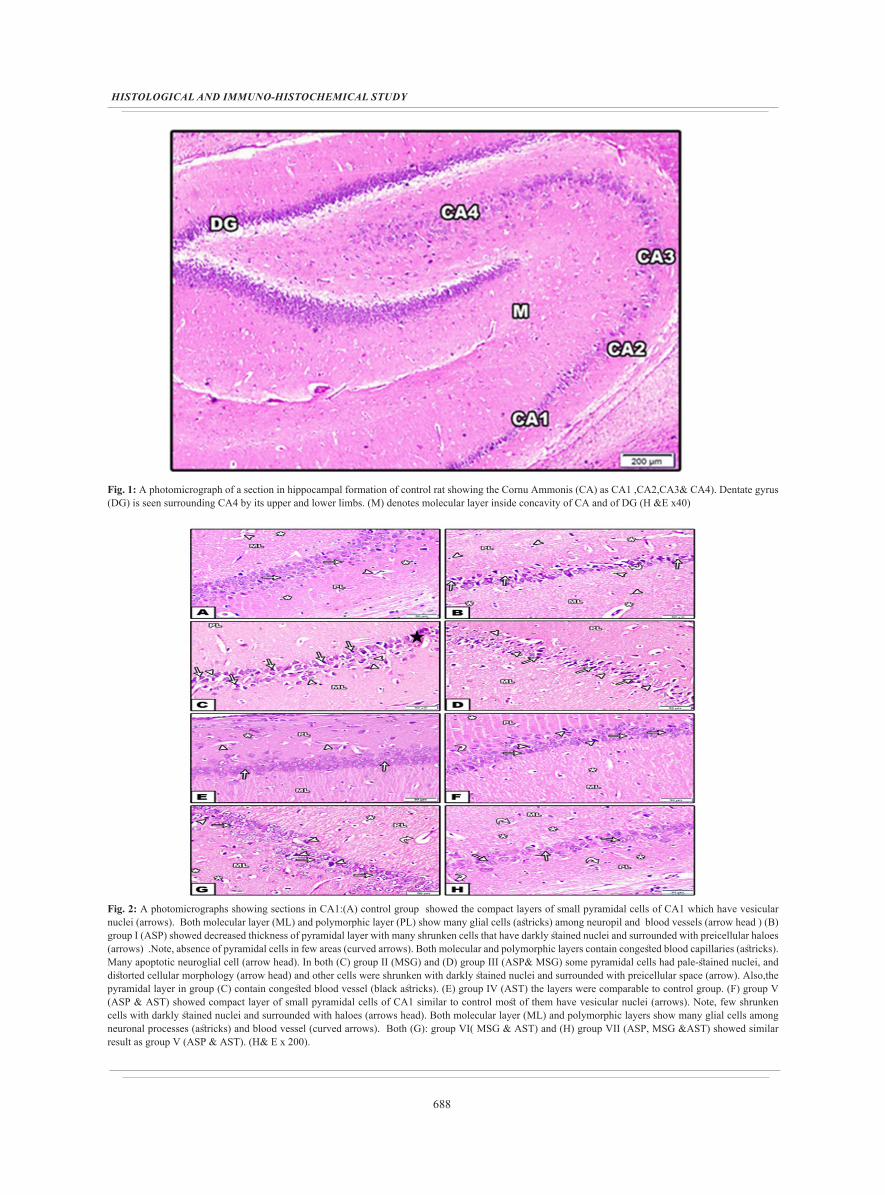
Fig. 1: A photomicrograph of a section in hippocampal formation of control rat showing the Cornu Ammonis (CA) as CA1 ,CA2,CA3& CA4). Dentate gyrus (DG) is seen surrounding CA4 by its upper and lower limbs. (M) denotes molecular layer inside concavity of CA and of DG (H &E x40)
Fig. 2: A photomicrographs showing sections in CA1:(A) control group showed the compact layers of small pyramidal cells of CA1 which have vesicular nuclei (arrows). Both molecular layer (ML) and polymorphic layer (PL) show many glial cells (astricks) among neuropil and blood vessels (arrow head ) (B) group I (ASP) showed decreased thickness of pyramidal layer with many shrunken cells that have darkly stained nuclei and surrounded with preicellular haloes (arrows) .Note, absence of pyramidal cells in few areas (curved arrows). Both molecular and polymorphic layers contain congested blood capillaries (astricks). Many apoptotic neuroglial cell (arrow head). In both (C) group II (MSG) and (D) group III (ASP& MSG) some pyramidal cells had pale-stained nuclei, and distorted cellular morphology (arrow head) and other cells were shrunken with darkly stained nuclei and surrounded with preicellular space (arrow). Also,the pyramidal layer in group (C) contain congested blood vessel (black astricks). (E) group IV (AST) the layers were comparable to control group. (F) group V (ASP & AST) showed compact layer of small pyramidal cells of CA1 similar to control most of them have vesicular nuclei (arrows). Note, few shrunken cells with darkly stained nuclei and surrounded with haloes (arrows head). Both molecular layer (ML) and polymorphic layers show many glial cells among neuronal processes (astricks) and blood vessel (curved arrows). Both (G): group VI( MSG & AST) and (H) group VII (ASP, MSG &AST) showed similar result as group V (ASP & AST). (H& E x 200).

689
Aboul Fotouh et. al.
Fig. 3: photomicrographs showing sections in CA3: (A) control group showed the three layers of CA3 region of hippocampus proper, molecular layer (ML), pyramidal layer, polymorphic layer (PL). Pyramidal layer contain large pyramidal cells (arrow) with vesicular nuclei .Both molecular and polymorphic layers show neuroglial cells (astrisks) and blood vessel (curved arrows).(B) group I (ASP) showed many shrunken pyramidal cells with dark stained nuclei (arrow)and vacuolated neuropil (astricks) .Both molecular and polymorphic layers contain many apoptotic neuroglial cells (arrow head) surrounded by empty spaces. In both (C) group II (MSG) and (D) group III (ASP& MSG), the pyramidal cell layer showed areas of cell loss (arrows). Most of the large pyramidal cells were shrunken with dark stained nuclei while, few cells had pale nuclei (arrow head) and neuropil showed vacuolation. However, there are clumping of neuronal process (curved arrows). (E) group IV (AST) the layers were comparable to control group. (F) group V (ASP & AST) most of the large pyramidal cells had vesicular nuclei (arrows). In Both (G): group VI (MSG & AST) and (H) group VII (ASP, MSG &AST) showed similar result as group V (ASP & AST). In addition group VI had few cells with pyknotic nuclei and surrounded by halo (arrow head). There are also dilated blood vessel (curved arrow). Group VII showed scattered pyramidal cells. (H& E x 200)
Fig. 4: photomicrographs showing sections in denentate gyrus: (A) control rats showed dentate gyrus forms of molecular layer (ML), granular layer (G) and polymorphic layer (PL). It was formed of dense aggregate of granular cells that appeared rounded with vesicular nuclei (arrow) .Dark cells are seen in subgranular zone (arrow head). The polymorphic layer contains scattered large pyramidal cells (p) with vesicular nuclei among the neuroglia while the molecular layer was formed of neuroglial cells (astricks). (B) group I (ASP) showed many granular cells shrunken (arrow) and some areas with cell loss (curved arrow) . Note, marked vacuolation (V) in neuropil and apoptotic dark stained neuroglia (astricks) in molecular (ML) layer. In both (C) group II (MSG) and (D) group III (ASP& MSG) were showed similar results as group I (ASP). In addition to dark stained apoptotic large pyramidal cells (p) in the polymorphic layer (E) group IV (AST) had similar results as control one. (F) group V (ASP & AST) , (G) group VI( MSG & AST) and (H) group VII (ASP, MSG &AST) were comparable to control group. Note, vacuolation (V) in both groups VI &VII. (H& E x 200)

690
HISTOLOGICAL AND IMMUNO-HISTOCHEMICAL STUDY
Fig. 5: photomicrographs of control sections of rat brain showing weak positive immunostaining for GFAP in hippocampus (A) CA1, (B) CA3, (C ) dentate gyrus. (Anti GFAP immunostain x200).
Fig. 6: photomicrographs sections of control rats showing negative immune staining for caspase 3 with few positive immune reactive cells (arrows) in hippocampus proper; (A) CA1 , (B) CA3 and (C) dentate gyrus. (Anti caspase 3 immunostain x 200)
Fig. 7: photomicrograph of ASP administrated sections of hippocampus proper (A) CA1 , (B) CA3, (C) dentate gyrus showing strong immunereactive star – shaped astrocytes (arrows) with intense brownish immunoreaction in glial fibers which appeared twisted and had irregular course (arrow head) (Anti GFAP immunostain x200).
Fig. 8: photomicrographs of ASP administrated rats showing strong reaction for caspace 3 in rat (A) CA1 ,(B) CA3 and (C) dentate gyrus (arrow). (Anti caspase 3 immunostain x 200 )

691
Aboul Fotouh et. al.
Fig. 9: photomicrographs of MSG administrated sections in (A) CA1 , (B) CA3, (C) dentate gyrus showed strong immunereactive star – shaped astrocytes (arrows) with intense brownish immunoreaction in glial fibers which appeared twisted and had irregular course (arrow head). (Anti GFAP immunostain x200).
Fig. 10: photomicrograph in sections of MSG administrated rats showing strong positive immune staining for caspace 3 in (A)CA1, (B) CA3 and (C) dentate gyrus (arrows), some other with moderate reaction (crossed arrow) (Anti caspase 3 immunostain x 200
Fig. 11: photomicrographs of ASP and MSG administrated sections of hippocampus proper (A) CA1 , (B) CA3, (C) dentate gyrus Showing strong immunereactive star – shaped astrocytes (arrows) with intense brownish immunoreaction in glial fibers which appeared twisted and had irregular course (arrows head) . (Anti GFAP immunostain x200).
Fig. 12: photomicrographs section of ASP and MSG administrated rats showing moderate positive immune staining for caspace 3 in hippocampus proper (A) CA1, (B) CA3 and (C) dentate gyrus (crossed arrow) and few cells showing strong reaction (arrow) (Anti caspase 3 immunostain x 200)

692
HISTOLOGICAL AND IMMUNO-HISTOCHEMICAL STUDY
Fig. 13: photomicrographs of AST treated rat hippocampus showing weak positive immunostaining for GFAP (A) CA1 , (B) CA3, (C) dentate gyrus (Anti GFAP immunostain x200).
Fig. 14: photomicrographs of sections in AST treated rats showing negative immune staining for caspase 3 with few positive immune reactive cells (arrows) in hippocampus proper; (A) CA1 , (B) CA3 and (C) dentate gyrus. (Anti caspase 3 immunostain x 200 )
Fig. 15: photomicrographs of ASP and AST sections showing moderate positive immunostaining in the astrocytes for GFAP in (A) CA1 , (B) CA3, (C) dentate gyrus (Anti GFAP immunostain x200)
Fig. 16: photomicrographs of ASP and AST treated rats showing few cells with moderate positive immune staining for caspace 3 in (A) CA1, (B) CA3 and (C) dentate gyrus (Anti caspase 3 immunostain x 200)

693
Aboul Fotouh et. al.
Fig. 17: photomicrographs of MSG and AST administrated sections of hippocampus proper (A) CA1 , (B) CA3, (C) dentate gyrus Showing moderate immunereactive star – shaped astrocytes (arrows) with faint brownish immunoreaction in glial fibers (arrows head) .(Anti GFAP immunostain x200)
Fig. 18: photomicrographs of MSG and AST treated rats showing few cells with moderate positive immune staining for caspace 3 in (A) CA1, (B) CA3 and (C) dentate gyrus (Anti caspase 3 immunostain x 200).
Fig. 19: photomicrographs of ASP ,MSG and AST sections of hippocampus (A) CA1 , (B) CA3, (C) dentate gyrus Showing normal distribution as weak positive reaction in the immuno-reactive astrocytes. (Anti GFAP immunostain x200)
Fig. 20: photomicrograph of ASP , MSG and AST administrated rats showing few cells with moderate positive immune staining for caspace 3 in hippocampus proper in (A) CA1, (B) CA3 and (C) dentate gyrus (Anti caspase 3 immunostain x 200).

694
HISTOLOGICAL AND IMMUNO-HISTOCHEMICAL STUDY
Table 1: The mean and SD of GSH and TNFα levels in the control and experimental group
Control group
ASPgroup
MSGgroup
ASP+MSG group
ASTgroup
ASP+AST group
MSG+AST group
ASP+MSG+AST group
GSH Mean±SD 68.1±4.3 25.7±8.4 36.8±9.1 23.5±7.8 64.35±8.5 48.8±11.4 55±5.6 45±4.11
TNFα Mean±SD 27.5±2.4 116.25±25.6 94.25±25.1 115±20.8 28±2.2 60±15.6 46.8±13.9 66±5.4
Table 2: The mean ± SD of body weight of the control and experimental group
Control group
ASPgroup
MSGgroup
ASP+MSG group
ASTgroup
ASP+AST group
MSG+AST group
ASP+MSG+AST group
Body Weight Mean±SD 27.5±2.4 116.25±25.6 94.25±25.1 115±20.8 28±2.2 60±15.6 46.8±13.9 66±5.4
Table 3: The mean +-SD area percent of positive GFAP in rats hippocampus of control and experimental groups
% area of GFAP Control group
ASPgroup
MSGgroup
ASP+MSG group
ASTgroup
ASP+AST group
MSG+AST group
ASP+MSG+AST group
CA1
Mean±SD
1.5±0.1 5.12±0.15 5.59±0.28 7.46±0.13 1.56±0.9 1.98±0.49 1.91±0.4 1.87±0.35
CA3 1.46±0.18 8.23±0.7 8.57±1.3 7.84±0.85 1.22±0.17 1.88±0.21 1.5±0.3 1.5±0.2
Dentate gyrus 0.97±0.09 8.17±0.8 7.5±0.52 8.03±0.68 1.7±0.19 1.88±0.06 1.8±0.089 2.18±0.17
Hippocampus of control and experimental groups
Table 4: The mean ± SD of area percent of positive Caspase 3 in rats ‘hippocampus of the control and experimental groups
% area of Caspase 3 Control group
ASPgroup
MSGgroup
ASP+MSG group
ASTgroup
ASP+AST group
MSG+AST group
ASP+MSG+AST group
CA1
Mean±SD
0.54±0.09 2.25±0.12 2.47±0.13 2.8±0.18 0.75± 0.17 0.7± 0.12 0.65± 0.2 0.69± 0.22
CA3 0.25±0.1 2.39±0.8 2.86±0.9 3.59±0.8 0.35± 0.09 .82± 0.07 0.46± 0.08 0.81± 0.09
Dentate gyrus 0.32±0.17 6.30±0.94 5.62±0.29 7.32±0.92 0.44± 0.05 1.07± 0.17 0.97± 0.2 1.1± 0.15
Histogram 1: The mean GSH levels in the control and experimental group
Histogram 2: The mean TNF α level in control and experimental group

695
Aboul Fotouh et. al.
Histogram 3: The mean body weight of the control and experimental groups
Histogram 4: The mean area percent of positive GFAP in rats` hippocampus of the control and experimental groups
Histogram 5: The mean area percent of positive Caspase 3 in rats` hippocampus of the control and experimental groups

696
HISTOLOGICAL AND IMMUNO-HISTOCHEMICAL STUDY
DISCUSSION
Aspartame (ASP) is one of the most widely used sweeteners in the world. Aspartame is used frequently nowadays to reduce sugar consumption and decrease caloric intake in healthy people as well as in diabetic patients[15]. Several studies reported that chronic use of ASP leads to different neuropsychiatric and neurobehavioral disorders. It also shows marked histopathological changes in different regions of the brain by inducing oxidative stress[16].
Monosodium glutamate (MSG) is one of the most controversial food additives locally and globally in term of the safety in its usage[17]. MSG was proved to be toxic for both humans and experimental animals[18].
In the present study, light microscopic examination of H&E stained sections of hippocampus of group І (ASP) revealed marked histopathological changes . The CA1 and CA3 showed gradual cell loss with a decrease in width of the pyramidal layer and there WAS no evidence of edema; however, in the dentate gyrus, there was cell death in the granular layer. Similar results were obtained by Onaolapo et al. 2017[19].
ASP administration alone or in combination was associated by changes in the brain morphology which had been occurred secondary to many factors. These were including; generation of large numbers of free-radical species (both nitrogen and oxygen types) that induced disintegration of cellular proteins and DNA, mainly the mitochondrial DNA and excitotoxicity effect of Aspartate[20].
Histological examination of hippocampus sections in group II (MSG) showed degenerative changes in the pyramidal and granule cells of CA1, CA3 and dentate gyrus however some cells appeared with pale-staining nuclei. Similar findings were reported by Owoeye and Onwuka, 2015[21].
Halo of empty spaces surrounding neuronal cells in groups (I.II&III) could be explained by the shrinkage, necrosis and apoptosis of cells leaving pericellular spaces. A similar observation was reported in other studies[22]. Pericellular spaces are formed secondary to cytoskeletal affection with subsequent shrinkage of cells and withdrawal of their processes. This finally led to vacuolation in the surrounding neuropil as recorded by Auer and Sutherland, 2002[23]. In the same groups, shrinkage of the cells and acidophilic cytoplasm of the cells could be attributed to ASP and MSG induced apoptosis. The neuronal death might be triggered by excitotoxins. A similar explanation has been reported by other authors as El-Samad, 2010[24]
who reported that excitotoxicity in the neurons is due to the ability of aspartate to open cationic channels leading to uncontrollable calcium (and to a lesser extent sodium) influx, triggering a cascade of intracellular enzyme-controlled reactions culminating in cell death. Most of cells appeared small, darkly stained and surrounded by empty spaces. The failure of the antioxidant mechanisms led to
accumulation of free radicals and uncompensated oxidative stress with subsequent accumulation of denatured proteins and dark staining of the degenerated cells. Some authors agreed with this explanation[25,19].
In the present study, GFAP stained sections of group І (ASP) revealed an apparent increase in the number of the star-shaped astrocytes with intense GFAP immunoreaction. Similar results with ASP treatment were observed by many authors[26]. Increased GFAP-reactivity has also been associated with neuronal injury[27]. Aspartame induces injury in the central nervous system which is compensated by an increase in the astrocytic number (reactive astrogliosis). These findings were in accordance with recent studies Cho et al. 2016[28]. Similar results obtained in both group II (MSG) and group III (ASP and MSG). It was reported that an increase in astrocyte immunoreactivity usually occur secondary to toxic insult, because astrocytes share widely in glutamate homeostasis and are important in the reuptake of free glutamate/prevention of glutamate excitotoxicity[29].
Programmed cell death (apoptosis) might have occurred in response to exposure to toxins[30]. ASP and MSG individually or in combination have been found to be toxic, especially to the nervous system, even in small doses[29]. This was supported by caspase 3 results which revealed strong positive immunoreactions in hippocampus. Similar finding was reported by Omar et al. 2016[31].Different mechanisms have been proposed for cell death and apoptosis may include the glutathione levels allowing the generation of neurotoxic ROS[28] and aspartame is also capable of damaging the essential cellular components such as nucleic acid, gene and gene repair activity that can lead to apoptosis[32].
The reduction in GSH content in groups (I,II and III) might be due to mitochondrial dysfunction and oxidative stress that increase the accumulation of toxicity[33]. While, there was elevation in the level of TNFα in this groups comparing to control group which was supported by statistical study. This finding was similar to the results of Riazi et al. 2010[34]. Which explained by Ashok and Sheeladevi, 2014[35] who observed that aspartame causes increase in the brain TNF-α level (a potent proinflammatory cytokine) that is produced by glial cells. Pro-inflammatory cytokines mediate the inflammation and contribute to excitotoxic neuronal damage. High TNF α level in MSG administered group due to accumulation of extracellular glutamate to a level that was high enough to inhibit synaptic activity and kill neurons[36].
Our results showed that groups treated by AST associated by an increase in the level of GSH. Chan et al. 2009[37] found that astaxanthin treatment showed neuroprotective effect via the restoration of glutathione level and reduction of ROS formation. Mattei et al. 2011[38] also demonstrated how AST administration leads to improved antioxidant capacity including increase ratio of GSH: GSSH in the plasma. . Results of the present

697
Aboul Fotouh et. al.
study indicate that AST decreased TNF-α concentration in all groups (VI.VII and VIII) treated with it. TNF-α inhibition by AST resulting from inhibited NOS activity and decreased NO production. The mechanism of the NO-induced suppression of TNF synthesis is not known this finding supported by Ohgami et al. 2003[39].
AST is potent antioxidant activity, reported to be much more effective than other similar compounds. As it work by different mechanisms as it can reduce radicals by absorption, donation of electrons, and formations of adducts with the reactive species due to presence of hydroxylated ion rings that cap both ends of the carbon backbone distinguish the molecule from other carotenoids in its class, and enables AST energetically favorable spanning the phospholipid bilayer of cell membranes. This orientation and chemical structure effectively protects the membrane against lipid peroxidation[40].
In our present study, we found that astaxanthin treated groups (V, VII and VII) reduce the caspase 3 activity. Lu et al. 2010[41] found that astaxanthin decreases the extent of ischemic infarction in rats by increasing the activities of antioxidants and by maintaining mitochondrial membrane potential (MMP). It is highly possible that these agents via increasing activity of antioxidant enzymes and/or GSH level decreased oxidative stress, which consequently enhanced the stability of mitochondrial membrane, plasma membrane, and transmembrane proteins. Therefore, the application of this agent might provide greater neuroprotection.
In present study, GFAP stained sections of rats treated with AST revealed fewer star-shaped astrocytes with less intense GFAP immunoreaction. This finding coincided with El-Samad (2010)[42] an author who reported that neuroglial cells might support the neuronal recovery through release of some cytokines and growth factors. It seemed that the recovery period was not enough to restore completely the normal structure of the cerebellar cortex. The reduction in the number of astrocytes could be attributed to a gradual return to the normal structure of neurons, which was reflected by the decrease in the number of astrocytes. Astrocytes are a central component of the brain’s antioxidant defense system and activation of astrocytes involves neurotoxic actions induced by oxidative insults[43].
In group II (MSG) showed significant alteration in body weight compared with control group. The effects of MSG on body weight were variable and conflicting according to the previously reported results whether increase[44], decrease[45] or reveal no change[29]. This could be explained by change in the body metabolism that occur secondary to increased adipocyte capacity to transport glucose and synthesize lipids, leading to increased insulin sensitivity[46]. On the contrary, there was no change in the body weight of rats that were given MSG (in food or water) in the study conducted by Tordoff et al. 2012[47]. There was a significant reduction of weight in aspartame administrated
group compared with control group. Similar finding was observed by Magalhães et al. 2018[48] because aspartame showed interference with metabolism, liver function and appetite of animals, increasing satiety and thus reducing food intake .However, in the current study, the body weight of the animals that were treated with MSG and ASP were lower than those within the control group. These results came in accordance with other reported studies[49]. On the other hand, some studies showed opposite results to these reported in the current study[50]. The explanations for these differences could be due to the use of different doses or different duration of exposure and use of animals from other stains.
CONCLUSIONS
From this study, it could be concluded that1. Aspartame (ASP) and Monosodium glutamate
(MSG) individually or in combination induced histopathological and immunohistochemichal alterations in hippocampus.
2. Astaxanthin (AST) administration ameliorate those associated changes in rat hippocampus through possible neuroprotective role.
RECOMMENDATION
It is recommended to avoid using ASP and MSG individually or in combination by avoiding the food and products containing it. Astaxanthin should be taken regularly as antioxidant to protect us against neuronal damage.
CONFLICTS OF INTEREST
There are no conflicts of interest.
REFERENCE
1. Kokoski CJ, Henry SH, Lin CS, Ekelman KB (1990). Methods used in safety evaluation. In: Branen AL, Davidson PM, Salminen S, editors. Food additives. New York: Marcel Dekker, Inc. pp. 579-616.
2. Lin, C. S., Shoaf, S. E. & Griffiths, J. C. 1992. Pharmacokinetic data in the evaluation of the safety of food and color additives. Regulatory Toxicology and Pharmacology, 15: (1), 62-72.
3. Mattson, M. P. 2008. Glutamate and Neurotrophic Factors in Neuronal Plasticity and Disease. Annals of the New York Academy of Sciences, 1144: (1), 97-112.
4. Horio, Y., Sun, Y., Liu, C., Saito, T. & Kurasaki, M. 2014. Aspartame-induced apoptosis in PC12 cells. Environmental Toxicology and Pharmacology, 37: (1), 158-165.
5. Mourad, I. M. & Noor, N. A. J. I. J. P. B. S. 2011. Aspartame (a widely used artificial sweetener) and oxidative stress in the rat cerebral cortex. 2: (1), 4-10.

698
HISTOLOGICAL AND IMMUNO-HISTOCHEMICAL STUDY
6. Chang, C.-S., Chang, C.-L. & Lai, G.-H. J. T. K. j. o. m. s. 2013. Reactive oxygen species scavenging activities in a chemiluminescence model and neuroprotection in rat pheochromocytoma cells by astaxanthin, beta-carotene, and canthaxanthin. 29: (8), 412-421
7. Yonden Z.,Ozcan o., Cimen A.Y.,Delibas N.,(2016): The effects of monosodium glutamate and aspartame on rat hippocampal N-methyl-D-aspartate receptor subunits and oxidative stress biomarkers. nt J Clin Exp Med;9(2):1864-1870.
8. Fernstrom, J. D. 1989. Oral aspartame and plasma phenylalanine: pharmacokinetic difference between rodents and man, and relevance to CNS effects of phenylalanine. Journal of Neural Transmission, 75: (2), 159-164.
9. Ottona R.,b., Marina D.P., Bolina A. P., Santosa R.,Polotowa T.G., Sampaioc S.C., Barrosa M.P.(2010): Astaxanthin ameliorates the redox imbalance in lymphocytes of experimental diabetic rats. Chemico-Biological Interactions 186 :306–315.
10. Celik I, Seker M and Salbacak A. 2018: Histological and histomorphometric studies on the cerebellar cortex and silver stained nucleolus organizer regions of Purkinje neurons in chronic morphine-treated rats. Veterinarski Arhiv; 88(1), 75-88.
11. Kushawaha S, Malpani A, Aswar UM, Bodhankar SL, Malpani A and Shivakumar S I (2011): Effect of different anaesthetic agents on cardiovascular parameters in male Wistar rats. RJPBCS; 2(2): 685-690.
12. Bancroft, J.D. and Layton, C. (2013): The hematoxylins and eosin. In: Bancroft's Theory and Practice of Histological Techniques. (Eds, Suvarna, S.K., Layton, C. and Bancroft, J.D.). 7th ed. Churchill Livingstone Elsevier. Oxford, P: 173-186.
13. Jackson, P. and Blythe, D. (2013): Immunohistochemical techniques. In: Bancroft's Theory and Practice of Histological Techniques. (Eds, Suvarna, S.K., Layton, C. and Bancroft, J.D.). 7th ed. Churchill Livingstone Elsevier. Oxford, P: 381-426.
14. Emsley R, Dunn G and White IR 2010: Mediation and moderation of treatment effects in randomized controlled trials of complex interventions. Statistical Methods in Medical Research.; 19(3), 237-270.
15. Khidr, B. M., El-Sokkary, G. H. & Saleh, S. M. M. 2017. Study on morphological changes induced by aspartame on liver of normal and diabetic male albino rats. Journal of Histology and Histopathology, 4: (1), 1.
16. Choudhary, A.K. and Lee, Y.Y. 2018: Neurophysiological symptoms and aspartame: What is the connection? Nutritional Neuroscience: 1-11
17. Khaled, F. A., Yousef, M. I. & Kamel, K. I. J. I. 2016. The protective role of propolis against the reproductive toxicity of mono-sodium glutamine in male rabbits. 4: (2), 4-9
18. Kazmi, Z., Fatima, I., Perveen, S. & Malik, S. S. 2017. Monosodium glutamate: Review on clinical reports. International Journal of Food Properties, 1-9.
19. Onaolapo, A. Y., Abdusalam, S. Z. & Onaolapo, O. J. 2017. Silymarin attenuates aspartame-induced variation in mouse behaviour, cerebrocortical morphology and oxidative stress markers. Pathophysiology, 24: (2), 51-62.
20. Abdul-Hamid, M. & Gallaly, S. R. 2014. Ameliorative Effect of Pimpinella anisum Oil on Immunohistochemical and Ultrastuctural Changes of Cerebellum of Albino Rats Induced by Aspartame. Ultrastructural Pathology, 38: (3), 224-236.
21. Owoeye, O. & Onwuka, S. K. 2015. Tomato pomace powder ameliorated cisplatin-induced microanatomical alterations in brain of Wistar rats. International Journal of Biological and Chemical Sciences, 9: (1), 1
22. Abdel Baky, F. A. 2016. Immunohistological study of the effect of extravirgin olive oil on aspartame-treated cerebellum of male albino rat. Menoufia Medical Journal, 29: (3), 728
23. Auer, R. N. & Sutherland, G. 2002. Hypoxia and related conditions. Greenfield’s neuropathology, 1: 233-80.
24. El-Samad, A. J. E. J. H. 2010. Light and electron microscopic study on the effect of aspartame on the cerebellar cortex of male albino rat. 33: (3), 419-30
25. Ali, E. M. T. & Sonpol, H. M. A. 2017. Neuroprotective and Ameliorating Impacts of Omega-3 Against Aspartame-induced Neuronal and Astrocytic Degeneration. The Anatomical Record, 300: (7), 1290-1298
26. Mohamed, N. A. 2013. Chronic effect of aspartame versus stevioside on the cerebellar cortex of the adult albino rat. The Egyptian Journal of Histology, 36: (1), 213-232.
27. Eng, L. F., Ghirnikar, R. S. & Lee, Y. L. 2000. Glial fibrillary acidic protein: GFAP-thirty-one years (1969–2000). Neurochemical Research, 25: (9-10), 1439-1451.

699
Aboul Fotouh et. al.
28. Cho, H.-S., Kim, T.-W., Ji, E.-S., Park, H.-S., Shin, M.-S. & Baek, S.-S. 2016. Treadmill exercise ameliorates motor dysfunction through inhibition of Purkinje cell loss in cerebellum of valproic acid-induced autistic rats. Journal of exercise rehabilitation, 12: (4), 293.
29. Onaolapo, A. Y., Onaolapo, O. J. & Nwoha, P. U. 2016a. Alterations in behaviour, cerebral cortical morphology and cerebral oxidative stress markers following aspartame ingestion. Journal of Chemical Neuroanatomy, 78: 42-56.
30. Iorga, A., Dara, L. & Kaplowitz, N. 2017. Drug-Induced Liver Injury: Cascade of Events Leading to Cell Death, Apoptosis or Necrosis. International Journal of Molecular Sciences, 18: (5), 1018.
31. Omar, A. I., Farag, E. A. & Yousry, M. M. 2016. The possible protective effect of piperine versus vitamin C on monosodium glutamate-induced cerebellar toxicity in adult male rats. The Egyptian Journal of Histology, 39: (4), 362-371.
32. Shivasharan, B., Nagakannan, P., Thippeswamy, B. & Veerapur, V. 2013. Protective effect of Calendula officinalis L. flowers against monosodium glutamate induced oxidative stress and excitotoxic brain damage in rats. Indian Journal of Clinical Biochemistry, 28: (3), 292-298.
33. Villareal, L. M. A., Cruz, R. A. M., Ples, M. B. & Vitor, R. J. S. 2016. Neurotropic effects of aspartame, stevia and sucralose on memory retention and on the histology of the hippocampus of the ICR mice ( Mus musculus ). Asian Pacific Journal of Tropical Biomedicine, 6: (2), 114-118.
34. Ashok, I., Sheeladevi, R. & Wankhar, D. 2013. Long term effect of aspartame (artificial sweetener) on membrane homeostatic imbalance and histopathology in the rat brain. Free Radicals and Antioxidants, 3: S42-S49.
35. Riazi, K., Galic, M. A. & Pittman, Q. J. 2010. Contributions of peripheral inflammation to seizure susceptibility: Cytokines and brain excitability. Epilepsy Research, 89: (1), 34-42.
36. Ashok, I. & Sheeladevi, R. 2014. Biochemical responses and mitochondrial mediated activation of apoptosis on long-term effect of aspartame in rat brain. Redox Biology, 2: 820-831
37. Clark, I. A. & Vissel, B. 2016. Excess cerebral TNF causing glutamate excitotoxicity rationalizes treatment of neurodegenerative diseases and neurogenic pain by anti-TNF agents. J Neuroinflammation, 13: (1), 236
38. Chan, K. c., Mong, M. c. & Yin, M. c. 2009. Antioxidative and anti-inflammatory neuroprotective effects of astaxanthin and
canthaxanthin in nerve growth factor differentiated PC12 cells. Journal of food science, 74: (7), H225-H231.
39. Mattei, R., Polotow, T. G., Vardaris, C. V., Guerra, B. A., Leite, J. R., Otton, R., et al. 2011. Astaxanthin limits fish oil-related oxidative insult in the anterior forebrain of Wistar rats: Putative anxiolytic effects? Pharmacology Biochemistry and Behavior, 99: (3), 349-355.
40. Ohgami, K., Shiratori, K., Kotake, S., Nishida, T., Mizuki, N., Yazawa, K., et al. 2003. Effects of Astaxanthin on Lipopolysaccharide-Induced Inflammation In Vitro and In Vivo. Investigative Opthalmology & Visual Science, 44: (6), 2694.
41. Kidd, P. 2011. Astaxanthin, cell membrane nutrient with diverse clinical benefits and anti-aging potential. Altern Med Rev, 16: (4), 355-64.
42. Lee SH, Földy C, Soltesz I, 2010. Distinct endocannabinoid control of GABA release at perisomatic and dendritic synapses in the hippocampus, J Neurosci, ,30(23):7993–8000
43. El-Samad, A. J. E. J. H. 2010. Light and electron microscopic study on the effect of aspartame on the cerebellar cortex of male albino rat. 33: (3), 419-30.
44. Rycerz, K. & Jaworska-Adamu, J. E. 2013. Effects of aspartame metabolites on astrocytes and neurons. Folia Neuropathologica, 1: 10-17.
45. Park, C. H., Choi, S. H., Piao, Y., Kim, S.-H., Lee, Y.-J., Kim, H.-S., et al. 2000. Glutamate and aspartate impair memory retention and damage hypothalamic neurons in adult mice. Toxicology Letters, 115: (2), 117-125.
46. Maletínská, L., Maixnerová, J., Matyšková, R., Haugvicová, R., Pirník, Z., Kiss, A., et al. 2008. Synergistic effect of CART (cocaine- and amphetamine-regulated transcript) peptide and cholecystokinin on food intake regulation in lean mice. BMC Neuroscience, 9: (1), 101.
47. Tordoff, M. G., Aleman, T. R. & Murphy, M. C. 2012. No effects of monosodium glutamate consumption on the body weight or composition of adult rats and mice. Physiology & Behavior, 107: (3), 338-345.
48. Magalhães, P. C. G., Abadie-Guedes, R., da Costa Mendonça, M. A. B., de Souza, A. D. & Guedes, R. C. A. 2018. Behavioral and electrophysiological brain effects of aspartame on well-nourished and malnourished rats. Metabolic Brain Disease, 34: (2), 651-658.
49. Collison, K. S., Makhoul, N. J., Inglis, A., Al-Johi, M., Zaidi, M. Z., Maqbool, Z., et al. 2010. Dietary trans-fat combined with monosodium glutamate induces dyslipidemia and impairs spatial memory. Physiology & Behavior, 99: (3), 334-342.

700
HISTOLOGICAL AND IMMUNO-HISTOCHEMICAL STUDY
50. Wong, P. T. H., Neo, H., Teo, W. L., Feng, H., Xue, Y. D. & Loke, W. H. 1997. Deficits in Water Escape Performance and Alterations in Hippocampal Cholinergic Mechanisms Associated With
Neonatal Monosodium Glutamate Treatment in Mice. Pharmacology Biochemistry and Behavior, 57: (1-2), 383-38

701
Aboul Fotouh et. al.
الملخص العربى
الوقايه المحتمله من عقار الاستاذنسين على التغيرات المستحدثه بجلوتامات أحادي الصوديوم والاسبرتام على مخ الجرذان البيضاء)دراسه هستولوجيه ومناعيه هيستوكيميائيه(
1جيهان ابراهيم ابو الفتوح، 1صافيناز صلاح الدين سيد، 2زينب محمد الطيب، 2ايمان عادل محمود زاهر
قسم الانسجه والخلايا - كليه الطب - 1جامعه القاهره - 2جامعه حلوان
الغذائية هي مواد مضافة إلى الطعام للحفاظ عليه و لتحسين سلامته ، أو طعمه، أو ملمسه، أو المحسنات المقدمه: مظهره. وهي تشمل مُحسِّن الرائحة مثل: جلوتامات أحادي الصوديوم أو مواد تحلية مثل: الأسبارتام .
الهدف من البحث: دراسه التغيرات المجهريه والمرفومتريه التي يسببها كلا من الاسبرتام وجلوتامات احادي الصوديوم على حده او في مركب علي الحصين واظهار التاثير الوقائي الممكن من عقار الاستاذنسين على هذه التغيرات المستحدثه. طرق ومواد البحث: في هذه الدراسه تم استخدام تسعة وأربعون من ذكور الجرذان البيضاء البالغة تتراوح اوزانها من 200-150 جرام. وقد تم تقسيمهم عشوائيا إلى المجموعة الضابطة التي تضمنت 14جرذ ثم تم تقسيمها بحيث ان كل مجموعه من المجموعات التجريبية لها جرذان ستة جرذان تلقت 0.5 ملم من الماء المقطر وسته جرذان تلقوا ملم من زيت المقطر 0.5 الماء ملم من تلقوا كلا من 0.5 اخرون البكر وستة جرذان الزيتون ملم من زيت 0.5الزيتون) و المجموعه التجريبيه التي تم تقسيمها الى سبع مجموعات فرعيه (خمس جرذان لكل مجموعه): تضمنت 1. مجموعه الاسبرتام.2 مجموعه احادي جلوتامات الصوديوم 3- مجموعه الاسبرتام وجلوتامات احادي الصوديوم 4- مجموعه الاستاذنسين 5- مجموعه الاسبرتام والاستاذنسين 6- مجموعه احادي جلوتامات الصوديوم والاستاذنسين 7- مجموعه الاسبرتام واحادي جلوتامات الصوديوم والاستاذنسين. تم تناول المواد عن طريق الفم في الصباح يومياً لمدة 6 أسابيع لجميع المجموعات. . وفي نهايه التجربه تم سحب عينات من الدم من ذيل الجرذان لتقييم مستوى عامل نخز الخلايا ومستوي هرمون الجلوتاثيون المختزل. وتم الحصول على كتل البارافين ثم تقطيعها بسمك 5ميكروميتر. الليفي الغروي البروتين للكشف عن كلا من مناعيه هيستوكيمايئيه بالهيماتوكسيلين والايوسين وصبغات وصباغتها
الحمضي في (الخلايا النجميه ) وعن الكسبيس 3الأجسام المضادة للنواة الخلوية.النتائج: لقد اظهرت المجموعات التي تلقت الاسبرتام وجلوتامات احادي الصوديوم بشكل فردي او في مركب انكماش في الخلايا مع انويه داكنه ومحاطه بفراغات واماكن اخري اظهرت فقدان للخلايا مع زيادة في نشاط المناعة لصبغة البروتين الغروي الليفي الحمضي والكسبيس 3 وهذه المجموعات ايضا سجلت ارتفاعا في مستوي عامل نخز الخلايا النسيجيه للحصين في جرذان البنيه وانخفاضًا في مستوى هرمون الجلوتاثيون المختزل. اظهرت هذه الدراسه ان الليفي البروتين لصبغه المناعي النشاط ونقص اقل داكنه انويه الاستاذنسين بعقار والمعالجه الضابطه المجموعه الحمضي والكسبيس 3 وكذلك سجلت انخفاضا في مستوي عامل نخز الخلايا وزياده في مستوي هرمون الجلوتاثيون.
الخلاصه: نستنتج من ذلك ان اعطاء عقار الاسبرتام وجلوتامات احادي الصوديوم بشكل فردي او في مركب يحدث تغيرات في الحصين وان عقار الاستاذنسين يقلل من تلك التغيرات.