astaxanthin in microalgae: pathways, functions and ... · astaxanthin in microalgae: pathways,...
TRANSCRIPT

Algae 2013, 28(2): 131-147http://dx.doi.org/10.4490/algae.2013.28.2.131
Open Access
Review
Copyright © The Korean Society of Phycology 131 http://e-algae.kr pISSN: 1226-2617 eISSN: 2093-0860
Astaxanthin in microalgae: pathways, functions and biotechnological implications
Danxiang Han1, Yantao Li2 and Qiang Hu1,*1Laboratory for Algae Research and Biotechnology, College of Technology and Innovation, Arizona State University Poly-technic Campus, Mesa, AZ 85212, USA2The Institute of Marine and Environmental Technology (IMET), The University of Maryland, Baltimore, MD 21202, USA
Major progress has been made in the past decade towards understanding of the biosynthesis of red carotenoid astax-
anthin and its roles in stress response while exploiting microalgae-based astaxanthin as a potent antioxidant for hu-
man health and as a coloring agent for aquaculture applications. In this review, astaxanthin-producing green microal-
gae are briefly summarized with Haematococcus pluvialis and Chlorella zofingiensis recognized to be the most popular
astaxanthin-producers. Two distinct pathways for astaxanthin synthesis along with associated cellular, physiological,
and biochemical changes are elucidated using H. pluvialis and C. zofingiensis as the model systems. Interactions between
astaxanthin biosynthesis and photosynthesis, fatty acid biosynthesis and enzymatic defense systems are described in the
context of multiple lines of defense mechanisms working in concert against photooxidative stress. Major pros and cons
of mass cultivation of H. pluvialis and C. zofingiensis in phototrophic, heterotrophic, and mixotrophic culture modes
are analyzed. Recent progress in genetic engineering of plants and microalgae for astaxanthin production is presented.
Future advancement in microalgal astaxanthin research will depend largely on genome sequencing of H. pluvialis and
C. zofingiensis and genetic toolbox development. Continuous effort along the heterotrophic-phototrophic culture mode
could lead to major expansion of the microalgal astaxanthin industry.
Key Words: astaxanthin biosynthesis; Chlorella zofingiensis; genetic engineering; Haematococcus pluvialis; mass cul-ture; photooxidative stress
INTRODUCTION
Astaxanthin (3,3′-dihydroxy-β,β-carotene-4,4′-dione)
is a ketocarotenoid synthesized by limited numbers of
microalgae, plants, bacteria, and fungi. In microalgae,
unlike primary carotenoids (e.g., β-carotene, zeaxanthin,
and lutein) which constitute structural and functional
components of the photosynthetic apparatus, astaxan-
thin is a secondary carotenoid accumulating in cytosolic
lipid bodies (LBs) only under environmental stress or ad-
verse culture conditions, such as high light, high salinity,
and nutrient deprivation (Johnson and An 1991, Johnson
and Schroeder 1996).
Astaxanthin has thirteen conjugated double bonds ar-
ranged as alternating single-double bonds. This configu-
ration confers astaxanthin strong antioxidant properties
thereby scavenging reactive oxygen species (ROS) and
neutralizing free radicals (Miki 1991, Vershinin 1999). Be-
cause of this, astaxanthin has been used as a nutraceu-
tical and a pharmaceutical, for example, to fight against
Received April 15, 2013, Accepted May 21, 2013
*Corresponding Author
E-mail: [email protected]: +1-480-727-1484, Fax: +1-480-727-1236
This is an Open Access article distributed under the terms of the Creative Commons Attribution Non-Commercial License (http://cre-ativecommons.org/licenses/by-nc/3.0/) which permits unrestricted non-commercial use, distribution, and reproduction in any medium, provided the original work is properly cited.

Algae 2013, 28(2): 131-147
http://dx.doi.org/10.4490/algae.2013.28.2.131 132
grown on glucose) and adopt a photoautotrophic, mixo-
trophic, or heterotropic culture mode (Orosa et al. 2000,
Del Campo et al. 2004, Ip et al. 2004, Sun et al. 2008).
The biosynthesis of astaxanthin in microalgae is usu-
ally accompanied by the transformation of the algae from
a green vegetative form into a red cyst (Droop 1954). The
cellular and molecular responses of H. pluvialis and C.
zofingiensis and to stress are proposed to result, at least in
part, from differential expression of carotenoid synthesis
genes at the transcriptional level (Hershkovits et al. 1997,
Li et al. 2008a, 2010). Recently, many genes responsible
for carotenoids, particularly astaxanthin biosynthesis
have been cloned and characterized in H. pluvialis and
C. zofingiensis, which has provided the opportunities to
study the pathways and regulation of carotenoid biosyn-
thesis and understand the biological role of astaxanthin
in stress response. This review summarizes the cell biol-
ogy, physiological, and biochemical characteristics of H.
pluvialis and C. zofingiensis, with emphasis on recent
progress on the pathways for and the physiological role of
astaxanthin synthesis under stress. The biotechnological
implications of these studies are discussed and recent ef-
forts on genetic engineering of algal genes in microalgae
and higher plants for overproduction of astaxanthin are
described.
HAEMATOCOCCUS PLUVIALIS
Cell biology and physiology
The life cycle of H. pluvialis consists of four types of
distinguishable cells, including macrozooids (or zoo-
spores), microzooids, palmella, and aplanospores (or
haematocysts) (Elliot 1934). Macrozooids are spherical,
ellipsoidal, or pear-shaped cells with two flagella and a
cup-shaped chloroplast. Exhibited by rapid growth and
cell division producing 2-8 daughter cells, macrozooids
predominate in the early vegetative growing stage of a
batch culture under favorable culture conditions. When
macrozooids are subjected to unfavorable environmental
or culture conditions, they develop into a non-motile ‘pal-
mella’ form by losing their flagella while expanding the
cell size. This process is defined as encystment. When the
stress persists, ‘palmella’ will develop into the non-motile
asexual aplanospores with thick and rigid cell walls. Dur-
ing the maturation of aplanospores, large amounts of
astaxanthin accumulate, which brings a bright red color
to the cells. It is worth noting that macrozooids of some
H. pluvialis strains are capable of accumulating astaxan-
free-radical associated diseases like oral, colon and liver
cancers, cardiovascular diseases, and degenerative eye
diseases (Lorenz and Cysewski 2000, Guerin et al. 2003).
Astaxanthin is also a common coloring agent in aquacul-
ture to impart red pigmentation in animal bodies such
as salmon and rainbow trout (Lorenz and Cysewski 2000,
Guerin et al. 2003).
The current world astaxanthin market is dominated by
synthetic astaxanthin. The price for synthetic astaxanthin
is above $2,000 kg-1, and the total market value of astaxan-
thin is over $240 M per year (Misawa 2009, Li et al. 2011).
Synthetic astaxanthin typically contains a mixture of 3S,
3’S; 3R, 3’S; and 3R, 3’R isoforms with a ratio of 1 : 2 : 1. In
contrast, astaxanthin from microalgae is predominantly
the 3S, 3’S isomer. The 3S, 3’S astaxanthin isomer is a
preferable form as a feed additive in aquaculture because
it imparts a higher extent of pigmentation in rainbow
trout than the synthetic astaxanthin (Barbosa et al. 1999).
There are growing concerns about the safety of using syn-
thetic astaxanthin for aquaculture or direct human con-
sumption and therefore natural astaxanthin represents a
preferred and premium choice for human consumption
(Li et al. 2011).
Astaxanthin can be produced in various amounts by
a number of microalgae including Botryococcus braunii
(up to 0.01% astaxanthin by dry weight [dwt]) (Grung et
al. 1994), Chlamydocapsa spp. (0.04% astaxanthin by dwt)
(Leya et al. 2009), Chlamydomonas nivalis (Bidigare et
al. 1993, Remias et al. 2005), Chlorella zofingiensis (0.7%
astaxanthin by dwt) (Bar et al. 1995, Orosa et al. 2000),
Chlorococcum sp. (0.7% astaxanthin by dwt) (Liu and Lee
2000, Ma and Chen 2001), Chloromonas nivalis (0.004%
astaxanthin by dwt) (Leya et al. 2009, Remias et al. 2010),
Eremosphera viridis (Vechtel et al. 1992), Haematococ-
cus pluvialis (4% astaxanthin by dwt) (Droop 1954, Lee
and Ding 1994), Neochloris wimmeri (1.9% astaxanthin
by dwt) (Orosa et al. 2000), Protosiphon botryoides (1.4%
astaxanthin by dwt) (Orosa et al. 2000), Scenedesmus
sp. (0.3% astaxanthin by dwt) (Orosa et al. 2000, Qin et
al. 2008), Scotiellopsis oocystiformis (1.1% astaxanthin
by dwt) (Orosa et al. 2000), and Trachelomonas volvo-
cina (Green 1963). Of these species, the green microalga
H. pluvialis is recognized as one of the most promising
producer of astaxanthin in nature due to its exceptional
ability to accumulate large amounts of astaxanthin under
stress conditions (Boussiba 2000, Lemoine and Schoefs
2010). Recently, C. zofingiensis has attracted some inter-
est as an alternative astaxanthin producer, due to its abil-
ity to grow rapidly (with a maximum specific growth rate
of 1.03 d-1 and biomass concentration of 53 g L-1 when

Han et al. Astaxanthin in Microalgae
133 http://e-algae.kr
red aplanospores (Boussiba et al. 1999, Han et al. 2012),
and yet red aplanospores were photosynthetically ac-
tive with only moderate declines in the maximal photo-
synthetic rate (Pmax) and the maximum quantum yield of
photosystem II (PSII), presumably attributable to the rel-
atively stable photosynthetic unit size and the activated
D1 repairing mechanism (Zlotnik et al. 1993, Wang et al.
2003, Qiu and Li 2006). Attenuated linear electron trans-
port as indicated by significantly reduced cytochrombe
b6f content and the concomitant enhanced plastid ter-
minal oxidase (PTOX)-mediated alternative electron
transport were considered to prevent the photosynthetic
electron transport chain from being over-reduced when
H. pluvialis cells are under photooxidative stress (Tan et
al. 1995, Han et al. 2012). On the other hand, the PSI activ-
ity was significantly enhanced during the transformation
of green vegetative cells to red aplanospores (Hagen et al.
1993, Tan et al. 1995, Qiu and Li 2006).
CHLORELLA ZOFINGIENSIS
The unicellular microalgae Chlorella spp. have been
widely used in basic biology research and various fields
of biotechnology, such as animal feeds, health food and
bioenergy production (Huss et al. 1999). C. zofingiensis
(Chlorellaceae) has a rigid cell wall containing glucose
and mannose, and is able to accumulate secondary ca-
rotenoids, whereas other species of Chlorella, such as C.
vulgaris, have a glucosamine-containing cell wall and no
capability of accumulating secondary carotenoids (Take-
da 1991).
In response to stress, C. zofingiensis accumulates astax-
anthin and canthaxanthin in the cytosolic LBs with con-
comitant degradation of chloroplast membranes (Rise et
al. 1994, Bar et al. 1995). Degradation of thylakoids can be
observed within 4 h when C. zofingiensis was exposed to
high light and nitrogen deprivation, and the grana struc-
ture of the thylakoids disappeared with drastic decrease
of thylakoid membranes after 24 h under stress. After 3
days, the chloroplast of C. zofingiensis lost its typical
membrane structure (Bar et al. 1995). A thin lipid-rich
layer containing the ketocarotenoids was observed at day
3, possibly function as a sunshade filter to reduce light
penetration into the chloroplast and thus prevent forma-
tion of excess ROS, a mechanism also found in H. pluvia-
lis (Hagen et al. 1994, Bar et al. 1995). Like H. pluvialis, a
very thick cell wall was observed in C. zofingiensis cells
under stress, which was resistant to concentrated sulfu-
ric acid treatment. The close proximity of the ketocarot-
thin without forming alpanospores (Brinda et al. 2004,
Hagen et al. 2004). If the environmental or culture con-
ditions are back to normal, aplanospores will germinate
to form flagellated zoospores to start a new vegetative
growth cycle. In some cases, after exposure to extreme
adverse conditions, e.g., freezing, desiccation, or nutrient
starvation, followed by favorable culture conditions, ga-
metogenesis may occur in aplanospores, giving rise to up
to 64 gametes, known as microzooids. The microzooids
appear as smaller cells (<10 µm) as compared to the zoo-
spores (20-50 µm), and exhibit high-speed motility after
their release from gametocysts (Triki et al. 1997).
Accumulation of astaxanthin in H. pluvialis cells is
accompanied by profound ultrastrutral changes. One of
the most remarkable changes is the formation of a large
number of cytosolic LBs where bulks of astaxanthin mol-
ecules are stored. Initially, LBs appear as tiny electron-
dense spots scattered near the rough endoplasmic reticu-
lum cisternae and eventually are coalesced to form large
mature LBs (Lang 1968, Santos and Mesquita 1984).
Flagellated zoospores of H. pluvialis exhibit a volumi-
nous extracellular matrix, resembling the typical Vovoca-
lean multilayered architecture with a tripartite crystalline
layer between two separate layers of gelatinous matrices.
The tripartite crystalline layer is connected to the cyto-
plasma membrane by fine radiating strands, which is only
observed in the genus of Haematococcus. Encystment of
the flagellated zoospores into non-motile palmella cells
is characterized by the formation of the primary cell wall
within the extracellular matrix (Hagen et al. 2008). The
primary cell wall can be stained with calcofluor white, in-
dicative of the presence of β-1,4-glycosidic linkages. Fur-
ther formation of red aplanospores is concurrent with the
development of secondary cell wall, which contains 3%
acetolysis-resistant materials suggested to be algaenan,
an aliphatic biopolymer with great resistance to chemi-
cals (Damiani et al. 2006, Hagen et al. 2008). Recently, the
proteomes of H. pluvialis cell wall at different develop-
mental stages were characterized, suggesting possible
involvement of cellulose synthesis in the primary cell for-
mation (Wang et al. 2004b).
The most noticeable response of H. pluvialis to ad-
verse culture conditions is to synthesize and accumulate
large amounts of astaxanthin in the form of mono- and
di-esters. While the initial astaxanthin synthesis utilizes
the existing β-carotene as a precursor, produce a bulk
of astaxanthin esters will depend on de novo β-carotene
synthesis (Schoefs et al. 2001).
Nearly 50% reduction in chlorophyll content occurred
during the transformation of green vegetative cells into
Algae 2013, 28(2): 131-147
http://dx.doi.org/10.4490/algae.2013.28.2.131 134
that IPI2 is responsible for synthesis of the secondary ca-
rotenoids, whereas the IPI1 is responsible for primary ca-
rotenoid synthesis in the chloroplast of H. pluvialis (Sun
et al. 1998).
Phytoene synthase (PSY) catalyzes the first committed
step for carotenoid biosynthesis through condensation of
two 20-carbon geranylgeranyl pyrophosphate molecules
to form a 40-carbon phytoene, the precursor for all other
carotenoids (Cunningham and Gantt 1998). Two classes
of PSY were found in certain green algae like Ostrecoccus
and Micromonas, while some other green algae like C.
reinhardtii and C. vulgaris only possess one class of PSY
(Ttran et al. 2009a) One copy of PSY gene has been cloned
and characterized from a number of microorganisms in-
cluding H. pluvialis (Steinbrenner and Linden 2001) and
C. zofingiensis (Cordero et al. 2011). The Haematococcus
PSY has an N-terminal extension similar to a chloroplast
targeting sequence, indicating that PSY is likely to be tar-
geted to the chloroplast in H. pluvialis (Steinbrenner and
Linden 2001).
Through a series of dehydrogenation reactions, two
structurally similar enzymes, phytoene desaturase (PDS)
and ζ-carotene desaturase (ZDS) convert the colorless
phytoene into red lycopene. Specifically, PDS catalyzes
the first two dehydrogenation reactions to form phyto-
fluene and ζ-carotene, whereas ZDS catalyzes two further
reactions converting ζ-carotene to neurosporene and ly-
copene (Cunningham and Gantt 1998). These dehydro-
genation reactions extend the conjugated carbon-carbon
double bonds to form the chromophore of carotenoids.
These FAD-containing enzymes require PTOX and plas-
toquinone (PQ) as electron acceptors (Carol et al. 1999,
Wu et al. 1999). The PDS and two PTOX genes (i.e., PTOX1
and PTOX2) have been cloned and characterized in H.
pluvialis (Grunewald et al. 2000, Wang et al. 2009, Li et al.
2010). High light illumination and nitrogen deprivation
increase the transcripts of PDS and PTOX simultaneously
in H. pluvialis, suggesting that PDS and PTOX may act in
concert to dehydrogenate phytoene and remove excess
electrons under stress, thereby preventing over-reduc-
tion of the photosynthetic electron transport chain and
the formation of excess ROS (Grunewald et al. 2000, Wang
et al. 2009, Li et al. 2010). Western-blot and immunogold
labeling experiments showed that PDS is located exclu-
sively in the chloroplast in H. pluvialis (Grunewald et al.
2000).
The cyclization of lycopene catalyzed by lycopene
β-cyclase (LCY-b) and lycopene ε-cyclase (LCY-e) is a
branching point in the carotenoid biosynthesis (Cunning-
ham and Gantt 1998). LCY-b catalyzes two β-cyclization
enoids layer to the cell wall suggested that the secondary
carotenoids may be used as substrates for synthesis of
sporopollenin in the cell walls (Bar et al. 1995).
The secondary carotenoids accumulated in C. zofingi-
ensis under stress are canthaxanthin and astaxanthin in
the forms of free, monoesters and diesters of the pigments
(Rise et al. 1994). Canthaxanthin, free astaxanthin and
monoesters were detected within 4 h flowing nitrogen
deprivation and high light stress, while astaxanthin dies-
ters appeared at 12 h. After 3 days, the main secondary
carotenoids accumulated in the cell were canthaxanthin
(ca. 25%) and astaxanthin monoesters (ca. 50%) (Bar et al.
1995). Interestingly, C. zofingiensis grown heterotrophi-
cally can also accumulate significant amounts of astax-
anthin (Ip and Chen 2005b). A survey of different carbon
sources showed that glucose, mannose, fructose, sucrose,
galactose and lactose can be used by C. zofingiensis, of
which glucose and mannose were the best carbon sourc-
es for growth and astaxanthin biosynthesis in the dark. A
high astaxanthin yield of 32 mg L-1 was achieved in a feed-
batch culture of this organism (Sun et al. 2008).
ASTAXANTHIN BIOSYNTHESIS PATHWAYS
Biosynthesis of astaxanthin in Haematococcus pluvialis
Isopentenyl pyrophosphate (IPP) is the precursor for
carotenoid synthesis (Lichtenthaler 1999). Two distinct
pathways for IPP biosynthesis have been found in higher
plants: the mevalonate pathway in the cytosol and the
non-mevalonate 1-deoxy-D-xylulose-5-phosphate path-
way in the chloroplast (DOXP pathway or MEP pathway)
(Lichtenthaler et al. 1997). In unicellular green microal-
gae H. pluvialis and Chlamydomonas reinhardtii, IPP is
believed to be synthesized solely from the non-mevalon-
ate DOXP pathway (Disch et al. 1998). Subsequently, the
isopentenyl pyrophosphate isomerase (IPI) catalyzes the
isomerization of IPP to dimethylallyl diphosphate (Lich-
tenthaler 1999). Introduction of a heterologous IPI gene
in Escherichia coli enhanced the accumulation of carot-
enoids, supporting a role of IPI in regulating carotenoid
biosynthesis (Kajiwara et al. 1997, Wang et al. 1999). Two
cDNAs of IPI, IPI1 and IPI2 have been cloned and char-
acterized in H. pluvialis (Sun et al. 1998). Transcripts of
both IPI genes increased in response to oxidative stress,
but only IPI2 was up-regulated at the translational level.
Moreover, only IPI2 protein was detected in mature red
cysts in which astaxanthin was accumulated, suggesting
Han et al. Astaxanthin in Microalgae
135 http://e-algae.kr
mechanism would be necessary for astaxanthin forma-
tion (Grünewald et al. 2001).
Heterologously expressed Haematococcus BKT1 was
not able to use dihydroxy carotenoid zeaxanthin as a sub-
strate, indicating that the oxygenation steps likely pre-
cede hydroxylation steps (Lotan and Hirschberg 1995).
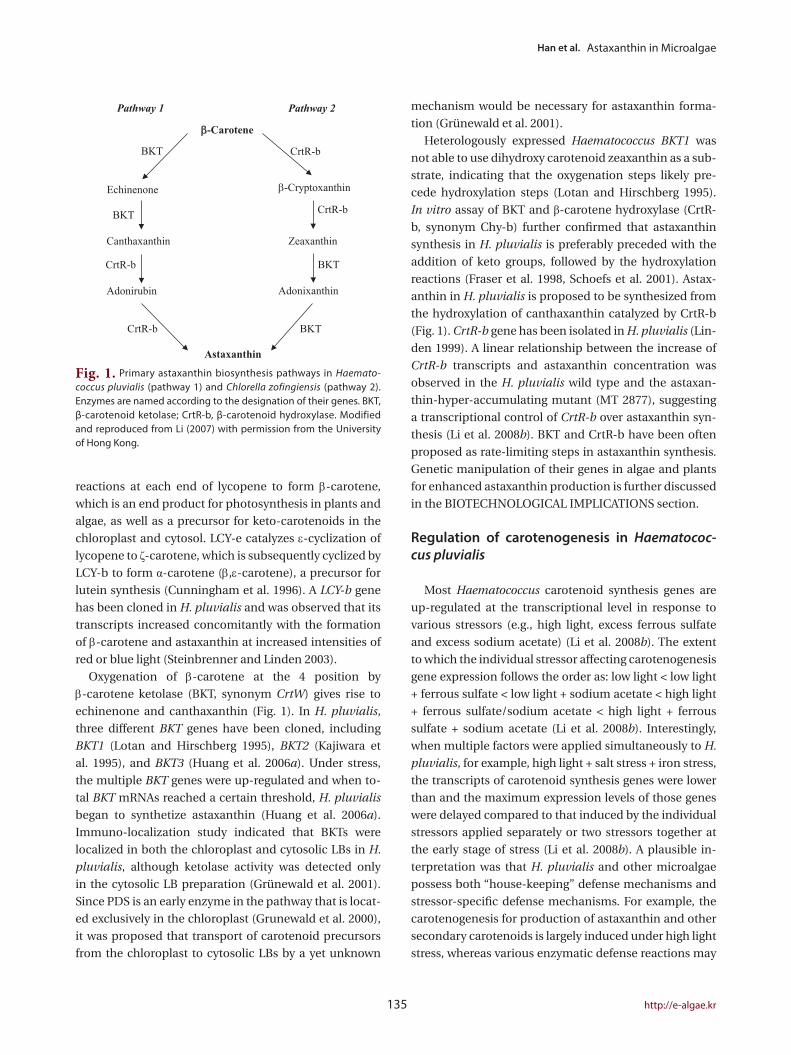
In vitro assay of BKT and β-carotene hydroxylase (CrtR-
b, synonym Chy-b) further confirmed that astaxanthin
synthesis in H. pluvialis is preferably preceded with the
addition of keto groups, followed by the hydroxylation
reactions (Fraser et al. 1998, Schoefs et al. 2001). Astax-
anthin in H. pluvialis is proposed to be synthesized from
the hydroxylation of canthaxanthin catalyzed by CrtR-b
(Fig. 1). CrtR-b gene has been isolated in H. pluvialis (Lin-
den 1999). A linear relationship between the increase of
CrtR-b transcripts and astaxanthin concentration was
observed in the H. pluvialis wild type and the astaxan-
thin-hyper-accumulating mutant (MT 2877), suggesting
a transcriptional control of CrtR-b over astaxanthin syn-
thesis (Li et al. 2008b). BKT and CrtR-b have been often
proposed as rate-limiting steps in astaxanthin synthesis.
Genetic manipulation of their genes in algae and plants
for enhanced astaxanthin production is further discussed
in the BIOTECHNOLOGICAL IMPLICATIONS section.
Regulation of carotenogenesis in Haematococ-cus pluvialis
Most Haematococcus carotenoid synthesis genes are
up-regulated at the transcriptional level in response to
various stressors (e.g., high light, excess ferrous sulfate
and excess sodium acetate) (Li et al. 2008b). The extent
to which the individual stressor affecting carotenogenesis
gene expression follows the order as: low light < low light
+ ferrous sulfate < low light + sodium acetate < high light
+ ferrous sulfate/sodium acetate < high light + ferrous
sulfate + sodium acetate (Li et al. 2008b). Interestingly,
when multiple factors were applied simultaneously to H.
pluvialis, for example, high light + salt stress + iron stress,
the transcripts of carotenoid synthesis genes were lower
than and the maximum expression levels of those genes
were delayed compared to that induced by the individual
stressors applied separately or two stressors together at
the early stage of stress (Li et al. 2008b). A plausible in-
terpretation was that H. pluvialis and other microalgae
possess both “house-keeping” defense mechanisms and
stressor-specific defense mechanisms. For example, the
carotenogenesis for production of astaxanthin and other
secondary carotenoids is largely induced under high light
stress, whereas various enzymatic defense reactions may
reactions at each end of lycopene to form β-carotene,
which is an end product for photosynthesis in plants and
algae, as well as a precursor for keto-carotenoids in the
chloroplast and cytosol. LCY-e catalyzes ε-cyclization of
lycopene to ζ-carotene, which is subsequently cyclized by
LCY-b to form α-carotene (β,ε-carotene), a precursor for
lutein synthesis (Cunningham et al. 1996). A LCY-b gene
has been cloned in H. pluvialis and was observed that its
transcripts increased concomitantly with the formation
of β-carotene and astaxanthin at increased intensities of
red or blue light (Steinbrenner and Linden 2003).
Oxygenation of β-carotene at the 4 position by
β-carotene ketolase (BKT, synonym CrtW) gives rise to
echinenone and canthaxanthin (Fig. 1). In H. pluvialis,
three different BKT genes have been cloned, including
BKT1 (Lotan and Hirschberg 1995), BKT2 (Kajiwara et
al. 1995), and BKT3 (Huang et al. 2006a). Under stress,
the multiple BKT genes were up-regulated and when to-
tal BKT mRNAs reached a certain threshold, H. pluvialis
began to synthetize astaxanthin (Huang et al. 2006a).
Immuno-localization study indicated that BKTs were
localized in both the chloroplast and cytosolic LBs in H.
pluvialis, although ketolase activity was detected only
in the cytosolic LB preparation (Grünewald et al. 2001).
Since PDS is an early enzyme in the pathway that is locat-
ed exclusively in the chloroplast (Grunewald et al. 2000),
it was proposed that transport of carotenoid precursors
from the chloroplast to cytosolic LBs by a yet unknown
β-Carotene
Echinenone
Canthaxanthin
Adonirubin
Astaxanthin
β-Cryptoxanthin
Zeaxanthin
Adonixanthin
Pathway 1
BKT
CrtR-b
CrtR-b
CrtR-b
CrtR-b
BKT
BKT
BKT
Pathway 2
Fig. 1. Primary astaxanthin biosynthesis pathways in Haemato-coccus pluvialis (pathway 1) and Chlorella zofingiensis (pathway 2). Enzymes are named according to the designation of their genes. BKT, β-carotenoid ketolase; CrtR-b, β-carotenoid hydroxylase. Modified and reproduced from Li (2007) with permission from the University of Hong Kong.
Algae 2013, 28(2): 131-147
http://dx.doi.org/10.4490/algae.2013.28.2.131 136
enzymatic activity of converting zeaxanthin to astaxan-
thin via adonixanthin (Huang et al. 2006b). When the BKT
inhibitor diphenylamine was applied, C. zofingiensis ac-
cumulated zeaxanthin while H. pluvialis accumulated
β-carotene (Wang and Chen 2008), suggesting that C.
zofingiensis synthesizes astaxanthin through zeaxanthin
(Fig. 1, Pathway 2).
Various environmental factors may differentially regu-
late carotenogenesis in C. zofingiensis. High irradiance
triggered up-regulation of PDS, BKT, CrtR-b genes in C.
zofingiensis, with most significant increase in CrtR-b
transcripts, leading to the accumulation of canthaxan-
thin, zeaxanthin and astaxanthin (Li et al. 2009). Under
salt stress, only BKT gene was up-regulated and can-
thaxanthin and astaxanthin were accumulated (Li et al.
2009). When fed with sugars, C. zofingiensis accumulated
astaxanthin in dividing cells. Glucose and mannose were
the best carbon sources to sustain growth and astaxan-
thin production in C. zofingiensis, followed by fructose,
sucrose and galactose, whereas lactose was the poor-
est carbon source for C. zofingiensis (Sun et al. 2008). In
heterotrophically grown C. zofingiensis, carotenogenesis
was regulated through the phosphorylation of glucose
by hexokinase, which was essential for up-regulation of
CrtR-b and synthesis of zeaxanthin, whereas the signals
from the mitochondrial alternative pathway may medi-
ate up-regulation of BKT and synthesis of astaxanthin (Li
et al. 2008a).
Two possible types of signals from the mitochondrial
alternative pathway may regulate carotenogenesis in C.
zofingiensis: ROS and organic acids from the tricarboxylic
acid cycle (TCA) cycle. Chemically generated ROS induce
carotenogenesis and astaxanthin formation in C. zofingi-
ensis (Ip and Chen 2005a, Li et al. 2009). Singlet oxygen
specifically induced CrtR-b whereas hydroxyl radical up-
regulated PDS and BKT, suggesting their distinct roles
in regulating carotenogenesis in C. zofingiensis (Ip and
Chen 2005a). In corroboration, the fatty acid synthesis
genes SAD (coding for stearoyl acyl carrier protein) and
BC (coding for biotin carboxylase) seemed to be associ-
ated with ROS (Liu et al. 2012). On the other hand, TCA
cycle acids such as citrate may act as a signal at the gene
expression level to induce mitochondrial alternative
pathway respiration to facilitate carotenogenesis (Van-
lerberghe and McIntosh 1996). In C. zofingiensis, citrate
was shown to induce BKT expression and astaxanthin
synthesis independent of ROS formation, suggesting that
the mitochondrial alternative pathway mediates BKT ex-
pression through modulation of TCA cycle with citrate as
a signal molecule (Li et al. 2008a).
be triggered by salinity or excess iron ion. When multiple
stressors were applied to H. pluvialis, multiple stress
protection mechanisms may all be activated and work
in concert, each contributing partially to overall cell pro-
tection and to a less extent carotenogenesis. However,
because the enzymatic defense systems mainly serve as
short-team cellular defense mechanisms in H. pluvialis
(Wang et al. 2004a), as the stress persists, the cells would
have to depend more upon the long-term defense mech-
anism, -carotenogenesis, for survival, which explained
why an delayed expression pattern of carotenogenesis
genes with greater expression levels occurred in the cul-
tures treated with the multiple stressors (Li et al. 2008b).
ROS play a role in regulation of carotenogenesis in H.
pluvialis. Treatment with ROS-generating compounds
such as methylviologen, methylene blue and Fe2+, in-
creased PDS and CrtR-b expression in H. pluvialis. They
also induced astaxanthin synthesis independent of de
novo protein synthesis (Kobayashi et al. 1993, Stein-
brenner and Linden 2001), suggesting ROS mediate
stress response and carotenogenesis in H. pluvialis at a
post-translational level (Steinbrenner and Linden 2003).
Unlike PDS and CrtR-b, however, treatment with various
transcriptional and translational inhibitors suggested
that BKT gene expression was dependent on de novo pro-
tein synthesis (Vidhyavathi et al. 2008). Considering PDS
is shown to be located in the chloroplast, whereas BKT
activity is found in the cytosolic LBs, this discrepancy can
be explained by the distinct subcellular localization of in-
dividual carotenoid biosynthetic enzymes.
Pathways and regulation of carotenogenesis in Chlorella zofingiensis
C. zofingiensis has the identical β-carotene synthesis
pathway found in H. pluvialis, but may take a different
route to make astaxanthin (Fig. 1) (Li 2007). Recently,
the genes involved in biosynthesis of astaxanthin in C.
zofingiensis have been cloned and characterized, includ-
ing PSY (Cordero et al. 2011), PDS (Huang et al. 2008),
LCY-b (Cordero et al. 2010), LCY-e (Cordero et al. 2012),
BKT (CrtO) (Huang et al. 2006b), CrtR-b (Chy-b) (Li et
al. 2008a). Under high-light conditions, PSY, PDS, BKT,
CrtR-b genes were up-regulated, whereas the mRNA lev-
els of LCY-b and LCY-e remained constant, leading to for-
mation of secondary carotenoids (Li et al. 2009, Cordero
et al. 2012).
Functional analysis of C. zofingiensis BKT demon-
strated that this enzyme not only converted β-carotene
to canthaxanthin via echinenone, but also exhibited high

Han et al. Astaxanthin in Microalgae
137 http://e-algae.kr
pression (Fey et al. 2005). Recently, a transcriptomic anal-
ysis has identified over 2,000 genes regulated under high
irradiance conditions, providing therefore more candi-
date genes for further study of the crosstalk between pho-
tosynthesis and carotenogenesis gene expression in H.
pluvialis (Kim et al. 2011).
In addition to the regulation at the gene expression
level, metabolic coupling between carotenoid and photo-
synthetic electron transport may be present in H. pluvia-
lis. The electrons produced in the sequential desaturation
reactions catalyzed by PDS and ZDS can be delivered to
the PQ pool, which is in turn oxidized by PTOX (Fig. 2).
Two genes encoding PTOX (PTOX1 and PTOX2) in H. plu-
vialis have been cloned and PTOX1 was found to be co-
regulated with the carotenoid biosynthesis genes under
various environmental stress conditions (Li et al. 2008b,
2010, Wang et al. 2009). Functional analysis of PTOX of
Chlamydomonas also suggested that PTOX1 was respon-
sible for regenerating oxidized PQ for phytoene desatura-
tion (Houille-Vernes et al. 2011).
Crosstalk between astaxanthin and fatty acid biosynthesis pathways
In H. pluvialis, over 90% astaxanthin molecules are
esterified with fatty acids (Yuan et al. 1997). Oleic acid is
Crosstalk between astaxanthin synthesis path-way and photosynthetic electron transfer chain
In H. pluvialis, the nuclear carotenoid biosynthetic
genes were suggested to be regulated by the redox state of
the PQ pool at the photosynthetic electron transfer chain,
as carotenoid biosynthesis genes are up-regulated under
high PSII excitation pressure (PQ reduced) (Steinbrenner
and Linden 2003), and the inhibition of photosynthesis by
photosynthetic electron transport inhibitor DCMU abol-
ished the up-regulation of the PSY gene induced by high
light (Steinbrenner and Linden 2001). However, how the
signal of redox state of the photosynthetic electron trans-
port chain can pass the chloroplast envelope and regulate
nuclear gene expression is not known. It was proposed
that protein phosphorylation cascade may be a possible
mechanism to mediate this retrograde signaling process
(Chandok et al. 2001, Chen et al. 2004). Furthermore, the
chlorophyll biosynthesis precursors, Mg-protoporphyrin
IX and Mg-protoporphyrin-methylesters, were proposed
to play a role in chloroplast-to-nucleus retrograde signal-
ing (Nott et al. 2006).
Besides the redox state of PQ, other photosynthetic
signals such as ROS, trans-thylakoid pH gradient and the
stoichiometry of PSI and PSII may also contribute to the
photosynthetic regulation of carotenogenesis gene ex-
Fig. 2. Multiple roles of the astaxanthin biosynthesis in protecting microalgae against oxidative stress. Astaxanthin biosynthesis pathway functions to 1) reduce subcellular oxygen levels via synthesis of oxygen-rich astaxanthin and its esters; 2) convert photosynthetically evolved oxygen into water via a coupled electron transport from carotenoid desaturation steps (phytoene desaturase [PDS] and ζ-carotene desaturase [ZDS]) to the plastoquinone pool to plastid terminal oxidase (PTOX), whereas astaxanthin itself; 3) serves as a “sunshade” to reduce excess light illumination on the photosystems; 4) functions as a powerful antioxidant against reactive oxygen species. PS, photosystem; PQ, plastoquinone. Modified and reproduced from Li (2007) with permission from the University of Hong Kong.
Algae 2013, 28(2): 131-147
http://dx.doi.org/10.4490/algae.2013.28.2.131 138
equally or more susceptible to high light stress than
astaxanthin-free vegetative cells (Fan et al. 1998). These
authors suggest that the multiple intermediate reactions
in astaxanthin synthesis pathway may consume exces-
sive electrons and thus prevent over-reduction of the
photosynthetic electron transport chain and reduce the
production of ROS. As such, astaxanthin was proposed as
an end-product rather than the protective agent itself in
the stress response process (Fan et al. 1998).
More recently, Li et al. (2008b) proposed that astaxan-
thin biosynthesis exerts multilevel protective roles against
photooxidative stress. In addition to the protective roles
summarized above, astaxanthin biosynthesis protects H.
pluvialis cells from stress through the consumption of
molecular oxygen (Li et al. 2008b). Up to 10% of molecular
oxygen evolved from photosynthesis under stress is con-
sumed by astaxanthin synthesis via two distinct routes:
1) use oxygen as a substrate for ketocarotenoids forma-
tion and 2) convert oxygen generated from photosynthe-
sis and electrons from the carotenoid desaturation steps
to water by PTOX connected to the PQ pool. Decrease in
subcellular molecular oxygen concentration reduces the
substrate available for oxygen dependent ROS forma-
tion, as is the case for the alternative oxidase which re-
duces the O2 concentration and prevents electrons from
reducing O2 to O2-1 (Mittler 2002). Re-oxidation of the PQ
pool by PTOX may further relax the photosynthetic elec-
tron transport chain and reduce ROS formation (Li et al.
2008b). The multiple roles of the astaxanthin biosynthesis
in protecting microalgae against oxidative stress are sum-
marized in Fig. 2 (Li 2007).In addition to the ability to accumulate large amounts
of astaxanthin, microalgae such as H. pluvialis have
evolved other mechanisms for ROS scavenging, for ex-
ample, by the antioxidative enzyme defense system. Pro-
teomics analysis revealed several superoxide dismutases
(SOD), peroxidases, and catalases were up-regulated in
response to photooxidative stress (Wang et al. 2004a, Tran
et al. 2009b). Transcriptomic analysis revealed that the
up-regulation of SOD, catalase, and glutathione peroxi-
dase genes occurred under high light and nutrient starva-
tion conditions (Eom et al. 2006, Kim et al. 2011, Wang et
al. 2011). All the antioxidative enzymes studied showed a
transient up-regulation pattern and then reverted to the
basal or below basal level, suggesting that the enzyme de-
fense system is an early response mechanism to oxidative
stress, and as stress persists, the cells will adopt astaxan-
thin biosynthesis and accumulation as a long-term de-
fense mechanism (Wang et al. 2004a).
the major fatty acid species that is conjugated to astax-
anthin molecules (Holtin et al. 2009). A linear correlation
between the cellular fatty acid content and astaxanthin
content in H. pluvialis under stress conditions has been
reported in several independent studies (Zhekisheva
et al. 2002, Chen 2007), leading to a hypothesis that the
biosynthesis of astaxanthin and fatty acids are coupled in
this organism. This hypothesis was further supported by
the fact that with the addition of the fatty acid synthesis
inhibitor cerulenin or the carotenoid synthesis inhibitor
norflurazon, both astaxanthin and fatty acid biosynthesis
were abolished (Schoefs et al. 2001, Chen 2007). Astax-
anthin esterification must be the reaction that links the
two pathways. Acyl-CoA, the presumable substrate for
astaxanthin esterification, when in excess, may feedback-
inhibit acetyl-CoA carboxylase, a rate-limiting enzyme
for fatty acid synthesis (Ohlrogge and Jaworski 1997).
Presumably, the inhibition of astaxanthin synthesis by
norflurazon may result in building up relatively excess
amounts of acyl-CoA which may in turn inhibit fatty acid
synthesis through a feedback inhibition mechanism.
Likewise, the enzymes responsible for astaxanthin syn-
thesis may also be under feedback control when fatty acid
biosynthesis is inhibited by cerulenin. Astaxanthin esteri-
fication, the key point for flux control, needs to be further
characterized at the gene, enzyme and subcellular levels.
Physiological role of astaxanthin biosynthesis
In algae and higher plants, ROS are mainly generated
through photosynthesis in chloroplasts (Asada 2006). Un-
der stress conditions, e.g., high light, nutrient starvation,
and high salinity, the imbalance between generation and
detoxification of ROS within the chloroplast may cause
photooxidative stress.
Astaxanthin was proposed to act as a “sunshade” to
reduce the penetration of blue light into the chloroplast,
thereby reducing photooxidative damage of the photo-
systems by excessive light (Hagen et al. 1994). Astaxan-
thin could also act as a physical-chemical barrier to pre-
vent DNA, RNA, enzymes and membrane lipids from ROS
attack (Hagen et al. 1993, Kobayashi et al. 1997, Kobayashi
and Sakamoto 1999). Astaxanthin esters were shown to
scavenge ROS, e.g., the superoxide anion radicals (O2-)
and singlet oxygen (1O2) and thus may act as an antioxi-
dant agent against ROS (Kobayashi et al. 1997, Kobayashi
and Sakamoto 1999).
The photoprotective role of astaxanthin has been chal-
lenged since astaxanthin-rich cysts of H. pluvialis were

Han et al. Astaxanthin in Microalgae
139 http://e-algae.kr
An alternative strategy for enhancing astaxanthin pro-
duction in microalgae is through random mutagenesis.
Through chemical mutagenesis, several Haematococ-
cus mutants with improved phenotypic traits have been
generated, and one such example is Haematococcus
astaxanthin-hyper-accumulating mutant strain MT 2877
(Hu et al. 2008). In the early vegetative growth stage, MT
2877 was identical to the wild type with respect to cell
morphology and physiology. However, the mutant grew
faster under stress conditions with about 100% more vi-
able cells. MT 2877 also accumulated 100% more astax-
anthin, leading to a 4-fold increase in volumetric astax-
anthin productivity compared to the wild type (Hu et al.
2008). Earlier, two H. pluvialis cell wall deficient mutants,
MT 537 and MT 2978, have been generated by chemi-
cal mutagenesis (Wang et al. 2005). Haematococcus wild
type cells possess thick and rigid cell walls that reduce
considerably the bioavailability of astaxanthin to human
and animals if whole Haematococcus cells are consumed
without cell disruption by chemical or physical means.
The cell wall mutants were demonstrated to significantly
improve the bioavailability of astaxanthin. Production
of astaxanthin using the cell wall mutants may also re-
duce downstream processing costs whereby eliminat-
ing or minimizing the cell wall disruption step which is
otherwise required for wild type Haematococcus (Wang
et al. 2005, Hu et al. 2006). Some mating-based breed-
ing techniques can also be applied to Haematococcus to
improve existing or introduce new desirable traits, given
that many Haematococcus strains vary in photosynthesis
efficiency, growth, astaxanthin content, and susceptibil-
ity to oxidative stress or parasite attack. Any efforts along
this line would be valuable.
Effects of environmental factors on growth and astaxanthin production
Growth and carotenogenesis of H. pluvialis are regulat-
ed by various environmental factors, such as light, tem-
perature and nutrients. The maximal specific growth rate
of H. pluvialis is 0.054 h-1, corresponding to a doubling
time of 12-13 h. Such a high growth potential occurs only
under favorable growth conditions, e.g., low light irradi-
ance (20-50 µE m-2 s-1), optimal temperature (25-28°C)
and replete nutrients (Fan et al. 1994). On the contrary,
high light, high temperature, and nutrient deprivation
(e.g., nitrogen and phosphorus) induce astaxanthin syn-
thesis while retarding cell division. High light is one of the
most effective factors to stimulate astaxanthin synthesis
and thus is often applied to increase astaxanthin produc-
BIOTECHNOLOGICAL IMPLICATIONS
Metabolic engineering for enhanced carotenoid production
Many higher plants exhibit enzymatic activity of carot-
enoid hydroxylase but not β-carotene ketolase, and as a
result, they produce 3-hydroxy carotenoids (e.g., lutein
and zeaxanthin) but no 4-ketocarotenoids such as astax-
anthin. Introduction of a Haematococcus β-carotene ke-
tolase gene (BKT) into a tobacco plant resulted in moder-
ate accumulation of astaxanthin (ca. 840 μg g-1 dwt) in the
nectary tissue (Mann et al. 2000). A similar effort was made
with a carrot plant, resulting in a comparable amount (ca.
916 μg g-1 dwt) of astaxanthin in root tissues (Jayaraj et
al. 2008). Expression of a Chlamydomonas BKT into Ara-
bidopsis led to the accumulation of greater amounts of
astaxanthin (up to 2,000 μg g-1 dwt) in leaves of the trans-
genic plant, whereas the expression of C. zofingiensis BKT
only resulted in 240 μg astaxanthin g-1 dwt (Zhong et al.
2011). A greater amount of astaxanthin (1,600 μg g-1 dwt)
in leaves of a transgenic tobacco was also obtained when
the Chlamydomonas BKT was introduced (Huang et al.
2012), suggesting that the catalytic capacity of BKT is spe-
cies-specific (Zhong et al. 2011). A much greater amount
of astaxanthin (5,440 μg g-1 dwt) was obtained in tobacco
leaves when a BKT and a CrtR-b gene from a marine bac-
terium Brevundimonas sp. were co-transformed into the
plant plastid genome (Hasunuma et al. 2008).
Lack of dominant selectable markers has hindered the
genetic engineering of astaxanthin production in mi-
croalgae. Recently, a stable nuclear transformation sys-
tem has been established in H. pluvialis (Steinbrenner
and Sandmann 2006) and in C. zofingiensis (Huang et al.
2008). A modified version of PDS gene in H. pluvialis and
C. zofingiensis has been developed as a dominant select-
able marker and was transformed into these two species
through biolistic transformation (Steinbrenner and Sand-
mann 2006, Huang et al. 2008, Liu et al. 2009). Agrobac-
terium-mediated transformation has also been reported
toexpress foreign genes in Haematococcus (Kathiresan et
al. 2009). The newly developed genetic transformation
tools for H. pluvialis and C. zofingiensis will provide met-
abolic engineering opportunities for overproduction of
astaxanthin in these two and perhaps other microalgae.
One possible application is to enhance astaxanthin pro-
duction in H. pluvialis or C. zofingiensis by overexpressing
PSY and CrtR-b genes, which previously have been shown
to be possible rate-limiting steps for astaxanthin synthe-
sis in H. pluvialis (Li et al. 2008b, 2010).

Algae 2013, 28(2): 131-147
http://dx.doi.org/10.4490/algae.2013.28.2.131 140
(Boussiba et al. 1992, Olaizola 2000, Fábregas et al. 2001,
Aflalo et al. 2007).
During the first two days of the red-stage, considerable
cell death (or photo-bleach) occurs, primarily to flagel-
lated zoospores, due to their higher susceptibility to pho-
tooxidative stress than non-motile palmella cells (Harker
et al. 1996a, Sarada et al. 2002, Han et al. 2012). To prevent
or reduce cell mortality caused by high light and nutri-
ent deprivation, the initial cell concentration in the cul-
ture must be optimized (Wang et al. 2013). The highest
astaxanthin productivity of 17.5 mg L-1 d-1 was obtained
at the red stage where an optimal initial biomass density
of 0.8 g L-1 dwt was applied to an outdoor glass column
photobioreactor (Wang et al. 2013). Greater tolerance of
palmella than zoospores to photooxidative stress resulted
in more than 10-fold increase in astaxanthin productivity
(Choi et al. 2011).
Besides the two-stage culture mode, a single-stage cul-
tivation mode has also been tested to produce astaxan-
thin in flagellates of some H. pluvialis strains in a chemo-
stat system (Del Río et al. 2005, 2008, García-Malea et al.
2009). Under optimal light irradiance, nutrient concen-
tration and dilution rate, algal biomass productivities of
0.7-1.9 g L-1 d-1 were obtained, corresponding to an astax-
anthin productivity of 5.6-21 mg L-1 d-1. The technical and
economic feasibilities of this single-stage culture mode
for mass culture of H. pluvialis remain to be seen.
Although a number of Chlorella species and strains
have been tested for carotenoid production on labora-
tory scales, little has been done on a large-scale (Liu et
al. 2011).
Heterotrophic and mixotrophic culture modes. H. plu-
vialis is capable of utilize organic carbon for growth in the
absence of light, which provides the means of heterotro-
phic and mixotrophic cultivation for astaxanthin produc-
tion. Under heterotrophic conditions, H. pluvialis grows
at a relatively low growth rate (0.22 d-1) and accumu-
lates ca. 0.5% dwt of astaxanthin (Kobayashi et al. 1992).
Growth and astaxanthin production can be enhanced
under mixotrophic culture conditions. A final cell density
of 0.9-2.65 g L-1 and a maximum astaxanthin content of
1-2% dwt were obtained from mixotrophic cultures of H.
pluvialis (Chen et al. 1997, Zhang et al. 1999, Wang et al.
2004a). A heterophotric-photoautotrophic culture mode
was also explored where heterotrophic culture produced
algal biomass, while astaxanthin production was induced
during photoautotrophic culture. As a result, a very high
cellular astaxanthin content of 7% by dwt, but low astax-
anthin productivities of 4.4-6.5 mg L-1 d-1 were obtained
(Hata et al. 2001, Kang et al. 2005).
tion (Choi et al. 2002). High temperature is rarely imple-
mented to induce astaxanthin production, as it was re-
ported to severely reduce biomass yield and thus overall
reduced astaxanthin productivity (Tjahjono et al. 1994).
Carotenogenesis in H. pluvialis can be stimulated by a
variety of metal ions such as Fe2+, Mn2+, and Cd2+ (Harker
et al. 1996b). All the tested metals exerted inhibitory effect
on cell growth except Fe2+. Fe2+ was suggested to stimu-
late ROS (especially hydroxyl radicals, HO·) production
via the Fenton reaction, which in turn trigger astaxanthin
synthesis (Tjahjono et al. 1994). This speculation was
supported by several lines of evidence: EDAT-chelated
FeCl3·6H2O (a form of iron which does not cause Fenton
reaction) did not induce astaxanthin synthesis, whereas
potassium iodide with a capability of scavenging HO· and
thus abolishing Fe2+-induced astaxanthin synthesis in H.
pluvialis (Boussiba and Vonshak 1991, Kobayashi et al.
1993).
Astaxanthin synthesis in H. pluvialis can also be in-
duced by salinity stress. NaCl (0.1-0.5%, w/w) was used
to increase astaxanthin accumulation in laboratory cul-
tures (Harker et al. 1996a, Sarada et al. 2002). However,
relatively high concentrations of NaCl (0.6-0.8%, w/w)
may cause severe cell mortality, in particularly for flagel-
lated zoospores, which limits the implementation of this
strategy in large-scale Haematococcus culture (Harker et
al. 1996a, Sarada et al. 2002, Cifuentes et al. 2003).
Mass cultivation of Haematococcus pluvialis and Chlorella zofingiensis
Photoautotrophic culture. Photoautotrophic culture
of H. pluvialis is carried out in open raceway ponds and
closed photobioreactors (Olaizola 2000, Carvalho et al.
2006, Ugwu et al. 2008). Because the culture conditions
for maximum growth and maximum astaxanthin content
are mutually exclusive, a two-stage batch culture mode is
commonly adopted for mass cultivation of H. pluvialis. In
the first stage (also called ‘green stage’ because the cells
are green), the cells are maintained in a nutrient-replete
growth medium and exposed to low light intensity to
promote biomass production. When the cells enter into
the stationary growth phase, the culture is then transited
into the second stage (also called ‘red stage’ because the
cells are turning red from green) where the cells are sub-
jected to high light intensity and nutrient deprivation to
induce astaxanthin production. The reported biomass
productivities range from 0.03 to 0.6 g L-1 d-1 at the green
stage and from 0.01 to 0.58 g L-1 d-1 at the red stage, and
astaxanthin productivity ranged from 0.4 to 17 mg L-1 d-1

Han et al. Astaxanthin in Microalgae
141 http://e-algae.kr
pretreated by a mechanical means to disrupt the rigid
cell walls, followed by a spray-drying process to produce
dry biomass powder. If astaxanthin is the final product, a
conventional solvent-based extraction method or super-
critical fluid extraction can be applied to wet or dry Hae-
matococcus biomass to obtain concentrated astaxanthin
extracts (Bubrick 1991, Mendes-Pinto et al. 2001, Nobre
et al. 2006, Sarada et al. 2006, Krichnavaruk et al. 2008).
CONCLUSIONS AND PERSPECTIVES
The occurrence and accumulation of astaxanthin in
microalgae is a strategy to cope with oxidative stress.
Astaxanthin synthesis pathways interact with multiple
metabolic pathways such as the photosynthetic electron
transport, fatty acid synthesis, and ROS generation. How-
ever, the molecular mechanisms mediating the crosstalk
between astaxanthin synthesis and related metabolic
pathways remain elusive. Recent progress in genome se-
quencing and genetic toolbox development of microalgae
has made it possible to further address these questions.
The new knowledge obtained from the studies of astaxan-
thin biosynthesis and stress response will be applied for
enhanced astaxanthin production. Until now, H. pluvialis
is still the best producer for astaxanthin although the bio-
mass productivity is relatively low. Future efforts should
focus on increasing biomass production of H. pluvialis or
screening for and manipulating other fast-growing algal
strains for enhanced astaxanthin production along with
improved phenotypic traits for various commercial ap-
plications.
ACKNOWLEDGEMENT
The first two authors contributed equally to this work.
REFERENCES
Aflalo, C., Meshulam, Y., Zarka, A. & Boussiba, S. 2007. On
the relative efficiency of two-vs. one-stage production of
astaxanthin by the green alga Haematococcus pluvialis.
Biotechnol. Bioeng. 98:300-305.
Asada, K. 2006. Production and scavenging of reactive oxy-
gen species in chloroplasts and their functions. Plant
Physiol. 141:391-396.
Bar, E., Rise, M., Vishkautsan, M. & Arad, S. 1995. Pigment
and structural changes in Chlorella zofingiensis upon
Much higher cell density can be achieved in hetero-
trophic culture of C. zofingiensis. For example, high cell
concentrations of 10 g L-1 (Ip and Chen 2005b) and 53 g
L-1 (Sun et al. 2008) were achieved in C. zofingiensis cul-
tures fed with glucose with a maximum specific growth
rate of 1.03 d-1. A moderate astaxanthin yield of 12.5 mg
L-1 was obtained in a mixotrophic culture of C. zofingien-
sis (Ip et al. 2004), and the yield was increased to 32 mg L-1
in a glucose fed-batch culture. However, the astaxanthin
productivity is low ranging from 0.9 to 2 mg L-1 d-1 (Sun et
al. 2008).
Microbial contamination and crop protection. Mass
culture of Haematococcus can be contaminated by fun-
gal parasites and zooplanktonic predators (e.g., amoe-
bas, ciliates, and rotifers), as well as other microalgae
and cyanobacteria, resulting in reduced biomass yield
and quality, and sometimes loss of culture all together. A
parasitic blastoclad fungus identified as Paraphysoderma
sedebokerensis is the most devastating disease respon-
sible for reduced astaxanthin productivity and frequent
culture collapses (Hoffman et al. 2008, Gutman et al.
2009). Hoffman et al. (2008) made detailed observations
on the infection of Haematococcus culture by the parasite
P. sedebokerensis (Hoffman et al. 2008). During the infec-
tion process, healthy green culture turns dark brown, ac-
companied by the formation of large clumps consisting
of living cells, cell debris, and particulate organic matters.
A more vivid red color of culture can be first observed if
infection occurred at the red stage, followed by gradual
bleaching of algal cells (Hoffman et al. 2008).
The infection process can be somewhat inhibited in
the presence of lectin-like carbohydrate-binding pro-
teins, which were assumed to bind to the surface of pal-
amelloids and aplanospores of Haematococcus (Gutman
et al. 2011). The recognition between the blastoclad para-
site and Haematococcus cells was suggested to be a lec-
tin-N-acetyl-D-glucosamine interaction. More research
along this line may better understand the molecular and
cellular mechanisms by which P. sedebokerensis recogniz-
es and infects Haematococcus, which may in turn lead to
develop a biology-inspired method to prevent or treat the
fungal disease in the mass culture of Haematococcus or
other related species.
Downstream processes and product formation
Haematococcus aplanospores can be readily harvested
by sedimentation and centrifugation prior to spray. If
whole Haematococcus cells are a final form of product,
Haematococcus pastes from centrifugation is usually

Algae 2013, 28(2): 131-147
http://dx.doi.org/10.4490/algae.2013.28.2.131 142
the microalgae Nitzschia laevis (Bacillariophyceae) and
Haematococcus pluvialis (Chlorophyceae). Ph.D. disser-
tation, The University of Hong Kong, Hong Kong, 150 pp.
Chen, Y. -B., Durnford, D. G., Koblizek, M. & Falkowski, P. G.
2004. Plastid regulation of Lhcb1 transcription in the
chlorophyte alga Dunaliella tertiolecta. Plant Physiol.
136:3737-3750.
Choi, Y. E., Yun, Y. -S. & Park, J. M. 2002. Evaluation of fac-
tors promoting astaxanthin production by a unicellular
green alga, Haematococcus pluvialis, with fractional fac-
torial design. Biotechnol. Prog. 18:1170-1175.
Choi, Y. -E., Yun, Y. -S., Park, J. M. & Yang, J. -W. 2011. Deter-
mination of the time transferring cells for astaxanthin
production considering two-stage process of Hae-
matococcus pluvialis cultivation. Bioresour. Technol.
102:11249-11253.
Cifuentes, A. S., González, M. A., Vargas, S., Hoeneisen, M.
& González, N. 2003. Optimization of biomass, total ca-
rotenoids and astaxanthin production in Haematococ-
cus pluvialis Flotow strain Steptoe (Nevada, USA) under
laboratory conditions. Biol. Res. 36:343-357.
Cordero, B. F., Couso, I., León, R., Rodríguez, H. & Vargas, M.
Á. 2011. Enhancement of carotenoids biosynthesis in
Chlamydomonas reinhardtii by nuclear transformation
using a phytoene synthase gene isolated from Chlorella
zofingiensis. Appl. Microbiol. Biotechnol. 91:341-351.
Cordero, B. F., Couso, I., León, R., Rodriguez, H. & Vargas, M.
A. 2012. Isolation and characterization of a lycopene ep-
silon-cyclase gene of Chlorella (Chromochloris) zofingi-
ensis: regulation of the carotenogenic pathway by nitro-
gen and light. Mar. Drugs 10:2069-2088.
Cordero, B. F., Obraztsova, I., Martín, L., Couso, I., León,
R., Vargas, M. A. & Rodriguez, H. 2010. Isolation and
characterization of a lycopene β-cyclase gene from the
astaxanthin-producing green alga Chlorella zofingiensis
(Chlorophyta). J. Phycol. 46:1229-1238.
Cunningham, F. X. & Gantt, E. 1998. Genes and enzymes
of carotenoid biosynthesis in plants. Annu. Rev. Plant
Physiol. Plant Mol. Biol. 49:557-583.
Cunningham, F. X., Pogson, B., Sun, Z., McDonald, K. A., Del-
laPenna, D. & Gantt, E. 1996. Functional analysis of the
beta and epsilon lycopene cyclase enzymes of Arabidop-
sis reveals a mechanism for control of cyclic carotenoid
formation. Plant Cell 8:1613-1626.
Damiani, M. C., Leonardi, P. I., Pieroni, O. I. & Cáceres, E. J.
2006. Ultrastructure of the cyst wall of Haematococcus
pluvialis (Chlorophyceae): wall development and be-
haviour during cyst germination. Phycologia 45:616-
623.
Del Campo, J. A., Rodríguez, H., Moreno, J., Vargas, M. Á., Ri-
light and nitrogen stress. J. Plant Physiol. 146:527-534.
Barbosa, M. J., Morais, R. & Choubert, G. 1999. Effect of ca-
rotenoid source and dietary lipid content on blood
astaxanthin concentration in rainbow trout (Oncorhyn-
chus mykiss). Aquaculture 176:331-341.
Bidigare, R. R., Ondrusek, M. E., Kennicutt, M. C., Iturriaga,
R., Harvey, H. R., Hoham, R. W. & Macko, S. A. 1993. Evi-
dence for a photoprotective function for secondary ca-
rotenoids of snow algae. J. Phycol. 29:427-434.
Boussiba, S. 2000. Carotenogenesis in the green alga Hae-
matococcus pluvialis: cellular physiology and stress re-
sponse. Physiol Plant. 108:111-117.
Boussiba, S., Bing, W., Yuan, J. -P., Zarka, A. & Chen, F. 1999.
Changes in pigments profile in the green alga Haeam-
tococcus pluvialis exposed to environmental stresses.
Biotechnol. Lett. 21:601-604.
Boussiba, S., Fan, L. & Vonshak, A. 1992. Enhancement and
determination of astaxanthin accumulation in green
alga Haematococcus pluvialis. Methods Enzymol.
213:386-391.
Boussiba, S. & Vonshak, A. 1991. Astaxanthin accumulation
in the green alga Haematococcus pluvialis. Plant Cell
Physiol. 32:1077-1082.
Brinda, B. R., Sarada, R., Kamath, B. S., & Ravishankar, G. A.
2004. Accumulation of astaxanthin in flagellated cells of
Haematococcus pluvialis - cultural and regulatory as-
pects. Curr. Sci. (Bangalore) 87:1290-1294.
Bubrick, P. 1991. Production of astaxanthin from Haemato-
coccus. Bioresour. Technol. 38:237-239.
Carol, P., Stevenson, D., Bisanz, C., Breitenbach, J., Sandma-
nn, G., Mache, R., Coupland, G. & Kuntz, M. 1999. Muta-
tions in the Arabidopsis gene immutans cause a varie-
gated phenotype by inactivating a chloroplast terminal
oxidase associated with phytoene desaturation. Plant
Cell 11:57-68.
Carvalho, A. P., Meireles, L. A. & Malcata, F. X. 2006. Micro-
algal reactors: a review of enclosed system designs and
performances. Biotechnol. Prog. 22:1490-1506.
Chandok, M. R., Sopory, S. K. & Oelmüller, R. 2001. Cytoplas-
mic kinase and phosphatase activities can induce PsaF
gene expression in the absence of functional plastids:
evidence that phosphorylation/dephosphorylation
events are involved in interorganellar crosstalk. Mol.
Gen. Genet. 264:819-826.
Chen, F., Chen, H. & Gong, X. 1997. Mixotrophic and hetero-
trophic growth of Haematococcus lacustris and rheologi-
cal behaviour of the cell suspensions. Bioresour. Tech-
nol. 62:19-24.
Chen, G. 2007. Lipid and fatty acid composition and their
biosyntheses in relation to carotenoid accumulation in

Han et al. Astaxanthin in Microalgae
143 http://e-algae.kr
316.
Grunewald, K., Eckert, M., Hirschberg, J. & Hagen, C. 2000.
Phytoene desaturase is localized exclusively in the chlo-
roplast and up-regulated at the mRNA level during ac-
cumulation of secondary carotenoids in Haematococcus
pluvialis (Volvocales, Chlorophyceae). Plant Physiol.
122:1261-1268.
Grünewald, K., Hirschberg, J. & Hagen, C. 2001. Ketocarot-
enoid biosynthesis outside of plastids in the unicellu-
lar green alga Haematococcus pluvialis. J. Biol. Chem.
276:6023-6029.
Grung, M., Metzger, P. & Liaaen-Jensen, S. 1994. Algal carot-
enoids 53: secondary carotenoids of algae 4. Secondary
carotenoids in the green alga Botryococcus braunii, race
L, new strain. Biochem. Syst. Ecol. 22:25-29.
Guerin, M., Huntley, M. E. & Olaizola, M. 2003. Haematococ-
cus astaxanthin: applications for human health and nu-
trition. Trends Biotechnol. 21:210-216.
Gutman, J., Zarka, A. & Boussiba, S. 2009. The host-range of
Paraphysoderma sedebokerensis, a chytrid that infects
Haematococcus pluvialis. Eur. J. Phycol. 44:509-514.
Gutman, J., Zarka, A. & Boussiba, S. 2011. Evidence for the
involvement of surface carbohydrates in the recognition
of Haematococcus pluvialis by the parasitic blastoclad
Paraphysoderma sedebokerensis. Fungal Biol. 115:803-
811.
Hagen, C., Braune, W. & Björn, L. O. 1994. Functional aspects
of secondary carotenoids in Haematococcus lacustris
(Volvocales). III. Action as a sunshade. J. Phycol. 30:241-
248.
Hagen, C., Braune, W. & Greulich, F. 1993. Functional-aspects
of secondary carotenoids in Haematococcus lacustris
[Girod] Rostafinski (Volvocales). IV. Protection from
photodynamic damage. J. Photochem. Photobiol. B
20:153-160.
Hagen, C., Grünewald, K., Schmidt, S., & Müller, J. 2000. Ac-
cumulation of secondary carotenoids in flagellates of
Haematococcus pluvialis (Chlorophyta) is accompanied
by an increase in per unit chlorophyll productivity of
photosynthesis. Eur. J. Phycol. 35:75-82.
Hagen, C., Siegmund, S. & Braune, W. 2008. Ultrastructural
and chemical changes in the cell wall of Haematococcus
pluvialis (Vovocales, Chlorophyta) during aplanospore
formation. Eur. J. Phycol. 37:217-226.
Han, D., Wang, J., Sommerfeld, M. & Hu, Q. 2012. Suscepti-
bility and potective mechanisms of motile and nonmo-
tile cells of Haematococcus pluvialis (Chlorophyceae) to
photooxidative stress. J. Phycol. 48:693-705.
Harker, M., Tsavalos, A. J. & Young, A. J. 1996a. Autotrophic
growth and carotenoid production of Haematococcus
vas, J. & Guerrero, M. G. 2004. Accumulation of astaxan-
thin and lutein in Chlorella zofingiensis (Chlorophyta).
Appl. Microbiol. Biotechnol. 64:848-854.
Del Río, E., Acién, F. G., García-Malea, M. C., Rivas, J., Molina-
Grima, E. & Guerrero, M. G. 2005. Efficient one-step pro-
duction of astaxanthin by the microalga Haematococ-
cus pluvialis in continuous culture. Biotechnol. Bioeng.
91:808-815.
Del Río, E., Acién, F. G., García-Malea, M. C., Rivas, J., Moli-
na-Grima, E. & Guerrero, M. G. 2008. Efficiency assess-
ment of the one-step production of astaxanthin by the
microalga Haematococcus pluvialis. Biotechnol. Bioeng.
100:397-402.
Disch, A., Schwender, J., Müller, C., Lichtenthaler, H. K. &
Rohmer, M. 1998. Distribution of the mevalonate and
glyceraldehyde phosphate/pyruvate pathways for iso-
prenoid biosynthesis in unicellular algae and the cyano-
bacterium Synechocystis PCC 6714. Biochem. J. 333:381-
388.
Droop, M. R. 1954. Conditions governing haematochrome
formation and loss in the alga Haematococcus pluvialis
Flotow. Arch. Microbiol. 20:391-397.
Elliot, A. M. 1934. Morphology and life history of Haemato-
coccus pluvialis. Arch. Protistenk 82:250-272.
Eom, H., Lee, C. G. & Jin, E. 2006. Gene expression profile
analysis in astaxanthin-induced Haematococcus pluvia-
lis using a cDNA microarray. Planta 223:1231-1242.
Fábregas, J., Otero, A., Maseda, A. & Domínguez, A. 2001.
Two-stage cultures for the production of astaxanthin
from Haematococcus pluvialis. J. Biotechnol. 89:65-71.
Fan, L., Vonshak, A. & Boussiba, S. 1994. Effect of tempera-
ture and irradiance on growth of Haematococcus pluvia-
lis (Chlorophyceae). J. Phycol. 30:829-833.
Fan, L., Vonshak, A., Zarka, A. & Boussiba, S. 1998. Does
astaxanthin protect Haematococcus against light dam-
age? Z. Naturforsch. C 53:93-100.
Fey, V., Wagner, R., Bräutigam, K. & Pfannschmidt, T. 2005.
Photosynthetic redox control of nuclear gene expres-
sion. J. Exp. Bot. 56:1491-1498.
Fraser, P. D., Shimada, H. & Misawa, N. 1998. Enzymic con-
firmation of reactions involved in routes to astaxanthin
formation, elucidated using a direct substrate in vitro
assay. Eur. J. Biochem. 252:229-236.
García-Malea, M. C., Acién, F. G., Del Río, E., Fernández, J. M.,
Cerón, M. C., Guerrero, M. G. & Molina-Grima, E. 2009.
Production of astaxanthin by Haematococcus pluvialis:
taking the one-step system outdoors. Biotechnol. Bio-
eng. 102:651-657.
Green, J. 1963. Occurrence of astaxanthin in euglenoid Tra-
chelomonas volvocina. Comp. Biochem. Physiol. 9:313-
Algae 2013, 28(2): 131-147
http://dx.doi.org/10.4490/algae.2013.28.2.131 144
astaxanthin-producing green alga Chlorella zofingiensis
(Chlorophyta). J. Phycol. 44:684-690.
Huang, J. C., Wang, Y., Sandmann, G. & Chen, F. 2006b. Iso-
lation and characterization of a carotenoid oxygenase
gene from Chlorella zofingiensis (Chlorophyta). Appl.
Microbiol. Biotechnol. 71:473-479.
Huang, J. C., Zhong, Y. J., Sandmann, G., Liu, J. & Chen, F.
2012. Cloning and selection of carotenoid ketolase
genes for the engineering of high-yield astaxanthin in
plants. Planta 236:691-699.
Huss, V. A. R., Frank, C., Hartmann, E. C., Hirmer, M., Klo-
boucek, A., Seidel, B. M., Wenzeler, P. & Kessler, E. 1999.
Biochemical taxonomy and molecular phylogeny of
the genus Chlorella sensu lato (Chlorophyta). J. Phycol.
35:587-598.
Ip, P. -F. & Chen, F. 2005a. Employment of reactive oxygen
species to enhance astaxanthin formation in Chlorella
zofingiensis in heterotrophic culture. Process Biochem.
40:3491-3496.
Ip, P. -F. & Chen, F. 2005b. Production of astaxanthin by the
green microalga Chlorella zofingiensis in the dark. Pro-
cess Biochem. 40:733-738.
Ip, P. -F., Wong, K. -H. & Chen, F. 2004. Enhanced production
of astaxanthin by the green microalga Chlorella zofingi-
ensis in mixotrophic culture. Process Biochem. 39:1761-
1766.
Jayaraj, J., Devlin, R. & Punja, Z. 2008. Metabolic engineer-
ing of novel ketocarotenoid production in carrot plants.
Transgenic Res. 17:489-501.
Johnson, E. A. & An, G. -H. 1991. Astaxanthin from microbial
sources. Crit. Rev. Biotechnol. 11:297-326.
Johnson, E. A. & Schroeder, W. A. 1996. Microbial carotenoids.
Adv. Biochem. Eng. Biotechnol. 53:119-178.
Kajiwara, S., Fraser, P. D., Kondo, K. & Misawa, N. 1997. Ex-
pression of an exogenous isopentenyl diphosphate
isomerase gene enhances isoprenoid biosynthesis in
Escherichia coli. Biochem. J. 324:421-426.
Kajiwara, S., Kakizono, T., Saito, T., Kondo, K., Ohtani, T.,
Nishio, N., Nagai, S. & Misawa, N. 1995. Isolation and
functional identification of a novel cDNA for astaxan-
thin biosynthesis from Haematococcus pluvialis, and
astaxanthin synthesis in Escherichia coli. Plant Mol. Biol.
29:343-352.
Kang, C. D., Lee, J. S., Park, T. H. & Sim, S. J. 2005. Compari-
son of heterotrophic and photoautotrophic induction
on astaxanthin production by Haematococcus pluvialis.
Appl. Microbiol. Biotechnol. 68:237-241.
Kathiresan, S., Chandrashekar, A., Ravishankar, G. A. & Sara-
da, R. 2009. Agrobacterium-mediated transformation in
the green alga Haematococcus pluvialis (Chlorophyce-
pluvialis in a 30 liter air-lift photobioreactor. J. Ferment.
Bioeng. 82:113-118.
Harker, M., Tsavalos, A. J. & Young, A. J. 1996b. Factors re-
sponsible for astaxanthin formation in the chlorophyte
Haematococcus pluvialis. Bioresour. Technol. 55:207-
214.
Hasunuma, T., Miyazawa, S. I., Yoshimura, S., Shinzaki, Y.,
Tomizawa, K. I., Shindo, K., Choi, S. K., Misawa, N. &
Miyake, C. 2008. Biosynthesis of astaxanthin in tobacco
leaves by transplastomic engineering. Plant J. 55:857-
868.
Hata, N., Ogbonna, J. C., Hasegawa, Y., Taroda, H. & Tanaka,
H. 2001. Production of astaxanthin by Haematococcus
pluvialis in a sequential heterotrophic-photoautotro-
phic culture. J. Appl. Phycol. 13:395-402.
Hershkovits, G., Dubinsky, Z. & Katcoff, D. J. 1997. A novel
homologue of the prokaryotic htrA gene is differentially
expressed in the alga Haematococcus pluvialis following
stress. Mol. Gen. Genet. 254:345-350.
Hoffman, Y., Aflalo, C., Zarka, A., Gutman, J., James, T. Y. &
Boussiba, S. 2008. Isolation and characterization of a
novel chytrid species (phylum Blastocladiomycota),
parasitic on the green alga Haermatococcus. Mycol. Res.
112:70-81.
Holtin, K., Kuehnle, M., Rehbein, J., Schuler, P., Nicholson,
G. & Albert, K. 2009. Determination of astaxanthin and
astaxanthin esters in the microalgae Haematococcus
pluvialis by LC-(APCI)MS and characterization of pre-
dominant carotenoid isomers by NMR spectroscopy.
Anal. Bioanal. Chem. 395:1613-1622.
Houille-Vernes, L., Rappaport, F., Wollman, F. A., Alric, J. &
Johnson, X. 2011. Plastid terminal oxidase 2 (PTOX2)
is the major oxidase involved in chlororespiration in
Chlamydomonas. Proc. Natl. Acad. Sci. U. S. A. 108:20820-
20825.
Hu, Q., Sommerfeld, M. & Lu, F. 2006. Extractability and bio-
availability of the natural antioxidant astaxanthin from
a green alga, Haematococcus pluvialis. WIPO Patent No.
2006107736.
Hu, Z., Li, Y., Sommerfeld, M. & Hu, Q. 2008. Enhanced pro-
tection against oxidative stress in an astaxanthin-over-
production Haematococcus mutant (Chlorophyceae).
Eur. J. Phycol. 43:365-376.
Huang, J. C., Chen, F. & Sandmann, G. 2006a. Stress-related
differential expression of multiple β-carotene ketolase
genes in the unicellular green alga Haematococcus plu-
vialis. J. Biotechnol. 122:176-185.
Huang, J., Liu, J., Li, Y. & Chen, F. 2008. Isolation and char-
acterization of the phytoene desaturase gene as a po-
tential selective marker for genetic engineering of the

Han et al. Astaxanthin in Microalgae
145 http://e-algae.kr
Planta 228:735-743.
Li, Y., Huang, J., Sandmann, G. & Chen, F. 2009. High-light
and sodium chloride stress differentially regulate the
biosynthesis of astaxanthin in Chlorella zofingiensis
(Chlorophyceae). J. Phycol. 45:635-641.
Li, Y., Sommerfeld, M., Chen, F. & Hu, Q. 2008b. Consump-
tion of oxygen by astaxanthin biosynthesis: a protective
mechanism against oxidative stress in Haematococcus
pluvialis (Chlorophyceae). J. Plant Physiol. 165:1783-
1797.
Li, Y., Sommerfeld, M., Chen, F. & Hu, Q. 2010. Effect of pho-
ton flux densities on regulation of carotenogenesis and
cell viability of Haematococcus pluvialis (Chlorophyce-
ae). J. Appl. Phycol. 22:253-263.
Lichtenthaler, H. K. 1999. The 1-deoxy-D-xylulose-5-phos-
phate pathway of isoprenoid biosynthesis in plants.
Annu. Rev. Plant Physiol. Plant Mol. Biol. 50:47-65.
Lichtenthaler, H. K., Rohmer, M. & Schwender, J. 1997. Two
independent biochemical pathways for isopentenyl di-
phosphate and isoprenoid biosynthesis in higher plants.
Physiol. Plant. 101:643-652.
Linden, H. 1999. Carotenoid hydroxylase from Haematococ-
cus pluvialis: cDNA sequence, regulation and functional
complementation. Biochim. Biophys. Acta 1446:203-
212.
Liu, B. -H. & Lee, Y. -K. 2000. Secondary carotenoids forma-
tion by the green alga Chlorococcum sp. J. Appl. Phycol.
12:301-307.
Liu, J., Huang, J. & Chen, F. 2009. Metabolic engineering of
Chlorella zofingiensis (Chlorophyta) for enhanced bio-
synthesis of astaxanthin. FEBS J. 276:S283.
Liu, J., Huang, J., Sun, Z., Zhong, Y., Jiang, Y. & Chen, F. 2011.
Differential lipid and fatty acid profiles of photoautotro-
phic and heterotrophic Chlorella zofingiensis: assess-
ment of algal oils for biodiesel production. Bioresour.
Technol. 102:106-110.
Liu, J., Sun, Z., Zhong, Y. J., Huang, J., Hu, Q. & Chen, F. 2012.
Stearoyl-acyl carrier protein desaturase gene from the
oleaginous microalga Chlorella zofingiensis: cloning,
characterization and transcriptional analysis. Planta
236:1665-1676.
Lorenz, R. T. & Cysewski, G. R. 2000. Commercial potential
for Haematococcus microalgae as a natural source of
astaxanthin. Trends Biotechnol. 18:160-167.
Lotan, T. & Hirschberg, J. 1995. Cloning and expression in
Escherichia coli of the gene encoding β-C-4-oxygenase,
that converts β-carotene to the ketocarotenoid canthax-
anthin in Haematococcus pluvialis. FEBS Lett. 364:125-
128.
Ma, R. Y. -N. & Chen, F. 2001. Enhanced production of free
ae, Volvocales). J. Phycol. 45:642-649.
Kim, D. -K., Hong, S. -J., Bae, J. -H., Yim, N., Jin, E. S. & Lee,
C. -G. 2011. Transcriptomic analysis of Haematococcus
lacustris during astaxanthin accumulation under high
irradiance and nutrient starvation. Biotechnol. Biopro-
cess Eng. 16:698-705.
Kobayashi, M., Kakizono, T. & Nagai, S. 1993. Enhanced ca-
rotenoid biosynthesis by oxidative stress in acetate-in-
duced cyst cells of a green unicellular alga, Haematococ-
cus pluvialis. Appl. Environ. Microb. 59:867-873.
Kobayashi, M., Kakizono, T., Nishio, N., Nagai, S., Kurimura,
Y. & Tsuji, Y. 1997. Antioxidant role of astaxanthin in the
green alga Haematococcus pluvialis. Appl. Microbiol.
Biotechnol. 48:351-356.
Kobayashi, M., Kakizono, T., Yamaguchi, K., Nishio, N. &
Nagai, S. 1992. Growth and astaxanthin formation of
Haematococcus pluvialis in heterotrophic and mixotro-
phic conditions. J. Ferment. Bioeng. 74:17-20.
Kobayashi, M. & Sakamoto, Y. 1999. Singlet oxygen quench-
ing ability of astaxanthin esters from the green alga Hae-
matococcus pluvialis. Biotechnol. Lett. 21:265-269.
Krichnavaruk, S., Shotipruk, A., Goto, M. & Pavasant, P. 2008.
Supercritical carbon dioxide extraction of astaxanthin
from Haematococcus pluvialis with vegetable oils as co-
solvent. Bioresour. Technol. 99:5556-5560.
Lang, N. J. 1968. Electron microscopic studies of extraplas-
tidic astaxanthin in Haematococcus. J. Phycol. 4:12-19.
Lee, Y. -K. & Ding, S. -Y. 1994. Cell cycle and accumulation of
astaxanthin in Haematococcus lacustris (Chlorophyta).
J. Phycol. 30:445-449.
Lemoine, Y. & Schoefs, B. 2010. Secondary ketocarotenoid
astaxanthin biosynthesis in algae: a multifunctional re-
sponse to stress. Photosynth. Res. 106:155-177.
Leya, T., Rahn, A., Lütz, C. & Remias, D. 2009. Response of
arctic snow and permafrost algae to high light and nitro-
gen stress by changes in pigment composition and ap-
plied aspects for biotechnology. FEMS Microbiol. Ecol.
67:432-443.
Li, J., Zhu, D., Niu, J., Shen, S. & Wang, G. 2011. An economic
assessment of astaxanthin production by large scale
cultivation of Haematococcus pluvialis. Biotechnol. Adv.
29:568-574.
Li, Y. 2007. The role of carotenogenesis in the response of the
green alga Haematococcus pluvialis to oxidative stress.
Ph.D. dissertation, The University of Hong Kong, Hong
Kong, 157 pp.
Li, Y., Huang, J., Sandmann, G. & Chen, F. 2008a. Glucose
sensing and the mitochondrial alternative pathway are
involved in the regulation of astaxanthin biosynthesis in
the dark-grown Chlorella zofingiensis (Chlorophyceae).

Algae 2013, 28(2): 131-147
http://dx.doi.org/10.4490/algae.2013.28.2.131 146
H. E. & Arad, S. M. 1994. Accumulation of secondary
carotenoids in Chlorella zofingiensis. J. Plant Physiol.
144:287-292.
Santos, M. F. & Mesquita, J. F. 1984. Ultrastructure study of
Haematococcus lacustris (Girod.) Rostafinski (Volvo-
cales) I. Some aspects of carotenogenesis. Cytologia
49:215-228.
Sarada, R., Bhattacharya, S., Bhattacharya, S. & Ravishankar,
G. A. 2002. A response surface approach for the produc-
tion of natural pigment astaxanthin from green alga,
Haematococcus pluvialis: effect of sodium acetate, cul-
ture age, and sodium chloride. Food Biotechnol. 16:107-
120.
Sarada, R., Vidhyavathi, R., Usha, D. & Ravishankar, G. A.
2006. An efficient method for extraction of astaxanthin
from green alga Haematococcus pluvialis. J. Agric. Food
Chem. 54:7585-7588.
Schoefs, B., Rmiki, N., Rachadi, J. & Lemoine, Y. 2001. Astax-
anthin accumulation in Haematococcus requires a cy-
tochrome P450 hydroxylase and an active synthesis of
fatty acids. FEBS Lett. 500:125-128.
Steinbrenner, J. & Linden, H. 2001. Regulation of two carot-
enoid biosynthesis genes coding for phytoene synthase
and carotenoid hydroxylase during stress-induced
astaxanthin formation in the green alga Haematococcus
pluvialis. Plant Physiol. 125:810-817.
Steinbrenner, J. & Linden, H. 2003. Light induction of carot-
enoid biosynthesis genes in the green alga Haematococ-
cus pluvialis: regulation by photosynthetic redox con-
trol. Plant Mol. Biol. 52:343-356.
Steinbrenner, J. & Sandmann, G. 2006. Transformation of the
green alga Haematococcus pluvialis with a phytoene de-
saturase for accelerated astaxanthin biosynthesis. Appl.
Environ. Microbiol. 72:7477-7484.
Sun, N., Wang, Y., Li, Y. -T., Huang, J. -C. & Chen, F. 2008.
Sugar-based growth, astaxanthin accumulation and
carotenogenic transcription of heterotrophic Chlorella
zofingiensis (Chlorophyta). Process Biochem. 43:1288-
1292.
Sun, Z., Cunningham, F. X. & Gantt, E. 1998. Differential ex-
pression of two isopentenyl pyrophosphate isomerases
and enhanced carotenoid accumulation in a unicellular
chlorophyte. Proc. Natl. Acad. Sci. U. S. A. 95:11482-
11488.
Takeda, H. 1991. Sugar composition of the cell-wall and
the taxonomy of Chlorella (Chlorophyceae). J. Phycol.
27:224-232.
Tan, S., Cunningham, F. X., Youmans, M., Grabowski, B., Sun,
Z. & Gantt, E. 1995. Cytochrome f loss in astaxanthin-
accumulating red cells of Haematococcus pluvialis
trans-astaxanthin by oxidative stress in the cultures of
the green microalga Chlorococcum sp. Process Biochem.
36:1175-1179.
Mann, V., Harker, M., Pecker, I. & Hirschberg, J. 2000. Meta-
bolic engineering of astaxanthin production in tobacco
flowers. Nat. Biotechnol. 18:888-892.
Mendes-Pinto, M. M., Raposo, M. F. J., Bowen, J., Young, A. J.
& Morais, R. 2001. Evaluation of different cell disruption
processes on encysted cells of Haematococcus pluvia-
lis: effects on astaxanthin recovery and implications for
bio-availability. J. Appl. Phycol. 13:19-24.
Miki, W. 1991. Biological functions and activities of animal
carotenoids. Pure Appl. Chem. 63:141-146.
Misawa, N. 2009. Pathway engineering of plants toward
astaxanthin production. Plant Biotechnol. 26:93-99.
Mittler, R. 2002. Oxidative stress, antioxidants and stress tol-
erance. Trends Plant Sci. 7:405-410.
Nobre, B., Marcelo, F., Passos, R., Beirão, L., Palavra, A., Gou-
veia, L. & Mendes, R. 2006. Supercritical carbon dioxide
extraction of astaxanthin and other carotenoids from
the microalga Haematococcus pluvialis. Eur. Food Res.
Technol. 223:787-790.
Nott, A., Jung, H. S., Koussevitzky, S. & Chory, J. 2006. Plastid-
to-nucleus retrograde signaling. Annu. Rev. Plant Biol.
57:739-759
Ohlrogge, J. B. & Jaworski, J. G. 1997. Regulation of fatty acid
synthesis. Annu. Rev. Plant Physiol. Plant Mol. Biol.
48:109-136.
Olaizola, M. 2000. Commercial production of astaxanthin
from Haematococcus pluvialis using 25,000-liter out-
door photobioreactors. J. Appl. Phycol. 12:499-506.
Orosa, M., Torres, E., Fidalgo, P. & Abalde, J. 2000. Production
and analysis of secondary carotenoids in green algae. J.
Appl. Phycol. 12:553-556.
Qin, S., Liu, G. -X. & Hu, Z. -Y. 2008. The accumulation and
metabolism of astaxanthin in Scenedesmus obliquus
(Chlorophyceae). Process Biochem. 43:795-802.
Qiu, B. S. & Li, Y. 2006. Photosynthetic acclimation and pho-
toprotective mechanism of Haematococcus pluvialis
(Chlorophyceae) during the accumulation of secondary
carotenoids at elevated irradiation. Phycologia 45:117-
126.
Remias, D., Karsten, U., Lütz, C. & Leya, T. 2010. Physiologi-
cal and morphological processes in the alpine snow alga
Chloromonas nivalis (Chlorophyceae) during cyst for-
mation. Protoplasma 243:73-86.
Remias, D., Lütz-Meindl, U. & Lütz, C. 2005. Photosynthe-
sis, pigments and ultrastructure of the alpine snow alga
Chlamydomonas nivalis. Eur. J. Phycol. 40:259-268.
Rise, M., Cohen, E., Vishkautsan, M., Cojocaru, M., Gottlieb,

Han et al. Astaxanthin in Microalgae
147 http://e-algae.kr
environmental stress responses of two plastid terminal
oxidases in Haematococcus pluvialis (Chlorophyceae).
Planta 230:191-203.
Wang, J., Sommerfeld, M. & Hu, Q. 2011. Cloning and expres-
sion of isoenzymes of superoxide dismutase in Hae-
matococcus pluvialis (Chlorophyceae) under oxidative
stress. J. Appl. Phycol. 23:995-1003.
Wang, S. B., Chen, F., Sommerfeld, M. & Hu, Q. 2004a. Pro-
teomic analysis of molecular response to oxidative
stress by the green alga Haematococcus pluvialis (Chlo-
rophyceae). Planta 220:17-29.
Wang, S. B., Chen, F., Sommerfeld, M. & Hu, Q. 2005. Isola-
tion and proteomic analysis of cell wall-deficient Hae-
matococcus pluvialis mutants. Proteomics 5:4839-4851.
Wang, S. B., Hu, Q., Sommerfeld, M. & Chen, F. 2004b. Cell
wall proteomics of the green alga Haematococcus plu-
vialis (Chlorophyceae). Proteomics 4:692-708.
Wang, Y. & Chen, T. 2008. The biosynthetic pathway of carot-
enoids in the astaxanthin-producing green alga Chlorel-
la zofingiensis. World J. Microbiol. Biotechnol. 24:2927-
2932.
Wu, D. Y., Wright, D. A., Wetzel, C., Voytas, D. F. & Rodermel,
S. 1999. The immutans variegation locus of Arabidopsis
defines a mitochondrial alternative oxidase homolog
that functions during early chloroplast biogenesis. Plant
Cell 11:43-55.
Yuan, J. -P., Gong, X. -D. & Chen, F. 1997. Separation and
analysis of carotenoids and chlorophylls in Haemato-
coccus lacustris by high-performance liquid chromatog-
raphy photodiode array detection. J. Agric. Food Chem.
45:1952-1956.
Zhang, X. W., Gong, X. -D. & Chen, F. 1999. Kinetic models for
astaxanthin production by high cell density mixotrophic
culture of the microalga Haematococcus pluvialis. J. Ind.
Microbiol. Biotechnol. 23:691-696.
Zhekisheva, M., Boussiba, S., Khozin-Goldberg, I., Zarka, A.
& Cohen, Z. 2002. Accumulation of oleic acid in Haema-
tococcus pluvialis (Chlorophyceae) under nitrogen star-
vation or high light is correlated with that of astaxanthin
esters. J. Phycol. 38:325-331.
Zhong, Y. -J., Huang, J. -C., Liu, J., Li, Y., Jiang, Y., Xu, Z. -F.,
Sandmann, G. & Chen, F. 2011. Functional characteriza-
tion of various algal carotenoid ketolases reveals that
ketolating zeaxanthin efficiently is essential for high
production of astaxanthin in transgenic Arabidopsis. J.
Exp. Bot. 62:3659-3669.
Zlotnik, I., Sukenik, A. & Dubinsky, Z. 1993. Physiological
and photosynthetic changes during the formation of red
aplanospores in the Chlorophyte Haematococcus plu-
vialis. J. Phycol. 29:463-469.
(Chlorophyceae): comparison of photosynthetic activ-
ity, photosynthetic enzymes, and thylakoid membrane
polypeptides in red and green cells. J. Phycol. 31:897-
905.
Tjahjono, A. E., Hayama, Y., Kakizono, T., Terada, Y., Nishio,
N. & Nagai, S. 1994. Hyper-accumulation of astaxanthin
in a green alga Haematococcus pluvialis at elevated tem-
peratures. Biotechnol. Lett. 16:133-138.
Tran, D., Haven, J., Qiu, W. -G. & Polle, J. E. W. 2009a. An up-
date on carotenoid biosynthesis in algae: phylogenetic
evidence for the existence of two classes of phytoene
synthase. Planta 229:723-729.
Tran, N. -P., Park, J. -K. & Lee, C. -G. 2009b. Proteomics analy-
sis of proteins in green alga Haematococcus lacustris
(Chlorophyceae) expressed under combined stress of
nitrogen starvation and high irradiance. Enzyme Mi-
crob. Technol. 45:241-246.
Triki, A., Maillard, P. & Gudin, C. 1997. Gametogenesis in Hae-
matococcus pluvialis Flotow (Volvocales, Chlorophyta).
Phycologia 36:190-194.
Ugwu, C. U., Aoyagi, H. & Uchiyama, H. 2008. Photobiore-
actors for mass cultivation of algae. Bioresour. Technol.
99:4021-4028.
Vanlerberghe, G. C. & McIntosh, L. 1996. Signals regulating
the expression of the nuclear gene encoding alternative
oxidase of plant mitochondria. Plant Physiol. 111:589-
595.
Vechtel, B., Kahmann, U. & Ruppel, H. G. 1992. Secondary
carotenoids of Eremosphaera viridis De Bary (Chloro-
phyceae) under nitrogen deficiency. Bot. Acta 105:219-
222.
Vershinin, A. 1999. Biological functions of carotenoids: diver-
sity and evolution. Biofactors 10:99-104.
Vidhyavathi, R., Venkatachalam, L., Sarada, R. & Ravishankar,
G. A. 2008. Regulation of carotenoid biosynthetic genes
expression and carotenoid accumulation in the green
alga Haematococcus pluvialis under nutrient stress con-
ditions. J. Exp. Bot. 59:1409-1418.
Wang, B., Zarka, A., Trebst, A. & Boussiba, S. 2003. Astaxan-
thin accumulation in Haematococcus pluvialis (Chlo-
rophyceae) as an active photoprotective process under
high irradiance. J. Phycol. 39:1116-1124.
Wang, C. W., Oh, M. K. & Liao, J. C. 1999. Engineered isopren-
oid pathway enhances astaxanthin production in Esch-
erichia coli. Biotechnol. Bioeng. 62:235-241.
Wang, J., Han, D., Sommerfeld, M. R., Lu, C. & Hu, Q. 2013.
Effect of initial biomass density on growth and astaxan-
thin production of Haematococcus pluvialis in an out-
door photobioreactor. J. Appl. Phycol. 25:253-260.
Wang, J., Sommerfeld, M. & Hu, Q. 2009. Occurrence and