the membrane filter technique for estimating numbers of viable
TRANSCRIPT

FILTER METHOD FOR COUNTING VIABLE ORGANISMS
several species of Salmonella. Appl. Microbiol., 1,296-301.
HURLEY, N. H. AND AYRES, J. C. 1953 A comparison of sixenrichment media for isolating Salmonella pullorum fromegg products. Appl. Microbiol., 1, 302-306.
OSBORNE, W. W. AND STOKES, J. L. 1955 A modified selenite
brilliant green medium for the isolation of Salmonella fromegg products. Appl. Microbiol., 3, 295-299.
STOKES, J. L. AND OSBORNE, W. W. 1955 A selenite brilliantgreen medium for the isolation of Salmonella. Appl. Micro-biol., 3, 217-220.
The Membrane Filter Technique for Estimating Numbers of ViableBacteria: Some Observed Limitations with Certain Species"2
H. WOLOCHOWThe Naval Biological Laboratory, School of Public Health, University of California, Berkeley, California
Received for publication November 11, 1957
The membrane filter, also known as molecular filter,is valuable for its ability to permit passage of largevolumes of fluid and yet retain on its surface particlesof bacterial size. The history of its development andsome of its properties have been reviewed by Goetzand Tsuneishi (1951). Other properties and specifi-cations are available from the manufacturers.3 Atpresent details of manufacture are not available to thepublic.While use of the membrane filter (MF) has not been
restricted to the field of bacteriology, this has been itschief area of application. Bacterial cells deposited fromliquid or air suspension on the upper surface of the MFare able in many instances to grow to individual, dis-crete colonies when the filter is placed on a suitablenutrient surface such as an agar, or a blotting papersaturated with a liquid medium. Successful use of theMF has depended on the development of suitable nu-trient media and this has progressed most rapidly in thefield of sanitary bacteriology. Aside from the coliformsand enterococci, growth of several other organisms hasbeen described. Rogers et al. (1955) and Morgante andMurray (1955) reported on cultivation of the tubercleorganism; Orlando and Bolduan (1953) demonstratedgrowth of a variety of bacteria and fungi imperfection the MF. Gaspar and Leise (1955, 1956) studiedgrowth of Brucella melitensis and Bacterium tularense
1 This work was supported by the Office of Naval Researchand the Bureau of Medicine and Surgery under ONR contractwith the University of California, Berkeley, California.
2 The opinions contained in this report are not to be con-
strued as reflecting the views of the Navy Department or theNaval Service at large. (Article 1252 U. S. Navy Regulations,1948.) Reproduction in whole or in part is permitted for anypurpose of the United States Government.
3 The Millipore Filter Corporation, Watertown, Massa-chusetts (Millipore filters); Carl Schleicher and Schull Co.,Keene, New Hampshire (Bactiflex filters).
(Pasteurella tularensis) on the MF. Goetz and Tsuneishi(1956) described a method for cultivation of Desul-fovibrio aestuarii on the filter.
In connection with other work in progress, it wasnoted that inconsistent and erratic viable counts ofseveral organisms were frequently obtained on the MF.This report presents attempts at elucidating possiblesources of this variability. No one factor seemed to beoperating. It would seem that medium satisfactory foruse in other applications is not necessarily useful forMF practice. A poor medium would help to explainthe poor recovery of viable cells frequently encounteredin this work, but it would not explain the variability inreplicate counts.
MATERIALS AND METHODS
Filters and filtration equipment. Most of the work wasdone with the commercially available Millipore filters.Filters made by Goetz were used in several comparativetrials. Filters have been sterilized by exposure to radi-ation from Sterilamps4 (or an equivalent make) or toethylene oxide or Carboxide5; however, the manufac-turers now recommend conventional steam sterilizationat 120 C for 10 min. To increase ease of handling, thefilters can be separated by the nutrient pads. A con-venient holder and dispenser for MF was constructedof stainless steel in the shape of a trough half the diam-eter of an MF in height and long enough to hold 100to 150 MF, interleaved with nutrient pads. These padswere notched to facilitate withdrawal of the MF andwere used repeatedly. A close-fitting sliding top, of thesame dimensions as the bottom, afforded adequate
4Westinghouse Electric Corporation, Bloomfield, NewJersey.
6Trade name for a mixture of 90 per cent carbon dioxide and10 per cent ethylene oxide, marketed by the Union Carbide andChemical Company, New York, New York.
1958] 201

H. WOLOCHOW
pr otection to the MF during sterilization andstorage.Because of certain shortcomings for the experiments
described, commercially availabe filtration equipmentwas not used; instead a unit was designed and builtlocally. Rogers et al. (1955) have depicted and describedthe usage of an earlier modification of the design finallychosen. The currently used model differs only in thatthe supporting screen is soldered to an annular metalring which in turn rests on a Neoprene "O" ring.Dimensions of the "O" ring (and the groove in whichit rests) were chosen so that it was under slight com-
pression when the top portion of the assembly was
clamped down by the 2 locking rings. In this way a
positive seal was readily obtained between the filterleaf and the metal portions of the holder. The funnelsupport, depicted by Rogers et al. (1955) has beenretained since it offers a firm, nontiltable base and re-
duced the over-all height of the filtering unit, a featureespecially desirable when work is being done in a
bacteriological safety hood.Other equipment. Glass Petri dishes were used in the
manner originally described by Goetz (1951) for some
of the work. This system, while adequate in mostrespects, was somewhat cumbersome. The availabilityof plastic dishes6 designed for the purpose overcame thisobjection, but introduced the requirement for steriliza-tion by ethylene oxide.7 To facilitate gaseous steri-lization without having to leave each dish open duringthe process, a small hole (no. 51 drill) was drilledthrough the side of the dish so as to pierce both topand bottom and so allow free access of the gas to theinterior. For subsequent incubation the top of the dishwas rotated slightly to minimize evaporation of themedium. Before admission of the sterilant (Carboxide)to the modified steam sterilizer, a vacuum of 15 to 20in. Hg was drawn to facilitate replacement of the airinside the dishes with the gas. On completion of theexposure period, alternate evacuation and return toatmospheric pressure served to flush out the sterilant.
Cornwall syringes8 were used to introduce mediuminto the dishes when large numbers of replicates were
needed.Other materials, such as media and organisms, will be
described with each experiment.Procedures. Counting of colonies on MF was done
under the 10X power of a binocular dissecting micro-
scope, using oblique incident illumination. Other pro-cedures will be described with each experiment.
6 Millipore Filter Corp., Watertown, Massachusetts; FalconPlastic Products, Culver City, California.
7 Presterilized dishes are now available from the above
suppliers.8 Becton Dickinson Co., Rutherford, New Jersey.
RESULTS
Concentration of the medium. The published formulafor a medium was termed the 1 X concentration.Fractions or multiples of this concentration were testedby placing filters, each of wh-iflh had received com-parable inocula of the organism under study, on padssoaked with medium. Data in table 1 demonstrate thatthere is probably no sharp optimum in medium con-centration for any of the seven bacterial species tested.
Volume of medium. Medium from 1.5 to 4.0 ml perpad was added to a number of replicate pads on whichwere placed filters previously inoculated with aliquotsof bacterial suspension. Serratia marcescens and Pas-teurella pestis A1122 were tested on two media each.One and a half to 2 ml of medium appeared to be opti-mum for the pads used.
Variability of the MF. Attempts were made to assessand explain the variability between replicate counts onthe basis of physical and biological factors.
Physical Factors
Fifteen to 20 filters of 6 different lot numbers wereweighed to the nearest mg. Each filter was then caston a dilute solution of carbol fuchsin in such a mannerthat it landed squarely on the surface of the dye. Thetime taken for about Y5 of the area of the filter to bepenetrated by dye was clocked with a stopwatch. Asseen from table 2, average weights on a lot basis rangedfrom 70 to 92 mg. Variability within lots was evengreater. Similarly, wetting times were variable, with
TABLE 1Effect of concentration of medium on growth of several bacterial
species
Limits* of Conc. ofMedium Supporting
Organism Medium Growth
Lower Upper
Malleomyces pseudo- Bacto nutrient broth <0.25X >4Xmallei +1% Glycerol <o. 25X 2-4X
+2% Glycerol <0.25X 2-4X+4% Glycerol <0.25X 2-4X+8% Glycerol <0.25X 2-4X
Malleomyces mallei Bacto nutrient broth <0.25X >4X+2% Glycerol <0.25X >4X+4% Glycerol 1-2X 2-4X
Brucella suis Albimi broth 0.25-0.5X 2-4XBrucella melitensis Albimi broth <0.25X 2-4XBacillus anthracis Nutrient broth 0.5-1.OX 24XPasteurella pestis Heart Infusion 0.5-1. OX 2-4X139L broth
Serratia marcescens Nutrient broth <0.25X >4XBunting medium 0.250. 5X 24X
after 24 hrBuinting medium <0.25X 4X
after 48 hr
* The published concentration is termed 1X; 0.25X isquarter strength, etc.
202 [VOL. 6

FILTER METHOD FOR COUNTING VIABLE ORGANISMS
the greatest variation taking place in those lots offilters which wetted the slowest. In comparing counts ofa bacterial suspension (table 3) done with these samelots of MF, definite correlation could not be observedbetween the physical measurements and the colonycounts since all lots were grossly unsatisfactory withthe organisms and media used. From table 4 there isa suggestion that those MF from lots with fast wetting
TABLE 2Physical variability in membrane filter lots
Weight (mg) Wetting Time (sec)MF Lot No. No. Tested
Avg Higb/Low Avg High/Low
251218 20 73.6 89/61 4.9 7.0/3.2250403 20 92.3 97/87 1.1 2.0/1.0250408 20 86.5 90/83 1.1 1.4/1.0251002 15 82.5 85/81 3.0 4.2/2.2350331 15 85.3 92/76 7.1 14.0/3.2251106 15 69.6 81/56 5.9 9.0/3.8
TABLE 3Biological variability tested with Pasteurella pestis A 1122
growing on heart infusion broth-TrB* medium
Replicate MF Giving Indicated No. of ColoniesMF Lot No.
0 1 2 3 4 >4
251218 6 7 1 1 0 0251002 9 5 0 0 1 0250408 8 4 3 0 0 0250403 7 2 5 1 0 0350331 9 3 2 0 0 0251006 6 5 2 1 0 0
* An extract of red blood cells treated with trypsin underslightly alkaline conditions.
Expected, on basis of viable count on agar, 74 colonies perMF.
times (250403 and 250408) were better than MF fromlot 251002.A number of different species were tested on various
batches of MF, obtained either from the MilliporeFilter Corporation or from Dr. Goetz. Growth of cellsof S. marcescens only was obtained in numbers ap-
proaching that expected on the basis of colony countson agar (table 4 and 5).
Biological Factors
Role of relative humidity. Goetz (1951) has stated thata saturated atmosphere over the growing culture was a
sine qua non for staisfactory application of the MF.Largely as a result of the observed variability of theMF counts, several trials were carried out to determineif humidity was playing a restricting role. Two suchtrials will be described. Plastic and glass dishes were
compared under 2 sets of incubation conditions, usingP. pestis as the test organism, in from 13 to 20 replicatesper variable. Glass Petri dishes were used according toGoetz (1951) while another set was further enclosedwith a glass tray sealed to the bottom one with tape.One set of plastic dishes was used normally, whileanother was further enclosed in a sealed can lined withwater-soaked towel. From the data in table 6, it ap-
pears that the original method of Goetz, that is, glassdishes on a soaked towel, prevented development ofmaximum number of colonies. The probability of get-ting a difference in the averages between the 2 glassdish series by chance, was calculated to be much lessthan 1 to 100. Similarly, the difference in averages
between the 2 plastic dish series proved to be statis-tically significant. No significance could be attached tothe difference between the plastic dishes in a humidatmosphere and glass dishes over a wet towel. However,the difference between plastic dishes as regularly used
TABLE 4
Variability of membrane filter. Evaluation of 6 lots of membrane filter with 4 strains of bacteria
Colony Count on MF Lot No. Specified ExpectedStrain Medium __Colony
250403 250408 450128 450112 450105 Goetz Count
Pasteurella pestis Heart infusion broth in No. MF 14 15 14 15 14 15nutrient pad Avg 80 26 22 84 4 2 370
Range 28-124 14-47 6-44 30-197 0-15 0-20Serratia marcescens Nutrient broth in pad No. MF 15 15 15 15 15 13
Avg 58 58 72 52: 66 43 73Range 36-78 42-114 59-80 41-61 45-82 0-67
Serratia marcescens No. MF 15 14 15 15 15 13Avg 28 31 28 33 29 25 32Range 20-35 20-51 18-34 22-43 21-39 21-33
Malleomyces pseudomallei Glycerol broth in pad No. MF 9 14 15 15 15(S) Avg 11 11 5 18 11 130
Range 0-54 1-30 0-11 3-38 2-33Malleomyces pseudomallei No. MF 8 15 15 13 15
(R) Avg 16 16 12 14 17 7500Range 6-24 5-27 0-20 5-22 12-23
1958] 203
H. WOLOCHOW
and glass dishes held in a sealed compartment wassignificant.
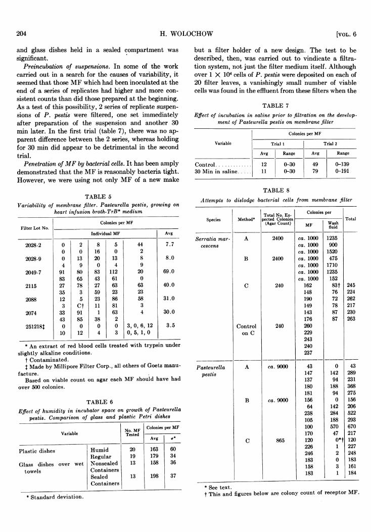
Preincubation of suspensions. In some of the workcarried out in a search for the causes of variability, itseemed that those MF which had been inoculated at theend of a series of replicates had higher and more con-sistent counts than did those prepared at the beginning.As a test of this possibility, 2 series of replicate suspen-sions of P. pestis were filtered, one set immediatelyafter preparation of the suspension and another 30min later. In the first trial (table 7), there was no ap-parent difference between the 2 series, whereas holdingfor 30 min did appear to be detrimental in the secondtrial.
Penetration ofMF by bacterial cells. It has been amplydemonstrated that the MF is reasonably bacteria tight.However, we were using not only MF of a new make
but a filter holder of a new design. The test to bedescribed, then, was carried out to vindicate a filtra-tion system, not just the filter medium itself. Althoughover 1 X 10⁴ cells of P. pestis were deposited on each of20 filter leaves, a vanishingly small number of viablecells was found in the effluent from these filters when the
TABLE 7
Effect of incubation in saline prior to filtration on the develop-ment of Pasteurella pestis on membrane filter
Colonies per MF
Variable Trial 1 Trial 2
Avg Range Avg Range
Control ............. 12 0-30 49 0-13930 Min in saline . ......110-30 79 0-191
TABLE 5Variability of membrane filter. Pasteurella pestis, growing on
heart infusion broth-TrB* medium
Colonies per MFFilter Lot No.
Individual MF Avg
2028-2 0 2 8 5 44 7.70 0 16 0 2
2028-9 0 13 20 13 8 8.04 9 0 4 9
2049-7 91 80 83 112 20 69.083 65 43 61 0
2115 27 78 27 63 63 40.035 3 59 23 23
2088 12 5 23 86 58 31.03 Ct 11 81 3
2074 33 91 1 63 4 30.043 85 38 2
251218t 0 0 0 0 3, 0, 6, 12 3.510 12 4 3 0, 5, 1, 0
* An extract of red blood cells treated with trypsin underslightly alkaline conditions.
t Contaminated.t Made by Millipore Filter Corp., all others of Goetz manu-
facture.Based on viable count on agar each MF should have had
over 500 colonies.
TABLE 6
Effect of humidity in incubator space on growth of Pasteturellapestis. Comparison of glass and plastic Petri dishes
Variable
Plastic dishes
Glass dishes over wettowels
HumidRegularNonsealedContainersSealedContainers
No. MFTested
201913
13
Colonies per MF
Avg or
163179158
198
603436
37
TABLE 8
Attempts to dislodge bacterial cells from membrane filter
Total No. Ex- Colonies perSpecies Method* pected Colonies Total
(Agar Count) MF Washfluid
Serratia mar- A 2400 ca. 1000 1235cescens ca. 1000 900
ca. 1000 1520B 2400 ca. 1000 475
ca. 1000 1710ca. 1000 1235ca. 1000 152
C 240 162 83t 245148 76 224190 72 262149 78 217143 87 230176 87 263
Control 240 260on C 229
243240237
Pasteurella A ca. 9000 43 0 43pestis 147 142 289
137 94 231180 188 368181 94 275
B ca. 9000 156 0 15664 142 206238 284 522105 188 293100 570 670170 47 217
C 865 120 °*t 120226 1 227246 2 248183 0 183158 3 161183 1 184
of receptor MF.* Standard deviation.
* See text.t This and figures below are colony count
204 [VOL. 6

9FILTER .METHOD FOR COUNTING VIABIE ORGANISMAIS
filtrate was re-run through fresh MIF or a portion tested(by the dropping pipette method) oni solid medium.Removal of cells impinged on MIF. Although not
strictly germane to the subject at hand, it was of interestto see if the bacterial cells once impinged on the filtersurface could readily be removed. To be of use as aconcentrating device by the bacteriologist, cells shouldbe removable, preferably quantitively. Three methods,with 2 different bacterial species were used to assessthis point. In method A (table 8), inoculated MF wererinsed with a pipette in 5 ml peptone water. In methodB, the MF were placed in a flask together with 5 mlpeptone water and the flask placed on a circular-motionshaker for about 15 min. In both instances, the bacterialcells in the peptone water were counted by dropping pi-pette method on appropriate solid medium; eachMF wasalso placed on a nutrient-soaked pad and incubated forcounts of the residual cells. In method C, a sterile MIFwas placed on the screen support of the MF holder; onthis was then placed another MF, face down, which hadreceived an inoculum just previously. After clampingdown the upper portion of the filter funnel, 25 ml ofsaline were poured through the 2 MF. These were thenremoved, separated and cultured as above. In table 8it is seen that the first two methods of washing the MFremoved not over 1 of the impinged cells of S. mar-cescens. With P. pestis, the total recovery was far lessthan that indicated by the viable count of the filteredsuspension. As it was, about ½1 the total of that whichwas recovered was found in the wash fluid. In the at-tempts to back-rinse MF, S. marcescens appeared lesstightly bound to the MF surface than did P. pestis.Of the numbers of the former organism, 2t were re-tained by the first MF, whereas with the latter, over95 per cent were so retained
DIscussIoN-
As with any new material and its application toestablished procedures, the membrane filter requiredconsiderable developmental work. From the foregoingresults it seems that there are still certain areas in thefield of bacteriology where further work is required be-fore the membrane filter can be considered to be entirelvsatisfactory.
Interpretation of much of the data presented is ren-
dered difficult because of the variability in replicatecounts and attempts to ascertain the cause(s) of this var-iability failed. However certain findings stand out. Cul-ture media devised on the basis of other uses may notbe satisfactory for use with MF; McCarthy (1955) hasexpressed the view that more satisfactory media foruse with the MF is a most urgent need. This may wellbe true in specialized instances, such as when a selectiveanid/or differential medium is required. Inadequate cul-ture media may be the basis for the variability in countsreported here; it is conceivable that some of the cells
in the population being filtered are borderline with re-spect to nutritional requirements and under the condi-tionsSwhich exist on the filter (in contrast to those on anagar surface) such cells are uiiable to reproduce to thepoint of producing countable colonies. There appearsto be no great restriction in concentration of ingredi-ents in the media tried. The desirability of using blotterpads impregnated with liquid media in preference to anagar surface seems to be questionable. Kutscher (1955),among others, is of the opinion that agar is to be pre-ferred. We too have observed in a number of instances,where comparisons were made, that agar supportedearlier growth, and somewhat more consistent quanti-tative results were obtained. Selection of the optimumvolume of liquid medium to be used in the nutrientpads must be governed by the thickness of the pad; it isnot desirable to have more than a drop of medium un-absorbed by the pad. Contamination of the medium bygrowth froin the upper side must be avoided and this isbest done by refraining from the use of too much liquidmedium and by employing aseptic technique.
It is obvious that some steps must be taken to preventdrying out of the medium; if this is not done the filterlifts off the surface and no flow of nutrient is possible.From the data presented it would seem that the sealafforded by the plastic dish is sufficient for incubationperiods of up to 2 days. On the basis of this work andaccording to Slanetz and Bartley (1955) saturatedhumidity, as described by Goetz (1951), may not berequired; on the other hand, Geldreich et al. (1955) areinsistent that saturated conditions are indispensable andsuggest the use of a parafilm wrap around Petri dishes.
Because of the variability in replicates, it cannot besaid with certainty that correlation existed betweenphysical characteristics and the suitability of the MFfor quantitative counts. However, unpublished work ofMleyers and Wolochow has indicated that, on certainfilters which were not wetted uniformly during fil-tration, growth of the inoculum subsequently failed toappear in the unwetted areas. On this basis, it is en-tirely possible that a similar situation existed wvithrespect to flow of nutrient to the impinged cell on thefilter surface, thus contributing to the v-ariability incounts which was so frequently encountered.
Loss of bacterial cells either through the ME itselfor around it was not found in these experiments; how-ever it seems that the cells do penetrate into the MF to
a certain extent or else are firmly held by some otherforce since it was not possible to rinse cells readily fromthe filter surface.
ACKN OWLEDGMENTSMuch of the work reported was carried out with the
excellent technical assistance of MIiss Eleanor Duffy and
MIiss Marjery Bailey.
1958] 205

H. WOLOCHOW
SUMMARYEquipment for use with the membrane filter is des-
cribed. It was found that the filter method for countingviable organisms was not reliable when certain organ-isms were tested. Some of the factors studied in anattempt to elucidate the cause(s) for the variabilityencountered were: concentration of medium, volume ofmedium in the nutrient pad, comparison of variationbetween lots of filters, humidity, and age of suspensionbefore filtration. None of these factors was found to beresponsible for the observed variability. The filtrationsystem (filter and funnel) was shown to retain over99 per cent of cells impinged on the filter; these cellswere thereafter removed with difficulty.
It was felt that the main sources of bias is probably afunction of the media used as well as variability inthe filter medium itself.
REFERENCES
GASPER, A. J. AND LEISE, J. M. 1955 Inhibitory effect ofgrid imprints on growth of Bacterium tularense on mem-brane filters. J. Bacteriol., 71, 728-731.
GASPER, A. J. AND LEISE, J. M. 1956 Inhibitory effect ofgrid imprints on growth of Brucella melitensis on mem-brane filters. Bacteriol. Proc. (Soc. Am. Bacteriologists),p. 31.
GELDREICH, E. E., KABLER, P. W., JETER, H. L., AND CLARK,H. F. 1955 A delayed incubation membrane filter test
for coliform bacteria in water. Am. J. Public Health, 45,1462-1474.
GOETZ, A. 1951 Early detection of bacterial growth. FinalReport. Contract W-18-064-CM-207, California Instituteof Technology, with Biological Department, ChemicalCorps, U. S. Army.
GOETZ, A. AND TSUNEISHI, N. 1951 Application of the mo-lecular filter membrane to the bacteriological analysis ofwater. J. Am. Water Works Assoc., 43, 943-984.
GOETZ, A. AND TSUNEISHI, N. 1956 A method for cultivationof Desulfovibrio aestuarii on molecular filter membranes.Bacteriol. Proc. (Soc. Am. Bacteriologists), p. 54.
KUTSCHER, U. 1955 Eine mikrobiologische Betrachtung zurmembranfilter methode. Brauerei Wiss. Beil., 8, 83-85.
MCCARTHY, J. A. 1955 Comparative coliform densities inwater by membrane filter test and multiple tube technic.Am. J. Public Health, 45, 1569-1577.
MORGANTE, 0. AND MURRAY, E. G. D. 1955 The isolation ofMycobacterium tuberculosis by filtration technique fromcerebrospinal fluid. Can. J. Microbiol., 1, 331-338.
ORLANDO, M. 0. AND BOLDUAN, 0. E. A. 1953 The applica-tion of the membrane filter to a variety of pathogenic bac-teria and fungi imperfecti. Bacteriol. Proc. (Soc. Am.Bacteriologists), pp. 26-27.
ROGERS, D. E., COOKE, G. M., AND MEYERS, C. E. 1955 Thedetection of tubercle bacilli in mouth wash specimens bythe use of membrane filter culture. Am. Rev. Tuberc.Pulmonary Diseases, 71, 371-381.
SLANETZ, L. W. AND BARTLEY, CLARA H. 1955 Evaluation ofmembrane filters for the determination of numbers ofcoliform bacteria in water. Appl. Microbiol., 3, 46-51.
206 [VOL. 6