the iraqi journal of veterinary medicine, 38(2):100-107
TRANSCRIPT

The Iraqi Journal of Veterinary Medicine, 38(2):100-107. 2014
100
Evaluation of general anesthesia induced by propofol, ketamine protocol in
rabbits premedicated with diazepam Ayad A. Amin and Areej Ali Atiyah
Department of Surgery and Obstetrics, College of Veterinary Medicine, University of Baghdad,
Iraq.
E-mail: [email protected]
Accepted on: 29/8/2014
Summary The present study was designed to evaluate the efficacy of diazepam-propofol-Ketamine
protocol for induction of general anaesthesia in rabbits. The experiment was conducted on
seventeen healthy male adult local rabbits weighting 0.9-1.5 kg. Rabbits were given protocol,
Diazepam 1mg/kg B.W. by intramuscular injection then 15 minutes later propofol 10 mg/kg B.W.
as bolus slow intravenous injection and ketamine 25 mg/kg BW by intramuscular injection. Several
parameters included respiratory rate, body temperature and heart rate were recorded before injection
of drugs and after giving the anesthetic protocol at 0, 5, 10, 15, 20, 30, 45, 60, 75 and 90 minutes.
The results showed that the anaesthesia with diazepam, propofol and Ketamine protocol in rabbits
was suitable as it produced reliable surgical anaesthesia, good analgesia and muscle relaxation with
minimal changes on the wave morphology of the cardiac muscle.
Keywords: Diazepam, propofol, ketamine, general anaesthesia, electrocardiogram, rabbit.
------------------------------------------------------------------------------------------------------------------------
Introduction
Rabbits are the third most anesthetized
animal species in the world, either as pets or as
research model. This gives these animals the
opportunity to conduct basic research with
direct application to the species and translation
to other species (1). Although rabbits have
more risks of anesthetic-related death when
compared to other species and they are easily
stressed by handling, and when excited may
struggle vigorously, this may lead to
musculoskeletal trauma, severe stress, shock
and cardiac arrest, in part due to difficulty in
endotracheal intubation, and also due to their
susceptibility to respiratory arrest once
adequate surgical anaesthesia has been
achieved (2 and 3). A variety of different
anesthetic techniques have been recommended
to overcome the various problems in rabbit
anaesthesia such as handling associated stress
and breath hold during induction with volatile
anaesthetic agent, apnoea is accompanied by
marked bradycardia, hypercapnia and hypoxia
and is not prevented by pre - anaesthetic
medication (4). General anaesthesia can be
induced by using a variety of drugs and
techniques, a single drug can be given to
produce all the required features of general
anaesthesia. Giving several drugs in
combination, at relatively low dose rates, can
often result in less effect on major body
systems than that following induction of
anaesthesia using a single anaesthetic agent
(5). Injectable anesthesia is more reliable than
inhalation anesthesia in clinical setting used
protocol of propofol and ketamine
administered for each can be reduced the dose
of other. Propofol is an alkylphenol, hypnotic
drug while ketamine is a dissociative non-
barbiturate anaesthetic agent. The
administration of propofol with ketamine is
producing a more stable hemodynamic and
respiratory profile (6). Premedication with
different sedative, hypnotic and muscle
relaxant drugs (e.g. diazepam which it is
classified as sedative, hypnotic and muscle
relaxant drugs of benzodiazepines derivative),
potentiates the effects of other anesthetic agent
in rabbits and facilitates a smooth induction
and recovery (7). The object of this study was
to evaluate the efficacy of diazepam-propofol-
Ketamine protocol for induction of general
anaesthesia in rabbits.
Materials and Methods
The study was performed on 17 local breed
adult males’ healthy rabbits. These rabbits
weight range from 0.9-1.5 kg, mean weight
was 1.24±0.04 kg, their ages ranged between
12-18 months. Animals were maintained in an
animal house and exposed for the same
The Iraqi Journal of Veterinary Medicine, 38(2):100-107. 2014
101
environment including climate, management
and feeding for 1 week to acclimatization and
adaptation on the place. Rabbits had free
access to water and food. Local anaesthetic gel
Lidocaine 2% was applied to the rabbits´ ears
for 5 mins to prevent pain during catheter
insertion (catheter gauge 27 was placed in the
marginal ear vein) and connected to a syringe
before induction of anesthesia. Prior to
induction of anaesthesia the animal was
premedicated with diazepam 1.0 mg/kg B.W.
I.M. (pharmaceutical industries. Aleppo-
Syria). After 15 minutes to induction of
anaesthesia by I.V. Propofol injection 10
mg/kg B.W. (Dong Kook pharmaceutical
/Korea) via marginal ear vein followed by
Ketamine (TEKAM 50, Gracure
pharmaceuticals Ltd, India) 25 mg/kg B.W.
I.M. The evaluation of the anesthetic protocol
was basically evaluated depending on the
monitoring of the following parameters which
recorded before injection of any drug as
control data and at zero, 5, 10, 15, 20, 30,
45, 60, 75 and 90 minutes. Monitoring
continued until the animal regained its righting
reflex. Anesthetic period was estimated from
the induction time of injection the anesthetic
agent to a satisfactory immobilization until
recovery. The recovery period was estimated
from the time of reappearance of the reflex
until complete consciousness (complete
righting reflex) (8). Degree of analgesia was
evaluated by (9): + (Mild degree of the
analgesia) ++ (Moderate analgesia) +++ (Deep
analgesia). Pinching toe-web region in the
hind limbs was alternatively pinched with
hemostatic forceps to the first, second and
third ratchet-lock for 5 sec, and was evaluated
at 0, 5, 10, 15, 20, 30, 45, 60, 75, and 90 min
from anesthetic injection. Depth of anesthesia
was going to be evaluated according to the
following data (Table 1).
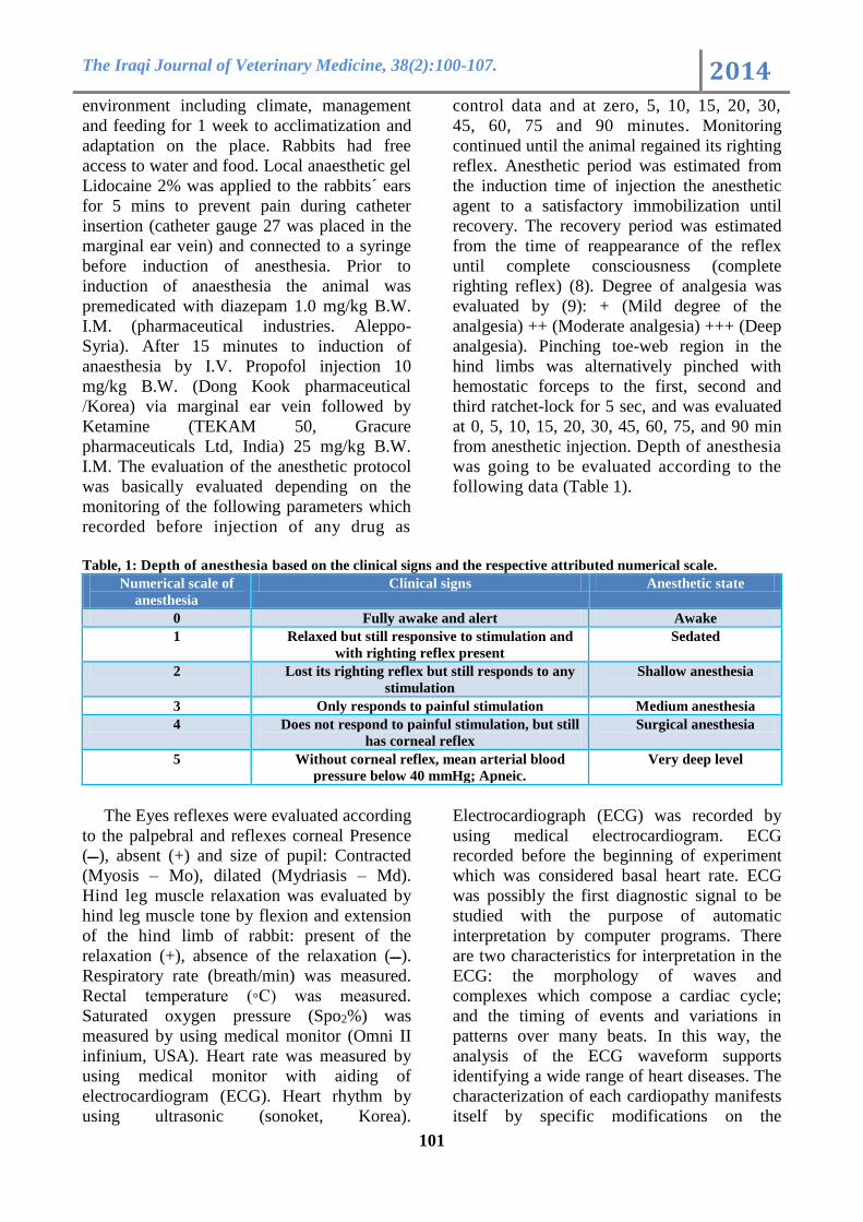
Table, 1: Depth of anesthesia based on the clinical signs and the respective attributed numerical scale.
Numerical scale of
anesthesia
Clinical signs Anesthetic state
0 Fully awake and alert Awake
1 Relaxed but still responsive to stimulation and
with righting reflex present
Sedated
2 Lost its righting reflex but still responds to any
stimulation
Shallow anesthesia
3 Only responds to painful stimulation Medium anesthesia
4 Does not respond to painful stimulation, but still
has corneal reflex
Surgical anesthesia
5 Without corneal reflex, mean arterial blood
pressure below 40 mmHg; Apneic.
Very deep level
The Eyes reflexes were evaluated according
to the palpebral and reflexes corneal Presence
absent (+) and size of pupil: Contracted ,(ـــ)
(Myosis – Mo), dilated (Mydriasis – Md).
Hind leg muscle relaxation was evaluated by
hind leg muscle tone by flexion and extension
of the hind limb of rabbit: present of the
relaxation (+), absence of the relaxation (ـــ).
Respiratory rate (breath/min) was measured.
Rectal temperature (◦C) was measured.
Saturated oxygen pressure (Spo2%) was
measured by using medical monitor (Omni II
infinium, USA). Heart rate was measured by
using medical monitor with aiding of
electrocardiogram (ECG). Heart rhythm by
using ultrasonic (sonoket, Korea).
Electrocardiograph (ECG) was recorded by
using medical electrocardiogram. ECG
recorded before the beginning of experiment
which was considered basal heart rate. ECG
was possibly the first diagnostic signal to be
studied with the purpose of automatic
interpretation by computer programs. There
are two characteristics for interpretation in the
ECG: the morphology of waves and
complexes which compose a cardiac cycle;
and the timing of events and variations in
patterns over many beats. In this way, the
analysis of the ECG waveform supports
identifying a wide range of heart diseases. The
characterization of each cardiopathy manifests
itself by specific modifications on the

The Iraqi Journal of Veterinary Medicine, 38(2):100-107. 2014
102
characteristics (10). Preparation of rabbit for
recording of ECG by using clip electrodes
which were attached to the skin at the triceps
brachi muscle (coputlongum and coputlaterale)
of the right and left limbs and biceps femurs
muscle of the right and left hips. Electrode gel
was rubbed into the skin in the area where the
clips were attached to act as a decreasing agent
and thereby decrease the resistance of the skin.
The rabbits were immobilized by wrapping
light cotton around them and then placed on a
wooden table. It waited about 5 min. for
rabbits to get calm. ECGs were recorded by a
direct writing electrocardiograph with a chart
speed of 50 mm/sec. Leads I, II, II, aVR, aVL
and aVF were recorded before and after
anesthetic protocol was given. The
morphological changes of the waves on the
trace were observed in all limb leads to detect
any effect of the anesthetic protocol on these
cardiac waves. The Statistical Analysis was
used to detect the effect of factor (group) in
study parameters. The least significant
difference (LSD) test and chi-square test were
used at the comparative between means and
percentage in this study (11).
Results and Discussion
The anaesthetic duration was 32.88±2.33
min. This result might due to inhibition effects
of CNS mediated by effect of diazepam,
propofol, and ketamine induced by deep
sedative effect mediated by diazepam and
anesthetic effects of propofol and ketamine
resulting in inhibition of CNS depression
mediated by inhibition of intranural
transmission of impulses. This result agreed
with (2). The recovery period was 42.27±1.95
min. Although this protocol is safe, it produces
a relatively long recovery period due to effect
of diazepam which is classified as sedative,
hypnotic and anticonvulsant muscle relaxant
properties drugs of benzodiazepines derivative
(12). The surgical anaesthesia was evaluated
according to the (Table, 1) in which the
anesthetic status was based on clinical signs
and the respective attributed numerical scale,
scale 4 in which rabbit status was
characterized by not responding to painful
stimulation, but still has corneal reflex. The
surgical anaesthesia (25 min duration)
extendes from 5 min to 30 min, the optimum
was at 15 min and there was significant
(P<0.01) difference between 0 time and 10–20
min, and there was significant (P<0.05)
difference between the 0 time and 30 min, but
there was non-significant (P˃0.01) difference
between the 0 time and 5, 45 min. The total
response percentage about 29% in diazepam,
propofol, and ketamine as in (Table, 2).
Table, 2: The effect of protocol (diazepam, propofol, and ketamine) on different parameters at ten time points.
parameter Time / Minutes significant
difference 0 5 10 15 20 30 45 60 75 90
R R
(bth/min)
(c)
37.82±
5.13
(c)
39.82±
4.33
(c)
43.00±
3.26
(bc)
46.29±
2.99
(bc)
51.47±
3.40
(abc)
60.64±
5.57
(ab)
69.52±
6.85
(a)
76.76±
7.73
(a)
79.53±
7.17
(a)
77.82±
6.52
23.91 *
Spo2(%) (abc)
88.88 ±
1.18
(bc)
85.35 ±
1.40
(c)
85.11 ±
1.97
(a)
90.70 ±
1.52
(abc)
87.76 ±
1.72
(abc)
88.23 ±
1.30
(abc)
87.88 ±
2.18
(bc)
85.70 ±
1.42
(ab)
89.47±
1.05
(abc)
89.23±
0.76
4.272 *
Temp(◦C) (a)
37.98 ±
0.17
(ab)
37.84 ±
0.17
(abc)
37.67 ±
0.17
(abc)
37.57 ±
0.18
(bc)
37.39 ±
0.19
(c)
37.26 ±
0.19
(c)
37.14 ±
0.18
(c)
37.03 ±
0.19
(c)
7.09 ±
0.25
(c)
37.15 ±
0.23
0.579 *
H.R.
(bt/min)
(a)
252.76
± 7.86A
(a)
248.23±
10.04
(a)
A248.58±
10.31
(a)
248.82
± 8.65
(a)
244.47
± 9.65
(a)
240.88
± 7.64
(a)
237.94
± 8.81
(a)
234.3 ±
7.09
(a)
238.12
± 6.63
(a)
230.29
± 4.80
22.684 NS
Analgesia
(scale 3)
(e)
0
(de)
5.88
(c)
47.06
(a)
82.35
(b)
58.82
(d)
11.76
(d)
0
---- ---- ---- 11.65**
Anesthesia
scale (4)
0 (c) 5.88 (ef) 47.06 (c) 82.35
(a)
58.82
(b)
11.76
(de)
0
(c)
---- ---- ---- 11.91**
Muscle
relaxation
(present)
(a)
100
(a)
100
(a)
100
(a)
100
(a)
100
(a)
100
(a) 100 (a)
100
(b)
5.29
(c)
23.53
16.72**
Size of
pupil (md)
(c)
29.41
(a)
100
(a)
100
(a)
100
(a)
100
(a)
94.12
(b)
58.82
(c)
23.53
(d)
5.88
(d)
0
12.40**
NS – non significant, small letters within group/rows are significantly different at ** (P<0.01), * (P<0.05). DPK (diazepam +
propofol + ketamine), R.R. (respiratory rate-breath/minute), bth-breath/minute, Spo2 (saturated pressure of oxygen-%),
Temp (rectal body temperature -◦C), ◦C- centigrade degree, H.R. (heart rate-beat/minute), bt- beat/minute, md (mydriases-
dilatation in the eye pupil).

The Iraqi Journal of Veterinary Medicine, 38(2):100-107. 2014
103
The degree of analgesia at scale (3) which
means no sense of pain extended from 5 min
to 30 min, the optimum was at 15 min, and
there was significant (P<0.01) difference
between the 0 time and 10, 15, 20, and
significant difference (P<0.05) between the 0
time and 30 min, but there was non-significant
(P˃0.01) difference between the 0 time and 5
min. The total response percentage about 29%
as in (Table, 2). Propofol in clinical doses was
weak at controlling pain and had no analgesic
effect (13), unless combined with analgesic
drug (14). The analgesic effects of ketamine
were thought to be mediated by binding of the
drug to N-methyl-D-aspartate (NMDA)
receptors which results in inhibition of these
receptors which mean inhibition of excitatory
glutaminergic transmission at spinal and
supraspinal sites (15). Although ketamine acts
on nicotinic and muscarinic receptors; it
blocks sodium channels in the peripheral and
human central nervous system and interacts
with opioid receptors, μ, δ and κ and with
calcium channels (16). Combination with
drugs such as diazepam is to improve
analgesia and muscle relaxation of anesthetic
regimes (17). It is important to ensure that
additional analgesic agents used when painful
procedures are required (18). The degree of
muscle relaxation started early after the animal
was premedicated with diazepam and reached
to the optimum degree in time control (the
time of propofol injection) extending to 60
min, while it was minimal at 75, 90 min until
loss of muscle relaxation at 105 min. There
was significant (P<0.01) difference between
the zero time and 75, 90, 105 min but there
was non-significant (P˃0.01) difference
between the 0 time and 5 min to 60 min. It had
a significant response in anesthetic protocol.
This result was in agreement with (19) which
revealed the use of diazepam with ketamine
aids muscle relaxation. The total response
percentage about 87%. In general
benzodizepines can be used to augment
sedation and muscle relaxation in combination
with other anaesthetic (e.g. ketamine). Muscle
relaxation effect of diazepam was mediated
through depression of polysynaptic
musculoskeletal reflexes (20). Propofol has
been shown to possess anticonvulsant activity
and antiseizure properties (18) and inhibit
calcium to entry in muscle cells (21).
Ketamine is always administered in
combination with diazepam to eliminate the
muscle rigidity, which occurs when ketamine
is used alone (22). The eye reflexes (palpebral
and corneal reflexes) were never abolished
completely. It became nearly sluggish at 5 min
to 30 min. These results were in agreement
with (23), the palpebral and corneal reflexes
may be consistently abolished only
immediately before fatal respiratory arrest.
The pupil size was dilated from time 0 and
extended to 75 min and it was found (P<0.01)
significantly dilated at at 5 min to 45 min.
There was significant (P<0.01) difference
between the 0 time and 5 min to 45 min and at
75, 90 min, but there was non-significant
(P˃0.01) difference between the 0 time and 60
min of the observation. This result was
consistent with (24). The total response
percentage was about (61%) in DPK as (Table,
2). This result may be due to the effect caused
by central inhibition of parasympathetic tone
to the iris and/or direct sympathetic
stimulation of alpha-2 adrenoceptors located in
iris and C.N.S. The respiratory rate
(breath/min) was decreased by the effect of
anesthetic protocol and this result was in
consistency with (25). The decreasing of
respiratory rate was from zero time, then it
increased gradually from 5 min. to 75 min.
then minimal decline at 90 min at the end of
the observation. There was significant
(P<0.05) difference between the 0 time and 45
min to 90 min but there was non-significant
(P˃0.05) difference between the 0 time and the
time of 5 min to 30 min as in (Table, 2).
Benzodiazepine did not significantly alter
respiratory rate (26). Ketamine administration
causes minimal depression of respiratory rate
(15), moderate respiratory depression (5),
bronchodilation (15) and high doses could lead
to serious respiratory depression including
apnea (27). The effect of anesthetic protocol
on the saturated pressure of oxygen was
(SPO2%): It was decreased in time 5 min, 10
min and fluctuating between the increasing
and decreasing to the end of the observation.
There was non-significant (P˃0.05) difference
between the 0 time and the other times to the

The Iraqi Journal of Veterinary Medicine, 38(2):100-107. 2014
104
end of the experiment as in (Table, 2). From
the observation, it was concluded that
saturated pressure of oxygen was not affected
by this protocol with high degree.
Anesthetized rabbits with propofol,
macroscopic findings of the lungs at necropsy
included lung enlargement and congestion and
pinky frothy edema fluid effusing from lung
sections and filling the tracheal cannula. The
lungs had also a milky tincture and
histological examination revealed interstitial
pneumonia and pulmonary edema. This last
finding was suggested as the most probable
cause of death, and the potential for pulmonary
embolism (28) are believed to be due in large
part to its oil-in-water emulsion formulation
(29). The effect of anesthetic protocol on the
body rectal temperature (◦C): It was gradually
decreasing in body rectal temperature in time
zero to 60 min then gradually increasing after
60 min to the end of the experiment. There
was significant (P˂0.05) difference between
the zero time and 20 min. to 105 min., but
there was non-significant (P˃0.05) difference
between the zero time and the time of 5 min to
15 min as in (Table, 2). From the observation,
it was concluded that body rectal temperature
was affected by sedative effect of diazepam
which induced reduction in metabolism,
muscle relaxation and depression of the CNS
which agreed with the study of other authors
(30). Reduction in cutaneous heat losses, in
contrast to the consistent reductions in body
temperature reported with the use of other
anesthetic agents that induce vasodilatation
(31). The heart rhythm was significantly
regular and this appeared from ECG paper.
There was minimal decreasing in heart rate
from 5 min to the end of the experiment
(Table, 2). From this observation,
cardiovascular and autonomic side effect of
diazepam is negligible and it has
antiarrhythmic action due to decrease
catecholamine release, which has proven
useful in treating certain kinds of myocardial
hyper excitability (32). Diazepam causes mild
decrease in heart rate (33).
T1
(control)
T2
(11 min)
T3
(26 min)

The Iraqi Journal of Veterinary Medicine, 38(2):100-107. 2014
105
T4
(33 min)
T5
(53 min)
T6
(78 min) Figure, 1: Electrocardiography results of anesthetic protocol, for local male rabbit in different times (T): T1
(control), T2 (11 min), T3 (26 min), T4 (33 min), T5 (53 min) and T6 (78 min) at the end of experiment.
The electrocardiogram (limb leads) of
rabbits in (Fig. 1), T wave in lead I showed
mild flatten after 26 minute (T3) comparing to
control (T1) and 11 min (T2) in the same
protocol. In addition there was a rise of QRS
complex wave peak in lead I and AVR lead at
26 min (T3), 33 min (T4), 53 min (T5), and
78 minute (T6) as compared to control (T1)
and 11 min (T2). The T wave is generated by
myocardial voltage gradients during the
repolarization phase of cardiomyocyte action
potentials (34). Myocardial ischemia may
cause T wave changes and abnormally tall T
waves (35). The majority of intraoperative
ischemic episodes occur in the absence of
hemodynamic aberrations, such as
tachycardia, hypertension, or hypotension.
This implies that many of the episodes of
intraoperative myocardial ischemia occur
because of a decrease in myocardial oxygen
supply rather than an increase in myocardial
oxygen demand (36). Modern anesthetic
interventions may help control ischemic
episodes during the intraoperative period (37).
The QT interval consists of two components:
the QRS complex and T wave interval (38),
Prolongation of the QT interval may be a
consequence of an unfavorable balance
between sympathetic and parasympathetic
activity. It has been noted that imbalance in
cardiac autonomic function (increased or
decreased sympathetic activity) shortens or
prolongs the QT interval of the
electrocardiogram (39). It was found from the
results that the study protocol is safe on the
cardiac muscle contractile because the
changes are minimal in the electrocardiogram.
Acknowledgements: We would like to
express our gratitude to several people who
helped us while performing this work,
especially Dr. Ahmed dawood; Dr. Raffal A.
Omar; Dr. Abed Fadhil Ali and Dr. Sameer
Ahmed Abid Al-Redah.
References
1. Campos, S. P. (2010). Assessment of propofol
anesthesia in the rabbit. Universidade de trás-
os-montes e. alto douro. p: 2.
2. Kilic, N. (2004). A Comparison between
Medetomidine – Ketamine and Xylazine –
Ketamine Anesthesia in Rabbits. Turk. J. Vet.
Anim. Sci. 28: 921-926.
3. Brodbelt, D. (2009). Perioperative mortality
in small animal anaesthesia. Vet J., 182(2):
152-161.
The Iraqi Journal of Veterinary Medicine, 38(2):100-107. 2014
106
4. Liles, J. H. and Flecknell, P. A. (1996).
Halothane anesthesia in the rabbit: a
comparision of the effect of medetomidine,
acepromazine and midazolam on breath –
holding during induction. Ass. Vet. Anesth.
23:11-14.
5. Flecknell, P. (2009). Laboratory Animal
Anaesthesia. Third Edition. P: 20.
6. Arora, S.; Gleed, F. S. and Loh, M. D. (2007).
Combining ketamine and propofol (ketofol)
for Emergency Department procedural.
Sedation and Analgesia Western J.
Emergency Med., 9:20-23.
7. Harcourt – Brown, F. (2001). Textbook of
Rabbit Medicine. Butterworth Heinemann.
Newton. M. A., USA. Pp: 148-153.
8. Ringer, S. K.; Kalchofner, K.; Boller, J.;
Furst, A. and Bettschart-Wolfensberger, R.
(2007). A clinical comparison of two
anesthetic protocols using lidocaine or
medetomidine in horse. Vet. Anesth. Analg.,
34(8): 257-268.
9. Choi, W.; Jang, H. S.; Yun, S. H.; Park, J. S.;
Kwon, Y. S. and Jang, K. H. (2011). Effect of
tramadol on medetomidine and ketamine
anesthesia in dogs. Pak. Vet. J., 31(2): 99-
104.
10. Andreão, R.; Pereira Filho, J. and Calvi, C.
(2006). “Tele Cardio - Telecardiology for
Patient Support in Hospital and Domicile
Environments” (in Portuguese). In X
Brazilian Conference in Health Informatics,
Florianópolis, Brazil.
11. SAS. (2004). Statistical Analysis System,
User's Guide. Statistical. Version 7th ed. SAS.
Inst. Inc. Cary. N.C. USA.
12. Harcourt-Brown, F. (2002): Text book of
rabbit medicine. 1st ed. Butterworth
Heinemann. Pp: 126-130.
13. Keath, L. M. (2005). Therapeutic agents. Ch.
9 in: Donald, C.P (ed.) Veterinary drug
handbook. 5th Ed., Pp: 285-315, 439-443,
670-672, 802-805.
14. Kanazawa, M.; Nitta, M.; Murata, T. and
Suzuki, T. (2006). Increased dosage of
propofol in anesthesia induction cannot
control the patient’s responses to Insertion of
a laryngeal mask airway. Tokai J. Exp. Clin.
Med., 31: 35-38.
15. Doherty, T. and Valverde, A. (2006). Manual
of Equine Anesthesia and Analgesia.
Pharmacology of drugs used in equine
anesthesia, Pp: 145-146.
16. Launo, C.; Bassi, C.; Spagnolo, L.; Badano,
S.; Ricci, C.; Lizzi, A., and Molinino, M.
(2004). Preemptive ketamine during general
anesthesia for postoperative analgesia in
patients undergoing laparoscopic
cholecystectomy. Minerva Anestesiol.,
70:727-738.
17. Mohammed, M. S. (2011). A comparison of
four injectable anesthetic regimes in
detomidine premedicated donkeys. Ms.c
thesis in Veterinary medicine/ Veterinary
surgery (Veterinary anesthesia).
18. Kotani, Y.; Shimazawa, M.; Yoshimura S.;
Iwama T. and Hara H. (2008). "The
experimental and clinical pharmacology of
propofol, an anesthetic agent with
neuroprotective properties." CNS Neurosci
Ther. 14(2): 95-106.
19. Ismail, Z. B.; Jawasreh, K. and Al-Majali, A.
(2009): Effects of xylazine–ketamine–
diazepam anesthesia on blood cell counts and
plasma biochemical values in sheep and
goats. Comp. Clin. Pathol., 10: 580-585.
20. Cantwell, S. L. (2001). Ferret, Rabbit, and
Rodent Anaesthesia. Veterinary Clinics of
North America: Exotic Anim. Prac., 4(1):
169-191.
21. Horibe, M.; Kondo, I.; Damron, DS. and
Murray, PA. (2001): Propofol attenuates
capacitative calcium entry in pulmonary
artery smooth muscle cells. Anesthesiology,
95:681-688.
22. Lumeij, J. T. and Deenik, J. W. (2003).
Medetomidine-ketamine and diazepam-
ketamine anaesthesia in racing pigeons – A
comparative study. J. Avian Med. Surgery.
17(4): 191–196.
23. Gonzalez, A.; Silvan, G.; Illera, M. and Illera,
J. C. (2004): The Effects of Anesthesia on the
Clinical Chemistry of New Zealand White
Rabbits. Comtemp. Top. Lab. Amin. Sci.,
43(3): 24-28.
24. Brown, E. T.; Umino, Y.; Loi, T.; Solessio, E.
and Barlow, R. (2005). Anesthesia can cause
sustained hyperglycemia in C57/BL6J mice.
Vis. Neurosci. 22(5): 615–618.
25. Cancho, M.; Lima, R.; Luis L.; Crisostomo,
V.; Carrasco, M. and Gargallo, JU. (2006).
Relationship of bispectral index values,

The Iraqi Journal of Veterinary Medicine, 38(2):100-107. 2014
107
haemodynamic changes and recovery times
during sevoflurane or propofol anaesthesia in
rabbits. Laboratory Animals. 40: 28–42.
26. Uzun, M.; Onder, F.; Atalan, G.; Cenesiz, M.;
Kaya, M. and Yildiz, S. (2006). Effects of
xylazine, medetomidine, detomidine, and
diazepam on sedation, heart and respiratory
rates, and cloacal temperature in rock
partridges (Alectoris graeca). J. Zoo and
Wildlife Med. 37: 135-140.
27. Sumitra, M.; Manikandan, P. and Reo, K.V.
(2004). Cardiorespiratory effect of diazepam
– ketamine, xylazine – ketamine thiopentone
anesthesia in male wistar rats. A comparative
Analysis, life Sci., 75: 1887 – 1896.
28. Barrientos-Vega, R.; Sanchez-Soria, M. M.
and Morales-Gracia, C.; Cuena-Boy R. and
Castellano-Hernández M. (2001).
Pharmacoeconomic assessment of propofol
2% used for prolonged sedation. Crit Care
Med., 29: 317–322.
29. Banaszczyk, M. G.; Carlo, A. T.; Millan, V.;
Lindsey, A.; Moss, R.; Carlo, D. J. and
Hendler, S. S. (2002). Propofol Phosphate, a
Water-Soluble Propofol Prodrug: In vivo
Evaluation by the International Anesthesia
Research Society. Anesth. Analg., 95:1285–
1292.
30. Kinjavdekar, P.; Singh, G. R.; Amarpal, H. P.
and Aithal AM. (2000). Pawde. Physiological
and biochemical effects of subarachnoidally
administered xylazine and medetomidine in
goats. Small Rum Res., 38: 217-228.
31. Sinclair, M. D. (2003). A review of the
physiological effects of alpha 2-agonists
related to the clinical use of medetomidine in
small animal practice. Can.Vet. J., 44:885–
897.
32. Flecknell, P. A. (2000). Manual of Rabbit
Medicine and Surgery. 1st ed. B. S. A. V. A.
Pp: 13.
33. Ghurashi, M. A.; Seri, H. I.; Bakheit, A. H.;
Ashwag, E. A. and Abakar, J. A. (2009).
Evaluation of Ketamine/ diazepam
Anaesthesia for Performing Surgery in Desert
Goats under Field Condition. Australian J.
Basic and Appl. Sci., 3(2): 455-459.
34. Antzelevitch, C. (2006). Cellular basis for the
repolarization waves of the ECG. Ann. N. Y.
Acad. Sci., 1080: 268 – 281.
35. Rowlands, D. J. (2002). The Resting Electro
cardio graph, Medicine Publishing Comp.,
Pp: 9-17.
36. Gotoh, K.; Minamino, T.; Katoh, O.;
Hamano, Y.; Fukui, S.; Hori, M.; Kusuoka,
H.; Mishima, M.; Inoue, M. and Kamada, T.
(1988).The role of intracoronary thrombus in
unstable angina: angiographic assessment and
thrombolytic therapy during ongoing anginal
attacks. Circulation. 77: 526-534.
37. Knight, A. A.; Hollenberg, M.; London, M. J.;
Tubau, J.; Verrier. E.; Browner, W. and
Mangano D. T. (1988). Perioperative
myocardial ischemia: importance of the
preoperative ischemic pattern.
Anesthesiology. Hollenberg M. London M. J.,
68: 681-688.
38. Reisner, A.; Clifford, G. and Mark, R. (2006).
The Physiological Basis of the
Electrocardiogram. Harvard-MIT Division of
Health Sciences and Technology J., 582: 11.
39. Bednar, M.; Harrigan, E. P.; Anziano, R. J.;
Camm, A. J. and Ruskin, J. N. (2001). The
QT interval. Prog. Cardiovasc. Dis., 43:1.
بالديازيبام تمهيديا المعالجة الأرانب في الكيتامين مع البروبوفول طريق عن العام برنامج التخدير تقييم اياد عبد الجبار امين و اريج علي عطية
.العراق بغداد، جامعة البيطري، الطب كلية والتوليد، الجراحة فرع
E-mail: [email protected]
لخلاصةا. لغرض إحداث التخير العام في الأرانب والكيتامين –البروبوفول –ديازيبام برنامج استعمال كفائة لتقييم الدراسة هذه تصميم تم
حقن تم. كجم 5.1 – 9.0وزنها يتراوح, المحلي النوع ذات صحة جيدة ومن البالغة الأرانب ذكور من سبعة عشر على التجربة أجريت
/ ملغم 59 البروبوفول دقيقة تم حقن 51وبعد العضلي الحقن طريق عن الجسم وزن كغم/ملغم 5 الديازيبام من المتكون بالبرنامج الأرانب
مجموعة تسجيل تم. العضلي الحقن طريق عن الجسم وزن كغم/ ملغم 51 الكيتامين و الوريد في البطئ الحقن طريق عن الجسم وزن كغم
التوالي. على 09, 51, 09, 51, 09, 59, 51, 59, 1 ,9بالدقائق البرنامج التخديري إعطاء بعد ثم ومن الأدوية حقن قبل القياساتمن
الجراحي، حيث التخدير من مناسب كان الأرانب في الكيتامين و البروبوفول و الديازيبام برنامج المتكون منال مع التخدير أن النتائج أظهرت
.القلب لعضلة الموجي الشكل على طفيفة تغيرات مع العضلات واسترخاء جيد في التسكين
نب.ابروبوفول، كيتامين، تخدير عام، تخطيط كهربائي للقلب، أر مفتاحية: ديازيبام،الكلمات ال