the distinctive electrocardiogram coronary … · the electrocardiogram resumea strictly...
TRANSCRIPT
THE DISTINCTIVE ELECTROCARDIOGRAM OF CORONARYARTERIOSPASM
BY
WILLIAM EVANS
From the Cardiac Department of the London Hospital
Received April 30, 1954
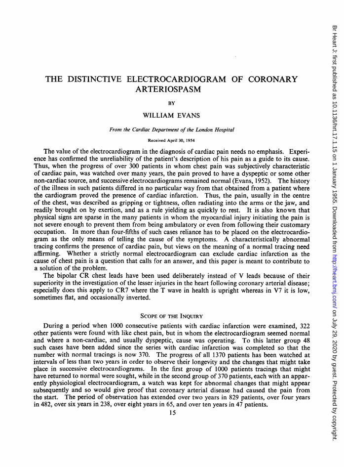
The value of the electrocardiogram in the diagnosis of cardiac pain needs no emphasis. Experi-ence has confirmed the unreliability of the patient's description of his pain as a guide to its cause.Thus, when the progress of over 300 patients in whom chest pain was subjectively characteristicof cardiac pain, was watched over many years, the pain proved to have a dyspeptic or some othernon-cardiac source, and successive electrocardiograms remained normal (Evans, 1952). The historyof the illness in such patients differed in no particular way from that obtained from a patient wherethe cardiogram proved the presence of cardiac infarction. Thus, the pain, usually in the centreof the chest, was described as gripping or tightness, often radiating into the arms or the jaw, andreadily brought on by exertion, and as a rule yielding as quickly to rest. It is also known thatphysical signs are sparse in the many patients in whom the myocardial injury initiating the pain isnot severe enough to prevent them from being ambulatory or even from following their customaryoccupation. In more than four-fifths of such cases reliance has to be placed on the electrocardio-gram as the only means of telling the cause of the symptoms. A characteristically abnormaltracing confirms the presence of cardiac pain, but views on the meaning of a normal tracing needaffirming. Whether a strictly normal electrocardiogram can exclude cardiac infarction as thecause of chest pain is a question that calls for an answer, and this paper is meant to contribute toa solution of the problem.
The bipolar CR chest leads have been used deliberately instead of V leads because of theirsuperiority in the investigation of the lesser injuries in the heart following coronary arterial disease;especially does this apply to CR7 where the T wave in health is upright whereas in V7 it is low,sometimes flat, and occasionally inverted.
SCOPE OF THE INQUIRYDuring a period when 1000 consecutive patients with cardiac infarction were examined, 322
other patients were found with like chest pain, but in whom the electrocardiogram seemed normaland where a non-cardiac, and usually dyspeptic, cause was operating. To this latter group 48such cases have been added since the series with cardiac infarction was completed so that thenumber with normal tracings is now 370. The progress of all 1370 patients has been watched atintervals of less than two years in order to observe their longevity and the changes that might takeplace in successive electrocardiograms. In the first group of 1000 patients tracings that mighthave returned to normal were sought, while in the second group of 370 patients, each with an appar-ently physiological electrocardiogram, a watch was kept for abnormal changes that might appearsubsequently and so would give proof that coronary arterial disease had caused the pain fromthe start. The period of observation has extended over two years in 829 patients, over four yearsin 482, over six years in 238, over eight years in 65, and over ten years in 47 patients.
15
on July 29, 2020 by guest. Protected by copyright.
http://heart.bmj.com
/B
r Heart J: first published as 10.1136/hrt.17.1.15 on 1 January 1955. D
ownloaded from
THE ELECTROCARDIOGRAM CUSTOMARILY FOUND IN CARDIAC PAINThe design of the electrocardiogram in the series of patients with cardiac infarction conformed
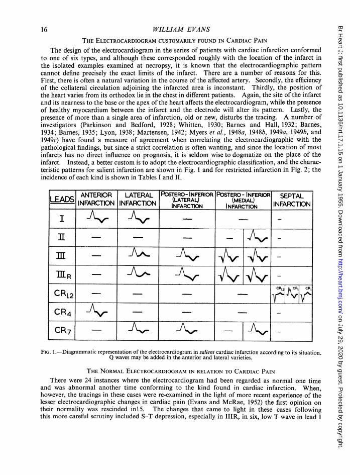
to one of six types, and although these corresponded roughly with the location of the infarct inthe isolated examples examined at necropsy, it is known that the electrocardiographic patterncannot define precisely the exact limits of the infarct. There are a number of reasons for this.First, there is often a natural variation in the course of the affected artery. Secondly, the efficiencyof the collateral circulation adjoining the infarcted area is inconstant. Thirdly, the position ofthe heart varies from its orthodox lie in the chest in different patients. Again, the site of the infarctand its nearness to the base or the apex of the heart affects the electrocardiogram, while the presenceof healthy myocardium between the infarct and the electrode will alter its pattern. Lastly, thepresence of more than a single area of infarction, old or new, disturbs the tracing. A number ofinvestigators (Parkinson and Bedford, 1928; Whitten, 1930; Barnes and Hall, 1932; Barnes,1934; Barnes, 1935; Lyon, 1938; Martensen, 1942; Myers et al., 1948a, 1948b, 1949a, 1949b, and1949c) have found a measure of agreement when correlating the electrocardiographic with thepathological findings, but since a strict correlation is often wanting, and since the location of mostinfarcts has no direct influence on prognosis, it is seldom wise to dogmatize on the place of theinfarct. Instead, a better custom is to adopt the electrocardiographic classification, and the charac-teristic patterns for salient infarction are shown in Fig. 1 and for restricted infarction in Fig. 2; theincidence of each kind is shown in Tables I and II.
ANTERIOR LATERAL P%STERO- INFERIOR POSTERO - INFERIOR SEPTALLED_IFRCIN_NARTO (LATERALQ (MEDIAL) INFARCTIONIEA|INFARCTION | INFARCTION | INFARCTION INFARCTION
CRI _
CRCR__|
C1.2 _ _"
CR4CR7- J
FIG. 1.-Diagrammatic representation of the electrocardiogram in salient cardiac infarction according to its situation.Q waves may be added in the anterior and lateral varieties.
THE NORMAL ELECTROCARDIOGRAM IN RELATION TO CARDIAC PAINThere were 24 instances where the electrocardiogram had been regarded as normal one time
and was abnormal another time conforming to the kind found in cardiac infarction. When,however, the tracings in these cases were re-examined in the light of more recent experience of thelesser electrocardiographic changes in cardiac pain (Evans and McRae, 1952) the first opinion ontheir normality was rescinded in15. The changes that came to light in these cases followingthis more careful scrutiny included S-T depression, especially in IIIR, in six, low T wave in lead I
16 WILLIAM EVANS
on July 29, 2020 by guest. Protected by copyright.
http://heart.bmj.com
/B
r Heart J: first published as 10.1136/hrt.17.1.15 on 1 January 1955. D
ownloaded from

ELECTROCARDIOGRAM OF CORONARY ARTERIOSPASM
DEPRESSION of S-T
FIG. 2.-Diagrammatic representation of the lesser electrocardiographic changes in restricted cardiac infarction.
TABLE IDISTRIBUTION OF THE INFARCT AND THE MORTALITY RATE AMONG 1000 CONSECUTIVE PATIENTS WITH CARDIAC
INFARCTION
DeathsDistribution of the infarct No. of patients
Number Mortality rate
Anterior 234 39 17Lateral 126 28 22
Salient Postero-inferior (lateral) 130 27 20Postero-inferior (medial) 132 24 18Septal 50 7 14
Restricted 328 60 19
Total 1000 185 19
and in CR4 or CR7 in three, the appearance of a significant Q wave in IIIR in three, a blunt T wavein CR4 in one, splintering of the RS in CR7 in one, and an inverted U wave in one.
There were left, therefore, nine electrocardiograms in patients with cardiac pain which onetime showed no departure from the design at present accepted as belonging to a healthy subject,but another time showed the characteristic changes found in cardiac infarction (Table III).The number is very small (9 out of 1004) and this fact alone emphasizes the hesitancy with whicha diagnosis of cardiac pain should be applied to a patient where the electrocardiogram fails tosupport it. Small as the number is, however, it calls for a close search for any special clinical or
cardiographic features that might help in the readier recognition of patients in this group.Eight of the patients were men and there was one woman. Their ages varied from 44 to 67
years and the average age was 52. The site and distribution of the pain was in no way distinctive,so that it was retrosternal in all, spreading into the left arm in four and into both arms in three.c
SICKLE CLAW TROUGH PLANE WING
OTHER CHANGESLOW TERMINAL NOTCHINGTi,40| BLUNT T DIPPING RQIflI| OF INVERTED UT1.4or7< < 1OFT RS7
ft%K%4 K K
17
on July 29, 2020 by guest. Protected by copyright.
http://heart.bmj.com
/B
r Heart J: first published as 10.1136/hrt.17.1.15 on 1 January 1955. D
ownloaded from

18 WILLIAM EVANSTABLE II
THE INCIDENCE OF THE LESSER ELECTROCARDIOGRAPHIC CHANGES AND THE MORTALITY RATE IN 328 PATIENTSWITH RESTRICTED CARDIAC INFARCTION
Cardiographic sign No. of patients Death as sequel tocardiac infarction
Depression of S-T segment 206 35Low T wave 86 23Blunt T wave 8 2Terminal dipping of T wave 5 0Q developing in IIIR 14 0Notching ofRS segment in CR7 4 0Inversion of U wave 5 0
TABLE IIITHE INCIDENCE OF A NORMAL ELECTROCARDIOGRAM AMONG PATIENTS WITH CHEST PAIN CLINICALLY CHARACTERISTIC
OF CARDIAC PAIN
Patients judged to have Patients with cardiac-cardiac infarction like pain
(1000) (370)
Cardiogram remaining Cardiogram returning Cardiogram remaining Cardiogram initiallyabnormal to normal normal normal, becoming
abnormal
995 5 366 4*
* These four cases were regarded at first as having cardiac-like pain on the grounds that their electrocardiogramswere strictly normal.
Similarly, the character of the pain did not vary from that met with in patients with cardiac infarc-tion, save that the pain seemed more liable to recur at night when it was often intense, lasting anhour or so, but without inducing a state of severe shock. On occasion also the relief of pain thatfollowed exertion was not so instantaneous on resting as is usual in patients with pain fromobstructive coronary arterial disease. In none of the nine patients was there hypertension, tripleheart rhythm, cardiac enlargement, or pulmonary congestion; indeed, they showed no abnormalsigns on clinical and radiological examination of the heart.
In four of the nine cases the normal cardiogram was the initial tracing and in five it was thesubsequent one. When the patients were watched after they had shown the abnormal tracing itrecovered completely in eight while in one the R wave remained absent in CR1 although the T wavehad recovered in all leads.
Among the 1000 patients with cardiac infarction there were 234 who showed a depression orinversion of the T wave in lead I and inversion of the T in CR4, and these changes were acceptedas meaning that the myocardial injury was situated for the most part in the anterior wall of the leftventricle (Table I). When the CR1 cardiogram was examined in these patients they were separatedinto two main groups (Table IV).
In the first group there were 140 patients in whom CR1 was a normal lead. During the timewhen the progress of these patients was watched there were 28 deaths. In no single instance didthe electrocardiogram resume a strictly normal pattern.
In the second group of 94 patients some abnormality was present in CR1 and according to thekind of deformity, these patients were divided into four classes.
In 44 a significant Q wave was the only abnormal finding in CR1 and in none of these did thecardiogram recover; during the period of observation there were seven deaths.
Similarly, among the 26 patients where the CR1 lead showed inversion of the T wave as well
on July 29, 2020 by guest. Protected by copyright.
http://heart.bmj.com
/B
r Heart J: first published as 10.1136/hrt.17.1.15 on 1 January 1955. D
ownloaded from

ELECTROCARDIOGRAM OF CORONARY ARTERIOSPASM
TABLE IVTHE PROGRESS OF ELECTROCARDIOGRAPHIC CHANGES IN 234 PATIENTS WITH DEPRESSION OR INVERSION OF THE T WAVE
IN LEAD I AND INVERSION OF THE T IN CR4 IN RELATION TO FINDINGS IN THE RIGHT PECTORAL CARDIOGRAM (CR1)
No. of Patients showing com-State of the CR1 cardiogram patients plete recovery of the Deaths
whole cardiogram
Normal (140) 140 0 28
Deep Q wave 44 0 7
Q wave present and T wave inverted 26 0 2
Abnormal (94) T wave inversion associated with changes in thegeneral electrocardiogram 14 0 1
Inversion of T wave only 10 8 0
as a Q wave, in none did the electrocardiogram recover completely. In one patient, however, aprevious cardiogram had proved to be a normal tracing at a time when chest pain on effort was asymptom, but following the appearance of a Q wave in CR1 and CR2 in addition to inversion of theT wave in these leads and in I and CR4, a significant Q wave remained in CR1 and CR2 althoughthe T waves had recovered (Fig. 3).
In a third group there were 14 patients where CR1 showed inversion of the T wave together withslight changes in the general cardiogram. These changes consisted of depression of the S-Tsegment in nine, a significant Q wave in leads I, CR4, or CR7 in three, a low T wave in leads I andCR7 in one, and an inverted U wave in one; such blemishes indicated injury to the lateral and/orpostero-inferior portions of the left ventricle in addition to the anterior wall. In none of the 14patients did the electrocardiogram recover completely during the period of observation whenone died.
The fourth group held ten patients and in these, inversion of the T wave was the only deformityin the CR1 cardiogram. The electrocardiogram recovered in its entirety in eight out of the tenpatients (Fig. 4 to 9). Even in the remaining two cases it had recovered at a subsequentexamination except for a Q wave in CR1 in one (Fig. 10) and notching of the RS stem in leadCR7 in the other (Fig. 11). Thus, when inversion of the T wave in CR1 and CR4 was accom-panied by significant Q waves or other lesser changes elsewhere in the tracing the cardiogram didnot recover completely. Further, when T wave inversion in CR4 was not accompanied by Tinversion in CR1 the cardiogram also failed to recover even in the absence of significant Qwaves or depression of the S-T segment. Such results show that here is a distinctive electro-cardiogram which possesses a remarkable facility to right itself even though at one time itsdeformity is so patent as to suggest an extensive myocardial injury. It is here proposedthat this distinctive tracing in a patient subject to cardiac pain should be named the mutableelectrocardiogram.
THE DESIGN OF THE MUTABLE ELECTROCARDIOGRAMAll ten patients whose electrocardiograms with two exceptions recovered wholly showed a
common pattern. Thus, the T wave was inverted in chest leads CR1 to CR4 and was sometimesflat or inverted in the left ventricular leads as far as CR7; the T was inverted in lead I and occasion-ally in lead II as well, and it often lost height in IIIR. An abnormal Q wave was always absent inevery lead, nor were there any changes customarily found in salient or restricted infarction graphi-cally represented in Fig. 1 and 2. This distinctive electrocardiogram bears no resemblance to theone described for coronary insufficiency by Buchner et al. (1935) and by Master et al. (1941), in thatdepression of the S-T segment, regarded here as meaning restricted cardiac infarction, was not afeature of any of the cases.
19
on July 29, 2020 by guest. Protected by copyright.
http://heart.bmj.com
/B
r Heart J: first published as 10.1136/hrt.17.1.15 on 1 January 1955. D
ownloaded from

WILLIAM EVANS
FIG. 3.-One-time normal electrocardiogramin cardiac pain. The tracing (A), re-corded in a patient liable to chest painon effort during the two previous weeks,is normal. (B) was taken a fortnightlater and shows inversion of T wave inleads I, CR1, and CR4 as well as a deepQ wave in CR1. Subsequently the Twaves recovered, but the significant Qwave remained in leads CR1 and CR2.
FIG. 4.-Coronary arteriospasm. In (A) the T wave is low inI, inverted in CR4, and shows terminal dipping in CR1.(B) recorded four months later is a strictly normal tracing;the exercise electrocardiogram (E) is abnormal.
The importance of terminal dipping of the T wave has already received emphasis (Evans andMcRae, 1952) for it proved as useful as frank inversion of the T wave in the recognition of a myo-cardial injury from coronary arterial disease. This change in CR1 was tested to see whether itspresence might indicate a special susceptibility to recover. This terminal dipping of the T in CR1appeared in 5 of the 14 cases where lesser changes in the general tracing occurred in company withthe features which characterize the mutable cardiogram; none of these tracings recovered completely.Again, the dipping T wave was present in 3 of the 9 cases where the cardiogram was onetimestrictly normal, so that the sign denotes the early age of the myocardial injury, and its presence orits absence does not predict the reversibility or permanence of any abnormal tracing so that in thisrespect it appears to have the same significance as frank inversion of the T wave.
The effect of exercise on the mutable electrocardiogram was sought in the nine patients thatexhibited it; the strictly normal tracing became temporarily deformed following strenuous exercise
20
on July 29, 2020 by guest. Protected by copyright.
http://heart.bmj.com
/B
r Heart J: first published as 10.1136/hrt.17.1.15 on 1 January 1955. D
ownloaded from

ELECTROCARDIOGRAM OF CORONARY ARTERIOSPASM>~~~~~~~~~~~~~~~~~~~~~~FIG. 5.-Coronary arteriospasm. In (A) the T wave is FIG. 6.-Coronary arteriospasm. In (A) the T wave
flat in leads I and CR7, low in lead II, and inverted shows terminal dipping in CR1, inversion in leadsin leads CR1 and CR4. (B) Recorded four years I and CR4, and is low in CR7. (B) Recordedlater is a strictly normal tracing; the exercise cardio- three years later is a strictly normal tracing; thegram (E) is abnormal. exercise cardiogram (E) is slightly abnormal.
in eight of the nine cases and regained its normal form within ten minutes of discontinuing theexercise. In most of the cases some months had elapsed since a severe paroxysm of pain hadovertaken them, although most of them were experiencing periodic episodes of chest discomfort onexertion. In the remaining patient although an exercise electrocardiogram had once showedchanges when the resting tracing was normal, a second test carried out when severe chest pain hadbeen absent for over a year failed to produce any blemish in the tracing. It appears, therefore, thatthe fully recovered mutable cardiogram yields a positive test if this is carried out within six months,but if a longer period has elapsed, provided no strong attacks of pain have visited the patient,strenuous physical exercise may fail to induce a pathological change in the cardiogram even thoughchest discomfort on walking quickly has continued as a symptom.
THE SIGNIFICANCE OF THE MUTABLE ELECTROCARDIOGRAMA watch over the progress of patients showing distinctive changes identified with this labile
electrocardiogram has established the benignity of the lesion. Over many years in spite of thepatients' liability to recurrent attacks of cardiac pain either following effort or while at rest, nonedied, and with two exceptions where the residual changes were slight, the electrocardiogram re-covered completely within a few months of experiencing a severe episode of pain and showedno permanent changes due to frank cardiac infarction.
The characteristic deformity in the tracing indicates a diffuse and intense cardiac ischaemiafrom interruption of the blood flow in the left coronary artery. The wide distribution of the T wave
21
on July 29, 2020 by guest. Protected by copyright.
http://heart.bmj.com
/B
r Heart J: first published as 10.1136/hrt.17.1.15 on 1 January 1955. D
ownloaded from
22 WILLIAM EVANS
JL~~~~~~
9... ,* ,, ,, .. .. _I II,.......-X-A-
..---
.I.7. rospsm In (Atewv IG .Crnrareisam IAtewv
CR,~~~~~~~~~~~~~~~~~~~~~~~~~~~~~~~.
zzi.-~~~~~~~~~~Iij!Hj.........
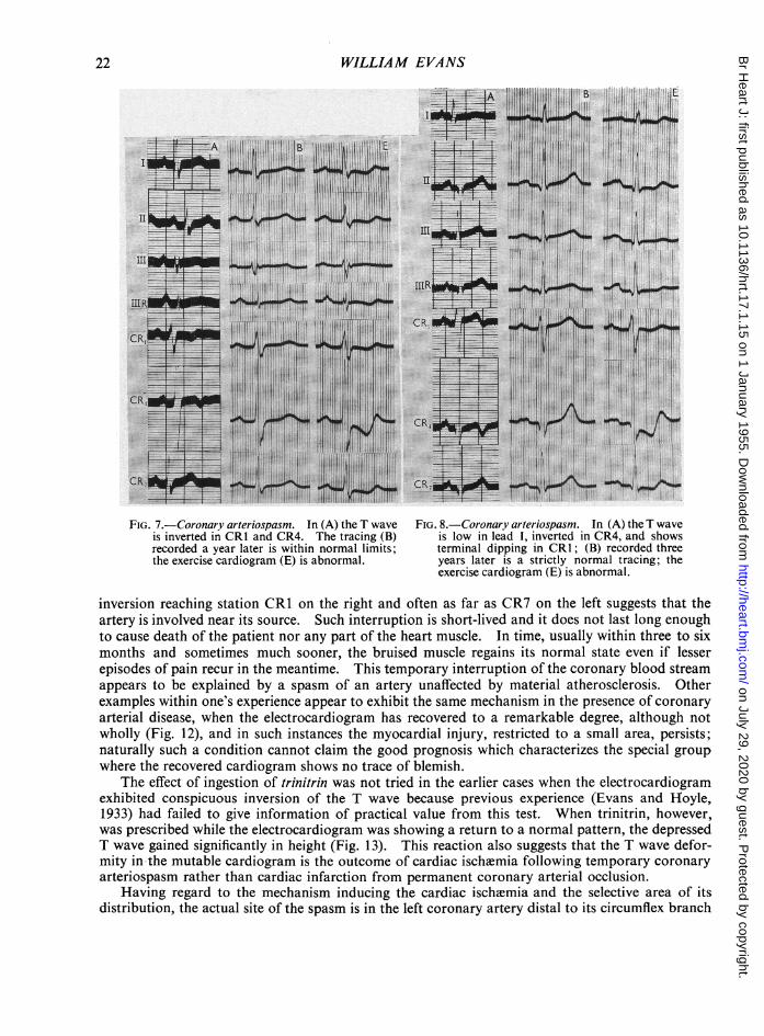
is inverted in CR1 and CR4. The tracing (B) is low in lead I, inverted in CR4, and showsrecorded a year later is within normal limits; terminal dipping in CR1; (B) recorded threethe exercise cardiogram (E) is abnormal. years later is a strictly normal tracing; the
exercise cardiogram (E) is abnormal.
inversion reaching station CR1 on the right and often as far as CR7 on the left suggests that theartery is involved near its source. Such interruption is short-lived and it does not last long enoughto cause death of the patient nor any part of the heart muscle. In time, usually within three to sixmonths and sometimes much sooner, the bruised muscle regains its normal state even if lesserepisodes of pain recur in the meantime. This temporary interruption of the coronary blood streamappears to be explained by a spasm of an artery unaffected by material atherosclerosis. Otherexamples within one's experience appear to exhibit the same mechanism in the presence of coronaryarterial disease, when the electrocardiogram has recovered to a remarkable degree, although notwholly (Fig. 12), and in such instances the myocardial injury, restricted to a small area, persists;naturally such a condition cannot claim the good prognosis which characterizes the special groupwhere the recovered cardiogram shows no trace of blemish.
The effect of ingestion of trinitrin was not tried in the earlier cases when the electrocardiogramexhibited conspicuous inversion of the T wave because previous experience (Evans and H-foyle,1933) had failed to give information of practical value from this test. When trinitrin, however,was prescribed while the electrocardiogram was showing a return to a normal pattern, the depressedT wave gained significantly in height (Fig. 13). This reaction also suggests that the T wave defor-mity in the mutable cardiogram is the outcome of cardiac ischemia following temporary coronaryarteriospasm rather than cardiac infarction from permanent coronary arterial occlusion.
Having regard to the mechanism inducing the cardiac ischemia and the selective area of itsdistribution, the actual site of the spasm is in the left coronary artery distal to its circumflex branch
on July 29, 2020 by guest. Protected by copyright.
http://heart.bmj.com
/B
r Heart J: first published as 10.1136/hrt.17.1.15 on 1 January 1955. D
ownloaded from
ELECTROCARDIOGRAM OF CORONARY ARTERIOSPASM
A
i
CR1
CR4
--..,... C,_ C ,,,..,,... ......, *.. ,.. . , C__. = ,, ,
.._ ff = _S. .,_. .::::: !_= ::::^:,, ^ ,_
_ _ ___. . , ._ _.: _ ,_ _^s ,_ ..,.,.. _ =_-,, . ,.,_,., ........g_v._. L ^_ ... _.__ ::_ _
_ ..,.__,,. ..__ _ ,__ ,..., ., , __, .._.. __. .........
___,....., ...... . ...... ... , ,,.,._ . _= , ^ ..... .... ,, ^ ,, __........... . ... _...^ . __. .,___ ....... ...... ........ .. , _, ._ +_ ':_''.'= *z.. _'::' =._ _ __ _ ...__.-.
_=I= P.,.,,...,,, ,_,,___ __w, _
= _= = :_.. ^ ::--'_ ...... _ _ _ ._ ... _ ._. . _ __ ...,,....,,.. ,.__..,, ._ _, .... ..O .. ...._
t ._'_. j _ ._ ,. _--- ^ .- .-- --.__ ._ __,. .,,_.__,. ._ ._ ,.___., ..______
..,. .. ... .... .. ,., .....,_ _._ ____ _,. ._=
= ::,_ __., ...__ _ -- .__,...._ _ _ =_ _ Q r I_ e , ._ t_ ._._._ 1= _t== a= =_ . _.
_ _ _ _,
== _C= L_ = _ ,_ _ ...... ._ __|._ _,.. _ .,,,._,,.. .. ,....,_,_ __ ,,. . ..... . r ,__.,, _.,,.._ _ .,,..,__.,.,. ._ ....,...._ _ i-_*.,... ....,..., ,,.w f .-._ S _ S *I _ ':s '.:::;L:.,- ,,. - . , ......... __* -.-. . . # - . t -__ l..... , . .e , ,,,_ t-.. --t- ... - i._. _ tw -t___,:_^ ^_+ _.v._,; _... _, ... _ # .... .. _ _ # v._ ^
,__.. w ....,_ _,,,... _, ..._ $ _ _,...,..._5= w. .,
[. -- 1---t-i--t -- t_4 _ ,,, __, .
i §9 . s § - i -.'Li'<":m,I=4''_--- 1--I- t'-r'-t- ---__
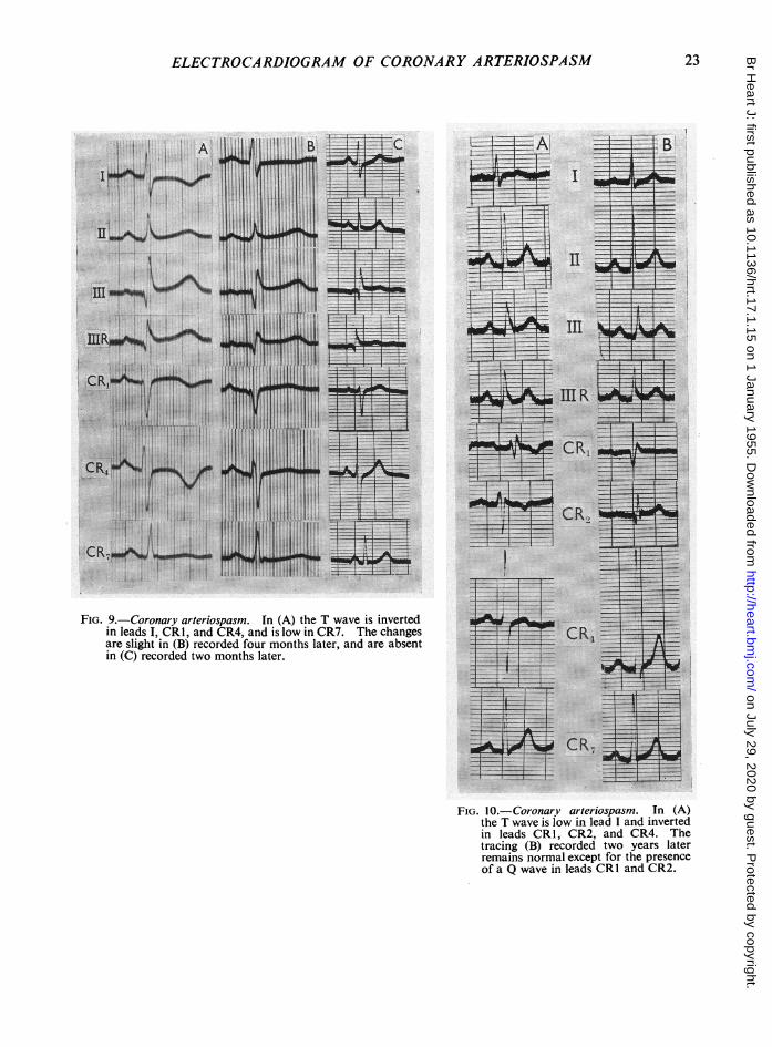
FIG. 9.-Coronary arteriospasm. fn (A) the T wave is invertedin leads I, CRI, and CR4, and is low in CR7. The changesare slight in (B) recorded four months later, and are absentin (C) recorded two months later.
1:--l--4= A -t ,< _& | J w _=t3 _
I --US-4- 1- -
11_- T __r= 1 s.--. -,- , s§,,,,,,,,,, t 2 -+- -t t--4 w_ _; 4 , I I -, -t r
_..1 - + --- t 8---1 ---4 +.-- -- t --- 1 -- f - 1 >-- 4 1 -_ | . } -i - t l- --4 - +>--t------ -__ | -s-n -- x t.
{ t 1H __ +_--- 1 ----t- . ,, ._, r_+ ............... , _|_ ___- --t --s 6_-4--t -_l_s s _, _
!. -..f.. _ s , _*1 4-lm1 IE |4. .. ..1.-4 --
I +-t.l i ,1* .. | t- | -'I
t r 1 1---:-t ' g_* _t I_sIER |, _ __1 r --t
-+- _.t_
n {S..r.=4l f- -a _ s_CRli.^ ._
H ,._-F-t_.
I.,,, ._
---t- t-- | --1t_ _ _- z _1, _, . t
g-gY rcy=-S \_0 a9
1lt-lI. . . . . .... .
ls- v ^ ^ t- s---t ._1_._t 1, --t ---- _ 1 .
i; ;; ;;;4----i---------fl 3=T-;r rn*>4'-t--t-----'1-- W oa
I t 1 t-t-t---t,H-t----
t-tt -----t + -^t-.H._,+. _ ........... ,.i ,., _ + ._.H.- ___....... ,,t._._{ .............. , ,,,.,.g,.,,{
-tu--1f- i-.f.. te 1-t -e --_ .+-_h -_-l-- 1.-t-- I . ......................_4 4
1# CRt,-t-..i._.-i-f-Jf_t ,__ -2............ ...
FIG. 10.-Coronary arteriospasm. In (A)the T wave is low in lead I and invertedin leads CR1, CR2, and CR4. Thetracing (B) recorded two years laterremains normal except for the presenceof a Q wave in leads CRI and CR2.
23
on July 29, 2020 by guest. Protected by copyright.
http://heart.bmj.com
/B
r Heart J: first published as 10.1136/hrt.17.1.15 on 1 January 1955. D
ownloaded from
24 WILLIAM EVANS
tB~~~~~~1~~~~~~~~~~~~~~~~~~~~~~~~~~~_
I~~~~~~~~~~~~~~~~~~~~~~~~~7
III~~~~~~~~~~~~~~~~~~~~~~~~~~~~~~~~~~~~~~~~~~~~~~~~~~~~~~
IIIR--~~~~~~~~~~~~~~~- - ~ ~ ~
CR11~~~~~~~~~~~~~~~~~~~~~~~~~~~~~~~~~~~~~~~~~~~~~~~~~~~~~~~~~
CR............-
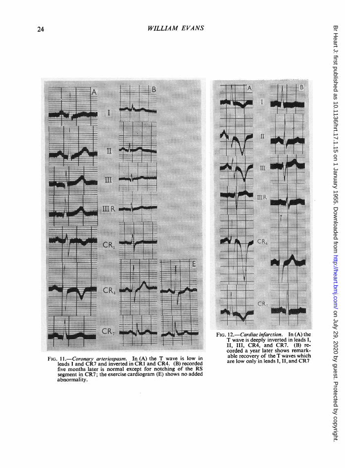
........................CR4.ad.CR7(B).r7"T~~ ~ ~ ~ ~ ~ ~~oddaya ltrsosrmr
FIG.11.-Coronary arwriospasm In (A) the T wave is low in able recoveo' of the Twaves..which.....led I..and.. CR7_an.netd.nC1ad.RB ecre r lo .olyi.lad.II.adRfivemonthslaterisnormalexcept for notching of the RS~~~~~~~~~~~~~~~~~~~~~~~~~~~~~~~~~~~~~~~~~~~~~~~~~~~~~~~~~~~~~~~~~~~~~~~~~~~~~~~~~~~~~~~.......segmentinCR7theexercise cardiogram (E) shows no added~~~~~~~~~~~~~~~~~~~~~~~~~~~..
abnormality.~~~~~~~~~~~~~~~~~~.
on July 29, 2020 by guest. Protected by copyright.
http://heart.bmj.com
/B
r Heart J: first published as 10.1136/hrt.17.1.15 on 1 January 1955. D
ownloaded from

ELECTROCARDIOGRAM OF CORONARY ARTERIOSPASM
*~~~~~~~~~~~~~~~~~~~~~~~~~~.........
* ~~~~~~~~~~~~~~~~~~~~~~~~~~~~~~~~~~~~~~~~~~.~~~~~~~~~~~~~~~~~~~~~~~~~~~~..:...-......-.._.,...........__.j..
..... .... ... ... ...
~~~~~~~~~~~~~~~~~~~~~~~~~~~~~.... ........ ................ ..........~--'-...CR > ~~~~~~~~~~~~~~~~~~~~~~~~~.._........
... . .... - _ _ ....... , , . _ ..~......... ._.
........ .___...............*~~~ ~ ~ ~ ~ ~~~~~~~~~~~~~~~~~~~~~~~~~~~~~~~~~~~~~~~~~~~~~~~~~~~~.....
.........
....f.....
*...............__.__......__..
FIG. 13.-Coronary arteriospasm. An inverted T wavein CR1 and a low T in CR2 and CR3 in (A) froma patient showing the mutable electrocardiogramare upright in (B) recorded four minutes afterchewing 1/25 gr. (2-6 mg.) of glyceryl trinitrate.
FIG. 14.-Distribution of the left coronary artery. The siteof the arteriospasm which gives rise to the mutable car-diogram is indicated by a circle and is about 1 cm.proximal to the seat of election for atheromatousobstruction and thrombosis, and lies between the cir-cumflex and the left marginal branches of the leftcoronary artery.
25
on July 29, 2020 by guest. Protected by copyright.
http://heart.bmj.com
/B
r Heart J: first published as 10.1136/hrt.17.1.15 on 1 January 1955. D
ownloaded from

and proximal to the left marginal branch unless this takes origin from the left circumflex (Fig. 14).No other part of the coronary circulation has been found to be so susceptible to such abrupt andtemporary closure for no example of complete recovery of an electrocardiogram has been metwith among patients in whom the tracing allocated the injury to either the lateral or postero-inferiorportions of the left ventricle. This distinctive segment of the coronary circulation possessing aspecial aptitude to spasm might be named the coronary floodgate for it can suddenly close andinterrupt the blood supply to a large area of the heart and as readily it can open and re-establishthe circulation.
It was late in the investigation that special attention was paid to the possible existence in thesame patient of a like-spasm in situations other than the coronary circulation. Two possibleexamples of this were met with, one involving the retinal artery and the other the transverse colon,but as such episodes might have been fortuitous, they do not in the meantime allow one to drawfirm conclusions from this association.
East and Oram (1948) published a valuable paper in which they recorded 34 cases of cardiacpain where a remarkable degree of recovery of the deformed T wave had taken place. Theydescribed 10 of these in detail and showed the electrocardiograms in nine. A right pectoral lead,however, was only available in one patient (their Case 3) and the tracing in this instance recoveredcompletely except that the T wave in VI remained slightly inverted; this demonstrates the inferiorityof lead VI with its tendency to show an inverted T in health, compared with CR1 in the investigationof these cases. In the bipolar lead, should deformity of the T wave remain, it would suggest thatcomplete recovery of the ischmic electrocardiogram had not taken place, although in the patientsdescribed here the deformity in the CR1 tracing usually disappeared earlier than the abnormalityin CR4. Complete recovery of the electrocardiogram also took place in two other patients in Eastand Oram's series (their Cases 2 and 7), but in neither was the right pectoral lead recorded. Intheir remaining six cases recovery of the tracing was only partial and signs remained that indicateda residual myocardial injury. They, too, considered that spasm of the coronary artery had causedthe temporary electrocardiographic changes. Thompson (1952) published three cases of cardiacpain to illustrate favourable results from treatment with anticoagulants. One of his patients(Case 2) showed a tracing identical in pattern with the mutable electrocardiogram described here;the tracing returned to normal and remained so during a period of three years when the patienthad also kept free from symptoms.
CONCLUSIONDoubt has remained on the true significance of a strictly normal electrocardiogram in a patient
in whom the diagnosis of cardiac pain is suspected. The time is not long past when a normaltracing in a patient whose pain apparently was caused by cardiac infarction, occasioned no surprise,but since the lesser electrocardiographic changes identified with coronary arterial disease havegained more general recognition the incidence of a normal cardiogram in such patients has decreased.
The question whether the electrocardiogram of cardiac infarction is ever normal still needs tobe answered. The present investigation supports the view that it is never strictly normal providedthe tracing has been a competent one, and provided the mutable electrocardiogram described hereand signifying coronary angiospasm is recognized. When cardiac pain arises from such spasm
the tracing becomes grossly abnormal from the ensuing cardiac ischaemia, always preserving a con-
sistent pattern, and thereafter gradually returning to assume its previous normal design even thoughlight attacks of cardiac pain continue to recur.
The mutable electrocardiogram is characterized by inversion of the T wave in chest leads CR1to CR4 and often beyond this as far as station CR7; early on, the deformity may be confined to theterminal portion of the T wave which may dip sharply below the iso-electric level. The T wave isusually inverted in lead I as well Signs of a permanent myocardial injury from coronary arterialdisease like a significant Q wave or depression of the S-T segment are absent from this distinctivecardiogram. When the tracing has recovered, strenuous physical exercise will re-introduce the
26 WILLIAM EVANS
on July 29, 2020 by guest. Protected by copyright.
http://heart.bmj.com
/B
r Heart J: first published as 10.1136/hrt.17.1.15 on 1 January 1955. D
ownloaded from

ELECTROCARDIOGRAM OF CORONARY ARTERIOSPASM
deformity as a rule unless the period of immunity from pain has been a long one. Only 10 suchtracings were found among the 1370 patients whose symptoms suggested cardiac pain, and it issignificant that the eight cardiograms which recovered completely were from this number, whileonly a small blemish remained in the other two tracings.
Because of the favourable prognosis associated with the mutable electrocardiogram, denotingas it does coronary arteriospasm with temporary cardiac ischemia rather than lasting coronaryocclusion with cardiac infarction, its recognition is a matter of considerable moment to the occa-sional patient who may exhibit it.
Sir John Parkinson made valuable suggestions on the preparation of this paper.
REFERENCESBarnes, A. R. (1934). Amer. Heart J., 9, 728.
(1935). Arch. intern. Med., 55, 457., and Ball, R. G. (1932). Amer. J. med. Sci., 183, 215.and Whitten, M. B. (1930). Med. Clin. N. Amer., 14, 671.
Buchner, F., Weber, A., and Haager, B. (1935). Koronarinfarkt und Koronarinsufficienz in vergleichender elektro-kardiographischer und morphologisher Untersuchung. Leipzig.
East, T., and Oram, S. (1948). Brit. Heart J., 10, 263.Evans, W. (1952). Lancet, 2, 1092.
, and Hoyle, J. C. (1935). Lancet, 1, 1109.and McRae, C. (1952). Brit. Heart J., 14, 429.
Lyon, R. M. M. (1938). Edin. med. J., 45, 285.Martensen, V. (1942). Acta med. Scand., 111, 503.Master, A. M., Gubner, R., Dack, S., and Jaffe, H. L. (1941). Arch. intern. Med., 67, 647.Myers, G. B., Klein, and Hiratzka, T. (1948). Amer. Heart J., 36, 838.
(1949). Amer. Heart J., 37, 205 and 720.(1949). Amer. Heart J., 38, 547 and 837.
and Stofer, B. E. (1948). Amer. Heart J., 36, 535.(1949). Amer. Heart J., 37, 374.
Parkinson, J., and Bedford, D. E. (1928). Lancet, 1, 4.Thompson, W. P. (1952). Med. Clin. N. Amer., 36, 991.Whitten, M. B. (1930). Arch. intern. Med., 45, 383.
27
on July 29, 2020 by guest. Protected by copyright.
http://heart.bmj.com
/B
r Heart J: first published as 10.1136/hrt.17.1.15 on 1 January 1955. D
ownloaded from