the complete integumentary system study guide
DESCRIPTION
PDF File of very helpful information in regards to the integumentary system. Excellent study resource for Anatomy and Physiology Students.TRANSCRIPT

The
Integumentary
System

The Integumentary System
1. The Skin and the hypodermis
2. Appendages of the skin

The Skin and the Hypodermis
Functions:
– Cushions and insulates deeper organs
– Protects from injury, chemicals, heat, and cold
– Excretes urea, salt & water through sweat
– Prevents water loss
– Regulates Body Temperature
– Regulates calcium and phosphate Levels
– Screens out UV rays from the sun
– Contains sensory receptors associated with nerve
endings
– Excretes sweat, sebum, and milk

The Skin and the Hypodermis
• Skin – the largest organ
– Varies in thickness form 1.5 to 4mm
– Divided into two distinct layers
i. Epidermis
ii. Dermis
– Hypodermis – fatty layer, lies deep to the
dermis

Skin Structure

Skin Structure

Epidermis
• Composed of keratinized
stratified squamous epithelial
cells
• Contains four main cell types
– Keratinocytes
– Melanocytes
– Merkel cells
– Langerhans cells (Epidermal
DENDRITIC cells)

Keratinocytes and Melanocytes

Epidermis
• Keratinocytes – most abundant cell type
– Arise from deepest layer of epidermis
– Produce keratin – a tough fibrous protein
– Keratinocytes at skin's surface are dead

Layers of the Epidermis
1. Stratum basale
2. Stratum spinosum
3. Stratum granulosum
4. Stratum lucidum
5. Stratum corneum
5 Layers: from inside outwards

Epidermal Cells and Layers

Layers of Epidermis 1. Stratum basale or Stratum
germinativum
Lies next to dermis
Most cells are kertatinocytes
Cells undergo mitosis
Merkel cells (Tactile cells)- associated with sensory nerve ending
Melanocytes- secrete the pigment melanin; long cytoplasmic processes branch out into layers above
Melanin pigment surrounds keratinocyte nuclei

Layers of Epidermis
1. Stratum basale or Stratum germinativum
2. Stratum spinosum – intermediate filaments, Langerhans cells (dendritic cells)
Receptor-mediated endocytosis;
communicate to lymph nodes
3. Stratum granulosum
4. Stratum lucidum –
5. Stratum corneum

Layers of Epidermis
1. Stratum basale or Stratum germinativum
2. Stratum spinosum
3. Stratum granulosum- Consists of flattened keratinocytes (dead cells), granules
Keratin & glycolipid
4. Stratum lucidum
5. Stratum corneum

Layers of Epidermis
1. Stratum basale or Stratum germinativum
2. Stratum spinosum
3. Stratum granulosum
4. Stratum lucidum – cells flatter, dead keratinocytes
Occurs only in hairless, thick skin (palm & sole)
Accumulate keratin
Farther away from blood supply dead
5. Stratum corneum

Thin vs Thick Skin

Layers of Epidermis
1. Stratum basale or Stratum germinativum
2. Stratum spinosum
3. Stratum granulosum
4. Stratum lucidum
5. Stratum corneum
Cornified dead cells
Filled with keratin
Protects against injury & desiccation

Melanin
Pigment produced by melanocytes
Color: yellow to brown to black
Melanocytes - stratum basale
Amount of melanin produced
• genetic control
• exposure to sunlight

Dermis
• Second layer of the skin
• Richly supplied with
nerves and blood
vessels (plexus)
• Has two layers
1. Papillary layer –
superficial 20%
2. Reticular layer – deeper
80%

Skin Structure

Skin Structure
1. Papillary layer
– Superficial layer, areolar connective tissue
– Dermal papillae project into epidermis
– Epidermal ridges formed
2. Reticular layer
– Dense irregular CT
– reticulum: network of collagen & elastic fibers

Skin Structure

Flexion Creases & Dermis
Continual folding of skin
over joints
Dermis is attached
tightly to underlying
structures

Fingerprints
Dermal ridges elevate the epidermis Epidermal Ridges

Reticular Layer of Dermis
Wrinkles
Lines of cleavage / Tension Lines: Less
dense regions
Represent the separations between
bundles of collagen fibers in reticular
dermis

Structures in the Dermis
Blood vessels

Tactile (Meissner’s) Corpuscles Sense of light touch
Lamellated (Pacinian) Corpuscles Pressure sensors
Structures in the Dermis
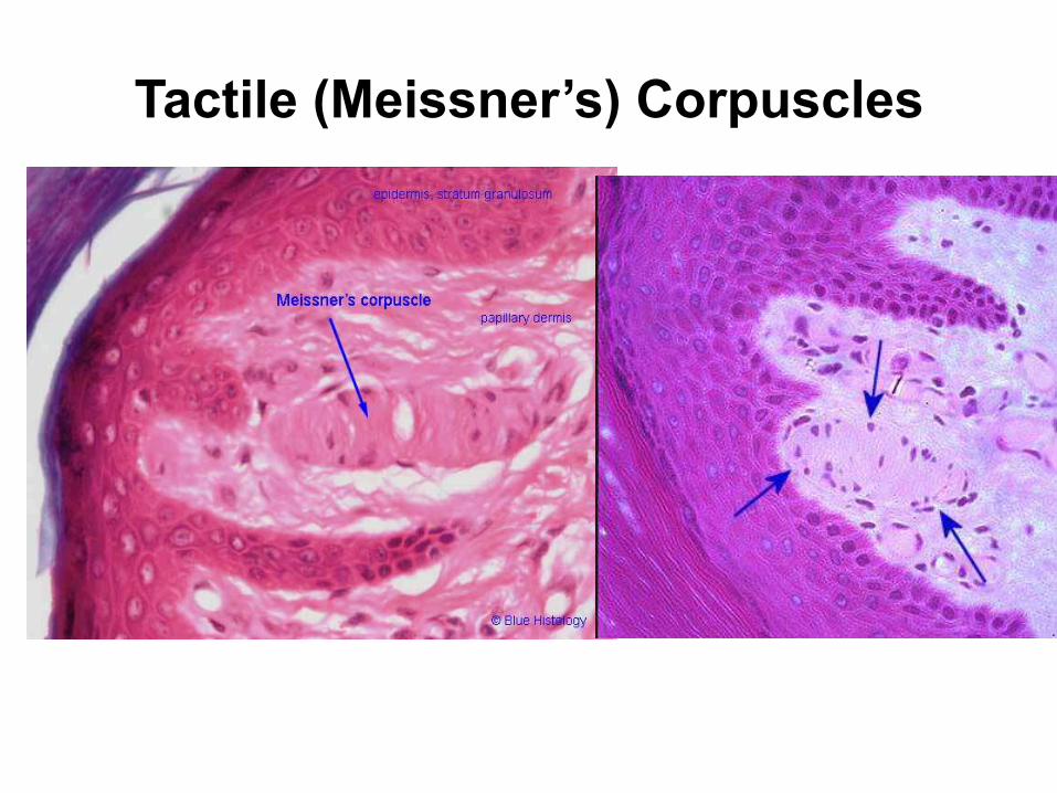
Tactile (Meissner’s) Corpuscles

Lamellated (Pacinian) Corpuscles

Free Nerve Endings
Sense of temperature and pain

Root Hair Plexus
Sensory nerves
around hair bulb
Sense movement of
hair and skin

Hypodermis
• Not part of skin
• Contains areolar and adipose connective
tissues
• Anchors skin to underlying structures
• Helps insulate the body

Hypodermis (Superficial Fascia or
Subcutaneous Fat)

Abnormal Skin Colors
• Cyanosis - blueness from deficiency of oxygen in the
circulating blood (cold weather)
• Erythema – abnormal redness due to dilated cutaneous
vessels (anger, sunburn, embarrassment)
• Jaundice - yellowing of skin and sclera due to excess of
bilirubin in blood (liver disease)
• Albinism - a genetic lack of melanin
• Hematoma - a bruise (visible clotted blood) showing
through skin
• Pallor – pale color under stress, shock, cold, severe
anemia

Skin Markings • Hemangiomas (birthmarks)
– discolored skin caused by benign tumors of dermal blood
capillaries
• Freckles and moles = aggregations of melanocytes
– freckles are flat; moles are elevated
• Friction ridges leave oily fingerprints on touched surfaces
– unique pattern formed during fetal development
• Flexion creases mark lines where skin folds during flexion of
joints

Appendages of the Skin
1. Hair & hair follicles
2. Sebaceous glands
3. Sweat glands
4. Nails

Accessory Structures of the Skin • Hair
• Muscles (Arrector pili)
• Sebaceous Glands
• Sudoriferous Glands
• Mammary Glands
• Ceruminous Glands
• Nails

Hair = Pilus

Hair & Hair Follicles
• Hair- long flexible strand of
keratinized cells
• Hair follicle – tubular
invagination of the epidermis
• Hair produced by hair follicle
• Daughter cells pushed up,
keratinized & die

Structure of Hair
• Three zones along the length of a
hair:
1. Hair bulb: a swelling at
the dermal base
2. Hair root: embedded in
the skin
3. Hair shaft: projects
above the skin surface

Hair Shaft and Hair Root have 3 layers
Cuticle
Cortex
Medulla

C.S. of a Hair
600X
• Three concentric layers of keratinized
cells
1. Cuticle – single layer of scale-like
cells that overlap
2. Cortex – several layers of flattened
cells
3. Medulla – large cells

Scanning EM of hair shaft
Scale-like cells of the cuticle

L.S. of Base of Follicle
Hair Shaft (Cuticle, cortex, Medulla)

Structure of Hair Follicle
Blood capillaries
• Extends into the dermis
• Deep end of follicle is
expanded: Hair Bulb
• Hair Papilla: Connective tissue
projecting into the bulb.
• Hair Matrix: epithelial cells
above the papilla in the bulb;
form the growth zone
• Cells of matrix divide
• Root hair plexus: Knot of
sensory nerves around hair bulb

Hair Bulb
Hair Papilla
Hair matrix

L.S. of Base of Follicle
1
2a
2b
Wall of hair follicle is composed of 2 sheaths:
1. Connective tissue root sheath: Outer (from Dermis)
2. Epithelial root sheath: Inner (Epidermis) surround hair root

L.S. of Base of Follicle
Melanocytes provide pigment for hair color

Hair Associated Structures
Arrector pili
Smooth muscle
Extend from dermal papillae
Contract- hair pulled upright
(PILOERECTION)

Arrector Pili

Functions of Hair
• Senses
• Guards
• Shields
• Protects

Hair Color
Eumelanin pigment in
black & brown hair Blond hair contain pheomelanin
pigment, but little eumelanin.
Hair color: different
melanin pigments
in cells of cortex

Red hair contains little
eumelanin but lots of
pheomelanin.
Hair Color
White hair - air bubbles in medulla
and lack of pigment in cortex.
Gray hair - mix of white & pigmented
hairs; lack tyrosinase

Hair Texture
Straight hair:
Round in C.S.
Wavy hair: Oval
in C.S.
Curly hair: Flat in
C.S.

Oil glands
Present all over, except palms & soles
Simple alveolar glands;
Holocrine secretion
Ducts empty into a hair follicle
Secretion~ SEBUM Lubricant for skin
Kills bacteria
Slows water loss
Sebaceous Glands
Sebaceous
Gland
Hair
Follicle

Sebaceous Glands

Acne
Excess sebum is produced. Duct is blocked.
Bacterial infection leads to inflammation.
May burst into the dermis.

Blackheads
Blocked sebum oxidizes & dries
Darkens to form blackhead

Produce sweat
Widely distributed in skin
Temperature regulation
Excretory product: is a blood filtrate, passes
through secretory cells of sweat glands, released
by exocytosis
Composition- 99% water, salts, ‘acidic’ excretory
products
Sweat Glands or Sudoriferous glands

Two types
Eccrine (or merocrine) glands – more numerous
Apocrine glands
Sweat Glands or Sudoriferous glands

Sweat Glands

Coiled simple tubular gland
Open via duct to a pore on skin
surface
Usually do not extend as far
into dermis
Produce acidic sweat (pH 4-6)
Involved in heat-regulating
system
Eccrine / Merocrine Glands
Dense in palms and
soles (~3000 per sq inch)

• Ducts empty into hair follicles
• Scalp, neck, axillary & genital areas
• Sweat also contains lipids, proteins
• Body odor
Apocrine Sweat Glands

Apocrine Sweat
Glands
(wide lumen)
Merocrine Sweat
Glands
(narrow lumen)

Modified Sweat Glands
• Ceruminous glands – external ear canal
• Mammary glands

Ceruminous Glands
Location
Secretion combines with sebum
earwax
Waterproof, keeps eardrum
flexible
Block foreign particles, kills
bacteria

• Milk-producing glands
• Modified apocrine sweat
gland
• Ducts open on the nipple
Mammary Glands

Mammary Glands

Nails
Scale-like modifications of the epidermis
Heavily keratinized
Each nail has
A free edge
A body- visible attached part
A root- embedded
Nail overlapped by folds of skin- Nail Folds
Thick proximal nail fold- Cuticle / Eponychium

Nails
Stratum basale of epidermis extends beneath
the nail as the nail bed Proximal thickened area of nail bed - nail matrix Responsible for growth
Cells produced become keratinized and die No pigment – colorless (pink?)

Nails
Region over thickened
nail matrix: White Crescent

Toe Nail