study guide for the integumentary system - littered boxlittered-box.com/study guide for the...
TRANSCRIPT

Study Guide for the Integumentary System
Thursday, November 29, 2012

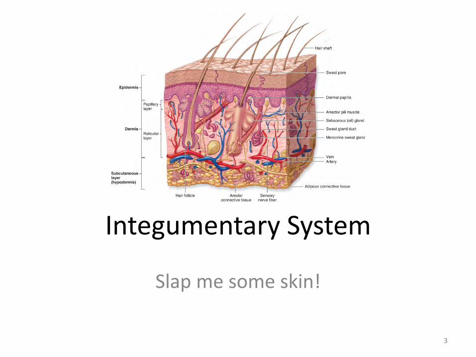
Integumentary System
Slap me some skin!
3
Integumentary System • The integumentary system consists of the skin, aka
cutaneous membrane, and its accessory structures: glands, hair, nails, muscle and nerves
• It is made up of different types of tissue that perform specific functions
• It is the heaviest organ with the largest surface area • Approximately 640,000 sensory receptors connected to
the spinal cord • An area of skin the size of a quarter contain roughly 3
million cells, 100 sweat glands, 50 nerve endings, and 3 feet of blood vessels
4
5
The Biochemical/Physiological/Anatomical functions of the skin: also discussed as “What does the Skin do
for Us?”
1. It helps our body control it internal and external temperature.
2. It is the first line of protection for our body. An intact skin keeps foreign matter out.
3. It is the receiver of sensation through the sensory nerves that reside in the skin.
4. It is involved in excretion . . . Sudoriferous glands assist in the release of certain salts and waste products of the cells.
5. It contributes to our immune function as the pH of both sweat and sebum are hostile to foreign organisms and their presence assists in preventing hostile organisms from entering the body.
6. It is a reservoir for blood. About 8 to 10% of the blood in the body is in the skin at any given time.
7. The skin is involved in the processing of Vitamin D, a Vitamin necessary to health and well-being.
Integumentary System
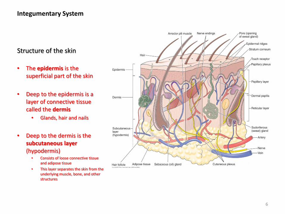
Structure of the skin
• The epidermis is the superficial part of the skin
• Deep to the epidermis is a layer of connective tissue called the dermis
• Glands, hair and nails
• Deep to the dermis is the
subcutaneous layer (hypodermis)
• Consists of loose connective tissue and adipose tissue
• This layer separates the skin from the underlying muscle, bone, and other structures
6
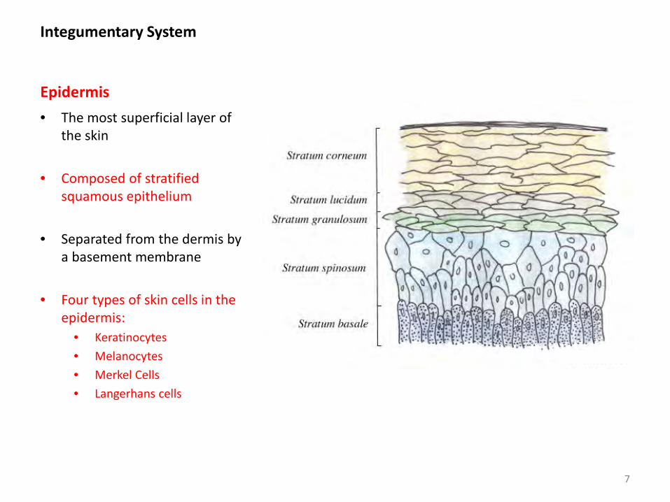
Integumentary System Epidermis • The most superficial layer of
the skin
• Composed of stratified squamous epithelium
• Separated from the dermis by a basement membrane
• Four types of skin cells in the epidermis:
• Keratinocytes • Melanocytes • Merkel Cells • Langerhans cells
7
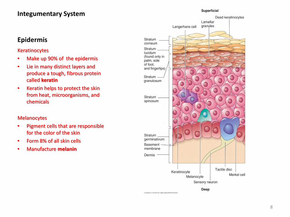
Integumentary System Epidermis Keratinocytes • Make up 90% of the epidermis • Lie in many distinct layers and
produce a tough, fibrous protein called keratin
• Keratin helps to protect the skin from heat, microorganisms, and chemicals
Melanocytes • Pigment cells that are responsible
for the color of the skin • Form 8% of all skin cells • Manufacture melanin
8
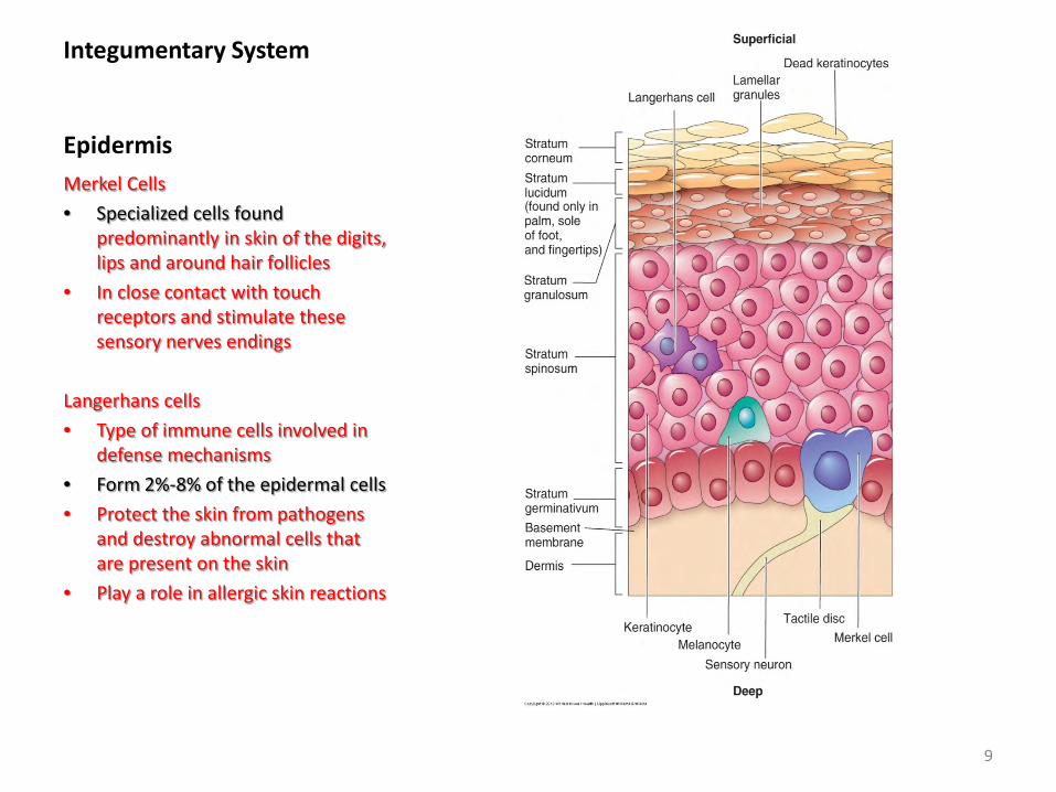
Integumentary System Epidermis Merkel Cells • Specialized cells found
predominantly in skin of the digits, lips and around hair follicles
• In close contact with touch receptors and stimulate these sensory nerves endings
Langerhans cells • Type of immune cells involved in
defense mechanisms • Form 2%-8% of the epidermal cells • Protect the skin from pathogens
and destroy abnormal cells that are present on the skin
• Play a role in allergic skin reactions
9
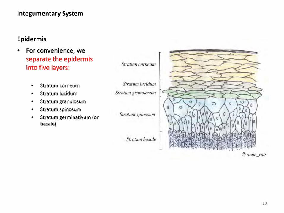
Integumentary System Epidermis
• For convenience, we separate the epidermis into five layers:
• Stratum corneum • Stratum lucidum • Stratum granulosum • Stratum spinosum • Stratum germinativum (or
basale)
10
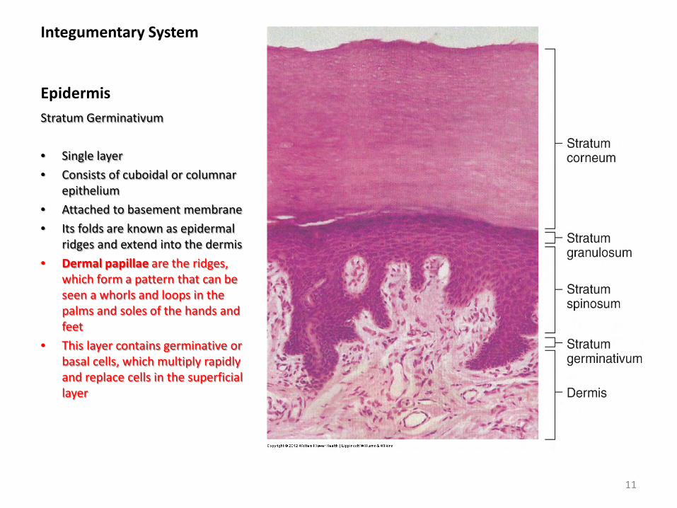
Integumentary System Epidermis Stratum Germinativum • Single layer • Consists of cuboidal or columnar
epithelium • Attached to basement membrane • Its folds are known as epidermal
ridges and extend into the dermis • Dermal papillae are the ridges,
which form a pattern that can be seen a whorls and loops in the palms and soles of the hands and feet
• This layer contains germinative or basal cells, which multiply rapidly and replace cells in the superficial layer
11
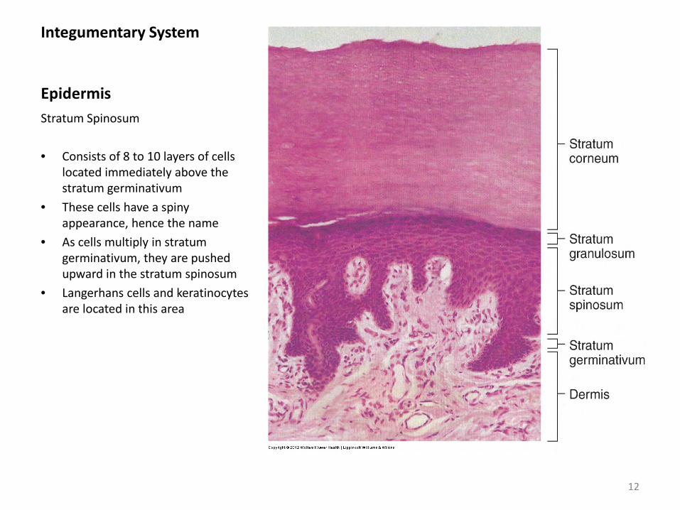
Integumentary System Epidermis Stratum Spinosum • Consists of 8 to 10 layers of cells
located immediately above the stratum germinativum
• These cells have a spiny appearance, hence the name
• As cells multiply in stratum germinativum, they are pushed upward in the stratum spinosum
• Langerhans cells and keratinocytes are located in this area
12
Integumentary System Epidermis
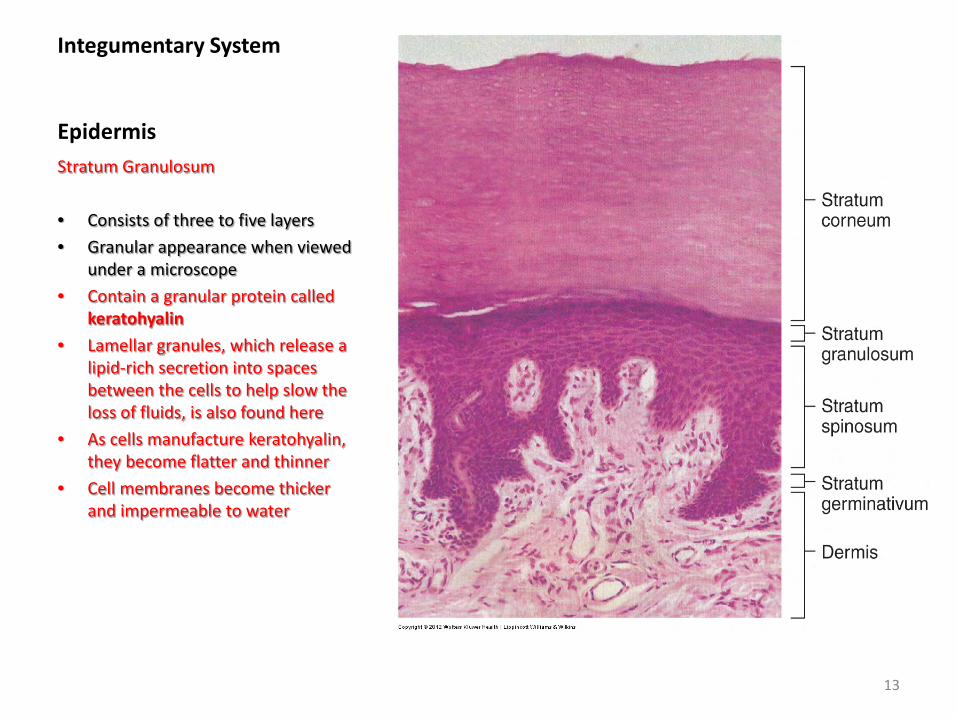
Stratum Granulosum • Consists of three to five layers • Granular appearance when viewed
under a microscope • Contain a granular protein called
keratohyalin • Lamellar granules, which release a
lipid-rich secretion into spaces between the cells to help slow the loss of fluids, is also found here
• As cells manufacture keratohyalin, they become flatter and thinner
• Cell membranes become thicker and impermeable to water
13
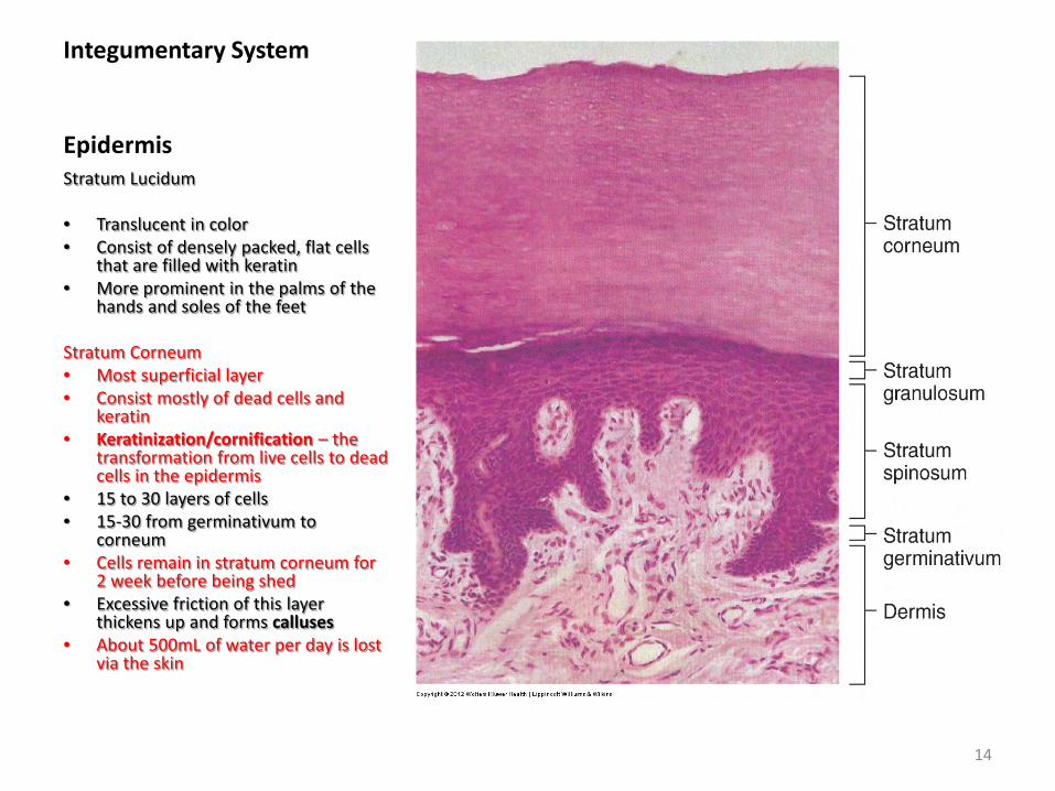
Integumentary System Epidermis Stratum Lucidum • Translucent in color • Consist of densely packed, flat cells
that are filled with keratin • More prominent in the palms of the
hands and soles of the feet
Stratum Corneum • Most superficial layer • Consist mostly of dead cells and
keratin • Keratinization/cornification – the
transformation from live cells to dead cells in the epidermis
• 15 to 30 layers of cells • 15-30 from germinativum to
corneum • Cells remain in stratum corneum for
2 week before being shed • Excessive friction of this layer
thickens up and forms calluses • About 500mL of water per day is lost
via the skin
14
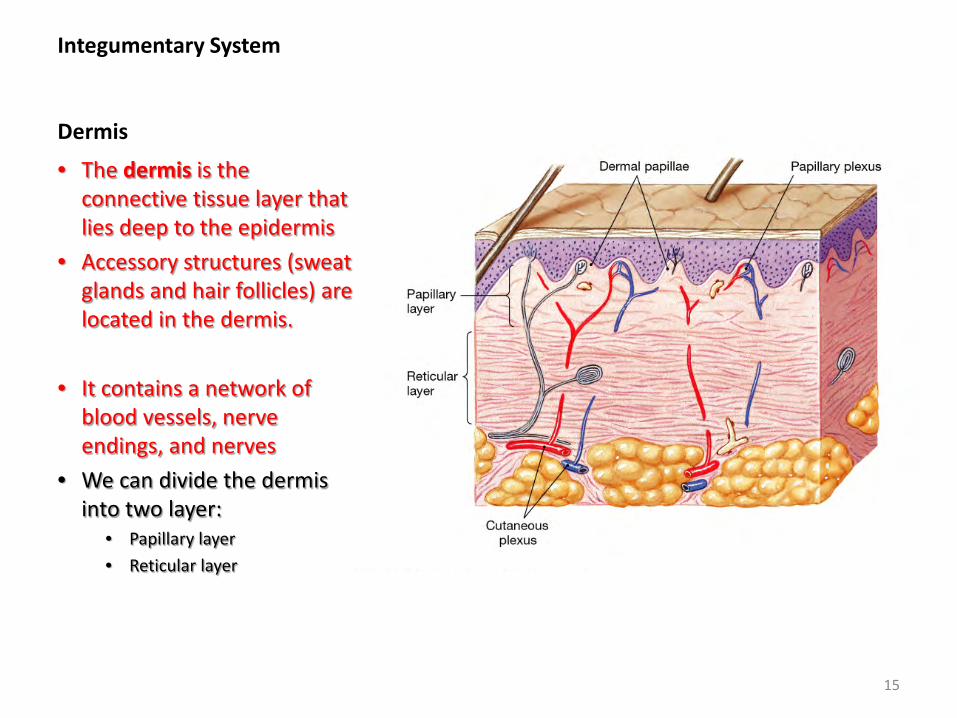
Integumentary System Dermis
• The dermis is the connective tissue layer that lies deep to the epidermis
• Accessory structures (sweat glands and hair follicles) are located in the dermis.
• It contains a network of blood vessels, nerve endings, and nerves
• We can divide the dermis into two layer:
• Papillary layer • Reticular layer
15
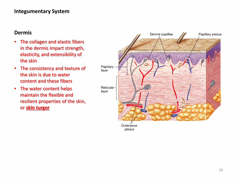
Integumentary System Dermis • The collagen and elastic fibers
in the dermis impart strength, elasticity, and extensibility of the skin
• The consistency and texture of the skin is due to water content and these fibers
• The water content helps maintain the flexible and resilient properties of the skin, or skin turgor
16
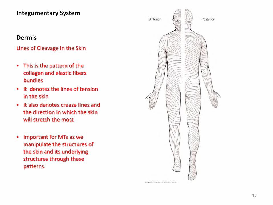
Integumentary System Dermis Lines of Cleavage In the Skin • This is the pattern of the
collagen and elastic fibers bundles
• It denotes the lines of tension in the skin
• It also denotes crease lines and the direction in which the skin will stretch the most
• Important for MTs as we manipulate the structures of the skin and its underlying structures through these patterns.
17
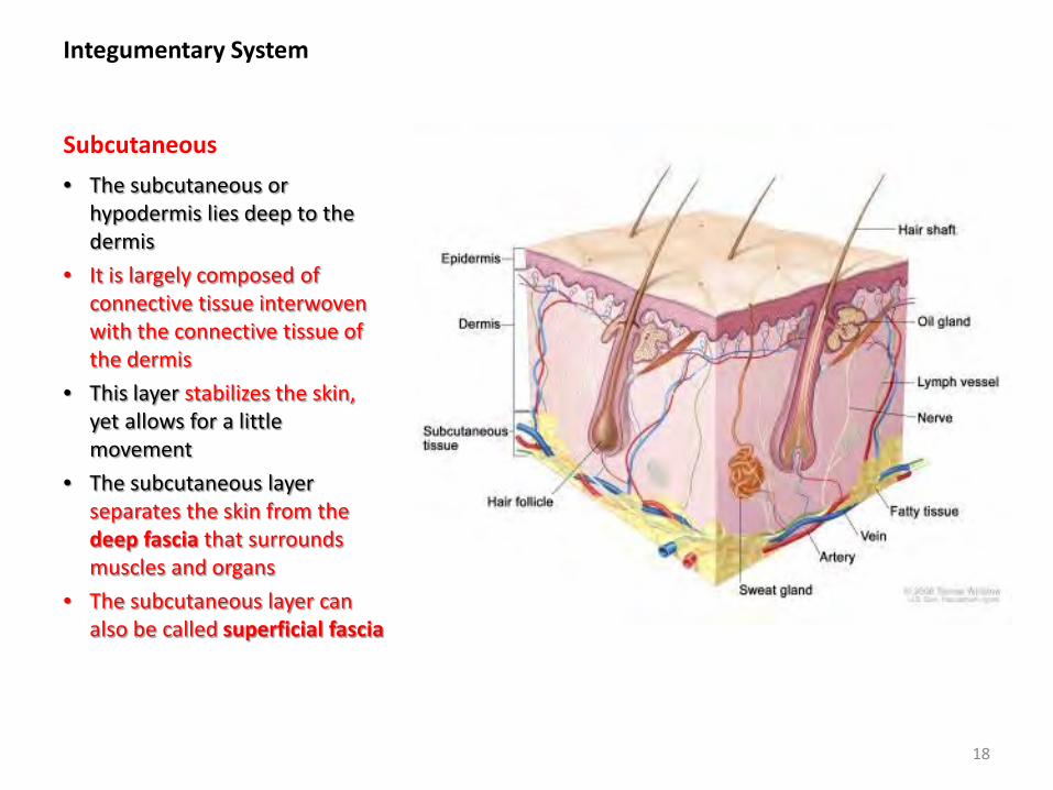
Integumentary System Subcutaneous • The subcutaneous or
hypodermis lies deep to the dermis
• It is largely composed of connective tissue interwoven with the connective tissue of the dermis
• This layer stabilizes the skin, yet allows for a little movement
• The subcutaneous layer separates the skin from the deep fascia that surrounds muscles and organs
• The subcutaneous layer can also be called superficial fascia
18
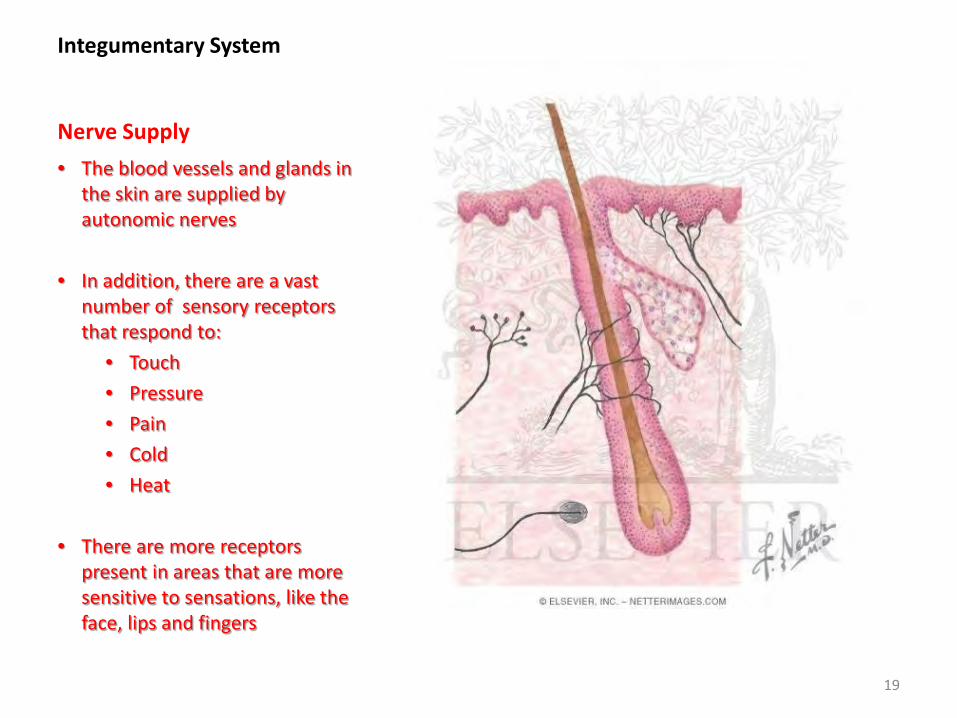
Integumentary System Nerve Supply • The blood vessels and glands in
the skin are supplied by autonomic nerves
• In addition, there are a vast number of sensory receptors that respond to:
• Touch • Pressure • Pain • Cold • Heat
• There are more receptors
present in areas that are more sensitive to sensations, like the face, lips and fingers
19
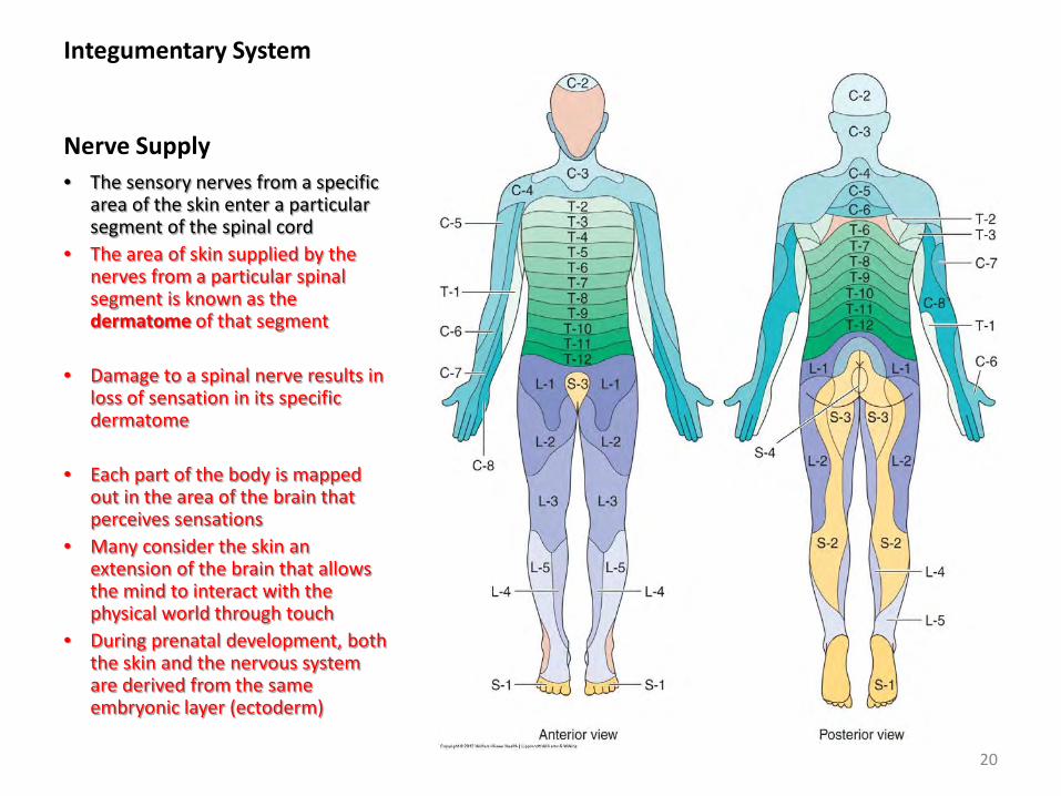
Integumentary System Nerve Supply • The sensory nerves from a specific
area of the skin enter a particular segment of the spinal cord
• The area of skin supplied by the nerves from a particular spinal segment is known as the dermatome of that segment
• Damage to a spinal nerve results in loss of sensation in its specific dermatome
• Each part of the body is mapped out in the area of the brain that perceives sensations
• Many consider the skin an extension of the brain that allows the mind to interact with the physical world through touch
• During prenatal development, both the skin and the nervous system are derived from the same embryonic layer (ectoderm)
20
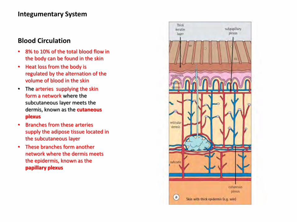
Integumentary System Blood Circulation • 8% to 10% of the total blood flow in
the body can be found in the skin • Heat loss from the body is
regulated by the alternation of the volume of blood in the skin
• The arteries supplying the skin form a network where the subcutaneous layer meets the dermis, known as the cutaneous plexus
• Branches from these arteries supply the adipose tissue located in the subcutaneous layer
• These branches form another network where the dermis meets the epidermis, known as the papillary plexus
21
Integumentary System Functions of the Skin • Protecting the underlying
organs and tissues from damage
• Excreting items like salt, water and organic wastes that are lost through sweat
• Maintaining body temperature • Detecting changes in the
surrounding environment with its sensory receptors
• Synthesizing vitamin D • Store nutrients • Acting as a reservoir of blood
in case it needs to be diverted for systemic needs
• Its most important function is to reflect emotional states, regardless of disease
22
Integumentary System Functions of the Skin
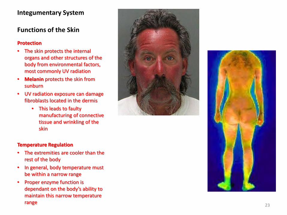
Protection • The skin protects the internal
organs and other structures of the body from environmental factors, most commonly UV radiation
• Melanin protects the skin from sunburn
• UV radiation exposure can damage fibroblasts located in the dermis
• This leads to faulty manufacturing of connective tissue and wrinkling of the skin
Temperature Regulation • The extremities are cooler than the
rest of the body • In general, body temperature must
be within a narrow range • Proper enzyme function is
dependant on the body’s ability to maintain this narrow temperature range 23

Integumentary System Functions of the Skin
Absorption • The skin can absorb or excrete
certain substances • Vitamin D synthesized in the skin
is transported to the liver and then to the kidneys
• Vitamin D increases calcium absorption in the intestines and is an important hormone in calcium metabolism
• A lack of vitamin D can lead to rickets in children and osteomalacia in adults
• Substances that are also lipid-soluble can penetrate the epidermis, therefore allowing for a slower absorption rate
• Systemic adverse effect can be produced if drugs are transmitted transdermally for prolonged periods
24
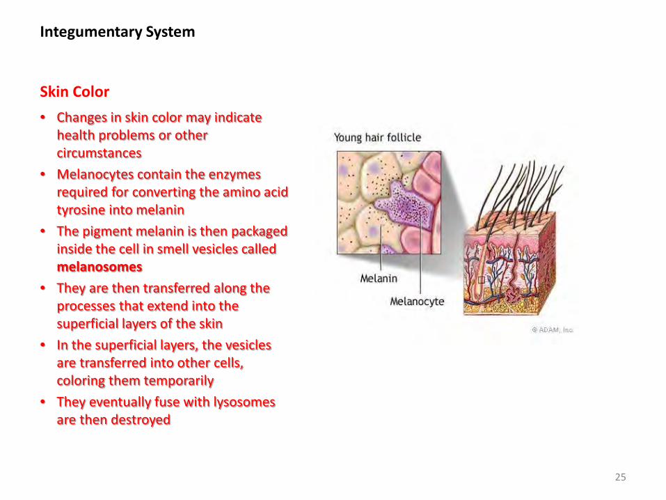
Integumentary System Skin Color • Changes in skin color may indicate
health problems or other circumstances
• Melanocytes contain the enzymes required for converting the amino acid tyrosine into melanin
• The pigment melanin is then packaged inside the cell in smell vesicles called melanosomes
• They are then transferred along the processes that extend into the superficial layers of the skin
• In the superficial layers, the vesicles are transferred into other cells, coloring them temporarily
• They eventually fuse with lysosomes are then destroyed
25
Integumentary System - correction undelined below Skin Color
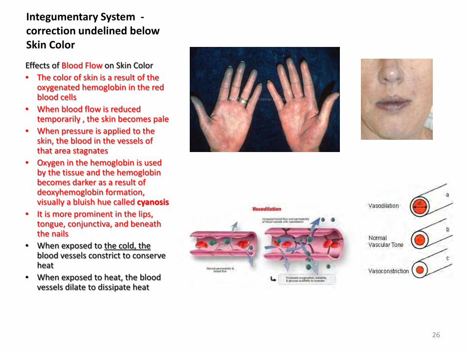
Effects of Blood Flow on Skin Color • The color of skin is a result of the
oxygenated hemoglobin in the red blood cells
• When blood flow is reduced temporarily , the skin becomes pale
• When pressure is applied to the skin, the blood in the vessels of that area stagnates
• Oxygen in the hemoglobin is used by the tissue and the hemoglobin becomes darker as a result of deoxyhemoglobin formation, visually a bluish hue called cyanosis
• It is more prominent in the lips, tongue, conjunctiva, and beneath the nails
• When exposed to the cold, the blood vessels constrict to conserve heat
• When exposed to heat, the blood vessels dilate to dissipate heat
26
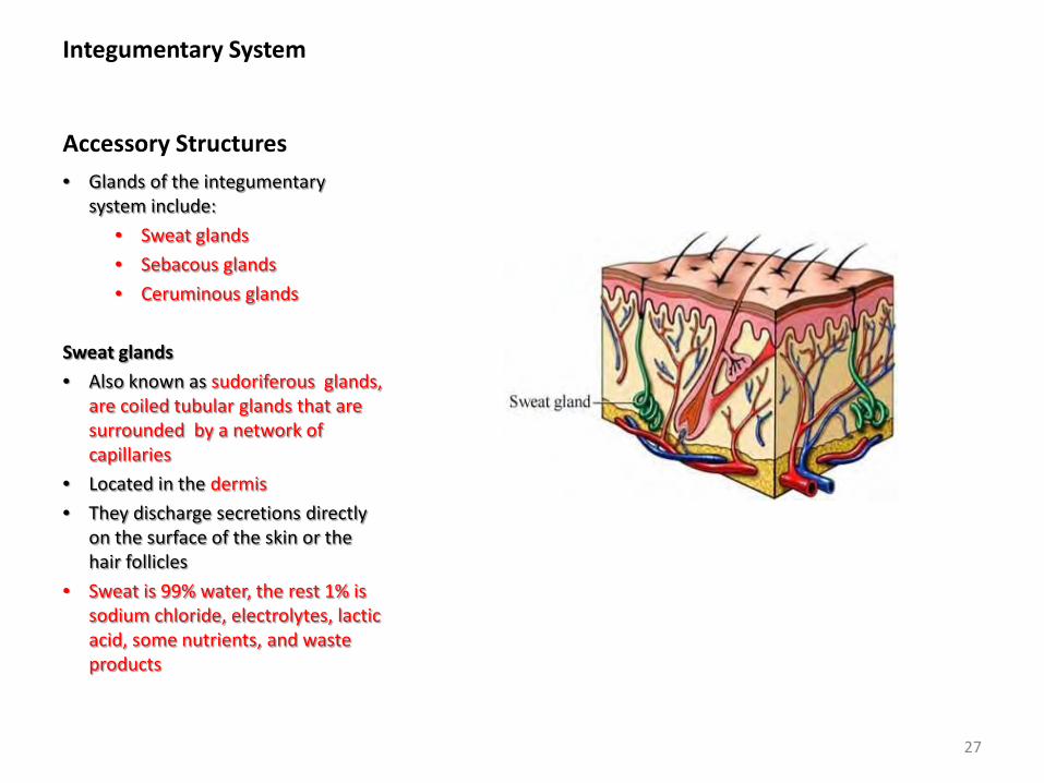
Integumentary System Accessory Structures • Glands of the integumentary
system include: • Sweat glands • Sebacous glands • Ceruminous glands
Sweat glands • Also known as sudoriferous glands,
are coiled tubular glands that are surrounded by a network of capillaries
• Located in the dermis • They discharge secretions directly
on the surface of the skin or the hair follicles
• Sweat is 99% water, the rest 1% is sodium chloride, electrolytes, lactic acid, some nutrients, and waste products
27
Integumentary System Accessory Structures
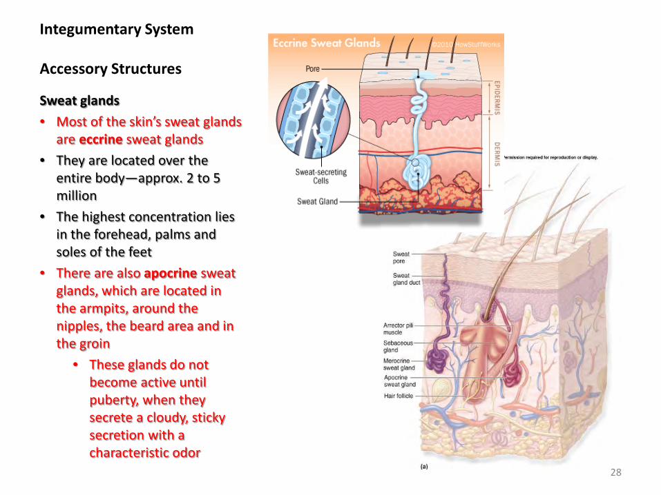
Sweat glands • Most of the skin’s sweat glands
are eccrine sweat glands • They are located over the
entire body—approx. 2 to 5 million
• The highest concentration lies in the forehead, palms and soles of the feet
• There are also apocrine sweat glands, which are located in the armpits, around the nipples, the beard area and in the groin
• These glands do not become active until puberty, when they secrete a cloudy, sticky secretion with a characteristic odor
28
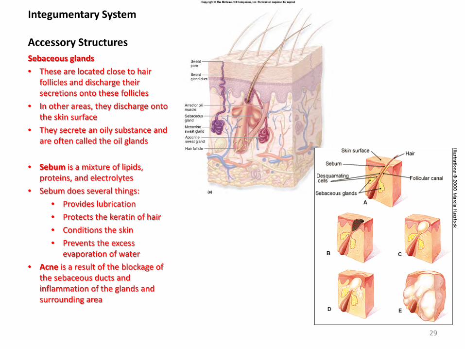
Integumentary System Accessory Structures Sebaceous glands • These are located close to hair
follicles and discharge their secretions onto these follicles
• In other areas, they discharge onto the skin surface
• They secrete an oily substance and are often called the oil glands
• Sebum is a mixture of lipids, proteins, and electrolytes
• Sebum does several things: • Provides lubrication • Protects the keratin of hair • Conditions the skin • Prevents the excess
evaporation of water • Acne is a result of the blockage of
the sebaceous ducts and inflammation of the glands and surrounding area
29
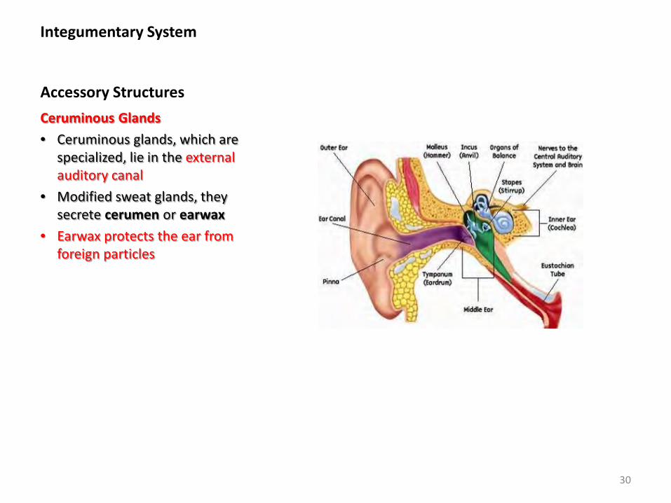
Integumentary System Accessory Structures Ceruminous Glands • Ceruminous glands, which are
specialized, lie in the external auditory canal
• Modified sweat glands, they secrete cerumen or earwax
• Earwax protects the ear from foreign particles
30
Integumentary System Mammary Glands
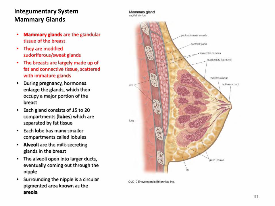
• Mammary glands are the glandular tissue of the breast
• They are modified sudoriferous/sweat glands
• The breasts are largely made up of fat and connective tissue, scattered with immature glands
• During pregnancy, hormones enlarge the glands, which then occupy a major portion of the breast
• Each gland consists of 15 to 20 compartments (lobes) which are separated by fat tissue
• Each lobe has many smaller compartments called lobules
• Alveoli are the milk-secreting glands in the breast
• The alveoli open into larger ducts, eventually coming out through the nipple
• Surrounding the nipple is a circular pigmented area known as the areola
31
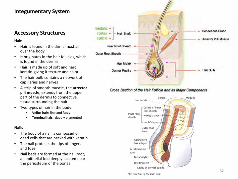
Integumentary System Accessory Structures Hair • Hair is found in the skin almost all
over the body • It originates in the hair follicles, which
is found in the dermis • Hair is made up of soft and hard
keratin-giving it texture and color • The hair bulb contains a network of
capillaries and nerves • A strip of smooth muscle, the arrector
pili muscle, extends from the upper part of the dermis to connective tissue surrounding the hair
• Two types of hair in the body: • Vellus hair: fine and fuzzy • Terminal hair: deeply pigmented
Nails • The body of a nail is composed of
dead cells that are packed with keratin • The nail protects the tips of fingers
and toes • Nail beds are formed at the nail root,
an epithelial fold deeply located near the periosteum of the bones
32
How does the skin react to inflammation?
• Inflammation is the reaction of living tissue to injury and is easily seen on the surface of the skin
• Inflammation helps to heal wounds and prevents and combats infection by helping the body adapt to stressors
33
Integumentary System Inflammation
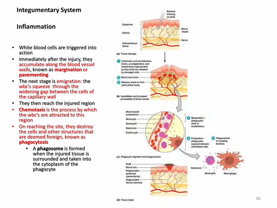
• White blood cells are triggered into action
• Immediately after the injury, they accumulate along the blood vessel walls, known as margination or pavementing
• The next stage is emigration: the wbc’s squeeze through the widening gap between the cells of the capillary wall
• They then reach the injured region • Chemotaxis is the process by which
the wbc’s are attracted to this region
• On reaching the site, they destroy the cells and other structures that are deemed foreign, known as phagocytosis
• A phagosome is formed when the injured tissue is surrounded and taken into the cytoplasm of the phagocyte
34
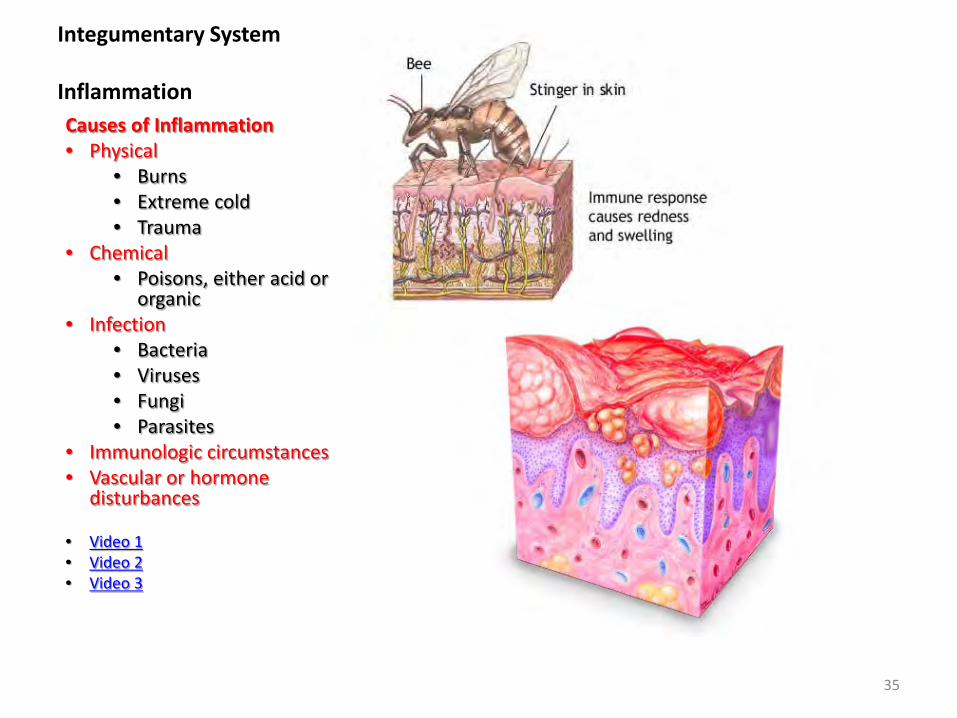
Integumentary System Inflammation Causes of Inflammation • Physical
• Burns • Extreme cold • Trauma
• Chemical • Poisons, either acid or
organic • Infection
• Bacteria • Viruses • Fungi • Parasites
• Immunologic circumstances • Vascular or hormone
disturbances
• Video 1 • Video 2 • Video 3
35
Effects of massage on the Integument
• Massage can help realign collagen fibers in the dermis during and after healing of deep skin wounds.
• Massage can increase blood flow locally and facilitate the removal of toxins released by injured tissue and speed up healing.
• Massage will also speed up the absorption of drug near the injection site.
• Massage can reduce pain perceived by the brain.
36