the anatomy, physiology, functional significance and evolution of
TRANSCRIPT

The Anatomy, Physiology, Functional Significance and Evolution of Specialized Hearing Organs of Gerbilline Rodents
DOUGLAS M. LAY M u s e u m of Zoology, Un iver s i t y of Mich igan , Ann Arbor, M i c h i g a n 48104
ABSTRACT Middle and inner ear anatomy correlates with neurophysio- logical responses to a wide range of sound frequencies for species of the Gerbil- linae representing generalized, intermediate, and specialized anatomical con- ditions. Neurophysiological data were recorded from 81 specimens of 13 species representing six genera. Anatomical parameters involved in the process of hearing were correlated with the neurophysiological data to assess the effects of different degrees of anatomical specialization on hearing. The 13 species tested in this manner have graphic curves of auditory sensitivity of remarkably similar disposition over the frequencies tested and to those published for Kan- garoo Rats. Ears with anatomical specializations show greater auditory sensitivity.
The natural history of the Gerbillinae, particularly the kinds of predators, degree of predation, and habitat is reviewed and utilized to interpret the sig- nificance of the degree of auditory specialization in the forms studied and to evaluate the prevailing hypothesis that these specializations enhance the abil- ity of these rodents to survive in open desert situations by detecting and evad- ing predators.
The middle ear anatomy of five additional genera and species was also studied. Thus, data on the entire spectrum of gerbilline middle ear morphology provide an evolutionary sequence. Certain anatomical parameters of the organ of Corti show a degree of specialization parallel to that of features of the middle ear. The morphological changes and possible functional roles of these features are considered.
A very high correlation exists for degree of specialization and aridity of habitat, thus specialization increases with increasing aridity. This increased specialization may result from more effective predation in open xeric environ- ments. Auditory acuity for a wide range of low frequency sounds augmented by auditory specialization is hence more advantageous here. There does not appear to be selection for hearing at particular frequencies in this range. The peaks of greatest auditory sensitivity appear to correspond to the resonant frequencies of the different components of the middle ear transformer and cavity.
Rodents of the subfamily Gerbillinae, family Cricetidae have the greatest num- bers of species of all mammals that in- habit the Great Palaearctic Desert. Eight genera including at least 46 species are endemic to this broad arid zone. The Dipodidae (jerboas) with 10-1 1 genera and about 26 species is the only other rodent family which has successfully ra- diated in this desert. The greatest taxo- nomic diversity of the Gerbillinae occurs in North Africa, while that of the Dipodi- dae is in Central Asia. However, both
groups occur throughout the Great Pa- laearctic Desert. These two assemblages, which represent evolutionary rodent lines divergent since the Oligocene (Wood, '65), possess a number of similar morphological features that appear to have evolved con- vergently as adaptations to desert habi- tats. The ear represents one of these adaptive complexes.
Considerable variation typifies the mid- dle and inner ear of the living gerbillines. Desert dwelling species are specialized relative to those that inhabit the savan-
J. MORPH., 138: 41-120. 41

42 DOUGLAS M. LAY
nah. The anatomy and physiological re- sponses of ears and the natural history of living species representing generalized, intermediate and specialized conditions were studied to: (1) provide numerical quantification of the anatomical param- eters effective in producing auditory re- sponses to sound in air; (2) correlate these anatomical parameters with physiological sensitivity to assess the effects of degree of anatomical specialization on hearing; (3) provide suggestions concerning the evolutionary sequence(s) by which spe- cialized ears may have been derived from generalized ears in the Gerbillinae; (4) evaluate the hypotheses which have been advanced to account for the specialized auditory structures characteristic of many desert rodents.
Earlier studies of rodent ear anatomy have been primarily descriptive. Hyrtl (1845), Doran (1878), Tullberg (1899), van Kampen ('05), Bondy ('08), Cockerell et al. ('14a,b), van der Klaauw ('31), Holz ('31), Howell ('32), Keen and Grobbelaar ('41), Vial ('62), and Hooper ('68) have described, in varying degrees of detail, the middle ear or the auditory ossicles of a variety of rodents.
Many histologists have examined the inner ear of Rattus rattus (e.g., lurato, '61, '62), and Cavia cobaya has played an important role in anatomical and modern neurophysiological studies of hearing (e.g., Bekesy, '53a,b; Tasaki et al., '52). Studies of inner ear anatomy and/or physiology of rodent ears, other than those species com- monly used by neurophysiologists, are limited to those of Legouix et al. ('54), Legouix and Wisner ('55), Webster ('60), and Pye ('65).
Middle ear ossicles andlor the audi- tory bullae of a few species of gerbilline rodents have been described in varying degrees of detail by Cockerell et al. ('14b), Turkevich ('39), Keen and Grobbelaar ('41), Wassif ('46, '48, '51, '57), and Sim- kin ('65).
The adaptive or functional significance of hypertrophied middle ears in rodents has been considered in a number of theoret- ical papers: Howell ('32); Heim de Balsac ('36); Zavattari ('38a,b); Petter ('53, '61, '68); Legouix and Wisner ('55); Vial ('62); Simkin ('65); Beecher ('69). Webster ('60, '62) demonstrated experimentally that the
hypertrophied bullae of kangaroo rats al- low them to detect low intensity sounds produced by the attack strike of owls and snakes. A very high percentage of normal kangaroo rats successfully evaded these attacks.
MATERIALS AND METHODS
Measurements
The middle ear bones, tympanum and accessory tympanum were measured with a 10 x' or 15 x' dissecting microscope fitted with an ocular micrometer with a 10 mm scale divided into 0.1 mm units. The ocu- lar micrometer was calibrated against a stage micrometer divided into 0.01 mm units. Measurements were rounded to the nearest 0.1. All measurements were con- verted using the determined calibration.
The following measurements were taken:
Footplate of the stapes. The longest diameter and the widest diameter perpen- dicular to the longest diameter, generally with the stapes in place in the oval win- dow of the cochlea. Occasionally, mea- surement was of the free intact stapes, the intact oval window, or silicone rubber casts. Casts produce an exact replica of the outline of the stapes footplate, but were only used when both footplate and oval window were damaged.
The malleus. The manubrial lever arm, i.e., the perpendicular from the dis- tal tip of the manubrium to the malleo- incudal axis of rotation. This axis of ro- tation is defined as a straight line passing between the point where the anterior process of the malleus articulates with the tympanic-mastoid fissure and the point where the short process of the incus articulates with the incudal fossa.
The perpen- dicular from the free tip of the processus brevis of the manubrium to the axis of rotation.
The incus. The incudal lever arm, i.e., the perpendicular distance from the center of the lenticular process of the incus to the malleo-incudal rotational axis.
The tympanum. The longest diameter of the ellipse; the greatest diameter per- pendicular to the longest axis (this runs from the superior rim of the tympanum
The neck of the malleus.

HEARING ORGANS OF GERBILLINE RODENTS 43
to the inferior rim at an angle to the axis of the manubrium that varies slightly from species to species).
The greatest antero-posterior diameter; the dorso-ventral diameter, i.e., the greatest distance from the superior rim of the tympanic bone to the free tip of the pro- cessus brevis of the manubrium.
Cochlear measurements were taken from sections through the midmodiolar axis, but the plane varied somewhat due to slight differences in orientation, and exact point to point comparisons are im- possible. Comparisons of variation along the cochlear spiral are based on approxi- mations to the same cochlear level in the various species studied. Measurements were made at 400 X using a Nikon micro- scope with a calibrated ocular micrometer and rotating stage.
The width of the basilar membrane was measured by the method of Fernandez ('52). The greatest height of the Hensen cells was read at right angles to the apical surface of the basilar membrane. The maximum thickness of the hyaline mass was determined within the pars pectinata of the basilar membrane.
A casting technique was employed to determine the volume of middle ear cavi- ties. Temporal bones were removed intact from 69 frozen or fresh specimens, two fixed specimens and three cleaned skulls. Dry bullae produce a better cast than moist bullae, therefore bullae removed from fresh, frozen, or fixed specimens were first allowed to air dry at room tem- perature for 30 minutes to several hours. Cavities were cast in RTV-11, a silicone rubber compound (General Electric Com- pany, Silicone Products Department, Wa- terford, New York) which is a low vis- cosity, readily pourable liquid, that after addition of Thermolite-12 curing catalyst, forms a resilient, flexible silicone rubber. This compound plus catalyst was intro- duced into the external auditory canal against the medially limiting intact tym- panic membrane. After the external cast had hardened, the middle ear cavity was force filled by means of a needleless hypo- dermic syringe placed against a hole drilled through the bullar wall, usually in one of the mastoid chambers. To pro- mote complete filling, pinpoint holes were
The accessory tympanum of Hyrt l .
drilled through the bullar wall a t a series of points where internal bony baffles formed blind pockets that tended to trap air.
After the medium had been cured the bone of the bullae was broken away from the cast with a pair of fine tipped forceps under a dissecting microscope. Vacuities detected during this process, were filled while the bullar walls still remained in- tact. Hardened RTV-11 has a specific gravity of 1.18 (General Electric Technical Data book S-3C). The volume of each cast was determined from its weight. Casts were weighed on a Sartorius electronic scale accurate to 0.001 grams. Bullar volume and tympanum area were deter- mined for 74 adult specimens represent- ing 17 species of nine genera.
The tympanum of the species studied is elliptical, although in a few cases it ap- proaches circularity. The following for- mula was used to calculate tympanum area, considered as a right elliptical cone:
m d F T F where r = d q where a and b are the semiaxes of the ellipse. Wever and Lawrence ('54) calcu- lated that the surface area of a human tympanic membrane with diameters of 9.2 mm and 8.5 mm and an umbo deflec- tion of 2 mm is 69 mm', but did not pro- vide their formula. Inserting these same values into the above formula, I obtained a value of 67.6 mm2, which is 2% smaller than their value. This difference seems almost inconsequential for the purposes of comparison. The stapedial footplate was measured in the cochlea of these speci- mens after the silicone cast was removed from the bulla.
Cochlear potentials were recorded and plotted graphically in the standard man- ner (cf. Wever and Lawrence, '54). These curves indicated that the limit of linearity of the response was reached at approxi- mately 90 db (re. 0.0002 dyneslcm') for all frequencies and species tested. The degree of the response at 90 db was se- lected arbitrarily for each of the frequen- cies recorded and these values were plotted to obtain a curve of overall sen- sitivity for each species. These data are plotted in figures 10-13. An intensity of 90 db constitutes a very powerful sound and it might be argued that sounds of such

44 DOUGLAS M. LAY
intensity should not be employed in a study of animals that are apparently un- dergoing selection for hearing very weak sounds. Because of the linearity of the response up to approximately 90 db (re. 0.0002 dyneslcmz)), the use of the 90 db response should not matter because any intensity between 0 and 90 db should produce a curve of very similar disposition to that of 90 db over the frequencies tested. In general, the degree of variation in the cochlear potential response among different specimens of all the species studied was large.
Sound stimuli for measuring cochlear potentials, CP, ( = cochlear microphonics) were pure tones generated by a Hewlett- Packard Model 302 oscillator. The fre- quencies used varied from 32 Hz to 16 KHz in octaves. The CP responses to the following frequencies were also recorded:
3.5; 4.5; 10 KHz. Stimulus intensity varied from 28.5 db to 123 db SPL. CP ampli- tudes were measured directly on the os- cilloscope and expressed in pV peak to peak. The data obtained for each fre- quency were plotted on semilog graph pa- per with intensity in decibels (db) on the abcissa and microvolts (pV) on the or- dinate. From these graphs the responses at 90 db for all frequencies were deter- mined and graphed separately. Responses at 90 db were selected for comparison, for this point was near the upper limit of the linear response for all frequencies tested.
1.2; 1.4; 1.6; 1.8; 2.2; 2.4; 2.6; 2.8; 3;
Histology
Several specimens of each species stud- ied were sacrificed at the conclusion of auditory tests by perfusion with 300-350 cm3 Heidenhain’s Susa fixative. Prior to fixation each animal’s circulatory system was flushed with 30-35 cm? of physiolog- ical saline. After perfusion the temporal bones were removed and left in the fixa- tive for 48 hours. Specimens were decalci- fied with 5% trichloroacetic acid changed daily. Subsequently each specimen was neutralized in 5% sodium sulfate solution for 24 hours and then washed in distilled water for 24 hours, dehydrated in graded alcohols and imbedded in 12% celloidin. Serial sections were cut at 20 p in the
midmodiolar plane and stored in 80% al- cohol until staining with hematoxylin and eosin, dehydration and mounting. Every tenth section of each specimen was stained except for selected specimens of each species in which every second section was stained.
Specimens studied
The origin, sex, age, details of testing or anatomical utilization, and museum or field number of each specimen is listed in Lay (’68) available from the University of Chicago Library, Chicago, Illinois 60637.
Neurophysiological tests were performed on 45 wild caught, 27 F1 progeny of wild caught stock and seven laboratory stock Meriones unguiculatus. Species with sam- ple sizes were: Tatera indica (5); Gerbil- lus pyramidum (5); Gerbillus gerbillus (1); Sekeetamys calurus (2); Psammomys obe- sus (3); Meriones hurrianae (5); M. persi- cus ( 6 ) ; M . tristrami (4); M. vinogradovi (5); M . shawi (15); M. crassus (13); M. unguiculatus (7) ; M. libycus (7) ; Pachy- uromys duprasi (1). Both ears were tested in 41 specimens while only one was tested in the remaining 38.
A total of 22 of the specimens studied neurophysiologically were studied histo- logically. Single specimens were sectioned for all species but P . obesus (3), M . vino- gradoui (3), M . shawi (2), M . crassus (3), and M . unguiculatus (2).
The malleus and incus were extracted and measured from wild caught speci- mens of: Taterillus congicus (1); Tatera indica (8); Gerbillus pyramidum (12); Sekeetamys calurus (6); Psammomys obesus (19); Rhombomys opimus (5); Meriones hurrianae (6); 111. persicus (11); M . tl-istrami (11); M. vinogradoui (7); M. shawi (8); M. crassus (17); M . ungui- culatus (8); M . libycus (11); Desmodillus auricularis (8).
Volume of the bulla and its component chambers and surface areas of the tym- panum and stapedial footplate were de- termined for: Taterillus congicus (1); Ta- tera indica (2); Gerbillus pyramidum (3); Sekeetamys calurus (2); Psammomys obe- sus (10); Rhombomys opimus (2); Meri- ones hurrianae (8 ) ; M . persicus (4); M. tristrami (4); M . vinogradovi (2); M. shawi (10); M. crassus (9); M . unguicu-

HEARING ORGANS OF GERBILLINE RODENTS 45
Fig, 1 Burrow of Meriones crassus in Western Desert of Egypt. The acacia tree in the center back-ground constituted the only vegetation in this region. Photograph courtesy of Dr. Dale J. Osborn, USNAMRU-3.
latus ( 5 ) ; M . libycus (7) ; Desmodillus auricularis (1); Pachyuromys duprasi ( 3 ) .
Ranges and habits of gerbilline rodents
It should be emphasized that knowledge of the biology of the Gerbillinae is mini- mal and generalizations concerning their natural history are a t best poor. The ma- jority of the species of the Gerbillinae in- habit open situations varying from true desert to savannah in Africa and Asia. Using annual precipitation, deserts are regions receiving less than ten inches, semi-deserts between 10-20 inches and savannah 10-30 inches. The actual biotic situation is not determined solely by rain- fall but by a balance between rainfall and potential evapo-transpiration (Odum, '59; Fay, '65). Most gerbils are nocturnal, though three species - Psammonys obe- sus, Meriones hurrianae and Rhombomys opimus - seem to be almost totally diur- nal. Meriones persicus, uinogradovi, tris- trami, shawi, and libycus are primarily nocturnal although each may be active above ground for varying periods during the day. Meriones crassus appears to be
completely nocturnal. The other species studied all seem to be predominantly noc- turnal but they are occasionally crepus- cular (Petter, '61; Golvan and Rioux, '61; Lay, '67; Heptner, '56; Allen, '40; Nur- gel'dyev, '69).
All the species studied are almost ex- clusively herbivorous or granivorous. Des- ert and semidesert inhabitants survive prolonged periods of drought lasting months or years. The carbohydrates of seeds may be partially metabolized to wa- ter by these rodents, allowing maintenance of water balance (Schmidt-Nielson, '64). True deserts, show meager or no plant life except following chance rainstorms, and their inhabitants must seek sustenance in the seeds then produced in great abun- dance by plants which have adapted to this stringent regime. Peripheral desert areas. only have rains during a brief an- nual cycle, and virtually all vegetation dies two or three weeks after the last rain (Happold, '67). Small rodents inhabiting such environs must forage on the exposed surface for periods, which vary from sev- eral months to years.
All gerbillids seem to live within bur-

46 DOUGLAS M. LAY
Fig. 2 Habitat of Tatera indica and Meriones hurrianae in December. Numerous burrow openings are visible. Arrow denotes match box positioned for scale. Locality - West Pakistan: 8 km E Karachi.
rows. Burrows of desert inhabiting species are deep (2-3 m), intricate and provide a favorable microenvironment in terms of humidity and temperature in contrast with the harsh conditions encountered at ground surface (fig. 1). Nocturnal activity allows these rodents to avoid the least favorable periods of high temperature and low humidity which occur during the day (Petter, '61 ; Bartholomew and Dawson, '68).
The genus Taterillus ranges across sub- Saharan Africa from Senegal to the Sudan and south to northwestern Congo, Kenya, and Uganda (Ellerman, '40). Taterillus species primarily inhabit semi-arid steppes or savannahs throughout the range of the genus (Dekeyser, '55; Setzer, '56; Hatt, '40; Bere, '62; St. Leger, '31).
All but one species of Tatera occur in sub-Saharan Africa. T. indica exists in Ceylon, the Indian peninsula north to Punjab, west across West Pakistan and southern Afghanistan, the southern half
of Iran, southern Iraq and Kuwait thence along the Euphrates River Valley to Syria (Ellerman, '40, '61; Lay, '67; Hatt, '59). T . indica occurs in semidesert conditions in that portion of its range west of and including the states of Rajasthan and Gujerat, India (Petter, '61; Lay, '67; fig. 2). The remaining species are confined essentially to the grasslands of sub-Sa- haran Africa but several species inhabit open woodlands and parts of the Kalahari Desert (Dekeyser, '55; Bere, '62; Ansell, '60; Hatt, '40; Shortridge, '34; Davis, '62).
Seheetamys calurus is endemic to the Sinai Peninsula and the Red Sea Moun- tains of Egypt where it lives in sparsely vegetated, arid, rocky hill and mountain- side situations (Ellerman and Morrison- Scott, '51 ; Hoogstraal, '63) (fig. 3).
Psammomys obesus ranges across the entire Sahara Desert and also occurs in southern Israel and at a single locality in the central part of western Arabia. Its habitat is limited to areas of succulent

HEARING ORGANS OF GERBILLINE RODENTS 47
Fig. 3 Sekeetamys calzirus inhabits these mountains, which were totally devoid of vege- tation in early December at the time this photograph was made. Acacia trees a t base of mountains stand about four meters high. Locality - Sinai: ca. 7 km W Eilat (Israel).
chenopodiaceous plants (Ellerman and Morrison-Scott, '51; Petter, '61; Hoog- straal, '63).
Rhombomys opimus occurs in the des- ert regions of Soviet Central Asia, north- ern China and southern Mongolia, eastern Iran and northern Afghanistan. This genus, like Psammomys, appears to be restricted to areas of halophytic vegetation (Eller- man and Morrison-Scott, '51; Gromov et al., '63; Bobrinskii et al., '65; Lay, '67; Allen. '40; Bannikov, '54) (fig. 4).
Gerbillus is composed of 35-55 species that occur in desert, semidesert and sa- vannah habitat of north and east Africa, the Arabian Peninsula (sensu Harrison, '64) southern Iran, West Pakistan and the states of Rajasthan and Gugerat, India (Ellerman and Morrison-Scott, '51 ; De- keyser, '55; Petter, '61; Hatt, '59; Hoog- straal, '63; Zahavi and Wahrman, '57). G. pyramidum occurs primarily in the desert - Nile Valley ecological interface on both sides of the river.
Species of Meriones live primarily in deserts and semideserts but occasionally inhabit near mesic situations in North Africa, the Arabian Peninsula (sensu Har- rison, '64), Iran, West Pakistan, Gugerat and Rajasthan states, India, Afghanistan, and the deserts of Central Asia as out- lined for Rhombomys (Ellerman and Mor- rison-Scott, '51; Petter, '61 ; Hoogstraal, '63; Zahavi and Wahrman, '57; Groinov et al., '63; Bobrinskii et al., '65; Lay, '67; Lewis et al., '67; Bannikov, '54; Allen, '40).
M. persicus inhabits dry, sparsely vege- tated, rocky hill and mountainside but has been observed in one instance to inhabit a mesic grassland. This species is con- fined to the Persian plateau. M . tristrami inhabits semi-arid steppe and cultivated areas from northeastern Iran and Trans- caucasia west along the southern edge of the escarpment of the Taurus Mountains of Turkey to Lebanon, thence south to Israel. M. uinogradoui is endemic to north-

48 DOUGLAS M. LAY
Fig. 4 Large colonies of Rhombomys opimus exist on grey lichen covered hills which support a fairly dense cover of a succulent, SaZsoZu sp., during the wet season. Photograph taken in late October during dry season. Burrows excavated at this time contained large stores of these succulents. Arrows denote burrow openings. Locality-Iran: 40 km N Pahlavi Dezh and ca. 25 km E of Caspian Sea.
western Iran, adjacent areas of Transcau- casia and southeast Turkey where it in- habits semi-arid valleys. M. unguicula tus occurs in arid steppes in northern China, Mongolia, and a very small part of the Soviet Union south of Lake Baikal.
M . hurrianae occupies a range within the deserts of Rajasthan and Sind in India and Pakistan and occurs along the south- ern coast of West Pakistan reaching ex- treme southeast Iran. It prefers areas of halophytic vegetation, upon which i t feeds extensively. M. crassus is strictly desert dwelling in its African and Arabian Pen- insula range, while on the Iranian plateau it is principally an inhabitant of semi-arid steppe, though it sometimes occurs in des- ert there (fig. 1). The range of M. Zibycus extends from eastern Morocco across the Sahara, Arabian Desert, Iranian plateau, and the Central Asian deserts to the West- ern edge of the Gobi Desert in Mongolia. It is a true desert inhabitant in North
Africa and Arabia; it inhabits semi-arid to arid steppe and desert elsewhere. M. shawi inhabits the Mediterranean littoral of North Africa from Morocco to Sinai. It occasionally ranges as far inland as the southern limit of the semi-arid steppes (20-240 km) along the northern edge of the true Sahara but has never been re- corded in the desert.
Desmodillus auricularis occurs in the desert and semi-arid regions of the west- ern two-thirds of South Africa throughout Botswana (Bechuanaland) and South West Africa, and penetrates into extreme south- west Angola, where it frequents open sandy desert or dry areas of the Karoo sandstone. However, peripheral popula- tions occur in savannah (Ellerman, Mor- rison-Scott and Hayman, '53; Shortridge, '34; Davis, '62; Meneses Cabral, '66).
Pachyuromys duprasi is known from 21 localities in and fringing the Sahara Des- ert ranging from western Algeria to the

HEARING ORGANS OF GERBILLINE RODENTS 49
eastern desert province of Egypt (Petter, ’61 ; Hoogstraal, ’63; Niethammer, ’63; Ranck, ’68).
Desmodilliscus braueri is known from less than a dozen places along the south- ern edge of the Sahara from eastern Sene- gal to central Sudan, suggesting that it probably occupies the “zone saheliane” and ranges into the southern edge of the Sahara Desert (Dekeyser, ’55; Nietham- mer, ’63; Heim de Balsac, ’67; Setzer, ’69).
Ammodillus imbellis, is known from about 17 specimens from six localities in the semidesert steppes of Somalia and ad- jacent Ethiopia (Roche and Petter, ’68).
The middle ear
The transformer mechanism
Sound waves do not pass readiIy from one medium to another of different acous- tic resistance, but are largely reflected at the boundary. Acoustic resistance is de- termined by the density and elasticity of the medium. The sensory transducers of the ear lie within the fluid filled cochlea, but respond to aerial sound waves. This system has the same transmission prop- erties of an air-seawater interface, so that only one-tenth of 1% of the energy con- tained in aerial waves is transmitted. The middle ear transformer system overcomes part of the resistance between these dif- ferent media (Wever and Lawrence, ’54).
The transformer consists of two kinetic units. One part is likened to “a sort of hydraulic principle” due to the surface area differences of the pars tensa of the tympanum relative to the small stapedial footplate. The other produces a lever ad- vantage by the differential force arms of the malleus and the incus and is additive to the effectiveness of the transformer mechanism (Wever and Lawrence, ’54). The air within the middle ear cavity re- sists incursions of the tympanum particu- larly at lower frequencies; the larger the volume for any given transformer the greater the sensitivity (Legioux and Wis- ner, ’55; Webster, ’61). Thus, the total volume of the middle ear air space plays a significant but passive role in the ac- tual transformer efficiency.
Sixty to 72% of the area of the tym- panum vibrates effectively in transmission
of airborne sound in man and Felis catus (Bekesy, ’60; Wever and Lawrence, ’54). Thus, two-thirds of the tympanum area is usually considered to constitute its effec- tive vibratory surface. The area of the stapedial footplate is calculated as an ellipse. The malleus and incus of the os- sicle chain act as joined first class levers. The manubrium of the malleus (resist- ance arm) is longer than the long process of the incus (force arm), hence the per- pendicular of these two lever arms from the axis of rotation, establishes the ratio of the incus arm length to that of the malleus. The ratio of the area of the sta- pedial footplate to the effective area of the tympanum multiplied by the ratio of the length of malleus lever to incus lever provides the transformer ratio (Wever and Lawrence, ’54).
The shape of the tympanum is a right circular cone or a right elliptical cone formed by the umbo, the medially in- dented portion of the tympanum attached to the tip of manubrium. The conical shape of the tympanum provides a mech- anism for enlarging the effective receptor area of the pars tensa (Wever and Law- rence, ’54). Consequently the higher the umbo, the greater the receptive area of the tympanum, and it is imperative to consider the height of the umbo in calcu- lating the tympanal area. Webster (’60, ’61, ’62) treated the tympanum of Dipo- domys merriami and spectabilis as a right circular cone. Wever and Lawrence con- sidered the human tympanum as a right elliptical cone. Oaks (’67) considered the tympanum as a flat surface.
I have measured the height of the umbo for intact tympani on silicone rubber casts obtained by filling the external auditory meatus. The measurements vary between individuals of the same species andlor bilaterally in individuals. These variations are probably inherent in the study ma- terial and methods of preparation (cf. p. 43 text). On drying, the factors deter- mining normal tympanum configuration, such as the tensor tympani muscle, the ligaments that attach to the short process of the incus and the anterior process of the malleus and the structure of the tym- panum may have been altered, resulting in deformation from the normal condition. Small variations in any of the constitu-
50 DOUGLAS M. LAY
ents of the transformer ratio are multi- plied by several factors in calculation of this ratio. Dissection of anesthetized spe- cimens or individuals freshly sacrificed would provide the best method for accu- rate calculation of the area of the tym- panic membrane, but such material was unavailable. In spite of the error due to my casting technique, the values provide a fairly reasonable and consistent mea- sure of the tympanum.
Table 2 provides the effective area, cal- culated as two-thirds of the total area, of the tympanic membrane, the surface area of the stapedial footplate, and the ratio of the former to the latter for the species studied.
It is not possible to measure the malleus and incus of specimens used for casting. Therefore, ossicles were extracted from museum specimens collected from the same areas or near the sources of the specimens studied. For Meriones ungui- culatus casts were made from individuals of the laboratory strain (cf. Schwentker, ’63) while the ear ossicles were taken from individuals caught wild in Mongolia.
A membrane is located dorsal to the pars tensa in a number of gerbilline and dipodid species. It is separated from the pars tensa by a thin band of connective tissue, the arcus terminalis, between the anterior and posterior spinae tympanicae. Most students of the rodent middle ear (e.g., Van Kampen, ’05; Bondy, ’08; Van der Klaauw, ’31; Oaks, ’67) have rejected Hyrtl’s (1845) observations and argue that this membrane is an enlarged pars flac- cida.
Hyrtl (1845) described a “Membrana tympani accessoria” in Dipus jerboa [sic] = Jaculus orientalis Erxleben, 1777, stat- ing that “owing to its tension it must con- vey vibrations and compression waves of the air to the upper tympanic cavity (and perhaps also to the middle ear bones), for the bodies of the malleus and the incus lie directly behind it.”
The tympanic membrane of the Gerbil- linae is composed of a large pars tensa and a small dorsal pars flaccida. Three layers constitute the pars tensa: an outer cuticular layer derived from the skin; a middle fibrous layer composed of super- ficial radiate and deep circular fibers; a deep mucous layer continuous with the
mucous lining of the tympanic cavity. The middle fibrous layer ‘is replaced by loose connective tissue in the pars flac- cida.
Bondy (‘08) and Van der Klaauw (’31) define the pars flaccida as: “The mem- brana Shrapnelli lies between (a) the skeletal element that closed the ‘Tym- panicumschenkel’ (the hiatus [incisura tympanica] lying between the two ‘Tym- panicumschenkel’), (b) these ‘Tympani- cumschenkel’ [anterior (cranial) and pos- terior (caudal) legs of the tympanic annulus] themselves and (c) the arcus terminalis; the latter is the band of con- nective tissue that connects the spinae tympanicae posterior and anterior and lies between the pars tensa of the tympanic membrane and the membrana Shrapnelli.” This statement only defines the morpho- logical boundaries of the pars flaccida. Van der Klaauw (’31) failed to note that Bondy (’08) distinguished the pars tensa and flaccida histologically by the same features enumerated in the preceding paragraph .
Van Kampen (‘05), Bondy (‘08), Van der Klaauw (’31), and Oaks (’67), have not studied the histological structure of the accessory tympanic membrane of Jac- ulus orientalis. Van Kampen (’05) homol- ogized Hyrtl’s accessory tympanic mem- brane with the pars flaccida and the rounded circumference to which the ac- cessory tympanic membrane attaches with the incisura tympanica.
The histological structure of the acces- sory tympanic membrane is identical with that of the pars tensa in Gipodidae (Sal- pingotus michaelis, Jaculus jaculus) and Gerbillinae (Meriones unguiculatus, M . libycus, Gerbillus pyramidum and Pachy- uromys duprasi, fig. 18). This evidence suggests that the architecture of the ac- cessory tympanic membrane of all gerbils and jerboas is the same.
Hyrtl’s accessory tympanic membrane is either present or absent. It is large in all species of (Dipodidae) Cardiocranius, I
Salpingotus, Euchoreutes,‘ Jaculus, Di- pus,’ Scirtopoda,I Eremodipus,‘ Paradi- pus, I (Gerbillinae) Desmodillus,’ Desmodil- liscus,‘ Ammodillus 1 and Pachyuromys.’ An accessory tympanic membrane exists
1 Monotypic genera.

HEARING ORGANS OF GERBILLINE RODENTS 51
in some species of Gerbillus and Meriones. In M . vinogradovi, the accessory tympanic membrane was found in two specimens, of a sample of 13, bilaterally in one and unilaterally in the other. Size of the ac- cessory tympanic membrane increases in a series of Meriones species from vino- gradovi to unguiculatus to zarudnyi to meridianus to libycus reaching maximal development in these last two species and provides suggestions as to the manner in which this membrane may have evolved in the Gerbillinae.
Specimens of M . vinogradovi have a rounded opening in the cranial tympanic lamella anterior to the incisura tympanica but the inferior portion of its posterior margin is confluent with the incisura (fig. 20). With enlargement to the size typical of M . unguiculatus and libycus its posterior rim obliterates the inferior por- tion of the incisura, reducing the dorso- ventral extent of this latter structure (fig. 21). With further enlargement as in Ger- billus nanus, gerbillus, pyramidum, and cheesmani, P a c ~ ~ u r o m y s duprasi and Des- modilliscus braueri, the incisura and the lateral wall of the epitympanic recess are almost entirely replaced by the accessory tympanum. The incisura persists as a small, distinct gap in the posterior upper edge of the rim of attachment of the ac- cessory tympanic membrane (figs. 19-23). This posterior relationship is maintained in all the gerbilline rodents studied.
In Desmodillus auricularis the acces- sory tympanum does not reach the pro- portions typical of Pachyuromys or Des- modilliscus, and the incisura is bridged by membrane, but its distal edge does not interrupt the rim of attachment of the accessory tympanum (fig. 23).
Study of four species provides data on the origins of an accessory tympanic mem- brane in Dipodidae. Allactaga elater (Al- lactaginae) does not process an accessory tympanum. The incisura tympanica is located in the posterior portion of the lat- eral bony wall of the epitympanic recess (fig. 24). In Jaculus jaculus and blanfordi (Dipodinae) a tympanic incisura persists, but this structure lies entirely posterior to the rounded emargination to which the accessory tympanum is attached (fig. 25). A unique attachment occurs in Salpingo- tus michaelis (Cardiocraniinae). The out-
growth of the anterior leg arcs superiorly and posteriorly over the surface of the mastoid and an incisura exists where the outgrowths of the two legs of the tym- panic annulus come into approximation, The major portion of the accessory tym- panum appears to attach directly to a rounded concavity of the mastoid but a small postero-ventral portion attaches to the posterior leg of the tympanic annulus, which articulates with the mastoid.
The extension of the anterior leg of the tympanic annulus arcs caudally to a much greater degree than does the posterior leg in the opposite direction. This difference accounts for the posterior displacement of the incisura tympanica in the species discussed above. The anterior process arcs and terminates much further posteriorly in the Dipodidae than in the Gerbillinae.
In gerbils and jerboas that lack an ac- cessory tympanic membrane, a thin plate of bone forms the lateral wall of the epi- tympanic recess. Two lamellae approxi- mate one another from the posterior and anterior legs of the tympanic annulus and a narrow slit-like opening usually sepa- rates them. This incisura is broadest at the lower free edge (distal) and narrows progressively in passing dorsally (fig. 19). A part of the true pars flaccida bridges the incisura. Fusion of the anterior and posterior lamellae may occasionally close the defect. Spinae tympanicae are indis- tinct in this morphological condition; only in species possessing accessory tympanic membranes do these spinae form discrete pointed processes.
A pars flaccida occupying a position similar to that of Hyrtl's membrane is present in most species of murid rodents (Bondy, '08; Oaks, '67). Sections through both pars tensa and flaccida of Peromys- cus leucopus show that the relatively wide, dorsally located pars flaccida, contains loose connective tissue in the middle layer, and is readily distinguishable from the pars tensa. This agrees with the results obtained by Bondy ('08). Thus, the ac- cessory tympanic membrane of Gerbillinae and Dipodidae does not fit the classically accepted criteria characterizing the pars flaccida. Hyrtl's (1845) designation of this structure as an accessory tympanum, though without histological evidence, should be regarded as correct. The error

52 DOUGLAS M. LAY
in interpretation began with Van Kampen ('05) and has persisted through the pa- pers of those authors that accept his opinion. The different relationships of the accessory tympanum with the incisura tympanica in the Gerbillinae and Gipodi- dae suggests that this membrane evolved independently in the two groups,
Ossicles
Middle ear bones of the Gerbillinae have been described or figured as line drawings by Tullberg (1899), Heptner ('40), Cock- ere11 et al. ('14a,b), Keen and Grobbelaar ('41), Wassif ('46, '48), Oaks ('67).
Several aspects of the ossicle chain show features for increasing vibrational sensitivity. The processes forming the ro- tation axis of the malleus and incus are connected to their respective articulation points by only three ligaments. A medial and lateral ligament holds the tip of the incudal processus brevis in the fossa in- cudis, and a single ligament connects the pointed tip of the lamina of the malleus to the adjacent tympanic and mastoid bones. Two Dipodomys species show the same condition (Webster, '60). The pro- cessus brevis and lamina exhibit a trend toward elongation and terminal attenua- tion in the morphological series typified by T. indica to M . shawi to P. duprasi. With attenuation, the area of ligament attachment to these processes is reduced, These features probably tend to reduce friction during the rotation of the malleus and the incus.
In all. the histological preparations the malleoincudal joint is held together by a capsular ligament. The joint is further rigidified by fine intracapsular ligaments arranged in medial, central (in the center of the sulci) and lateral bands. Intracap- sular ligaments should increase the rigid- ity, thus reducing energy loss due to me- chanical deformation at this articulation.
The manubrium of the malleus extends perpendicular to the axis of rotation in all forms studied, in Dipodomys (Webster, '60, '61) and in many other rodents (Oaks, '67). Oaks ('67) distinguishes this manu- brium as a perpendicular type as opposed to a parallel type typical of most murine rodents. The perpendicular type increases the length of the lever arm because the
distal tip of the manubrium is further vertically from the axis of rotation.
The neck and manubrium of the mal- leus of Taterillus e m i n i and Tatera indica extend ventrally, in a uniaxial line, from the body of the malleus. In all the other forms studied, the neck passes laterally from the body, and the manubrium, at- tached to the distal extent of the neck, angles medially along a different axis at varying angles to the axis of the neck (fig. 26). The processus brevis remains close to the neck in Taterillus but is pro- gressively displaced laterally and ventrally in a morphological series typified by T . indica to M . tr is trami to P. duprasi (fig. 26). Other species studied bridge the gaps between these selected types,
A series of remarkable morphological modifications take place in the malleus with the development of an accessory tym- panum, including (1) lengthening of the neck, (2) medial rotation of the manu- brium, ( 3 ) increase in the length of the processus brevis manubrii and (4) the development of a laterally directed ridge on the upper third to half of the neck, to which the accessory tympanum attaches (fig. 18). Table 1 provides the mean and extremes of neck length for 17 species, only five of which possess an accessory tympanum. The average of mean neck length in the group lacking an accessory tympanum is 0.77 mm compared with 1.37 mm in the group with an accessory tympanum. Considerable variation exists in each of these groups, particularly since there are large size differences between some of the species and there is some overlap when only neck length is consid- ered. However, the ratio of length of man- ubrium lever arm to that of neck clearly distinguishes these groups (cf. table 1). Pachyuromys and Desmodilliscus are the most specialized in these respects.
The malleus and incus of all the Ger- billinae have large, well developed heads, the major portions of which lie above the axis of rotation. The caput mallei is two to three times larger than the caput inci. Considerable variation exists in size and morphology of the heads of the malleus and incus among the forms studied and I am unable to discern any trends in this variation. The mass of the malleus and incus is distributed symmetrically about

TA
BL
E 1
Mea
ns a
nd e
xtre
mes
of
leng
ths in
mil
lim
eter
s an
d ra
tios
of
leve
r ar
ms
of th
e m
alle
us a
nd i
ncu
s
Sp
ecie
s R
atio
R
atio
L
engt
h m
anu
bri
um
N
eck
of
mal
leu
s In
cud
al
LM
A
LM
A
S'
arm
(L
MA
) le
ngt
h (
NA
) ar
m (I
A)
IA
NA
Tat
eril
lus
emin
i
Tat
era
ind
ica
Sek
eeta
mys
cal
uru
s
Rho
mbo
mys
opi
mus
Psa
mm
om
ys
obes
us
Ger
bill
us p
yra
mid
um
Mer
ione
s h
urr
ian
ae
tris
tram
i
pers
icus
vino
grad
ovi
shaw
i
(Ira
n)
cras
sus
(Egy
pt)
cras
sus
un
gu
icu
latu
s
liby
cus
Des
mod
illu
s au
ricu
lari
s
Pac
hyur
omys
du
pra
si
2 8 6 5 10
12
5 11
10
7 8
8 9 8
11
3
6
2.32
, 2.6
3
3.26
(3
.1S
3.3
6)
2.96
(2
.8s3
.13
)
3.96
(3.
74-4
.21)
3.76
(3.
61-3
.85)
2.70
(2.
54-2
.88)
3.33
(3.2
3-3.
42)
3.49
(3.3
G3.
69)
3.51
(3.
28-3
.69)
3.51
(3.
44-3
.61)
3.62
(3.5
3-3.
69)
3.87
(3.
73-4
.10)
4.28
(4
.1g
4.4
3)
3.29
(3.2
1-3.
36)
4.04
(3.
94-4
.18)
3.8
1 (3
.68-
3.90
)
4.14
(4
.0u
1.2
6)
0.53
, 0.5
3
0.68
(0.
53-0
.82)
0.54
(0.
45-0
.57)
0.93
(0.
84-1
.OO
)
0.68
(0.
53-0
.82)
1.03
(0.9
0-1.
07)
0.86
(0.
86-0
.86)
0.82
(0.7
4-0.
90)
0.81
(0.
70-0
.90)
0.70
(0.5
7-0.
82)
0.85
(0.8
2-0.
98)
0.84
(0.
74-0
.98)
0.93
(0.
82-1
.03)
1.1
3 (0
.98-
1.23
)
1.55
(1.3
9-1.
64)
1.40
(1.3
7-1.
47)
1.72
(1
.56
1.8
0)
1.0
5, 1
.00
1.17
(1.0
7-1.
23)
0.98
(0.
90-1
.07)
1.42
(1.2
6-1.
47)
1.17
(1.0
7-1.
23)
0.97
(0.
90-1
.07)
1.08
(1.0
5-1.
44)
1.14
(1.0
7-1.
23)
1.12
(1.0
7-1.
23)
1.08
(0.9
8-1.
1 1)
1.06
(0.9
8-1.
1 1)
1.08
(0.9
8-1.
19)
1.15
(1.0
7-1.
23)
0.99
(0.9
0-1.
07)
1.17
(1.0
7-1.
23)
1.18
(1.1
1-1.
26)
1.26
(1.2
3-1.
31)
2.20
, 2.6
3
2.79
3.02
2.79
3.21
2.78
3.08
3.06
3.13
3.25
3.42
3.58
3.72
3.32
3.45
3.23
3.29
4.38
, 4.9
6
4.79
5.48
4.26
5.53
2.62
3.87
4.26
4.33
5.01
4.26
4.61
4.60
2.91
2.61
2.71
2.41
I S,
sam
ple
.

TABLE 2
Tw
o-th
irds
area
of
tym
pan
um
, tw
o-th
irds
are
a of
acce
ssor
y ty
mpa
nu
m, a
nd a
rea
of s
tape
s foo
tpla
te in
mm
z (m
ean
s an
d ex
trem
es o
f sa
mpl
es)
Acc
esso
ry
Sp
ecie
s S
Sid
e T
ymp
anu
m
S S
ide
tym
pa
nu
m
S S
ide
Sta
pes
Tat
eril
lus
con
gicu
s
Tat
era
ind
ica
Sek
eeta
mys
cal
uru
s
Rho
mbo
inys
op
imu
s
Psa
mm
omys
ob
esu
s
Ger
billu
s py
ram
idum
Mer
ione
s h
urr
ian
ae
tris
tram
i
per
sicu
s
vino
grad
ovi
shaw
i
(Ira
n) c
rass
us
(Egy
pt)
cra
ssu
s
un
guic
ula
tus
lib
ycu
s
Des
mod
illu
s au
ricu
lari
s
Pac
hyu
rom
ys d
up
rasi
1
L
3
L
2 R
1
L
9 L
3 L
9 L
3
L
3
L
2 L
9 L
5 L
4 L
4 L
6 R
1 R
3 L
8.61
16.9
9 (1
6.48
-17.
77)
18.9
2 (1
8.86
, 18.
97)
27.9
0
27.9
2 (2
5.76
-30.
96)
12.4
5 (i
2.2
ai2
.64
)
18.9
6 (1
6.51
-20.
57)
19.5
8 (1
9.01
-20.
10)
21.9
3 (2
1.51
-22.
23)
23.2
2 (2
1.94
,24.
49)
25.7
5 (2
2.74
-27.
86)
26.5
1 (2
3.47
-29.
21)
34.7
0 (3
1.79
-3 7
.22)
17.2
4 (1
6.67
-1 7
.80)
31.6
3 (2
9.2
63
2.9
9)
21.2
9
26.9
5 (2
2.88
-29.
83)
~ ~
1
L
3 L
2 R
1
L
9 L
3 L
2.02
(1.
84-2
.09)
3
L
9 L
3
L
3 L
2 L
9 L
4 L
5 L
4 L
1.09
(1.0
3-1.
16)
5 L
6 R
2.
46 (
2.23
-2.7
0)
6 R
1
R
1.82
1
R
3 L
6.85
(5.7
7-7.
63)
3
L
0.37
0.50
(0.4
8-0.
54)
0.64
(0.
64, 0
.64)
0.80
0.84
(0.
74-0
.91)
2
0.39
(0.3
7-0.
40)
8 0.
62 (0
.51-
0.71
) h
0.65
(0.6
4-0.
67)
0.64
(0.
54-0
.70)
0.75
(0.7
3, 0
.77)
4
0.78
(0.
71-0
.9 1
)
0.79
(0.
71-0
.91)
0.93
(0.9
1-0.
97)
0.62
(0.
56-0
.64)
0.82
(0.7
7-0.
87)
0.64
1.06
(0.9
&1.
14)
HEARING ORGANS OF GERBILLINE RODENTS
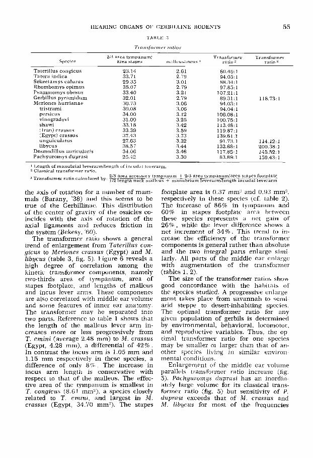
TABLE 3
55
Species area stapes malleuslincus 1 ratio 2 ratio
Taterillus congicus Tatera indica Sekeetamys calurus Rhombomys opimus Psammomys obesus Gerbillus pyramidum Meriones hurrianae
tristrami persicus vinogradovi shawi (Iran) crassus (Egypt) crassus unguiculatus libycus
23.14 33.71 29.35 35.07 33.40 32.01 30.73 30.08 34.00 31.00 33.18 33.39 37.43 27.63 38.57 34.06 25.42
2.61 2.79 3.01 2.79 3.21 2.79 3.06 3.06 3.12 3.25 3.42 3.59 3.73 3.32 3.44 3.46 3.30
107.21: 1 89.31 : 1 118.73:l 94.03:l 94.04:l
106.08: 1 100.75:l 113.48:l 119.87:l 139.61:l 91.73:l 114.12: 1
132.68: 1 200.38: 1 11 7.85: 1 145.52:1
I Length of manubrial leverarinilength of incudal leverarm. 2 Classical tramformer ratio.
the axis of rotation for a number of mam- mals (Barany, '38) and this seems to be true of the Gerbillinae. This distribution of the center of gravity of the ossicles co- incides with the axis of rotation of the axial ligaments and reduces friction in the system (Bekesy, '60).
The transformer ratio shows a general trend of enlargement from Taterillus con- gicus to Meriones crassus (Egypt) and M . Zibycus (table 3, fig. 5 ) . Figure 6 reveals a high degree of correlation among the kinetic transformer components, namely two-thirds area of tympanum, area of stapes footplate, and lengths of malleus and incus lever arms. These components are also correlated with middle ear volume and some features of inner ear anatomy. The transformer may be separated into two parts. Reference to table 1 shows that the length of the malleus lever arm in- creases more or less progressively from T. emini (average 2.48 mm) to M . crassus (Egypt, 4.28 mm), a differential of 42%. In contrast the incus arm is 1.05 mm and 1.15 mm respectively in these species, a difference of only 8 % . The increase in incus arm length is conservative with respect to that of the malleus. The effec- tive area of the tympanum is smallest in T. congicus (8.61 mm2), a species closely related to T. emini, and largest in M. crassus (Egypt, 34.70 mm2). The stapes
footplate area is 0.37 mm2 and 0.93 mm2, respectively in these species (cf. table 2). The increase of 86% in tympanum and 60% in stapes footplate area between these species represents a net gain of 26%, while the lever difference shows a net increment of 34%. This trend to in- crease the efficiency of the transformer components is general rather than absolute and the two integral parts enlarge simi- larly. All parts of the middle ear enlarge with augmentation of the transformer (tables 1, 2).
The size of the transformer ratios show good concordance with the habitats of the species studied. A progressive enlarge- ment takes place from savannah to semi- arid steppe to desert-inhabiting species. The optimal transformer ratio for any given population of gerbils is determined by environmental, behavioral, locomotor, and reproductive variables. Thus, the op- timal transformer ratio for one species may be smaller or larger than that of an- other species living in similar environ- mental conditions.
Enlargement of the middle ear volume parallels transformer ratio increase (fig. 5). Pachyuromys duprasi has an inordin- ately large volume for its classical trans- former ratio (fig. 5) but sensitivity of P. duprasi exceeds that of M. crassus and M . libycus for most of the frequencies

+O
pd
1 I
I 1
I I
I I
I I
J 70
90
110
130
150
Mid
dle
ea
r tr
an
sfo
rmer
ra
tio
o T
ran
sfo
rmer
ra
tio
wit
h a
cces
sory
tym
pa
nu
m
Fig
, 5
Rat
io o
f bu
lla
volu
me
to t
rans
form
er r
atio
. G
p, G
erbi
llus
pyr
amid
urn
; D
a, D
esm
oclil
lus
nu
ricu
lari
s;
Mc,
Mer
ione
s cr
assu
s (E
) E
gypt
, (I
) Ir
an;
Mh,
M.
hu
rria
nae
; M
1, M
. li
bycu
s; M
p, M
. pe
rsic
us;
Ms,
M.
shaw
i;
Mt,
M. t
rist
ram
i; M
u, M
. u
ngu
icu
latu
s; M
v, M
. vin
ogra
dovi
; P
d, P
ach
yuro
mys
du
pras
i; P
o, P
sam
mom
ys o
besu
s;
Ro,
Rh
ombo
mys
opi
mu
s; S
c, S
ekee
tam
ys c
alu
rus;
Tc,
Tat
eril
lus
con
gicu
s; T
i, T
ater
a in
dica
. O
pen
circ
les
ind
icat
e tr
ansf
orm
er
rati
os o
btai
ned
whe
n th
e ac
cess
ory
tym
pan
um
an
d n
eck
of
mal
leu
s ar
e in
clud
ed i
n th
e ca
lcul
atio
ns,
cf.
p.
57 t
ext.
N
ote
that
sam
ples
of
six
Mer
ion
es s
peci
es s
how
an
alm
ost
lin
ear
incr
ease
in
th
is r
atio
(d
ash
ed li
ne).

HEARING ORGANS OF GERBILLINE RODENTS 57
All species N = 17 Volume Two-thirds area tympanum Malleus arm length Area stapes footplate Incus arm length Area acc. tympanum Malleus neck length
1 .oo 0.743' 1.00 0.769' 0.915 I 1.00 0.868' 0.898 1 0.904 I 1.00 0.430 0.5172 0.6032 0.541 * 1.00 0.559 0.114 0.676 I 0.510 2 0.054 1.00 0.5642 0.228 0.920 0.973 0.390 0.917 1.00
Vol 213 AT MAL ASF IAL AAT MNL
' p, 0.01. 2 p, 0.05.
Fig. 6 Correlation coefficients of middle ear components for all 17 species studied
tested (figs. 10-14). Yet, the transformer ratio of the first is only 60 and 63% as great as of the last two species, and the three species have similar organs of Corti. This suggests that the accessory tym- panum functions to augment the middle ear transformer.
The central region of the accessory tympanum attaches to the upper half of the neck and lower-portion of the body of the malleus and to the upper third of the long process and lower part of the body of the incus in living specimens of G. pyra- m i d u m , M . unguicula tus , M . libycus and P. duprasi. The membrane is stretched taut and is histologically like the tym- panum, which suggests that it is capable of vibration in response to airborne sound, and, by virtue of attachment to the mal- leus and incus, is capable of transmitting vibrations to the ossicle chain.
The manubrium is disposed at nearly right angles to the neck in Pachyuromys (fig. 27). The attachments of the tym- panum and accessory tympanum thus lie at sharply different angles and this allows these membranes to function independ- ently by vibrating in different planes but both would displace the neck and manu- brium medially. This arrangement seem- ingly increases the transformer ratio by enlarging the vibratory receptor surface and lengthening the malleus lever arm by a small factor. If the transformer ratio of Pachyuromys is calculated, using the functional area of the tympanum plus two- thirds the area of the accessory tympanum and the length of the manubrium lever arm plus one-half the length of the neck, the value obtained (159:l) agrees well with the transformer ratio: volume trend of the other 16 species studied (fig. 5).
The transformer ratio values deter- mined in this manner for the four other
species with accessory tympani are dis- proportionately high relative to volume. If the accessory tympanum functions to enhance the transformer, the system aug- ments the transformer of these four spe- cies much less than that of Pachyuromys since the parallel increase of volume and transformer ratio is consistent though nonlinear for all species lacking an ac- cessory tympanum.
The effective area of the tympanum relative to that of the stapedial footplate is much lower in Pachyuromys than in the other species for which the accessory tympanum was measured (table 2). The greater elaboration of its accessory tym- panum may compensate for this difference.
The anatomical data concerning the accessory tympanum lend some support to the hypothesis that this structure func- tions as a part of the transformer. That this membrane characterizes desert in- habiting species and is largest in those inhabiting the most desertic regions seems a good indication that it is an adaptive feature.
Auditory bullae
The mastoid air cells
Three primary mastoid air chambers occur in the Gerbillinae: anterior; pos- terior superior; posterior inferior.
Anterior mastoid chamber (fig. 29). The anterior chamber (AM) lies directly above the external auditory meatus. It is usually triangular in dorsal view. A bony partition extends dorsally from the full span of the crista parotica to the superior cover of the bulla and separates the an- terior chamber from the superior poste- rior mastoid cell. The anterior third of the anterior (superior) semicircular canal ex-

58 DOUGLAS M. LAY
tends into the anterior chamber through the medial edge of this septum. This chamber opens ventrally into the epitym- panic recess of the tympanic cavity via a semicircular opening in its inferior wall, the lateral straight border of which is es- sentially demarcated by the fossa incudis posteriorly and the petrotympanic fissure anteriorly. The posterior half of the semi- circle is formed by the crista parotica and the anterior half is demarcated by a bony shelf directed anterolaterally.
Posterior mastoid chambers. There are one or two (the superior posterior SP, and inferior posterior IP) posterior mastoid chambers in most gerbillines. Consider- able variation exists in these pneumati- zations.
Superior posterior chamber (fig. 29). The SP cavity generally forms a crude pyramid, the base of which can be con- sidered as the posterior limiting bullar capsule while the apex lies just posterior to the anterior junction of the lateral and superior semicircular canals. The lateral canal and the bony shelf that projects posteriorly from this canal to the bullar wall form the ventral border of this cham- ber. Portions of the petrous and squamous temporal bones constitute its medial bound- ary. The septum between this chamber and the anterior cavity forms the lateral border of the pyramid, is oriented postero- laterally and inclines toward the occiput. The superior canal penetrates this sep- tum at a right angle. A postero-medially directed shelf of bone projects from that portion of the posterior canal to the pos- terior border of the bulla, separating a small medial chamber from the remainder of the superior posterior chamber. These are named medial (MSP) and lateral su- perior posterior (LSP) cells, respectively. The medial connects with the lateral cell through an opening bounded vertically by the posterior canal and dorsally by the lateral canal. The lateral superior pos- terior chamber opens ventrally into the tympanic cavity by an almost circular orifice bounded posteriorly by the lateral canal and anteriorly by the bony covering of the sacculus.
Inferior posterior chamber (fig. 29). This chamber occurs in most gerbilline rodents and lies immediately ventral to
the superior posterior cell. The inferior border of this latter, namely the lateral canal and its bony lamina forms the roof of the inferior posterior chamber. A shelf formed by juxtaposed tympanic and mast- oid plates composes the inferior margin, which is inclined postero-ventrally at an angle of about 45" to the almost horizon- tal roof. The superior rim of the tympanic and mastoid plate junction forms the ventral limitation. The IP chamber con- nects dorsally with the SP through an opening bounded by the lateral canal. The tympanic mastoid shelf has a free border anteriorly by means of which the IP is connected to the tympanic chamber.
The posterior auricular branch of the vagus nerve (X) traverses this chamber in the genera Sekeetamys, Gerbillus, Psam- momys, Rhombomys, and Meriones. This nerve enters the mastoid cavity via a minute foramen located at the tympano- mastoid junction posterior and dorsal to the foramen of the stapedial artery, and then courses either almost straight medio- laterally or parallel to the curvature of the posterior end of the bulla. Laterally it enters the facial canal and emerges with the facial nerve through the stylomastoid foramen. The posterior auricular nerve lies between the overlapping mastoid and tympanic plates (figs. 29, 39). It may lie completely in a bony canal as in Psam- momys, Gerbillus pyramidurn, G. gerbillus, G. nanus, and Seheetamys or in a groove in the upper surface of the tympanic plate as in Rhombomys and certain Meriones, where the mastoid plate fails completely to cover the tympanic plate.
This chamber exists as a single cell in many species (cf. table 4) but i t may be divided into as many as three compart- ments. A small medial cell, here named the accessory medial inferior posterior (AMIP), occurs when a bony shelf extends from the ventral portion of the posterior canal to reach the medial bullar wall. This cell, when present, communicates via an opening bounded by the lateral canal dor- sally and the posterior canal ventrally with the orifice by which both principal poste- rior (SP and IP) chambers connect to the tympanic cavity. A dorso-ventrally ori- ented partition in the lateral third of the compartment may divide the IP chamber

HEARING ORGANS OF GERBILLINE RODENTS 59
into lateral inferior posterior (LIP) and medial inferior posterior (MIP) compart- ments. When such a separation is absent, the single large chamber is termed the common medial inferior posterior (CMIP).
Enlarged auditory bullae are one of the most distinctive features of the Gerbil- linae. Two basic components derived from the mastoid and tympanic ( = ectotym- panic) bones form the auditory bulla (figs. 32, 33). A trend toward enlargement of the middle ear cavity, the degree of which correlates with the size of the transformer ratio and habitat characterizes the Ger- billinae. Major differences in the compo- nent parts of the bulla characterize the groups of species studied and provide some insight into the manner by which the spe- cialized forms may have evolved from a generalized ancestral type.
The mastoid of the gerbilline progeni- tor was probably somewhat similar to that of Rattus rattus. In this species the mast- oid faces postero-laterally and lies primar- ily posterior to the tympanic bulla (fig. 34). The anterior end of the mastoid roofs the epitympanic recess and only at this point does the mastoid make contact with the tympanic. The subarcuate fossa, which houses the petrosal lobe of the cerebellar paraflocculus, forms the most significant feature of the mastoid in Rattus. The subarcuate fossa, which is connected to the brain cavity through the arch of the anterior semicircular canal, is formed by a thin layer of lamellate bone that balloons out into the mastoid lateral to the plane of the canal arc. Small unconnected air cells are present within a thin layer of cancellous bone interposed between the wall of the fossa and the external lamel- late wall of the mastoid. A very small, hollow anterior mastoid cell exists but has connection neither with the epitympanic recess nor with the small cells surround- ing the subarcuate fossa (figs. 34, 35).
The mastoid of Tateriltus is approxi- mately the same size as in Rattus. In T. emini and congicus the AM cell, 0.003 cm3 in volume, is four or five times more capacious than that of Rattus and opens ventrally into the epitympanic recess by an aperture which is completely sur- rounded by mastoid elements. The floor of the AM in Rattus is very thin and trans-
lucent to light at several places; this con- dition might be considered as adumbrating the development of the connection be- tween the tympanic and AM chambers. The nature of the posterior region in T. emini is not unlike that described for Rattus. However, T . congicus has devel- oped a small (0.002 cm?) chamber in the posterior mastoid, above the level of the lateral canal between the wall of the subarcuate fossa and outer mastoid wall. No connection exists between it and the AM because the lateral wall immediately posterior to the anterior chamber adheres to the border of the subarcuate fossa.
The anterior and posterior mastoid chambers of Tatera indica are each ten times larger than the respective cells of T . congicus. This enlargement has been accomplished by reduction of the subar- cuate fossa, lengthening the mastoid an- tero-posteriorly, displacement of the lateral mastoid wall laterally, and pneumatiza- tion of the cancellous bone between these boundaries. The small segment of mast- oid which extends inferior to the plane of the lateral canal is composed of inner and outer layers of lamellar bone between which is a thin layer of cancellous bone containing minute air cells.
The AM and PM cell morphology of Gerbitlurus paeba does not differ in any important respects from that of Tatera. The mastoid below the plane of the lateral canal, however, is considerably larger than that found in either Tatera or Tater- illus due to expansion dorso-ventrally. The cancellous bone between the inner and outer walls has been replaced by a single narrow chevron-shaped chamber which extends around the posterior angle of the mastoid (figs. 36, 37). This chamber does not connect with either the tympanic cavity or the posterior superior cell, but because of its relations this cavity proba- bly represents the initial stage of develop- ment of a posterior inferior cell typical of most of the more specialized Gerbillinae discussed below.
Taterillus, Tatera, and Getbillurus are characterized by the most generalized mastoid morphology among the extant Gerbillinae. The mastoid of all other ge- nera is highly pneumatized and these segregate into two groups on the basis of
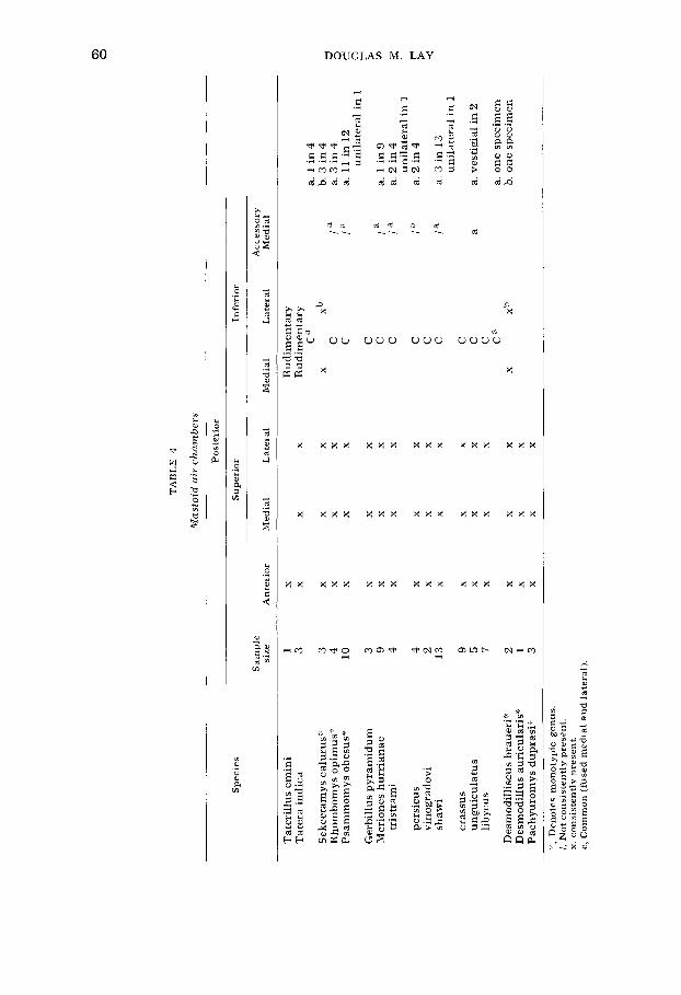
Q, 0
TA
BL
E 4
Mas
toid
air
cha
mbe
rs
Sp
ecie
s
Pos
teri
or
Su
per
ior
Infe
rior
Sam
ple
A
cces
sory
si
ze
An
teri
or
Med
ial
Lat
eral
M
edia
l L
ater
al
Med
ial
Tat
eril
lus
emin
i T
ater
a in
dica
Sek
eeta
mys
cal
urus
" R
hom
bom
ys o
pim
us':
Psa
mm
omys
obe
sus"
Ger
bill
us p
yra
mid
um
M
erio
nes
hu
rria
nae
tr
istr
ami
pers
icus
vi
nogr
adov
i sh
awi
cras
sus
ungu
icul
atus
li
bycu
s
Des
mod
illi
scus
bra
ueri
" D
esm
odil
lus
auri
cula
ris"
P
achy
urom
ys d
upra
si"'
1 3 3 4
10 3 9 4 4 2 1
3
9 5 7 2 1 3
X X X
X
X
X
X X X
X X X
X X
X
X X X
Rud
imen
tary
R
udim
enta
ry
C"
X
Xb
C
c
L
C
C
C
C
C
C
C
C
C
C"
X
Xb
a. 1
in
4
b. 3
in
4
U
la
a.
11
in 1
2 C
/a
a.
3 i
n 4
0
unil
ater
al i
n 1
la
a.
1 i
n 9
% s F
la
a. 2
in
4
la
a. 2
in
4
la
a. 3
in
13
unil
ater
al i
n 1
4
unil
ater
al i
n 1
a a.
ves
tigi
al i
n 2
a. o
ne
spec
imen
b.
on
e sp
ecim
en
::c,
Den
otes
mon
otyp
ic g
enu
s.
/, N
ot c
onsi
sten
tly
pre
sen
t.
x.
con
sist
entl
y p
rese
nt.
c,
Com
mon
(fu
sed
med
ial
and
late
ral)
,

HEARING ORGANS OF GERBILLINE RODENTS 61
morphology. The presence of only a PS chamber in Ammodillus, Desmodillus and Pachyuromys differentiates these from all the other genera, which are characterized by both SP and IP cells (cf. table 4).
Considerable variation in posterior mast- oid morphology characterizes this last group with regard to the number of cham- bers present and the volumes of each (ta- bles 4, 5).
Because the inferior chamber is absent or rudimentary in Tatera, Taterillus and Gerbillurus, while the superior cell is rela- tively well developed, 1 believe that the former cell probably arose after the latter. However, the alternative that those forms with SP and IP chambers may represent a distinct evolutionary line cannot be ruled out. Further, it seems that inferior cells of small volume may be indicative of degree of specialization when considered with other parameters suggesting varying degrees of auditory specialization. The inferior chamber is smaller than the su- perior in 9 of 13 species studied. Of these, S. calurus seems to be least specialized and the inferior chamber is partitioned into three parts in three specimens and two parts in a fourth. The medial and lat- eral inferior chambers have fused to form a single common inferior cavity in this fourth specimen, the condition typical of the other 12 species of this group. Vestiges of the bony partition that originally di- vided this chamber occur commonly in M. shawi as a raised ridge oriented dorso- ventrally along the middle of the posterior wall of the cell; the same condition exists in Desmodilliscus. A minute accessory medial cavity is present in all specimens of Sekeetamys studied, a large proportion of Rhombomys and Psammomys and a smaller percentage of four species of Meriones. This cell is present unilaterally in single specimens of M. tristrami and M. shawi and vestigial in M. unguiculatus (table 4). The AMIP cell is incorporated into the medial inferior cell when the wall formed by the horizontal lamina of the lateral canal fails to develop. The general trend of change in the inferior cell is toward fusion of the two or three small cells into a single large unit con- comitant with expansion of this chamber. The fenestration connecting the IP with the tympanic cavity is smallest in Sekee-
tamys calurus, among all 13 species, by a factor of at least five. Among the other 12 species, the size of this opening seems to be closely correlated with the magnitude of the transformer ratio. This connection is smallest in Meriones hurrianae, persi- cus and tristrami and largest in M. cras- sus and libycus (figs. 39,40).
The superior chamber is highly pneu- matized in all the Gerbillinae but Tateril- lus, Tatera, and Gerbillurus paeba and is always divided into a small medial com- partment that varies much less in size than does a large lateral chamber (tables 5, 6). In the generalized condition of Ta- tera and Gerbillurus the superior cham- ber connects only with the anterior cell. In all other forms studied the SP opens ventrally either directly into the tympanic cavity (Desmodillus and Pachyuromys) or into the inferior chamber directly above the opening of this chamber into the tym- panic cavity. The arc of the lateral canal and the sacculus form the limits of this opening in all species but Desmodillus auricularis and Sekeetamys calurus. Though no measurements have been made to verify this, the opening is remarkably uniform in size, except in Pachyuromys duprasi in which the diameter of the arc of the lateral canal is about twice that of any of the other species examined.
The subarcuate fossa extends into the medial side of the superior chamber where it completely fills the inside of the lateral canal arc in Tatera indica and Gerbillurus paeba. However, in all other forms, it is reduced relative to this condition and is progressively eliminated from the SP chamber with increasing pneumatization. It persists as a small nodule in most spe- cies but is absent in M. crassus, M. libycus, and P. duprasi (figs. 41, 42).
The morphology of Desmodillus and Sekeetamys provides indications as to how this mastoid tympanic connection may have developed, The superior chamber of Desmodillus connects with the anterior cell as in Tatera and Gerbillurus. The subarcuate fossa is virtually absent and a sheet of thin bone fills the arc enclosed by the lateral canal and its connection with the utriculus. A very small semicir- cular opening adjacent to the utriculus perforates this sheet, thus joining the SP with the tympanic cavity. This connection

TA
BL
E 5
Vol
um
es of
bu
llae
and
th
e va
riou
s se
para
te c
ham
bers
in
cubi
c ce
nti
met
ew
Vol
um
es o
f ce
lls
det
erm
ined
for
sp
ecim
en a
nd
bu
lla
ind
icat
ed i
n f
irst
tw
o co
lum
ns
at le
ft
Sp
ecie
s T
otal
vol
um
e T
otal
T
otal
C
IP
Sp
ecim
en
mea
n a
nd
ext
rem
es
bu
llar
po
ster
ior
Tot
al
no.
Sam
ple
of
sam
ple
vo
lum
e A
M
mas
toid
M
SP
LS
P
MIP
L
IP
AM
IP
Tym
pan
ic
mas
toid
Tat
eril
lus
con
gicu
s
Tat
era
ind
ica
Sek
eeta
mys
cal
uru
s
Rho
mbo
mys
op
imu
s
Psa
mm
omys
ob
esu
s
Ger
billu
s py
ram
idum
Mer
ione
s hu
rria
nae
tris
tram
i
per
sicu
s
vino
grad
ovi
shaw
i
(Egy
pt)
cra
ssu
s
(Ira
n)
cras
sus
un
guic
ula
tus
lib
ycu
s
Des
mod
illu
s au
ricu
lari
s
Pac
hyur
omys
du
pra
si
5046
9
2647
2550
10
72
2587
2625
2648
2522
2548
2545
2675
2684
2538
2568
2543
3877
5
2670
1 3 2 2 9 3 8 4 4 2 10
4 5 5 7 1 3
0.14
5
0.27
0 (0
.26G
0.28
9)
0.35
6 an
d 0
.371
0.35
3 an
d 0.
404
0.43
8 (0
.38
€~
0.5
10
)
0.1
76 (
0.1
5S
0.2
01
)
0.23
4 (0
.181
-0.2
33)
0.21
5 (0
.191
-0.2
36)
0.26
5 (0
.248
-0.2
91)
0.34
7 an
d 0
.358
0.45
2 (0
.370
-0.5
82)
0.79
4 (0
.755
-0.9
32)
0.36
3 (0
.27
S0
.44
2)
0.21
8 (0
.201
-0.2
37)
0.58
3 (0
.508
-0.7
01)
0.38
3
1.01
7 (0
.79
g1
.13
5)
0.14
5
0.26
7
0.35
6
0.40
4
0.43
9
0.17
1
0.22
5
0.21
4
0.25
9
0.34
7
0.45
5
0.75
3
0.36
3
0.21
8
0.58
3
0.38
3
1.09
0
0.00
3
0.03
4
0.02
5
0.05
7
0.05
7
0.01
2
0.03
0
0.01
7
0.02
4
0.02
2
0.04
9
0.08
5
0.03
1
0.01
7
0.03
1
0.01
3
0.13
0
0.00
2 I
0.02
4
0.08
5
0.11
0
0.09
7
0.02
7
0.05
2
0.03
6
0.05
0
0.07
1
0.10
3
0.18
6
0.10
3
0.05
2
0.12
4
0.13
4
0.45
1
0.00
9
0.00
7
0.00
5
0.00
3
0.00
4
0.00
3
0.00
5
0.01
0
0.00
9
0.00
5
0.00
6
0.00
7
0.01
0
0.00
9
0.01
5
0.04
5
0.04
5
0.03
6
0.01
5
0.03
1
0.02
1
0.03
4
0.01
9
0.03
1
0.10
3
0.06
4
0.02
9
0.07
1
0.12
5
0.43
6
0.02
4 0.
004
0.05
6
0.05
2
0.00
9
0.01
8
0.01
2
0.00
9
0.04
2
0.06
3
0.07
8
0.03
3
0.01
6
0.04
3
0.14
2
0.20
9
0.00
3 0.
246
0.00
2 0.
237
0.00
3 0.
285
0.13
2
0.14
3
0.16
1
0.00
2 0.
189
0.25
3
0.30
3
0.48
1
0.22
9
0.14
8
0.42
4
0.23
6
0.50
9
0.00
3
0.05
8
0.11
0
0.16
7
0.15
4
0.03
9
0.08
2
0.05
3
0.07
4
0.09
3
0.15
2
0.27
1
0.13
4
0.06
9
0.15
6
0.14
7
0.58
1
I N
ot c
onn
ecte
d w
ith
rem
ain
der
of
bu
llar
cav
itie
s an
d n
ot c
onsi
der
ed i
n t
otal
vol
um
e.

TA
BL
E 6
Rat
ios
of m
iddl
e ea
r ch
amb
er v
olum
es
-
Sp
ecie
s T
ota
l T
ota
l v
olu
me
Ty
mp
anic
m
asto
id
in c
m?
To
tal
To
tal
An
teri
or
mas
toid
T
ota
l
To
tal
po
ster
ior
mas
toid
T
ota
l M
SP
T
ota
l --
LS
P
MIP
T
ota
l T
ota
l L
IP
CIP
T
ota
l T
ota
l ~
-
Tat
eril
lus
cong
icu
s
Tat
era
ind
ica
Sek
eeta
mys
cal
uru
s
Rho
mbo
mys
op
imu
s
Psa
mm
om
ys
obes
us
Ger
bill
us p
yra
mid
um
Mer
ione
s h
urr
ian
ae
tris
tram
i
pers
icus
vino
grad
ovi
shaw
i
(Egy
pt)
cras
sus
(Ira
n) c
rass
us
un
gu
icu
latu
s
liby
cus
Des
mod
illu
s au
ricu
lari
s
Pac
hyur
omys
du
pra
si
0.1
45
0.26
7
0.35
6
0.40
6
0.43
9
0.17
1
0.2
25
0.21
4
0.25
9
0.34
7
0.45
5
0.75
3
0.36
3
0.21
8
0.58
3
0.38
3
1.09
0
0.98
0.78
0.6
9
0.59
0.65
0.77
0.6
4
0.75
0.73
0.7
3
0.67
0.64
0.63
0.68
0.73
0.62
0.47
0.02
0.22
0.31
0.41
0.35
0.2
3
0.36
0.25
0.27
0.27
0.33
0.36
0.3
7
0.32
0.27
0.38
0.53
0.02
0.1
3
0.0
7
0.14
0.1
3
0.07
0.1
5
0.08
0.09
0.0
6
0.11
0.11
0.0
9
0.0
8
0.05
0.0
3
0.12
0.09
0.24
0.27
0.22
0.16
0.21
0.17
0.19
0.21
0.2
3
0.25
0.28
0.24
0.21
0.35
0.41
0.03
0.02
0.01
0.02
0.01
0.01
0.02
0.03
0.02
0.01
0.02
0.03
0.02
0.02
0.01
0.13
0
.07
0.11
0.08
0.09
0.13
0.10
0.1
3
0.06
0.07
0.1
4
0.18
0.1
3
0.12
0.33
0.40
0.0
1
0.14
0.12
0.05
0.07
0.06
0.04
0.12
0.14
0.10
0.09
0.07
0.0
7
0
0.01
P 0
0.01
9 5 %
0.01

64 DOUGLAS M. LAY
has enlarged in Seheetamys to encompass two-thirds the area enclosed by the lateral canal, but a thin bony partition still fills the lateral one-third of the arc. However, a partition separates the posterior supe- rior and anterior cells in this species and in Pachyuromys, which is the closest re- lated form of all Gerbillinae to Desmodzl- lus. The septum between the anterior and superior cells, characteristic of all the species with greatly pneumatized mastoids with the exception of Desmodillus, may have developed as an internal buttress for the following reasons. The entire outer layer of mastoid is composed of thick la- mellar bone in Tatera. The outer layer is much thinner than in Tatera in all forms with pneumatized mastoids. The anterior cell of Desmodillus is small in volume and the ratio to total bullar volume is the smallest of all specialized forms (tables 5, 6). Desmodillus, however, possesses what appears to represent initial stages in the development of a partition between the anterior and superior chambers. A small sickle-shaped buttress extends ven- trally from the roof at the juncture of the anterior and posterior superior cells. The thicker medial portion of this brace at- taches to the anterior canal at a point slightly posterior to the anteriormost ex- tent of this canal. A larger wedge-shaped plate arises from the entire length of the crista facialis lateral to the point from which this ridge emerges from the lateral canal, is oriented dorso-ventrally and is highest laterally where it fuses to the lateral wall of the mastoid. The upper free edge of this plate and the lateral extent of the upper buttress meet to form a slightly raised ridge along the lateral roof between the anterior and posterior su- perior cells. This incomplete septum lies at the boundary of these two cells (cf. p. 58 text) and is in good accord with the boundaries of the complete partition of other specialized species (fig. 38).
A number of muscles insert entirely or partly into the mastoid along the outside boundary between the anterior and pos- terior superior cells. These are the cleido- mastoid, occipitoscapularis, splenius cap- itis, longissimus capitis, and temporalis. The partition between the anterior and posterior cells rests upon a beam formed
by the laterally directed anterior part of the lateral canal superimposed on the facial canal and the thick crista facialis lateral to the point where these two canals diverge. Pneumatization of the anterior cell produces concentric expansion and thinning of the bony roof. It seems that under these conditions a vertical, trun- cated, triangular partition would reduce compressile forces on the broad thin mast- oid roof and provide a stable insertion for these muscles. If such is the case, then a connection of the posterior superior chamber with the tympanic or posterior inferior cells is required in order to achieve maximum bullar volume.
The evidence suggests that the anterior cell developed first and was followed by pneumatization of the posterior mastoid. It is not clear when the division of this latter region into superior and inferior chambers first occurred or if this event may have occurred independently in one or several lines. If the double chambered posterior mastoid line is derived from a Tatera-like ancestor then the evidence available suggests that the inferior cham- ber evolved subsequent to the superior cell.
The tympanic portion of the bulla is conservative in the degree of change ob- served between generalized and specialized forms relative to the degree of mastoid modifications described above. This cell composed 47 to 98% (mean 69.1% of the total middle ear volume among the forms studied) (table 6).
This chamber constitutes 18% of total middle ear volume in Dipodomys merriami and spectabilis (Webster, '61). Although I have made no volumetric determinations, study of the auditory bullae of specimens of the heteromyids Microdipodops pallidus and M . megacephalus suggests that a similar size relationship holds for these specialized forms. Likewise, examination of specimens of the specialized Gipodidae, Jaculus jaculus and blanfordi and Sal- pingotus michaelis suggests that the tym- panic volume constitutes considerably less than half of total bullar volume and probably less than 25 % . Illustrations of the skulls of Eremodipus and Cardiocra- nizis (Vinogradov, '37; Gromov et al., '63; Ognev, '48) indicate that the same rela-

HEARING ORGANS OF GERBILLINE RODENTS 65
tionship holds for these specialized forms. Specimens of Ctenodactylus and Massou- teria (Ctenodactylidae), inhabitants of the rocky highlands of the Sahara region, also show the tendency of greater pneu- matization of the mastoid cells relative to the tympanic. The middle ear structure of Heteromys and Perognathus is less spe- cialized than that of Dipodomys and Mi- crodipodops. As the ears become special- ized the mastoid component enlarges much more than does the tympanic com- ponent (Howell, '32).
Specimens of Atlactaga, Alactagulus, Dipus and Scirtopoda and illustrations of Pygerethymus, Scarturus, Paradipus, and Euchoreutes (Vinogradov, '37) show a trend in the enlargement of the middle ear cavity very similar to that which ap- pears to have occurred among the Hetero- myidae described above. The middle ears of these dipodids show virtually every intermediate stage between the most gen- eralized and most specialized conditions. In this trend the mastoid component ex- pands at a much greater rate than the tympanic. No generalized ctenodactylids are known.
In the Gerbillinae the mastoid volume increases from 2% of total bullar volume in the generalized Taterillus to 53% in the highly specialized Pachyuromys (mean of forms studied 30 %I ) (table 6).
Any discussion of evolutionary trends in the gerbilline ear must regard this organ as a unit composed of the middle and in- ner ears, which function together to con- vert airborne sound to neural responses. Enlargement of the tympanic cavity has in general followed two directions. The most obvious pathway is that of overall increase in volume by expansion in all directions. Table 6 illustrates the overall changes in tympanic size among the spe- cies studied.
Dissections of bullae and/or examination of endobullar casts reveal that pneumati- zation of the tympanic bone around the anterior edge of the external auditory meatus is achieved by a partitioning of the tympanic wall. The tympanic wall in this region is formed of a single lamina in generalized forms (e.g., Taterillus, Tatera, Meriones persicus) but separates into me- dial and lateral plates in intermediate
and specialized forms. The medial lamina then forms the anterior wall of the ex- ternal auditory meatus and helps support the tympanic annulus. The lateral wall becomes continuous with the lateral bul- lar wall.
The space between these plates is pneu- matized in different ways in the gerbillines studied. Volume has been added to the middle ear cavity by expanding this space so that the anteroventral part becomes confluent with the tympanic chamber as in Meriones crassus, sacramenti and liby- cus (fig. 46). The tympanic may also sep- arate into dorsal and ventral sheets just anterior to the superior extent of the ex- ternal auditory meatus. This cavity when formed is purely tympanic in origin, but in most cases opens into the lateral side of the anterior mastoid chamber. This condition occurs in Desmodillus, Pachy- uromys, Psammomys, Rhombomys, and Meriones shawi, hurrianae, and crassus. Both pathways of pneumatization of this region occur in a number of these same forms. The inferior enlargement predomi- nates in Psammomys, 111. shawi and M. crassus, the superior enlargement char- acterizes Desmodillus and M. hurrianae and superior and inferior cavities are nearly equal in Pachyuromys (figs. 43-46). Five casts of Meriones crassus from Iran show that both centers have enlarged and are in virtual contact, separated by a single bony lamina. Three of four casts of M. crassus from Egypt show that the internal lamina separating these centers has been lost and that the two chambers are continuous but a pronounced external groove demarcates the former point of division.
The tympanic anterior to the external auditory meatus is pneuinatized in a unique manner in the two specimens of Desmodilliscus braueri studied. The tym- panic wall has been inflated to a degree equal or similar to that found in Pachy- uromys, M . crassus and libycus. How- ever, this cell opens into the tympanic chamber via a small fenestra (0.63 mm x 0.32 mm) just below the point of articu- lation of the anterior process of the mal- leus with the tympanic. In these other mentioned forms this cell is not cut off from the tympanic chamber but consti-

66 DOUGLAS M. LAY
tutes a direct unconstricted continuation of it. The tympanic annulus
The tympanic annulus supports the tympanum. In the adult condition it lies within the tympanic cavity. The annulus does not form a complete ring. Approxi- mately the upper one-third to one-fourth of the ellipse remains open as the dorsal arms never meet. The ventral portion ex- tends considerably further medially than the dorsal part so that the ring rests at a rather sharp angle to the vertical. This angle varies among the forms studied but has not been quantified.
The attachment of the tympanic an- nulus to the lateral bullar wall (tympanic) has been modified in all of the Gerbil- linae, but exists as a raised ridge on the medial surface of the lateral tympanic wall in the Dipodidae and Heteromyidae.
In the Gerbillinae the tympanic annulus retains connection to the rest of the tym- panic, but the bulla has been extensively remodelled in this region. The attachment of the free ends of the annulus to the tym- panic at the dorsal edge of the external auditory meatus constitutes what is essen- tially the primitive condition or the con- dition as occurs in Taterillus, Tatera, Sekeetamys, Rhombomys, and the less specialized species of Gerbillus and Meri- ones. The points of attachment here rep- resent raised ridges on the inside of the lateral bullar wall. In the more specialized species of Gerbillus and Meriones, Des- modillus, Psammomys, Desmodilliscus and Pachyuromys the condition remains es- sentially the same but is obscured by the pneumatizations of the tympanic anterior and dorsal to the external auditory meatus andlor by the development of the acces- sory tympanic membrane. Distal to the points of attachment of the free ends of the annulus the situation changes strik- ingly. The diameter of the external part of the auditory canal is approximately one-half that of the tympanic annulus. Thus, the canal widens in its medial ex- tent. The cylindrical canal opens opposite the upper two-thirds of the tympanum and its upper margin is flush with the superior portion of the annulus. Because the inferior extent of the tympanum in- clines sharply medially and its diameter
is greater than that of the canal, a bell- shaped lamella of bone joins these two. The meatal cylinder and bell-shaped sup- port of the annulus are connected to the lateral bullar wall by a series of buttresses (figs. 47, 48). These braces run essentially perpendicularly between the auditory canal and the lateral wall. With increase of bullar volume these buttresses lengthen and at the points of attachment to the lateral wall, each principle strut branches dendritically into smaller braces forming elaborate patterns. Dissection of the lat- eral wall reveals that in spite of rather complicated peripheral patterns a primary arrangement exists. There are three pri- mary braces which form a modified T, the arms of which form a well-opened chevron. The base of the T passes ventrally while the arms of the chevron are directed an- teriorly and posteriorly, respectively (figs. 47, 48).
This morphological arrangement allows the tympanic air space to expand into the area lateral to the tympanum. The great expansion of this area that is obvious by comparison of Taterillus or Tatera with Pachyuromys, Meriones crassus or several other forms suggests that the tympanic bulla has enlarged in this manner. The buttress system between the truncated cone and lateral wall probably functions to stabilize the tympanic annulus but also may act to maintain the integrity of the bulla by resisting compressile forces which might be produced by contractions of the posterior belly of the digastric, stylohyoid, stylopharyngeus, and styloglossus, which either originate on or pass over the in- ferior surface of the tympanic bulla. The buttress appears appropriately shaped to direct compressile forces upward along the various axes to the more stable region at the base of the external auditory mea- tus. These forces would likely be dissi- pated along this route and if not totally damped on reaching this point, the V- shaped floor of the external auditory mea- tus would seem ideally situated to transmit any excess forces to the structurally stable region at the tympanic mastoid junction above the meatus. These factors would most likely be functional in those species possessing greatly inflated bullae (e.g., Pachyuromys, M . crassus).
The internal relocation of the tympanic

HEARING ORGANS OF GERBILLINE RODENTS 67
annulus seems to be one, if not the key factor that has allowed pneumatization of the tympanic cavity of the Gerbillinae. By elaborating this modification a large vol- ume has been incorporated into the mid- dle ear chamber of these rodents. This volume probably represents between 30 and 40% of the total air space of the tympanic cavity in the species with great- est hypertrophy ( M . crassus, M. Libycus and P. duprasi).
Two other lines of evidence support the observation that the auditory bullae have expanded concentrically.
In Tatera indica the posterior auricular branch of the vagus nerve passes around the outer edge of the bulla in a groove along the tympanic-mastoid juncture and, on the lateral side of the bulla just pos- terior to the stylomastoid foramen, enters a bony canal which joins the facial canal anteriorly. The posterior auricular and facial nerves (VII) exit via the stylomast- oid foramen.
In all the gerbillids possessing inflated mastoid cells the posterior auricular nerve passes through the posterior mastoid cells as discussed above (p. 58 text). Exam- ination of an ontogenetic series of Meri- ones shawi and Pachyuromys duprasi con- firms that the adult posterior mastoid and posterior portion of the tympanic expand posteriorly and laterally. It seems likely that with the increased pneumatization of the mastoid cells the posterior auricu- lar nerve was surrounded and its course came to lie between the laminae of the tympanic and mastoid bones within the middle ear.
In Dipodomys and Jaculus this phenom- enon does not occur, apparently because the mastoid cells expand to a much greater degree than the tympanic bullae. Due to this differential enlargement in the spe- cialized heteromyids and dipodids the original course of the posterior auricular nerve outside the bulla is altered only little or not at all.
With great inflation of the anterior and superior mastoid cells as in M . crassus, Desmodilliscus and Pachyuromys the para- occipitals, parietal, and interparietal bones have been extensively remodelled and are reduced in width. Hypertrophy of the tym- panic results in a great reduction in width of the basioccipital and basisphenoid. An-
terior expansion of the tympanic modifies the squamosal and alisphenoid extensively. These changes are relative to the gener- alized condition typical of Taterillus, Ta- tera, and GerbiLLurus. The species other than the forms mentioned above display virtually all degrees of transition between the extremes of generalization and spe- cializa tion.
The functional significance of hyper- trophied bullae was referred to briefly above (p. 49 text). Impedance of the ear drum at low frequencies is greatly influ- enced by the cushion of the middle ear cavity (Bekesy, '60). Increase of air pres- sure wjthin the middle ear reduces coch- lear potential responses to low frequency tones more than for high frequency tones (Wever and Lawrence, '54). Cochlear po- tentials were greatly diminished relative to normal Dipodomys and Meriones eras- sus when bullar volume was reduced (Webster, '61; Legouix and Wisner, '55). This difference is attributed to increased resistance to incursions of the tympanum. Effective tympanum area and middle ear volume increase concomitantly (tables 2, 5). The species studied neurophysiologi- cally are most sensitive to sounds in the low frequency range of 500-2800 Hz (figs. 10-13). Species showing greatest sensi- tivity to these sounds are characterized by larger transformer ratios as discussed above, and this increase has resulted in part by enlargement of the tympanum. A larger tympanum will compress a middle ear air cushion of given volume propor- tionately more than will a small tym- panum. Low frequency sounds produce greater displacements of the tympanum than high frequencies. The characteristic hypertrophy of the middle ear volume of the specialized Gerbillinae has probably evolved as a means for reducing friction due to enlargement of the tympanum coupled with the auditory specializations for hearing low frequency sounds. Bullar hypertrophy, within the two chambered mastoid gerbils (8 genera, ca. 75 species) has resulted in a great deal of experi- mentation upon a single architectural plan.
A correlation matrix of the mean values of the six kinetic components of the ger- billine transformer ratio and the volume of the middle ear cavity for 17 species is

68 DOUGLAS M. LAY
presented in figure 6. A high degree of correlation exists among all of these fac- tors. Only five combinations are not well correlated at the 0.05 or 0.01% levels. Three components, two-thirds the area of the accessory tympanum, the length of the neck of the malleus, and the length of the incus lever arm, seem to account for these low values. The first two of these components are involved in an acquired system characteristic of only five of the 17 species. Both of these factors are os- tensibly least correlated with length of incus arm and the functional area of the tympanum, while each is highly corre- lated with the other four components and with each other.
It seems that the gerbilline middle ear represents a highly integrated organ, the components of which have evolved at very similar rates.
The inner ear
In the following discussion I have used the standard anatomical nomenclature for reference to inner ear anatomy (Bloom and Fawcett, ’68). Details of what is known concerning the manner in which the organ of Corti functions to transduce mechanical stimuli to neural responses are presented by Bekesy (’53a,b, ’54, ’56, ’60), Davis (‘54, ’59, ’Sl), Wever and Law- rence (‘54), and Wever (’66).
The basilar membrane is stiffer near the stapes than at the helicotrema, located at the opposite end of the cochlea (Bekesy, ’56). For man and most other mammals studied, this stiffness is about 100 times greater near the stapes than near the helicotrema. At very low frequencies the movement of the vibrating system is in- dependent of mass, and the displacement of the various parts of the membrane is determined solely by their elasticity. Damp- ing by the cochlear fluids affects move- ment of the basilar membrane. Traveling waves, observed along the basilar mem- brane, display a flat maximum that shifts its location along the membrane with change in frequency. In summary, these observations show that structurally the basilar membrane is adaptive for respond- ing to different frequencies in different regions and that this phenomenon actu- ally occurs. Thus, high frequencies are
“received’ by the stiff membrane of the basal turn, and lower frequencies by the more flexible axea away from the basal turn.
The organ of Corti acts as a mechan- ical impedance-matching device comple- menting that of the middle ear. Bekesy shows that “a pressure on an elastic mem- brane can be transformed not only into a pull but also into a shearing force.” Where a part of an elastic plate is covered with a thin layer of stiff material, then bending of the elastic plate will produce shearing forces under the stiff layer that may be large compared to the pressure forces act- ing to bend the plate (Bekesy, ’53a).
As the basilar membrane bulges “up- ward” or “downward” the stiff reticular lamina tends to rock on the support of the rods of Corti around an axis where the basilar membrane attaches to the osseous spiral lamina. In turn, the tectorial mem- brane swings on its attachment to the limbus spiralis. In characterizing the movements of these two structures, Be- kesy (’53a) has shown that the tectorial membrane is quite stiff and resistant to vibrations due to its small elastic forces and large friction forces, whereas the op- posite holds for the basilar membrane. The result of these two movements is a shearing force between the reticular la- mina and the tectorial membrane (cf. figs. 11 and 12 in Davis, ’59). The “hairs” extend from the hair cell cuticular plates, which are firmly attached within the re- ticular lamina, to the undersurface of the tectorial membrane to which they are firmly attached. Therefore, as the basilar membrane bends, the “hairs” are also bent due to different axes and character- istics of movement of the tectorial and basilar membranes. The remaining events in the auditory stimulation sequence are electrical.
Legouix and Wisner’s (’55) brief de- scription of the inner ear of Meriones crassus provides the only published infor- mation on the anatomy of the inner ear of the Gerbillinae.
The structure of the organ of Corti of the 14 species examined in this study, while somewhat modified in certain re- spects, is typical of the basic pattern de- scribed for Felis catus, Rattus rattus, Cauia cobaya, and Homo sapiens.

HEARING ORGANS OF GERBILLINE RODENTS 69
The cochlea contains two and one-half to three turns or six to seven half-turns respectively, in the six genera and 14 species studied histologically. While a seventh half-turn is present in a few species (e.g., T. indica, M . tristrami, M . libycus), the organ of Corti diminishes in size rapidly beyond the sixth half-turn. Legouix and Wisner ('55) reported two and one-half turns in the cochlea of M . crassus. Data concerning cochlear anat- omy are available for several species of desert inhabiting heteromyid rodents, that show anatomical convergence to the Gerbillinae in a number of middle and inner ear characters. Webster ('61) states that four and one-fourth turns are present in the cochlea of Dipodomys merr iami and spectabilis. Three full turns or seven half-turns are present in the cochlea of Dipodomys agilis and nitratoides, Micro- dipodops pallidus, and Perognathus Cali- fornicus (Pye, '65). Only Pye ('65) pro- vided an illustration from which the number of cochlear turns could be dis- cerned. I have studied sections of D. (cf. merr iami) and find three turns. Webster ('61) lists eight half-turns and another fraction of a turn for the cochlea of Di- podomys . In actuality three half-turns as viewed in midmodiolar section, the method Webster used, constitute a single complete turn. Thus, there are only three and one- half turns in the cochlea of the two spe- cies of Dipodomys studied by Webster, which is in much better agreement with the number of turns found by Pye (loc. cit.) and that presented here. The cochlea of this specimen is somewhat longer in midmodiolar axis than in any of the ger- billine species studied. However, the diam- eter of this Dipodomys cochlea is consid- erably narrower than that of the gerbilline species. The three scalae appear on gross examination to be more voluminous in all turns in the gerbilline species than in Dipodomys but these differences were not measured. The cochlea of Salpingotus michael is , a desert inhabiting jerboa of the family Dipodidae, has two complete turns and is similar in diameter to the Gerbillinae.
The organ of Corti displays several spe- cializations in the majority of the Gerbil- linae. Legouix and Wisner ('55) were the first to note that the Hensen cells are
much more hypertrophied than in the guinea pig and that a hyaIine mass lies within the basilar membrane.
The basilar membrane is narrowest in the basal turn and widens progressively to the fifth half-turn, then narrows some- what in the sixth half-turn for all the forms studied (fig. 7). This pattern seems to be typical of most mammals in which the basilar membrane has been measured (Guild, '27; Wever, '38; Fernandez, '52; Webster, '61; Pye, '65). Within the Ger- billinae the width of basilar membrane increases progressively along the cochlea as does increasing middle and inner ear specialization (fig. 7). Throughout the cochlear coil, the narrowest width was found in the two species of Gerbillus stud- ied, and the widest in Pachyuromys du- prasi.
The presence of a hyaline mass within the basilar membrane was first mentioned, but without detailed description, by Wis- ner et al. ('54). This was the first discov- ery of any such structure in any verte- brate. A similar mass occurs in Dipodomys (Webster, '61). The hyaline mass is con- tained within the zona pectinata of the basilar membrane; the fibrous components of the basilar membrane "splits" into two sheets which encompass the hyaline mass (fig. 49). Wisner et al. ('54) refer to i t in Meriones as "sub basilar," but, their fig- ure 7 clearly shows that this mass is con- tained between the dorsal and ventral fibrous lamellae of the pars pectinata. A hyaline mass within the pars pectinata has been noted for two species of Dipo- domys , Microdipodops pallidus, Perogna- t h u s californicus, and Heteromys anomalus (Pye, '65), but not in Liomys pictus .
A similar structure has been reported and measured within the zona pectinata of a variety of bats (Griffin, '58; Pye, '66a,b, '67), and odontocete whales (Rey- senbach de Haan, '56), where it is con- fined to the basal turn.
In 11 of 12 species of Gerbillinae stud- ied, a hyaline mass is present in the organ of Corti and is confined to the pars pectinata of the basilar membrane. Two fibrous strata are present in the pars pec- tinata. These two strata unite to form a single sheet in the pars tecta of the basi- lar membrane and at the junction with the spiral ligament. Comparison of the

70
350
310
' - a) 270
$ 230 6
'2 Q 190 9
5
110
j r
A
t
DOUGLAS M. LAY
A A
A
t 1 1
A
*
1 2 3 4 5 6 7 C o c h l e a r h a l f t u r n s
Fig. 7 Variation in basilar membrane width along the cochlear coil. Circles, Tatera indica, Gerbil- lus gerbillus, G. pyramidum, Seheetamys calurus. Squares, Psammomys obesus, Meriones persicus, M . tn'strami, M . shawi, M . crassus, M . libycus. Triangles, Pachyuromys duprasi. Asterisks, Dipodomys cf. merrinmi. Vertical lines with bars indicate one standard deviation.
variation of this structure along the coch- lear coil among the forms studied reveals several interesting trends.
Tatera indica lacks a hyaline mass. The species of Gerbillus and Sekeetamys studied, possess a hyaline mass that is only slightly elaborated. Meriones, Psam- momys, and Pachyuromys have greatly enlarged the hyaline mass beyond the con- dition of the previous two genera but the mass reaches greatest proportions in Psammomys. Figure 8 provides graphic representation of the variation in the
thickness of the hyaline mass of these groups along the cochlear spiral. Hyaline tissue is first encountered in the apical part of the basal half-turn. Because of the differences in specimen orientation no measurements of the hyaline tissue were recorded from the basal half-turn. Maximum thickness is reached in half- turns three, four, and five in each group, and declines in the apical half-turn or is absent, as in the specimen of Seheetamys examined. However, figure 8 does not reveal the relative position of the hya-

55
4 r: 45 ’.I)
0 v)
Q) 35 r:
v)
Q r: Y
ZE 15 h
5
HEARING ORGANS OF GERBILLINE RODENTS
#
0 * 0
A
T
#
0 #
O
71
0
C o c h l e a r half turns Fig. 8 Variation in thickness of hyaline mass along the cochlear coil. Circles, Gerbillus gerbillus,
G. p y r a m i d u m , Sekeetamys calurus. Closed squares, Meriones persicus, M. tristrami, M . shawi , M . l ibycus and M . crassus. Open squares, P s a m m o m y s obesus. Other symbols as in figure 7. Vertical lines with bars indicate one standard deviation.
line mass in the basilar membrane and may thus be somewhat misleading, be- cause positioning of this mass probably affects the characteristics of vibration of any given segment of the membrane. The position of the hyaline mass varies con- siderably along the cochlear turn. It may be located centrally, beneath the organ of Corti or take a more peripheral position toward the spiral ligament (figs. 50-56). In all species studied, the mass was most centrally located in half-turns three and four. In general, it was displaced towards the spiral ligament in half-turns two, five and six. In the two apical half-turns the tendency for positioning towards the spiral ligament begins, with the exception of Pachyuromys, in half-turn five and
reaches an extreme in half-turn six. Al- though a hyaline mass has been noted ( supra cit.) in a number of mammals, I am unaware of any attempt at explana- tion of its origin. The submicroscopic basilar membrane of R. rat tus figured by Iurato (’62) appears to be extremely simi- lar in structure to that of Tatera indica. The pars pectinata is formed by the fol- lowing components: (1) two anhistous fibrous layers; (2) anhistous “cottony” ground substance; ( 3 ) basilar membrane cells. These components present a most interesting arrangement in R. rat tus . A thin fibrous layer formed of medium-sized radially aligned fibers runs in a straight line beneath the organ of Corii from which it is separated by a 200 A mem-

72 DOUGLAS M. LAY
brane. A layer of “cottony” ground sub- stance intervenes between this thin fibrous layer and a second thicker fibrous layer composed of large size fibers, which do not run parallel to the first fibrous layer, but curve slightly, with the concave side toward the cochlear duct. A second layer of “cottony ground substance” occurs between the second fibrous stratum and the single layer of basilar membrane cells that cover the scala tympani surface of the membrane. The two fibrous strata unite, forming a single layer in the zona tecta and at the attachment of the basilar membrane to the spiral ligament. The pars pectinata is about twice as thick in the “middle cochlear whorl” than in the basal and apical whorls due to a greater amount of “cottony ground substance” (Iurato, ’62).
Iurato‘s study offers a number of im- plications for consideration regarding the origin of the hyaline mass. It seems pos- sible that the hyaline mass of the pars pectinata of the Gerbillinae may represent different degrees of elaboration of the “cottony ground substance” between the two fibrous strata. The presence and mor- phological disposition of the two fibrous strata of the pars pectinata are certainly similar to the fibrous strata found in those Gerbillinae having a hyaline mass in the pars pectinata. Knowledge of the ultra- structural organization of the basilar mem- brane of the Gerbillinae might provide a basis for a better understanding of the manner in which the hyaline mass has developed in this subfamily and in eluci- dating its functional significance.
The Hensen cells of the organ of Corti show considerable hypertrophy in the Ger- billinae in comparison to the condition in Rattus rattus, Cauia cobaya and Felis catus. Considerable variation in degree of hypertrophy exists among the species studied and along the cochlear coil in each species (fig. 9). In general, these cells are of minimal height in the basal half-turn and increase progressively in the apical direction reaching a maximum in the third or fourth half-turn, and de- cline beyond this point; however, height at the sixth half-turn considerably exceeds that of the first half-turn. Tatera and Sekeetamys represent exceptions to this generalization.
The Hensen cells of Tatera indica show an unbroken increase in height from the basal to the apical half-turn, reaching a maximum in the sixth half-turn. The graphic curve of Hensen cell height for Sekeetamys is bimodal, where height in- creases from one-half turn one to one-half turn two, decreases by 150 p in one-half turn three, then increases to maximum in one-half turn four, decreases by 165 p in one-half turn five, and increases slightly in one-half turn six.
The relatively distinct groups into which the various species segregate, on the basis of middle ear anatomy and the morphology of the hyaline mass, are much less apparent if only Hensen cell hyper- trophy is considered; the means of these groups, however, indicate differences.
In summary, all of the species studied demonstrate good concordance in the dis- position of the curve for Hensen cell height along the cochlear spiral. The amount of variation in cell height along the cochlear coil is considerably less among all the species studied than that found for thick- ness of the hyaline mass.
Because specialized forms of the Hetero- myidae and Gerbillinae show convergence in a number of features of ear structure, it seems appropriate to consider here the available data regarding certain charac- teristics of the organ of Corti of the Het- eromyidae.
A group of unusual supporting cells in the organ of Corti of Dipodomys have been described as Hensen cells (Webster, ’61). They are flask-shaped with long apical cytoplasmic processes, and do not rest upon the basilar membrane but upon a layer of supporting cells which rest on the basilar membrane. Pye (’65) described these cells in Perognathus, Microdipodops and two other species of Dipodomys, but referred to Heteromys, which lacked these unusual cells, as being of “conventional form.” In Heteromys, all Gerbillinae stud- ied, and numerous other mammalian species (Pritchard, lSSl), only the Deiters’ cells stain darkly (basophilic) while those of Claudius and Henson stain lightly (acidophilic).
My sections of Dipodomys and the plates of Webster and Pye reveal that the Hensen cells (sensu Webster) are distinctly baso- philic, in sharp contrast to all other sup-

HEARING ORGANS OF GERBILLINE RODENTS 73
A
T i
t
i
*
1 2 3 4 5 6 7 C o c h l e a r h a l f turns
Fig. 9 Variation of Henson cell height along the cochlear coil. Symbols as in figure 7 Vertical lines with bars indicate one standard deviation.
porting cells, except Deiters’ and those of Boetcher. Of the supporting cells in the organ of Corti, only the Deiters’ and pillar cells contain a considerable quantity of oriented fibrous material (Iurato, ’61, ’67). The Hensen cells (sensu Webster) of my specimen of Dipodomys show birefring- ence (anisotrophy) in the axis, from base to tip, of the “cytoplasmic process,” the same axis in which Deiters’ cells show birefringence. This indicates the presence of an oriented arrangement of intracellu- lar material in these cells. Birefringence was seen only in Deiters’ and pillar cells. In the Gerbillinae only the Deiters’ and pillar cells showed birefringence. These
unusual cells of the specialized hetero- myids may not be Hensen cells because their structure differs from classical Hen- sen cell morphology. They cannot be de- fined without more extensive study. A number of features of their internal mor- phology (oriented fibrous? material) and external (long apical processes in contact with the reticular lamina) are quite remi- niscent of Deiters’ cells. The Deiters’ cells farthest from the modiolus abut against the Hensen cells nearest the modiolus; the phalanx bearing necks of the former run close along the modiolar face of the first row of Hensen cells. Thus, the Dei- ters’ cells are in close proximity to the

74 DOUGLAS M. LAY
Hensen cells in the generalized ears of Rattus and Cavia (Engstrom and Wersall, ’58; Iurato, ’61).
Specialized structures generally orig- inate as modifications of preexisting struc- tures. The possibility that Deiters’ cells have proliferated and now rest upon the Hensen cells in the Heteromyidae should be investigated. The cochlea of the desert and semidesert inhabiting species of Liomys and Perognathus should obviously be studied. The supporting cells upon which these unique cells rest have been identified as “cup cells of Claudius” by Webster (’61). I believe that these “Clau- dius” cells probably represent the Hensen cells of other mammal species. The sup- porting cells of the organ of Corti of the specialized Heteromyidae appear to be unique among the mammals in which these structures have been studied.
Auditory sensitivity
Examination of figures 10-14 reveals a striking similarity in the disposition of the overall curves of sensitivity of the species studied electrophysiologically. With respect to all the species studied, the peak of maximum sensitivity occurs between 2200 and 3000 Hz. A second major peak, usually of slightly less magnitude, exists between 1200 and 1800 Hz. These two peaks are separated by a pronounced drop in sensitivity at either 2000 or 2200 Hz. The peak between 1200 and 1800 Hz does not seem to be present for Tatera indica, Meriones persicus, or M . hurrianae. T . indica shows a minor peak at 2000 Hz followed by a drop at 2200 Hz, but both persicus and hurrianae display a graded rise in responses from 1200 to 2200 Hz. At least three additional peaks occur, one at 4000 or 4500 Hz, a second at 8000 Hz (in all species but T. indica and M . hur- rianae), and the third usually at 800 Hz, but at 1000 Hz in M. hurrianae and un- guiculatus. This last peak does not appear for T . indica and S. calurus. A minor peak occurs at 500 Hz in a few species. The major impression obtained from plotting the sensitivity curves of all species on a single graph is that increasing anatom- ical specialization in the inner and middle ear, resuIts in a concomitant increase in the cochlear potential response (or sen-
sitivity) over the range of frequencies tested (fig. 14).
The exact details of the manner by which the mechanical energy of the fluids of the inner ear is imparted to the organ of Corti to produce neural sensation are unknown. It is widely acknowledged that bending of the “hairs” of the hair cells is critical for eliciting a neural response. The only direct observations in vivo of the vibrations of the cochlear partition, in response to sound have been provided by Bekesy (’53a,b, ’54). Bekesy (’53a) states:
“. . . that a fair description of the move- ments of the organ of Corti would be ob- tained by taking the movements of the basilar membrane as the primary given movement. To this movement are joined in a passive way the reticular membrane through the pillars of Corti and the Hensen cells. To the reticular membrane is loosely connected the stiff tectorial membrane damping the vibrations of the reticular membrane. The Reissner’s membrane has the loosest coupling of the vibrations of the basilar membrane, but by observing the stained nuclei of the cells in the Reissner’s membrane i t is easy to see that the longi- tudinal and radial displacements of the Hensen cells are transmitted even to the Reissner’s membrane . . . near the place of maximal transverse vibrations on the basi- lar membrane. On the side of the stapes the ends of the hair cells under the tectorial membrane and the Hensen cells perform vibrations in a direction which is close to the direction [radial] of the fibers of the tectorial membrane . . . the movements of the Hensen cells are always larger than the movements of the outer hair cells . . . in the case of the longitudinal vibrations the tips of the hair cells under the tectorial membrane are so much damped that i t seems to be correct to assume that the Hen- sen cells are moved by the longitudinal bending of the basilar membrane, and they in turn transmit their movements to the outer and inner hair cells. By separating the hair cells from the edge of the reticular membrane the longitudinal vibrations of the outer hair cells decrease their ampli- tude 1:4. Therefore. the Hensen cells con- tribute to originating the longitudinal vi- brations of the hair cells.”
The process of transducing mechanical energy to neural responses in the coch- lea is extremely complex and incompletely understood in many respects. In this study it is possible only to infer cochlear func- tion from a combined study of cochlear potential responses and histological sec- tions of the ears of the species examined. The techniques which I have employed,

HEARING ORGANS OF GERBILLINE RODENTS 75
1 t I
t n
P i
, d -5
x ? 2 e L i
r --7 0 0 0 0 * o N N
Ad

76
r 0
DOUGLAS M. LAY
----7--7
t Q
I,
c
2
1 c
3
7-
t
10
n
3
* 4

HEARING ORGANS OF GERBILLINE RODENTS 77
c
ti
\.
4 4
"i
Y) 3
m

78 DOUGLAS M. LAY
2800 4 1
1 I I 1 I I I I I
-031 .a62 -125 .25 .5 1 2 4 8 16
Frequency in m z per second
Pachyuromys d u p r a s i
400L I I I I \>- .031 .062 .125 .25 .5 1 2 4 8 16
Frequuency in KHz per second
Fig. 13 Auditory sensitivity curves at 90 db (re. 0.0002 dyneslcmz) for Psammomys obesus and Pachyuromys dupras i (n = 1). Vertical lines with bars indicate one standard deviation.
while admittedly limited, do permit one to make some fairly reasonable postula- tions regarding cochlear function.
There appears to be a general correla- tion between the height of the Heiisen cells and the thickness of the hyaline mass along the cochlear coil with the frequencies of maximum sensitivity de- termined by cochlear potentials. The am- plitudes of motion of the basilar mem- brane in cochleae o€ fresh human cadavers showed that the central portion of the cochlea responds to frequencies around 2000 Hz (Bekesy, '44). Frequency local- ization was mapped along the basilar
membrane for cats which were behavior- ally conditioned for pure tone threshold and pitch discrimination. Correlation of conditioned responses with the localization of cochlear lesions showed that 500 Hz, 1000 Hz, 2000 Hz, and 4000 Hz were lo- calized respectively at 81%, 70.5%, 58.2%, and 45.9% of the length of the basilar membrane from the basal end (Schuknecht, '60). Other techniques have produced similar results for the guinea pig (Tasaki et al., '52). As shown above, maximum cochlear potentials for the Ger- billinae occur in the range of 1200-3000 Hz and maximal development of the Hen-

HEARING ORGANS OF GERBILLINE RODENTS 79
2800
2400
> 2000 1
C .d
u) rl
.d U C P) U
0
Fi
d K U
m 1600
a 1200 m
8 800
400
-031 .062 ,125 .25 .5 1 2 4 8 16
Composite of mean auditory sensitivity curves at 90 db (re. 0.0002 dyneslcmz) of four species representing generalized to specialized ears. T.i., Tatera indica (generalized). G.p., Gerbillus pyramidurn (intermediate). M.c., Meriones crassus (specialized). P.d., Pachy- uromys duprasi (specialized).
Frequency in KHz per second
Fig. 14
sen cells and hyaline mass exists in the central three half-turns of the cochlea. Assuming that the frequency analysis of the gerbilline cochlea is similar to that found for other mammals, i t appears that the modifications of the organ of Corti are in some way related to increased auditory sensitivity. T. indica and M . hurrianae have similar transformer ratiolmiddle ear volume ratios but markedly different cochlear potential responses over all fre- quencies (cf. figs. 5, lo). This strongly implies that the pronounced differences in anatomy of the organ of Corti of these species may be responsible for these dif- ferential sensitivities rather than the slight difference in middle ear structure, which theoretically favors greater sensitivity for T. indica.
The size o f the shearing forces relative to the bending pressure on the surface of the membrane increases with the thick- ness of the elastic layer (Bekesy, ’53a).
The shearing forces to which Bekesy re- fers develop between the reticular lamina and the tectorial membrane. Bekesy stated “It may be that the thickness of the layer between the reticular membrane and the basilar membrane is important in this respect. ”
While it is not possible to explain the functional significance of the specializa- tions of the gerbilline organ of Corti be- cause of the incompletely known physi- ology of this structure, certain hypotheses may be drawn regarding these modifi- cations.
The Hensen cells contribute to the lon- gitudinal bending of the basilar mem- brane, and, because the reticular mem- brane is attached to the Hensen cells, this membrane is displayed proportionally ac- cording to the length of the movement arc of the Hensen cells. The length of the arc of a circle for an arc of 8 degrees in- creases in direct proportion to the length

80 DOUGLAS M. LAY
of the radius. In this case the height of the Hensen cells constitutes radius length. Displacement of the reticular membrane and the hair cells should be directly pro- portional to Hensen cell height if mechan- ical impedence is similar for Corti organs of differing mass. It follows that the greater the height of the Hensen cells, the greater will be the displacement of the reticular membrane for any given ampli- tude. If this mechanism should prove operative in the Gerbillinae, the hyper- trophy of the Hensen cells is obviously an important factor for producing shearing forces between the reticular lamina and tectorial membrane; seemingly a weaker stimulus would provide sufficient shear to produce a response than would be re- quired for animals with unhypertrophied Hensen cells. The histological and physio- logical data seem to support this inter- pretation.
The nature of the function of the hya- line mass is more obscure. It may simply add mass to the system in addition to that added by hypertrophy of the Hensen cells, which would tend to increase force if ac- celeration remained constant. An increase in mass might tend to damp movements of the cochlear partition and a larger hy- draulic force would seemingly be required to achieve a displacement of the same magnitude as produced in an organ of Corti of smaller mass. Even if displace- ment is diminished, there should be no reduction in the shearing forces produced because the increase in mass does not pro- duce a proportional decrease in accelera- tion. Indeed, the added mass probably improves responses to low frequency sound under appropriate conditions as discussed below.
The large diameters of the scala vesti- buli and tympani should reduce friction to the displacements of the cochlear fluids. The diameter of these scalae is larger in the Gerbillinae than in Cavia cobaya (Legouix and Wisner, '55) and Dipodomys ?merriami. It is possible that larger coch- lear scalae have evolved to reduce fric- tion in compensation for the increased mass of the cochlear partition. Indeed, the stimulus for increase of middle ear transformer ratio and volume might be the increased friction resulting from spe- cialization of the organ of Corti. Pneu-
matization of the mastoid did not occur in the species lacking a hyaline mass.
The weighting of the basilar membrane by the addition of a hyaline mass might serve to alter the vibratory characteristics of the cochlear partition. Weighting the center of circular diaphragms in water effectively narrows the resonance fre- quency but does not decrease sensitivity. Indeed increase in sensitivity is governed by the effective mass of the diaphragm, while the factors tending to damp the diaphragm are relatively insignificant. Maximum sensitivity of center loaded dia- phragms was achieved when the load was equal to the effective mass of the dia- phragm (Powell, '24; Powell and Roberts, '23). Though the basilar membrane is not circular, it vibrates in a sinusoidal man- ner as does a diaphragm. The center load- ing of the hyaline mass increases with increase in organ of Corti mass. It is pos- sible that there may be some analogy be- tween these two systems and loading the basilar membrane with a hyaline mass constitutes a means of increasing the vi- brational sensitivity of the organ of Corti.
The possibility exists that a hyaline mass might modify the vibratory charac- teristics of the basilar membrane for the frequencies to which it will respond max- imally. More data are needed concerning both the morphology of the hyaline mass and neurophysiology of the organ of Corti. In animals, under selection for hearing weak sounds in a particular frequency range, it seems possible that selective forces might work to augment either or both of these alternatives. The hyaline mass has been acquired independently in a number of mammalian families. In each it is present in that part of the basilar membrane known from neurophysiolog- ical data to respond to those sound fre- quencies for which these animals are most sensitive. Audition of these sounds plays an important biological role in Chir- optera (Griffin, '58), odontocete whales (Norris et al., '61) and, seemingly, in a number of desert-inhabiting rodents.
Functional significance of auditory specialization
The very large middle ear cavities of desert rodents have attracted the atten- tion of a number of investigators since

HEARING ORGANS OF GERBILLINE RODENTS 81
Lataste (1882, 1888) who noted mammals inhabiting the Sahara desert possessed much larger middle ears than closely re- lated North African species extralimital to the desert proper. Several explanations of the significance of hypertrophy have been offered. Howell (’32) concluded that the great inflations of Dipodomys, Jaculus and Dipus are developed as resonating apparatus. He also suggested that the ac- cessory tympanum in the latter two forms might act as an independent “sounding board.” Hatt (‘32) suggested that bullar hypertrophy might shift the center of gravity of the skull posteriorly and thus facilitate ricochetal locomotion, a hypoth- esis later shown to be invalid (Webster, ’62). Orr (‘40) invariably found among the wide ranging hares and rabbits of California, that those of the southern deserts possessed larger pinnae, and tym- panic bullae than did conspecifics inhab- iting less arid regions. He suggested that increase in size was correlated with greater auditory sensitivity, since (1) predators are more numerous in deserts, thus neces- sitating better hearing for predator eva- sion; and (2) sound reception is more dif- ficult in deserts.
Knudsen (’31, ’35) demonstrated that sound transmission is considerably re- duced for frequencies above 1000 Hz in air characterized by low humidity or high temperature or both. The absorption of sound in air is greatest at relative humid- ities of 10-20% for frequencies of 1500- 10,000 Hz. Absorption of 1500 and 3000 Hz was found to be considerably less than that of 6000 and 10,000 Hz. Decreased absorption was determined to be a func- tion of water vapor concentration. Absorp- tion of the frequencies 3000, 6000 and 10,000 Hz at 20°C was almost doubled when the temperature was raised to 55°C. The higher the frequency the greater the absorption (Knudsen, ’31, ’33). High tem- peratures and low humidities characterize the Great Palaearctic Desert for prolonged durations, lengths of which are dependent on geographical location. Such conditions probably favor airborne and wide rang- ing mammalian predators by minimizing sounds produced by their locomotion. This represents a factor €or which prey species must compensate if predators are to be avoided.
Heim de Balsac (’36), studied the ears of Saharan mammals and noted a high correlation between bullar volume and environmental desiccation. Because he failed to understand ear physiology, his functional explanations are obsolete. Equally outdated hypotheses were prof- fered by Zavatarri (‘38a,b).
Petter (‘53, ’Sl), Legouix et al. (‘54), Wisner et al. (‘54) and Legouix and Wis- ner (‘55) noted that bulla volume and population density in the Sahara are in- versely proportional and suggested that the hypertrophied “tympanic bullae” aug- ment auditory sensitivity, especially €or lower frequencies by decreasing middle ear impedance. This was supposed to en- able individuals to hear at great distances so that they may find mates. The premise of low population density in marginal por- tions of the range was not documented, nor was variation of bullar volume within populations inhabiting marginal and op- timal range determined for any species. Table 5 shows that bullar volume varies considerably within all populations in- cluding those of M . crassus and Pachy- uromys, upon which a large part of Pet- ter’s hypothesis is based. It was stated that the cry of these animals “seems to correspond to the same narrow band of frequencies [of greatest sensitivity = 2000 Hz] ,” but the only frequencies which they reported for these cries were 25-35 KHz.
Two species .of Dipodomys which show convergence in many respects of middle and inner ear anatomy to the condition of Mer iones have been studied by Webster (‘61). Webster (‘62) recorded a sensitivity curve with four major peaks; maximum between 1800-2400 Hz; another at 2600- 2800 Hz only slightly smaller than the maximum; a tertiary at 1200 Hz; a qua- ternary at 3500-4000 Hz, which was very similar to that which Legouix et al. (‘54) recorded for Meriones. Kangaroo rats with middle ears of normal and reduced volume (volume was reduced in a manner similar to that employed by Legouix and Wisner, ’55) were exposed to predation by owls, Otus asio, and rattlesnakes, Crotalus cerastes. All specimens with reduced vol- ume were quickly caught by these preda- tors in contrast to a very small percentage of the normals. Tape recordings revealed

82 DOUGLAS M. LAY
that the owl’s attack flight produced fre- quencies up to 1200 Hz and that each time a rattlesnake struck, a weak burst of sound was produced containing fre- quencies up to 2200 Hz. Webster suggests that auditory and locomotor specializations aid kangaroo rats in avoiding snakes or owls in the dark and have an important adaptive value. The only vocalizations he was able to record from these animals were chirps containing frequencies of 5000-7500 Hz, well above the range of greatest sensitivity.
Analysis of vocalizations of three gerbil species by Oaks (‘67) may be summarized as follows:
(1) A trilled squeak when annoyed or hurt contained frequencies between 2-5.5 KHz, with most of the components between 3-4 Khz. (2) One un- trilled distress squeak consisted of two short sounds which ranged between 8- 15 KHz. (3) Several annoyance squeaks carried “loud’ components over 20 Khz.
(1) Squeaks made by un- receptive females embodied frequencies from 13 KHz to more than 15 KHz with some components over 20 KHz. (2) Two or more individuals fighting emitted sounds in the range of 4-15 KHz, the major fre- quencies of which were 13-15 KHz, but some components up to 20 KHz were also present. (3) Distress calls made by very young newborn contained “a loud funda- mental which ranges in frequency from 2000-3000 cps, with several loud over- tones in the audible range.”
Pachyuromys. (1) Unreceptive females emitted “loud, trilled or raspy squeaks” between 2-10 KHz “mostly in the range between 3000 and 6000 cps.” (2) The mat- ing chirps of an estrous female embodied frequencies between 4-15 KHz, mostly from 10-15 KHz. (3) The squeaks of dis- tressed or abandoned babies contained a “loud” fundamental “of about 1500 to 3000 cps and several loud overtones.”
Oaks concluded, that “the most signifi- cant sounds for gerbils are probably those made by predators or by other individuals of the same species in warning of preda- tors. Sounds made by other individuals of the same species in connection with mat- ing are probably less important except at short distances.”
Psammomys.
M . crassus.
Oaks believes that high frequency sounds in the 10-20 KHz range are the most im- portant means of vocal interspecific com- munication in the Gerbillinae. Neverthe- less, she recorded a number of vocalizations in the 2 to 5 KHz range produced by young (1500-3000 KHz) or by annoyed or fighting individuals (2000-5000 KHz). Certainly, parts of these sounds fall within the range of maximum auditory sensi- tivity that I have found for the Gerbillinae. I do not believe that important adaptive significance can be attached to hearing these frequencies in these situations be- cause: (1) fighting or annoyed individuals are usually in close, often direct, contact with their antagonist and thus should be able to hear these sounds without diffi- culty: (2) reproduction is usually seasonal in wild Gerbillinae (Heptner, ’56; Boden- heimer, ’48; Kim, ’60); the female usually drives the male away from the nest and cares for the young until these become independent a t about four weeks of age in most of the species I have observed in captivity. It is doubtful that sounds pro- duced by abandoned newborn could be heard on the surface of the ground out- side the deep and intricate burrow. My observations of females of Pachyuromys duprasi, Meriones crassus, shawi, Zibycus, unguiculatus and hurrianae, Tatera indica and Gerbillus pyrarnidum with newborn litters indicate that the mother spends most of her time in the nest box with the litter, leaving occasionally at night to feed or exercise, until the young begin to feed independently at age 17-21 days. Thus, it seems that it is important that she be able to hear young within the burrow dur- ing this period. However, because of the confined nature of the burrow and the nature of the cries of the distressed young (audible at 10 to 20 feet in the laboratory) and the seasonal nature of the reproduc- tive period, it does not seem that speciali- zation of the ear to the degree character- istic of the desert species can be attributed to selection for hearing newborn better. Further, males which do not care for the young hear as well as females.
Recordings and analyses of 42 vocaliza- tions elicited from seven specimens of M. unguiculatus revealed three major am- plitudinal peaks in the spectrograms, a

HEARING ORGANS OF GERBILLINE RODENTS 83
first order peak in the region of 4-6 KHz, second order peak around 10 KHz, and third order peak about 15 KHz (Finck and Goehl, ’68). The auditory sensitivity as determined by cochlear potentials in the present study agrees well with the values obtained by Finck and Sofouglu (‘66) for this species. That no vocalizations were recorded in the range of 1-4 KHz in spite of very good auditory sensitivity in this range suggests that more data regarding predator prey interactions in natural sit- uations be obtained before assigning major adaptive significance to vocal com- munications in these rodents.
Beecher (‘69) postulates that the en- larged bullae of ricochetal rodents func- tion as motion detectors. This hypothesis is based upon a belief that the inertia of acceleration of rapid locomotion produces air movements within the auditory bullae. These air movements are said to impinge upon “air vanes” (anterior process and buttress of the manubrium of the mal- leus) situated at right angles to one an- other and are then transmitted via the ossicular chain to the cochlea where they elicit a response which somehow produces an appropriate postural response through connections in the central nervous sys- tem. The following objections apply: (1) The surface areas of the “vanes” are minute and it is unlikely that air imping- ing upon these would provide sufficient thrust to overcome resistance of the mass of the ossicular chain and attached tym- panum. (2) The “vane” of the manubrium is smaller than that of the anterior proc- ess and lies at right angles to the malleo- incudal axis of rotation, and is firmly attached to the tympanum. Even if the small forces that might strike it were cap- able of displacing it, only slight distortion would result because it is mechanically impossible to transmit forces in this plane to the stapes. (3) The largest bullae of desert rodents contain slightly more than 1 cm3 of air, only fractions of which would be capable of impinging upon the vanes because of bony baffles. Even then these air movements would not likely contain the energy of sinusoidal sound waves. (4) The “vanes” of richochetal rodents are characteristic of many other non-rico- chetal small-bullae rodents that were not
considered. (5) Surely the vestibular sys- tem of these animals is sufficient to pro- vide them with a motion sense far supe- rior to that proposed. Selection would certainly favor the cheapest means of improving motion sensitivity and rather than impose the already complex auditory system with additional new functions, selection should seemingly favor elabora- tion of the vestibular system.
This hypothesis is so weakly founded that it does not deserve further considera- tion or testing. None of the above cited papers have provided information regard- ing predators or degree of predation in the natural environment of any of the species studied. Considerable information is available concerning this aspect of ger- billine biology.
Relationship to owls
Gerbil remains have been found in owl pellets from: South Africa (Davis, ’58; Nel and HoIte, ’65; Coetzee, ’63; Nietham- mer, ’68); Malawi (Hanney, ’65); Mali (Heim de Balsac and Lamotte, ’54); Mor- occo (Brosset, ’60); Algeria (Niethammer, ’63); Israel (Dor, ’47); Iran (Misonne, ’59; Lay, ’67); Afghanistan (Niethammer, ’65); USSR: Kazakhstan; Uzbekistan; Turk- menistan (Dunaeva and Kucheruk, ’38; Mironov, ‘49; Gromov and Parfenova, ’50; Petrovskaya, ’51; Gusev and Chueva, ’51; Dement’ev and Gladkov, ’51 ; Dement’ev et al., ’53; Pavlov, ’62); Mongolia (Ko- walski, ’68).
These studies establish that at least five owl species of large (Bufo bubo), in- termediate (Tyto alba, Asio otus, A. flam- meus) and small size (Athene noctua) feed on at least 22 species of nine genera of Gerbillinae.
Certain Russian studies have correlated feeding habits with time of year or with specific localities in Central Asia. These, exemplified by Gusev and Chueva (’51) and Dement’ev et al. (‘53), show: (1) Prey items vary qualitatively and quantitatively among different ecological situations but are largely comprised of small mammals, especially rodents (usually 80% or more of total diet). (2) During the spring, sum- mer and fall a significant proportion of invertebrates, reptiles and birds is taken

84 DOUGLAS M. LAY
in addition to rodents. ( 3 ) During the win- ter the prey is almost exclusively com- prised of nonhibernating rodents, among which Gerbillinae and Murinae are the most abundant.
The data from only one study (Nel and Holte, '65) suggest that in areas where the two types occur sympatrically, owls may capture rodents with generalized ears more frequently than those with specialized ears. A sample of 1396 rodents recovered from owl pellets collected in Kalahari- Gemsbok Park, South Africa contained three species with generalized ears (Ger- billurus paeba, Tatera schinzi, T . brantsi) in a 1 O : l ratio to the single species having specialized ears, Desmodillus auricularis, but the authors state only that simul- taneous trapping data indicated that pop- ulation density of D. auricularis was con- siderably higher than indicated by the owl pellet data.
Relationship to snakes
There is limited information concerning predation of snakes on Gerbillinae. Golvan and Rioux ('61) report catching several dozen snakes during the course of exca- vating Meriones burrows in Kurdistan Province, Iran, one of which had an adult M. vinogradovi in its stomach.
Remains of Gerbillinae have been found in the digestive tracts of snakes from desert or semidesert areas of southern Uzbekistan SSR as follows: Coluber raver- gieri - M . libycus; Coluber tyria - M . libycus (1 of 2 specimens containing this species had eaten three young M . libycus); Naja oxiana - M . libycus (Bogdanov, '64). Agkistrodon halys feeds to some extent on Rhombomys and M . meridianus in northern Kazakhstan, but hibernates from October to April (Bogdanov, '70). This pattern is probably typical, with minor variations, for other snakes in Central Asia. Therefore, snakes are effective pred- ators only during the warm parts of the year. Bogdanov also found that small snakes consume small prey items (mostly invertebrates, birds and young rodents) and individuals 60 cm or longer feed al- most entirely on adult rodents. Darevsky ('66) notes that an adult viper, Vipera xanthina, consumes no fewer than 100
small rodents (Microtus) per season in Armenia.
These data suggest that snakes prey significantly on small rodents. Their ef- fectiveness as predators on Gerbillinae remains to be evaluated.
Relationship to carnivores
Felis margarita, the barkhan cat, feeds primarily on M. meridianus and R. opimus in the Kyzylkum desert of Uzbekistan. Re- mains of these rodent species were re- covered from feces of this cat, which is said to be totally dependent on its prey for water (Andrushko, '48).
In the Turkmen SSR F . margarita is a nocturnal predator during the hottest parts of the year, and primarily diurnal during the remainder. Analysis of 182 stomachs and fecal remains revealed that 65% of the diet was rodents comprising R. opimus (34%) and M . meridianus (19%). The remainder of the diet con- sisted of birds, reptiles, and arthropods (Sapozhenkov, '61a). A sample of 94 stom- achs and scats of Felis catus libyca in the eastern Karakum Desert of Turkmenia SSR contained 78% mammalian remains, and 22% birds, birds' eggs (11%) and reptiles. Gerbils accounted for the major portion of the mammal diet (Rhombomys 38%, M. meridianus 19% ) (Sapozhenkov, '61b). The Caracal, Lynx caracal, (102 stomachs and scats) diet contained 85% mammal remains. The major food ele- ments were hares (Lepus) - 41 % , Rhom- bomys - 16%, and jerboas - 11% (Sapo- zhenkov, '62).
Mambetzhumaev and Palvaniyazov ('68) found that the Felis libyca inhabiting the Kyzylkum desert and the dry Ust-Urt pla- teau had an entirely different dietary regimen than the population of the Amu Darya River delta. The steppe-desert ani- mals fed largely on Rhombomys and M . meridianus and to a small degree on birds and jerboas, while the Amu Darya delta population fed on a much wider variety of foods, which were available due to the less stringent climatic conditions of this area. Felis chaus, distribution of which is confined to watered areas of the Amu Darya delta, showed a highly variable diet. Analysis of 157 stomachs and scats

HEARING ORGANS OF GERBILLINE RODENTS 85
revealed that rodents comprised 77% of the contents of 130. M . tamariscincus, accounted for 75% of the remains in 126 of the specimens studied. Felis margarita, which occurred only in the Kyzylkum and Ust-Urt plateau preyed extensively on rodents (88% of the diet in 53 of the 61 specimens studied). Birds, reptiles, and insects formed the remainder. Among the rodent remains, Rhombomys was the most common species encountered while jer- boas were consumed to a lesser degree.
Vulpes vulpes feces collected in Septem- ber in Kurdistan, Iran contained 80% Meriones, 6% other rodents, 7% plants, 7% insects and grain. In October, foxes of the same region fed almost exclusively on grapes (Misonne, '59). Stomach con- tents of 25 V. vulpes collected from diverse localities in Iran contained only three ro- dents, none o€ which can be considered as desert inhabitants and the stomach of one V . ruppelli collected in arid steppe con- tained a jerboa, lizard, and centipede (Lay, '67). Lewis et al. ('68) found remains of M . tristrami in the stomachs of V. vulpes collected in Lebanon.
In summary, details concerning preda- tors of and degree of predation on the Gerbillinae, especially in the Great Palae- arctic Desert, are scarce and the role of predation in the biology of these rodents is at best only suggestive. Very little is known concerning mammalian and rep- tilian predators of these rodents and in- formation presently available indicates that owls prey extensively on them. Re- ports of owl predation virtually cover the range of the Gerbillinae, but none provide the information necessary to evaluate critically the degree to which owls prey on gerbils, especially in open desert situ- ations. The data provide some tantalizing bits of information, which are suggestive that gerbilline ear specializations may be important with respect to predator avoid- ance, but we need documentation by sim- ultaneous assays of rodent population densities and owl pellet contents from the same localities.
Data suggests that the variety of avail- able food resources of owls is greatly re- duced in the deserts of Central Asia in winter (Dunaeva and Kucheruk, '38; Pe- trovskaya, '51). Of the vertebrates that
occur in the Kyzylkum desert of Uzbek- istan and Kazakhstan SSR, 16 of 40 mam- mal, 15 of 251 bird, and none of 24 rep- tile and amphibian species are present or active the year round (Zakhidov, '58). All invertebrates are also said to be inactive in the winter months. Such seasonal fluc- tuations probably result in considerable alterations of predator-prey relations. Es- sentially, the mammals active during the winter are rodents and carnivores, and at least 30% of the bird species are raptors. Because gerbilline rodents constitute the major non-hibernating prey component of this fauna, they would likely incur greater pressure from owls, and other active pred- ators such as cats and foxes during this period. Certainly mammals, such as cats, are important predators on gerbils in the deserts of Central Asia.
A number of the owl species reported to prey on Gerbillinae occur throughout most of the Great Palaearctic Desert, e.g., Bubo bubo, Athene noctua and essentially Tyto alba. Both F . catus libyca and F . margarita also have a similar distribution. V . ruppelli occurs in this desert from Morocco east to India. This species is re- placed in Central Asian deserts by v. corsac. Colubrid snakes occur throughout the Great Palaearctic Desert.
It seems valid to assume that owls, cats, foxes, and snakes are likely to display similar feeding habits throughout the Great Palaearctic Desert and that rodents probably provide the primary prey, at least for the first three of these groups.
The seasonal changes in the faunal composition are less in the Saharo-Sindien portion of the Great Palaearctic Desert and for this reason there will probably be considerable differences in the predator- prey relationships from those seen in the Central Asian desert. Indeed, the distribu- tion of the more generalized ear forms, Gerbillus and Tatera, only extends to about 32"N in eastern Iran and southern Afghanistan, which corresponds to the southern extent of the frost line (Misonne, '59). No physiographic barriers exist to prevent northward dispersal. Species with specialized ears, R. opimus, M . liby- cus and M . meridianus occur on both sides of this line. It seems possible that one of the factors limiting northward extension

86 DOUGLAS
of Tatera and Gerbillus might be attribut- able to their inability to cope with the increased predation during the winter, assuming that these forms could tolerate the colder climate. Any differences in se- lective forces acting upon both predator and prey species in these two regions can only by evaluated by further field studies.
Webster ('62) did not consider mam- mals as important predators upon Dipodo- m y s even though he reported ('60) that bobcats, Lynx rufus, and coyotes, Canis latrans, proved extremely efficient in catching kangaroo rats in spite of the lat- ter's rapid leaps. The role of mammalian predators in the desert ecosystem demands study, before generalizations regarding the factors responsible for the evolution of specialized ear anatomy of desert ro- dents are accepted.
Nevertheless, a number of owls prey extensively on gerbilline rodents. The data of Nel and Holte ('65) provide the best indication that species with more special- ized ears have a selective advantage over species with more generalized ears in avoiding owl predation. All other informa- tion in this regard (supra cit.) is only sug- gestive that this phenomenon may occur. The auditory sensitivity curves show that, concomitant with anatomical specializa- tion, there is an increase in overall sen- sitivity, which includes an incrementation in the 1200-1800 Hz peak (fig. 14). The attack flight of the single owl species con- sidered by Webster ('61) contained fre- quencies up to 1100 Hz. Of the known owl predators on gerbils, Athene noctua is a small species, similar in size to Asio otus, the species Webster ('61) studied. Tyto alba and Otus asio are middle-sized species while Bubo bubo is large. It is possible that the flight of different owls produces different frequencies. The 1200- 1800 Hz peak of the gerbils may represent the 1100 Hz peak which Webster found for Dipodomys. The components of the human middle ear have different resonant frequencies (Onchi, '61). These are: stapes, 3500-4000 Hz; malleo-incudal body, 1500-1700 Hz; mastoid antrum, 2250 Hz. The middle ear transformer was shown to produce a maximum pressure gain of 25 db at 1000 and 2000 Hz. The antrum and mastoid air cells increased displace-
M. LAY
ment of the tympanum below 1000 Hz and decreased it in the range of 1000-3000 Hz. However, this effect was within 10 db.
These data correspond well with the peaks of maximum sensitivity for gerbils and kangaroo rats and suggest that nat- ural selection has favored increased sen- sitivity in the lower frequencies among the Gerbillinae, but not to particular fre- quencies in this range. The peaks in the sensitivity curves of these rodents are likely due to the vibrational characteristics of the components of the middle ear trans- former. Increased sensitivity correlates well with the degree of anatomical spe- cialization of the species. Specializations of the organ of Corti occur by a regular transition over most of the length of this organ and are not confined to narrow regions which would be expected if adap- tation to particular frequencies occurred.
The evidence indicates strongly that the auditory specializations of the desert- inhabiting Gerbillinae have probably evolved primarily in response to increased predation incurred when this open en- vironment was inhabited.
Considerations concerning the evolution of the specialized Gerbillinae
The Gerbillinae of the Great Palaearctic Desert are characterized as either herbi- vores, represented by three species, Rhom- bomys opimus, Psammomys obesus, and Meriones hurrianae, or granivores, com- prising the remainder of this subfamily (Petter, '61). These two groups also are distinct in basic activity patterns. Almost all the herbivores are active diurnally, while the granivores are almost completely nocturnal. The herbivores, all of which are allopatric, feed almost exclusively on halophytic vegetation of the family Chen- opodiaceae, which grows year round and is confined to soils characterized by rather high salinity. These plants provide a sta- ble localized food supply.
These three species are members of the gerbilline group characterized by the most specialized ears. This mode of life is a specialization, for it is dependent on a restricted variety of plants limited geo- graphically to salt pans and only a few gerbils have succeeded in utilizing it.
HEARING ORGANS OF GERBILLINE RODENTS 87
Psammomys, Rhombomys and M . hur- rianae have evolved dental specializations for this type of diet. These three species probably adapted to this alimentary re- gime after attaining auditory specializa- tions and their patterns of diurnal activ- ity developed subsequent to this dietary change.
Because the great majority of the Ger- billinae are primarily granivorous, includ- ing the less specialized desert-inhabiting forms, characterized by Gerbillus, this probably represents the primitive modus vivendi. Desert perennials are seasonal or have rapid growth and reproductive cycles which begin whenever rain falls. In many parts of the Great Palaearctic Desert, water requirements for stimulating growth and fruiting of perennials may not be available for periods of several years. As a response to such environmen- tal extremes, these plants produce an in- ordinately high seed yield which enhances survival over periods of prolonged drought. While these plants occur over extensive areas, they probably do not provide the stable localized food supply or cover fur- nished by saltworts. With the progressive invasion of deserts the Gerbillinae proba- bly encountered increasingly stringent conditions characterized by diminishing plant densities. Survival in such conditions would more likely require greater move- ment over larger, more exposed areas to secure sufficient quantities of seeds. Pred- ators would constitute a greater threat to survival in these more open situations than in places with sufficient ground cover. Thus for the prey species to survive in this environment, natural selection would favor any means by which these animals could better balance the effects of predation. The ear of modern desert- inhabiting Gerbillinae has probably evolved in response to this mode of life.
Joleaud (’38) and Moreau (’55) have shown that the formation of the Sahara has been characterized by at least three major climatic fluctuations, roughly cor- responding to the Pleistocene pluvials and interpluvials, which have resulted in suc- cessive expansions and contractions of the desert proper. Similar cyclic climatic instability has probably prevailed in the Sahara since middle Pliocene time (Butzer,
pers. comm.). Climatic fluctuations have also characterized the western part of the Iranian plateau during the late Quater- nary (Megard, ’67).
Stebbins (’52) considered unstable des- ert environment and plant evolution and provides three reasons why evolution might be expected to be relatively rapid in arid or semi-arid regions: (1) Local diversity in topography, soil, and other factors will have a much greater effect on the character of the vegetation than in regions where moisture is adequate. (2) The population structure of the con- ponent species is likely to be particularly favorable to promote rapid evolution. ( 3 ) Plants can evolve a number of different specialized structures as adaptations to dry conditions.
Only the first two of these need concern this discussion. As Stebbins points out “regional diversity of species and genera in arid or semi-arid regions has two im- portant effects on evolution . . . the fre- quent geographic or spatial isolation pro- vides many situations favorable to the origin of reproductive isolating mecha- nisms . . . climatic fluctuations will cause frequent merging of previously isolated and differentiated races or species, and hence give many opportunities for the increase of’ genetic diversity through hy- bridization and introgression, as suggested by Anderson (’49):’ Wright (’40) demon- strated that the most favorable situation for rapid evolution exists when a large or medium sized population is divided into many subunits which are largely isolated from each other, but can interchange genes through occasional migrations be- tween these subunits. This kind of popu- lation structure permits new gene com- binations to become established in different subunits by natural selection and/or chance fixation, without the swamping effect that occurs in large continuous populations. Migrations between subunits prevent stagnation within these and, at the same time, allow the population as a whole to draw upon a large pool of genes. Stebbins ( ’52) states “in arid or semi-arid regions, with their regional diversity, this type of population structure may be ex- pected much more frequently than in areas with abundant moisture.” Stebbins be-
88 DOUGLAS M. LAY
lieves that divergent evolution will not necessarily be limited to adaptations toward xerophytism in semi-arid regions. He found evidence of readaptation of xerophytes to more mesic conditions in regions where climate was becoming pro- gressively moister.
Details of past climatic regimes are sketchy for most of the Great Palaearctic Desert. In the Saharan region west of a line between Tripoli, Libya and Khartoum, Sudan there is considerable evidence of Pleistocene climatic amelioration roughly correlated with the glacial maxima of Europe, while the area northeast of this line appears to have remained desertic throughout this period (Moreau, '63). These climatic oscillations undoubtedly produced ecological and faunal fluctua- tions over a vast region of a type favoring speciation among the fauna and flora.
The desert-adapted Gerbillinae show much diversity in ear anatomy, tooth struc- ture, and locomotor apparatus (hind-limb and tail). On the basis of dentition and ear structure Rhombomys, Psammomys, and Brachiones are all closely related to Meriones. These closely related genera form a group which I shall refer to as the Meriones-group. Only two genera Meri- ones and Gerbillus, of the desert-inhabit- ing gerbiIs are polytypic, the former with 15 species, the latter with about 55 spe- cies. These two genera are characterized by ears representing different levels of specialization, yet within each group there appears to have been considerable experi- mentation with ear structure. All 15 species of Meriones inhabit semi-arid or desert situations. Most differ considerably in the degree and manner of bullar hyper- trophy. Five have evolved an accessory tympanum with its associated modifica- tion of the malleus. Initial stages in the development of an accessory tympanum have been found in two of 13 specimens of M. vinogradovi. The accessory tym- panum has been evolved at least twice and possibly three times in Meriones. Eleven of 14 species of Gerbillus, ranges of which are confined to the Sahara or the deserts of the Middle East, have evolved an acces- sory tympanum and the concomitant mod- ifications of the malleus. Though only one has been studied in detaiI., consider-
able variation in size of accessory tym- panum appears to exist among these sug- gesting that this structure may have evolved more than once in this genus. Only one of four East African species of Gerbillus studied has an accessory tym- panum. The accessory tympanum char- acterizes three other genera, none of which appears to be closely related to either the Meriones-group or Gerbillus. Pachyuromys and Desmodillus are closely related and probably represent an inde- pendent evolutionary line characterized by a single-chambered posterior mastoid cell and other morphological features (Lay, unpublished data) and each possesses an accessory tympanum. Desmodilliscus, hav- ing a double-chambered posterior mastoid differs strikingly from the Meriones-group in dentition and locomotor features, yet possesses an accessory tympanum. Pachy- uromys, Desmodilliscus, and Desmodillus are the only forms in which a laterally- directed protuberance has been developed on the neck of the malleus for the attach- ment of the accessory tympanum, which reaches greatest relative proportions in the former two genera. Petter ('61), Ranck ('68) and Osborn (pers. comm.) have called attention to the isolated and fluctuating nature of gerbil populations within dif- ferent regions of the Sahara and Bannikov ('54) states that great fluctuations in density characterize M. unguiculatus in Mongolia and Transbaikalia SSR. My own cursory observations indicate that such populations may also typify the gerbilline species inhabiting Iran. While a great deal of documentation is required €or evalua- tion of these population characteristics, these observations are suggestive that populations of widespread Saharan spe- cies of Gerbillinae are composed of many small isolated subunits of the type that Wright ('40) thinks are most conducive to rapid evolution. Adaptation of small popu- lations of gerbils to factors associated with environmental fluctuations has doubtlessly played a significant role in the develop- ment of the genetic diversity responsible for the auditory specializations and the origin of features in parallel, as exempli- fied by the accessory tympanum, which characterize this group of rodents.
A posterior mastoid morphology like

HEARING ORGANS OF GERBILLINE RODENTS 89
that of any of the more specialized Ger- billinae does not characterize any of the generalized savannah-inhabiting forms of Taterillus, Gerbillurus or Tatera. The similarities between these two groups sug- gest that the specialized gerbillines were derived from an ancestral gerbilline(s) not greatly unlike the modern generalized group.
The Gerbillinae are represented very poorly in the early fossil record but late Pleistocene remains of Meriones and Ger- billus are fairly common in Israeli and USSR collections. Schaub (’34) described Pseudomeriones abbreuictus from “Plio- cene” deposits in Mongolia. Bogachev (’38) described bones of Gerbillus sp. from Late Pliocene beds in Armenia. It is impossible to determine any details concerning the ear anatomy from the descriptions pro- vided by these authors. Tedford (pers. comm.) has collected Plio-Pleistocene specimens of Tatera cf. indica in Afghan- istan. From such scanty information it is impossible to attempt reconstruction of the evolutionary events in the radiation of the Gerbillinae with certainty. It is pos- sible to state from a study of the middle ears of modern forms that a generalized ancestor(s) gave rise to at least two mod- ern lines. One line is characterized by a single-chambered posterior mastoid cavity and represented by three living monotypic genera, Pachyuromys, Desmodillus and Ammodillus. The first two share consid- erable specializations not only in ear anat- omy but in many other respects. The other linage contains the remainder of the liv- ing Gerbillinae, all of which possess a two- chambered posterior mastoid cell. Study of living forms does not provide indications of whether or not this group constitutes a monophyletic or polyphyletic assemblage.
It seems likely that the ancestor of the specialized Gerbillinae was probably a savannah inhabitant somewhere in the region that became the Great Palaearctic Desert. I suspect that these rodents orig- inated in Africa because of the major dis- tribution and speciation of all recent forms but the Meriones-group occurs on this continent. However, the less likely possibility that the African Gerbillinae may have been derived from immigrants cannot be ruled out. Greatest species di-
versity in the Meriones-group occurs in Asia. The geographical center where the greatest number of species exist is on the Iranian plateau.
Evidence exists that much of the region presently known as the Sahara desert was largely savannah in late Pliocene times and in parts of the Pleistocene (Moreau, ’63). Similar conditions seem to have ex- isted in the asiatic parts of the modern Great Palaearctic Desert. All subpolar des- erts grade into grassland peripherally where climatic factors become less ex- treme. These savannahs usually provide stable food and water sources for rodent inhabitants. The location of grasslands along an ecological gradient provides op- portunities for adaptive experimentation within progressively arid situations. It seems possible that the intermittent late Plio-Pleistocene climatic fluctuations cul- minating in the present dessication may have provided the conditions which pro- duced the modern desert-inhabiting spe- cies from an ancestral savannah form not greatly different from modern Tatera. That the group of generalized species which occur at the climatically more sta- ble southern periphery of the arid zone have maintained, with relatively slight modification, their generalized nature lends considerable support to this hy- pothesis.
ACKNOWLEDGMENTS
This paper derives from a PhD thesis in Anatomy at the University of Chicago. I am greatly indebted to many who helped, particularly to Drs. Caesar Fernandez and Ronald Singer, who generously made their laboratories available and provided much valuable counsel and assistance. Drs. Harry Hoogstraal, Mahmoud Bahmanyar, George Rabb and Mr. Jerry Anderson pro- vided live animals. Miss Mary Cernius and Mrs. Addye Brown rendered assistance with the histology. Drs. Karel Liem, Rich- ard Van Gelder, Henry Setzer and Joseph Moore allowed me to dissect or study spe- cimens in their care.
Dr. David Wake provided much sound counsel and the use of facilities of his laboratory. Mr. Leslie Siemens and Louis Martonyi assisted with the plates and

90 DOUGLAS M. LAY
figures. Last, but by no means least, my wife Nelda has given me moral support above and beyond the call of duty.
Financial assistance for this study was received from the U.S. Public Health Ser- vice (NIH grant A1 07384), the Wallace C. and Clara A. Abbott Memorial Fund of the University of Chicago and the National Science Foundatioii (grant GB 6190).
LITERATURE CITED
Allen, G , M . 1940 The nianinials of China and Mongolia. Natural History of Central Asia, Part 2. Amer. Mus. Nat. Hist., New York, pp. 621-1350.
Anderson, E. 1949 Introgressive hybridization. 109 pp., Wiley, New York.
Andrushko, A. M. 1948 K rasprostraneniyu biologii barkhannogo kota. Priroda. 1948 (10). 81-82.
Ansell, W. F. H. 1960 Mammals of Northern Rhodesia. 155 pp. , 19 figs.. 17 range inaps. Govt. Printer. Lusaka.
Bannikov, A. G 1954 Mlekopitayushchie Moil- gol‘skoi Narodnoi Respubliki. Izd. Akad. Nauk SSR, no. 53, 669 pp.. 44 tab., 54 maps. 99 figs.
Barany, E. 1938 A contribution to the physiol- ogy of bone conduction. Acta Oto~laryngol. Suppl. 26, 223 pp., 57 figs.
BarthoIoniew, G. A,, and W. R. Dawson 1968 Temperature regulation in desert mammals. !:I: nesert Biology. Vol. 1. Brown, ed. Academic Press, New York, pp. 39,1421.
Beecher, W. J. 1969 Possible mution detection in the vertebrate middle car. Bull. Chicago Acad. Sci., 1 1 : 1 5 5 2 1 0 .
Bektsy, G. von 1944 Ubcr die Frequenzauflo- sung in der menschlichen Schnecke. Acta Oto- laryngol., 32: 6G-84.
1953a Description of some mechanical properties of the organ of Corti. J. Acoust. Soc. America. 25: 770-785.
1953b Shearing microphonics produced by vibrations near the inner and outer hair cells. J. Acoust. Soc. America. 25: 786-790.
1954 Some electro~inechanical proper- ties of the organ of Corti. Ann. Otol., Rhinol., Laryngol., 63: 448-468.
__ 1956 Current status of theories on hearing. Science, 123: 77G783.
___ 1960 Experiments i n hearing. x + 745 pp., 691 figs. McGraw Hill. New York.
Bere, R. M. 1962 The wild mammals of U g a n ~ da. 148 pp., Longmans. London.
G100171, W., and D. Fawcett 1968 A textbook of histology. Saunders, Philadelphia. 720 pp.
Bobrinskii, N. A , . B. A. Kuznetzov and A. P. K u z ~ yakin 1965 Opredelitel Mlekopitayushchikh SSSR, Izd. “Prososhchenae” Moscow. 2nd ed., 382 pp., 129 figs., 111 distribution maps.
Bodenheimer, F. S. 1948 Ecological and phys- iological studies on some rodents. Physiol. Comp. et Oecol., I : 37e-389.
Bogachev, V . V. 1938 Paleontologicheskie za- metki, Tr. Azerb. AN SSSR, Geol. Ser., 9(39): 1-96.
Bogdanov, 0. P. (ed.) 1964 Pozvonochnye zhi- votnye yuga Uzbekistana: Ekologiya i khwyaist vennoe znachenis pozvonochnykh zhivotnykh yuga Uzbekistana (Bassein Surkhandarvi). Akad. Nauk Uzbekskoi SSSR, Inst. Zool. and Parisi- tol. Izd. “Nauka” Uzbekskoi SSSK, Tashkent. 158 pp.
1970 Pitanie obykhovennogo shchi to~ mordnika (AxcistTodon knlysj v srednei azii. Zool. Zhur.. 49: 1851-1856.
Bond?, G. 1908 Beitrage zur vergleichenden Anatomie des Gehororgans der Sauger., Anat. Hette, Abt. 1 , 106: 2 9 5 4 0 8 .
Brosset, A. 1960 Les ~nammiteres du Maroc oriental. Leur repartition. Leur status actuel. Bull. Soc. Sci. Nat. Phys. Maroc, 40: 243-263.
Buxton, P. A. 1923 Animal life i n deserts. E . Arnold and Co., London. 176 pp., 43 figs.
Cockerell, T. D. A,. L. I Miller and M . Printz 1914a The auditory ossicles of American ro- dents. Bull. Amer. Mus. Nat. Hist., 33: 347-380.
1914b The auditory ossicles of some African rodents. Zool. A i m , 44: 433-440.
Coetzce, C. G 1963 The prey of owls in the Kruger National Park as indicated by owl pel- lets collected during 1960-1961. Kocdoe. 6: 1 1 5 1 2 5 .
Darevsky. I . S. 1966 Ecology of rock-viper ( V i - pera xaiztkinn mdrlei Boettger) in the natural surroundings 0 1 Armenia. Mem. Inst. Butantan. 33: 81-83.
Davis, D. H. S. 1958 Notes on small mammals in the Kalahari Gemsbok National Park, with special reference to those preyed upon by Barn Owls. Koedoe, I : 184-188.
1962 Distribution patterns of Southern African Muridac with notes on some of their fossil antecedents. Ann. Cape Prov. Mus., 2 : 5&76.
Davis, H. 1954 The excitation of nerve im- pulses in the cochlea. Ann. Otol. Rhinol. Lar- yngol., 63: 469-480.
1959 Excitation of auditory receptors. pp. 5 6 5 5 8 4 in Field. Magoun and Hall. Hand- book of physiology, Ser. 1. Neurophysiology. Am. Physiol. Soc., vol. 1.
1961 Some principles of sensory re- ceptor action. Physiol. Rev., 41: 381-418.
Dekeyser, P. L. 1955 Les mammifercs dc 1’Afrique Noire Francaise (Initiations Africaincs 1). Grand inipreni. Africaincs. Dakar. 426 pp. (2nd cd.).
Deinent’ev, G. P., and N. A. Gladkov 1951 Ptitsy Sovetskogo Coyuza. Vol. 1. 652 pp.. Gos. lzd. “Sovctskaya Nauka.” Moscow.
Dement’ev, G. P.. N. N. Kartashev and A. N Soldatova 1953 Pitanie i prakticheskoe znachenie nekotorykh khshchnykh ptitz v yugo- zapadnoi Turkmenii. 2001. Zhur., 32: 361-375.
Dor, M. 1947 Observations sur les micromam- iniferes trouves dans les pelotes de la chouette effraye (Tyto n l b a ) en Palestine. Mainnialia, 1 I : 44-54.
Doran, A. H. G. 1878 Morpholoqy of the niani malian ossicla auditus. Trans. Linn. Soc. Lon- don. Ser. 2. I ; 371-498.
Dunaeva, T. H., and V. V. Kucheruk 1938 Oso- bennosti pitaniya domovogo sycha v svyazi s geograficheskimi i statzionarhymi ysloviyami i sezonanii goda. Zool. Zhur., 17: 108G1090.

HEARING ORGANS OF GERBILLINE RODENTS 91
Ellerman, J. R. 1940 The families and genera of living rodents. 690 pp., Brit. Mus. (Nat. Hist.) Publ.. London.
1961 The fauna of India. Mammalia (2nd ed.). vol. 3. part 2, pp. 483-884.
Ellerman, J. R . , and T. C. S. Morrison-Scott 1951 A checklist of palaearctic and Indian mammals. 810 pp.. 1 fold out map, Brit. Mus. (Nat. Hist.) Publ., London.
Ellerman, J. R., T. C. S. Morrison-Scott and R. W. Hayman 1953 Southern African Mammals. 363 pp.. 2 fold out maps. Brit. Mus. (Nat. Hist.) Publ., London.
Engstrom, H.. and J. Wersall 1958 The ultra- structural organiation of the organ of Corti and of the vestibular sensory epithelia. Exp. Cell Res. Suppl.. 5: 46Ck492.
Fay, G. S. 1965 Physical geography. 391 pp. Doubleday, New York.
Fernandez, C. 1952 Dimensions of the cochlea (Guinea pig). J . Acoust. SOC. America, 24: 51% 523.
Field, J.. H. W Magoun, and V. E. Hall 1959 Handbook of Physiology. Sec. 1, Neurophysiol~ ogy. Am. Physiol. Soc., Washington. D. C. , vol. 1 . 779 pp.
Finck, A,. and H. Goehl 1968 Vocal spectra and cochlear sensitivity in the Mongolian gerbil. J Aud. Res., 8: 65-69.
Finck, A,. and M. Sofouglu 1966 Auditory sen- sitivity of' the Mongolian gerbil. J. Aud. Res.. 6: 313-319.
Golvan, Y. J . , and J A. Rioux 1961 Ecologie des Merions du Kurdistan Iranien. Ann. Parasit. Hum. et Comp.. 36: 44S588 .
Griffin, D. K. 1958 Listening in the dark. Yalc Univ. Press. New Haven. xviii + 413 U U . . 16 - . ~~
pls., 15 figs. Gromov, I. M., A. A. Gureev, G. A. Novikov, I. I.
Sokolov, P P. Strelkov and K. K. Chapskii 1963 Mlckopitayushchie Fauny SSSR. Izd. Akad. Nauk SSSR. vol. 1 ,638 pp., 295 figs.
Gromov, I . M., and N M. Parfenova 1950 Novye materialy po pitaniyu filina [Bicbo bubo L.) v polypustynyakh severnogo prikaspiya. Zool. Zhur., 29: 389-398.
Guild, S. R 1927 The width of the basilar niembraiie. Science, 65: 67-69.
Gusev, V. M.. and G. I. Chueva 1951 Materialy po pitaniyu nekotorykh ptitz del'ty reki Ili. Zool. Zhur., 30: 594-601.
Hanney. P. 1965 The Muridae of Malawi (Afri- land). J. Zool., 146: 577-633.
Happold, D. C. D. 1967 Biology of the jerboa, J u c u l 7 ~ s J ' C I C I ~ ~ Z L S bzrtleri (Kodentia. Dipodidae), in the Sudan. J. Zool., 151: 257-275.
Harrison, D. L 1964 The mammals of Arabia. Vol. 1, Insectivora, Chiroptera, Primates. Ernest Ben Ltd., London. 192 pp., 1 fold out map.
Hatt. R. T. 1932 Vertebral columns of ricoche- tal rodents. Bull. Amer. Mus. Nat. Hist.. 63: 599-738.
1940 Lagomorpha and rodentia other than Sciuridae. Anaomaluridae and Idiuridae. collected by the American Museum Congo Ex- pedition. l3ull. Amer. Mus. Nat. Hist., 76: 457- 604.
~. 1959 The mammals of' Iraq. Misc. Publ. Mus. Zool., Univ. Mich., No. 106, 113 pp., 6 pl.
Heim de Balsac, H. 1936 Biogeographie dcs Mammiferes et des Oiseaux de L'Afrique du Nord. Bull. Biol. de France et d e Belgique. Suppl. 21, 446 pp.. 16 figs., 17 maps, 7 pl.
1967 La distribution reelle de Desnzo- dilliscus (Gerbillinae). Mammalia. 31 : 160-164.
Heim d e Balsac, H., and M. LaMotte 1954 Les pelotes de rejection de la chouette effraye en Afrique Occidentale et leur interet pour les recherches faunistiques. Notes Africaines, N. 64: 124-125.
Heptner, V. G. 1940 Fauna peschanok (Mam malia, Glires) Irana. Nov. Mem. Mosk. Obshch. Isp. Prirody., 29: 5-72. 73 figs.
1956 Fauna pozvonochnykh zhivotnykh Badlchyza (Yuzhnyi Turkmenistan). Izd. Akad. Nauk Turkmenskoi SSSR. (Inst. Biol.) Ashkha- bad. 335 pp., 22 figs.
Holz, K. 1931 Vergleichende anatomische und topographische studien uber das Mittelohr der Saugetiere. Z. Anat. Entw. Gesch., 94: 757- 791,22 figs.
Hoogstraal, H. 1963 A brief review of the coil- temporary land mammals of Egypt (including Sinai). 2. Lagomorpha and Rodentia. J . Egyp- tian Publ. Health Assoc., 38: 1-35.
Hooper, E. T. 1968 Anatomy of middle-ear walls and cavities of microtine rodents. Occ. Pap. Mus. Zool., Univ. Mich., No. 657, 28 pp.
Howell, A. B. 1932 The saltatorial rodent Di- podo??zys: The functional and comparative anat- omy of its muscular and osseous systems. Proc. Amer. Acad. Arts and Sci.. 67: 377-536.
Hyrtl, J. 1845 Vergleichend - anatoinische Untersuchungen uber das innere Gehororgen des Menschen und der Saugethiere. Prague. F. Ehrlich, 139 pp.. 9 pl.
Iurato, S . 1961 Su1)microscopic structure of' the membranous labyrinth. 11. The epithelium of Corti's organ. Z. Zellforsch., 53: 250-298.
1962 Submicroscopic structure of the membranous labyrinth. 111. The supporting structure of Corti's organ (Basilar membrane. liinbus spiralis and spiral ligament). Z. Zell- forsch., 56: 40-96.
1967 Submicroscopic structure of the inner ear. Pergamon Press. New York, 367 pp.. 196 figs.
Joleaud, L. 1938 Histoire de la formation d'un desert: paleogeographie du Sahara. Memoires de la Societe de biogeographie, Paris. 6. 21-47.
Keen, J. A, . and C. S. Grobbelaar 1941 The Comparative anatomy of the tympanic bulla and auditory ossicles. with a note suggesting their function. Trans. Roy. Soc. S. Afr., 28: 307--329, 78 text figs.
Kim, T. A. 1960 Materialy po ekologii tamar- iskovii peschanki (Meriones tanztrrisciims Pall.) v pustyni kyrylkum. Zool. Zhur., 39: 759-765.
Knudsen, V. 0. 1931 The effect of humidity upon the absorption of sound in a room, and ;I
determination of' the coefficients of absorption of sound in air. J. Acoust. Soc. America. 3: 126--138.
___. 1933 The absorption of sound in air. in oxygen. and in nitrogen - effects of humid- ity and temperature. J. Acoust. SOC. America, 5: 112-121.
1935 Atmospheric acoustics and the weather. Sci. Monthly. 40: 485486 .

92 DOUGLAS M. LAY
Kowalski, K. 1968 New data on the distribu- tion of mammals in the Mongolian People’s Republic. Acta Zool. Cracoviensia, 13: 1-1 1.
Lataste, F. 1882 Mammiferes nouveaux d’Al- gerie. Le Naturaliste, 1882: 107-109.
1888 Le Pachyu~oniys et ses Moeurs. Le Naturaliste, 1888: 181-182; 191-193.
Lay, D. M. 1967 A study of the mammals of Iran. Fieldiana Zool., 54: 282 pp.
The functional anatomy and evo- lution of the ear i n rodents of the subfamily Gerbillinae. PhD Thesis Univ. of Chicago, 208 pp.
Legouix, J. P.. F. Petter, and A. Wisner 1954 Etude de l’audition chez des mammiferes a bulla tympaniques hypertrophiees. Mammalia. 18: 2 6 s 2 7 1 . 5 figs.
Legouix, J. P., and A. Wisner 1955 Role fonc- tionnel des bulles tympaniques geantes de cer- tains rongeurs (Meriones). Acoustica. 5: 208- 216.
1967 A review of Lebanese mammals. Lagomorpha and Rodentia. J. Zool., 153: 45-70.
-. 1968 A review of Lebanese mammals. Carnivora, Pinnipedia, Hyracoidea and Artio- dactyla. J. Zool.. 154: 517-531.
Mambetzhumaev, A. M., and M. Palvaniyazov 1968 E‘kologiya, rasprostraneniei praktiches- koe znachenie nekotorykh koshck (Carnivora, Felidae) v Karakalpakskoi ASSR. Zool. Zhur., 47: 423-431.
Megard, R. 0. 1967 Late-quaternary cladocera of Lake Zeribar. Western Iran. Ecology, 48: 179-189.
Mcneses Cabral. J C. de 1966 Some new data 5: Angolian Muridae. Zool. Africaiia, 2: 193- 203.
Mironov. N. P. 1949 K voprosy o pitanii filina v usloviyakh polupostyni. Priroda, No. 9, 7G-77.
Misonne, X . 1959 Analyse zoogeographique des mammiferes de l’lran. Mem. Inst. Roy. SOC. Nat. Belgique, (ser 2), 59, 157pp., 3 pls., 8 pp.
1968
Lewis, R. E., J . H. Lewis and S. I. Atallah
dist. maps. Moreau, R. E 1955 Ecological changes in the
Palaearctic Region since -the Pliocene. Proc. Zool. SOC. London, 125: 255-259.
Vicissitudes of the African biomes in the late Pleistocene. Proc. Zool. SOC. London,
1963
141 : 395-421. Niethammer, J. 1963 Nagetiere und Hasen aus
der zentralen Sahara (Hoggar). Zeits. fur Sauge- tierk., 28: 350-369.
1965 Die Saugetiere Afghanistans (Part 2). Insectivora, Lagomorpha, Rodentia. Science, Quart. J. Fac. Sci. Kabul Univ. (Afghan- istan). August, 1965: 18-41.
1968 Gewolluntersuchungen aus Sud- westafrika. S.W.A. Wissensch. Gesellsch., 23: 5-34.
Nel, J. A. J.. and N. Holte 1965 Notes on the prey of owls in the Kalahari Gemsbok National Park, with special reference to the small maim mals. Koedoe. pp. 75-81.
Norris. K. S. , J. H. Prescott. P. V. Asa-Dorian and P. Perkins 1961 An experimental demon- stration of echolocation behavior in the por- poise, Trcrsiops t r u ~ ~ a t t ~ s (Montaga). Biol. Bull., 120: 163-176.
Nurgel’dyev, 0. N. 1969 Ekologiya mlekopitay- ushchikh ravninnoi turkmenii. Akad. Nauk
Turknien SSR Izd. “Ylym,” Ashkhabad. 258 pp., 96 tab., 46 figs.
Oaks, E. C. 1967 Structure and function of in- flated middle ears of rodents. Ph.D. Thesis, Yale Univ., 41 1 pp., 99 figs.. 13 tables.
Odum, E. P. 1959 Fundamentals of ecology. xviii + 546 pp.. 160 figs. W. B. Saunders Co., Philadelphia.
Ognev, S. I. 1948 Zveri SSSR i prilezhashchikh stran. vol. 6. pp. 559. Izd. Akad. Nauk SSSR, Moscow.
Onchi, Y . 1961 Mechanism of the middle ear. J. Acoust. SOC. America, 33: 794-805.
Orr, R. 1940 The rabbits of California. Occ. Pap. Calif. Acad. Sci., 19: 207 pp., 9 pl.
Pavlov, A. N. 1962 0 khozyaistvennom znach- enii domovogo sycha v polupustyne severo- zapadnogo prikaspiya. Zool. Zhur., 41 : 1898- 1901.
Petrovskaya, E. P. 1951 Pitanie khishchnykh ptitz v svyzi s raspredeleniem gryzunov na poberezhe Aral’skogo Morya. Bull. Mask. Obshch. Isp. Prirody, 56: 2 5 3 0 .
Petter, F. 1953 Remarques sur la signification des bulles tympaniques chez les mammiferes, C. R. Acad. Sci., 237: 848-849.
1961 Repartition geographique et eco- logic. des rongeurs desertiques de la region palaearctique. Mammalia supplement. 219 pp.
Retour au gite et nomadisme chez un rongeur a bulles tympaniques hypertrophiees. Mammalia, 32: 537-549.
Powell, J. H. 1924 The sensibility of circular diaphragms for the reception of sounds in water. Proc. Phys. SOC. London. 37: 8 4 9 6 .
1923 On the frequency of vibrations of circular diaphragms. Proc. Phys. Soc. London, 35: 170-1232,
Pritchard. U. 1881 ? h e cochlea of the Omi- tkorhy??crrs plntgpzrs compared with that of ordinary mammals and of birds. Trails. Roy. Soc. London, vol. 172. Part 2 : 267-282.
Pye, A. 1965 The auditory apparatus of the Heteromyidae (Rodentia, Sciuromorpha) J. Anat.. 99: 161-174.
1966a The structure of the cochlea in Chiroptera. I. Microchiroptera: Emballonuroi- dea and Rhinolophoidea. J. Morph., 118: 495- 510.
1966b The structure of the cochlea in Chiroptera. 11. The Megachiroptera and Ves- pertilionoidea of the Microchiroptera. J. Morph., 119: 101-119.
~. 1967 The structure of the cochlea in Chiroptera. 111. Microchiroptera: Phyllosto- matoidea. J. Morph., 121: 241-254.
Ranck. G. L. 1968 The rodents of Libya: Tax- onomy, ecology and zoogeographical relation- ships. Bull. U. S. Nat. Mus. 275: 1-264, 8 pl.
Reysenbach de Haan, F. W. 1956 De Ceti Au- ditu. Schotanus and Jens, Utrecht. 160 pp.
Roche, J., and F. Petter 1968 Faits nouveaux concernant trois gerbillides ma1 connus de So- malie: Ainmodillus imbellis (De Winton), Mi- crodillus peeli (De Winton). Monodia juliani (Saint Leger). Monitore Zool. Ital. (N.S.) 2 ( suppl.). 18 1- 198.
St. Leger, J. 1931 A key to the families and genera of African rodents. Proc. Zool. Soc. Lon- don, 1931 : 957-997.
1968
Powell, J. H., and J. H. T. Roberts
~.
HEARING ORGANS OF GERBILLINE RODENTS 93
Sapozhenkov, Yu. F. 1961a K rasprostraneniy i ekologii barkhannogo kota (Fel is nmrgqnritnj v vostochnoi Turkmenii. Zool. Zhur., 40: 1086- 1089.
Ob ekologii stepnogo kota (Fe l i s libyca Forst.) v vostochnykh karakumakh. 2001. Zhur., 40: 1 5 8 5 1 586.
1962 Ob ekologii karakala (Fel is car- ccccd M ~ i l l . ) v karakumakh. Zool. Zhur., 41: 1111-1112.
Schaub, S. 1934 Uber cinige fossile Simplici- dentatten aus China und der Mongolei. Abh. Schw. Pal. Gesel., 54: 100-121.
Schmidt-Nielsen, K. 1964 Desert animals. Clarendon Press, London. 277 pp., 51 figs., 36 tabs.
Schuknecht, H. F. 1960 Neural mechanisms of the Auditory and Vestibular Systems. Chapt. VI. G. L. Rasniussen and W. F. Windle. eds. Thomas. Springfield, pp. 7&90.
Schwentker, V. 1963 The gerbil. A new labora- tory animal. Ill. Vet., 6: 5 9 .
Setzer. H. W. 1956 Mammals of the Anglo- Egyptian Sudan. Proc. U. S. Nat. Mus., 106: 447--587.
1969 A review of the African mice of the genus Desmoclilliscus Wettstein, 1916. Univ. Kansas Mus. Nat. Hist.. Misc. Pub]., 51:
Shortridge. G. C. 1934 The mammals of South West Africa. William Heinernann Ltd., London. Vol. 1. 437 pp., 1 fold out map.
Simkin, 6. N. 1965 Tipy slukhovykh polostei mlekopitayushchikh v svyazi s osobennostyami ikh obraza zhizni. Zool. Zhur., 44: 1538-1545.
Stebbins, G. L., Jr. 1952 Aridity as a stimulus to plant evolution. Am. Nat., 87: 35-44.
Tasaki, J. , H. Davis and J. P. Legouix 1952 The space-time pattern of the cochlear microphonics (guinea pig). as recorded by differential elec- trodes. J. Acoust. Soc. America, 24: 502-519.
Tullberg. T. 7899 Ueber daz system der Nage- tiere: eine phylogenetische Studie. Reg. Soc. Sci. Upsaliensis, Nova Acta, 18: 1514.
Turkevich. V. G. 1939 Kostnoe vnutrennee i srednee ykho gryzunov srednei azii. K anatomii organa slukha mlekopitayushchikh. Tr. Sred. Az. Univ., Ser. Biol., 12: 1-116.
Van der Klauw, C. J. 1931 The auditory bulla in some fossil iuammals. Bull. Amer. Mus. Nat. Hist., 62: 1-352, 18 figs.
Van Kampen, P. N. 1905 Die Tympanalgegend des Saugetierschades. Morpholog. Jahrbuch., 34: 321-722.
Vial, J. L. 1962 The auditory bulla of Dipo- d o m y s cleserti (Rodentia) and evidence of its adaptive significance. Rev. Trop., 10. 11-17.
1961b
283-288.
Vinogradov, B. S. 1937 Tushkanchiki (Jerboas). Fauna SSSR, Mlekopitayushchie. Zool. Inst. Acad. Nauk SSSR, N. S. . 13, 3, 197 pp.. 68 figs.
Wassif, K. 1946 Anterior process of the mal- leus in rodents. Nature, 157: 630.
1948 Studies on the structure of the auditory ossicles and tympanic bone in Egyp- tian Insectivora, Chiroptera and Rodentia. Bull. Fouad I Univ. Fac. Sci.. 27: 177-213.
~. 1951 The anterior process and the os- sification of the malleus in mammals. Science, 113: 48&487.
1957 The development of auditory os- sicles and tympanic bone in mammals with special reference to the part played by the an- terior process in the ossification of the malleus. Ain Shams Sci. Bull., 2: 259-281, 2 pl.
Webster, D. B. 1960 The morphology and func- tional significance of the ear of the kangaroo rat, Dipodomys. Ph.D. Thesis, Univ. Microfilms, Inc., Ann Arbor. 135 pp., 37 figs.
1961 The ear apparatus of the kanga- roo rat, Dipodomys . Am. J. Anat., 108: 123-148.
~. 1962 A function of the enlarged mid- dle-ear cavities of the kangaroo rat, Dipodomys . Physiol. Zool., 35: 248-255.
Wever. E. G. 1938 The width of the basilar membrane in man. Ann. Otol. Rhinol. Laryn- gol., 47: 37-47.
~. 1966 Electrical potentials of the coch- lea. Physiol. Rev., 46. 102-127.
Wever. E. G.. and M. Lawrence 1954 Physio- lozical Acoustics. Princeton Univ. Press. Prince-
Y
ton, pp. 454. Wisner, A , J P. Legouix and F. Petter 1954
Etude histologique de l’oreille d’un rongeur a’bulles tympanique hypertrophies: Meriones crass~cs. Mammalia, 18: 371-374, 2 figs.
Wood, A. E. 1965 Grades and clades among rodents. Evol., 19: 115130 .
Wright, S. 1940 Breeding structure of’ popu- lations in relation to speciation. Amer. Nat.. 74: 232-248.
Zahavi, A, , and J. Wahrman 1957 The cyto- taxonomy, ecology and evolution of the gerbils and jirds of Israel, (Rodentia: Gerbillinae). Mammalia. 21 : 341-380.
Zakhidov, 1’. Z. 1958 Ecological survey of the vertebrate fauna in the Kyzylkum Desert. Proc. XV Int. Cong. Zool., Sec. 10 Ecology, pp. 773- 775.
Zavattari. E. 1938a Significato e funzione delle bulle timpaniche ipertrofiche dei inammiferi sahariani. Rev. Biol. Colon. Rome, 1 : 249-259.
~. 1938b Essai d’une interpretation physi- ologique de I’hypertrophie des bulles tympa- niques des mammiferes sahariens. Mammalia. 2: 173-176.

(D
4
PL
AT
E 1
EX
PL
AN
AT
ION
O
F
FIG
UR
ES
15
Acc
esso
ry
tym
pan
um
of
P
achy
urom
ys
dupr
asi
at l
evel
of
he
ads
of
mal
leus
an
d
incu
s.
X
48.
(AT
) ac
cess
ory
tym
pan
um
, (B
I) b
ody
of
incu
s, (
BM
) bo
dy o
f m
alle
us,
(EA
M)
exte
rnal
aud
itor
y m
eatu
s, (
ET
R)
epit
ympa
nic
rece
ss,
(FN
) fa
cial
ner
ve,
(L)
ante
rior
lam
ina
of m
alle
us).
16
A
cces
sory
ty
mp
anu
m o
f P
. du
pras
i at
lev
el o
f ne
ck
of m
alle
us
and
long
pro
cess
of
incu
s.
X
48.
(B)
mid
dle
conn
ecti
ve
tiss
ue l
ayer
, no
te
that
th
is
laye
r do
es
no
t ex
tend
in
to t
he
acce
ssor
y ty
mp
anu
m,
(FS
) fo
otpl
ate
of
stap
es,
(J)
join
t be
twee
n le
ntic
ular
pro
cess
of
incu
s an
d he
ad
of s
tape
s, (
LP
I)
long
pr
oces
s of
inc
us,
(NM
) ne
ck
of
mal
leus
, (S
A)
stap
edia
l art
ery,
oth
er a
bbre
viat
ions
as
in fi
gure
15.
Tym
panu
m o
f P
. du
pras
i at
th
e up
per
one
four
th o
f th
e m
anu
bri
um
of
the
mal
leus
. X
48
. (A
TA
) an
teri
or r
im o
f ty
mpa
nic
ann
ulu
s, (
MM
) m
anub
rium
of
mal
leus
, (P
T)
par
s te
nsa
of t
ym
pan
um
, (P
TA
) po
ster
ior
rim
of
tym
pani
c an
nu
lus,
(T
C)
tym
pani
c ca
vity
. O
ther
ab
brev
iati
ons
as in
fig
ure
15.
Hig
her
mag
nifi
cati
on
of r
egio
n la
bell
ed
B o
n r
ight
sid
e of
fig
ure
16.
X
160.
(A
) m
ucou
s m
emb
ran
e of
mid
dle
ear
cavi
ty f
orm
ing
inte
rnal
la
yer
of a
cces
sory
ty
mp
anu
m,
(B)
mid
dle
conn
ecti
ve t
issu
e la
yer
whi
ch
fail
s to
pe
netr
ate
acce
ssor
y ty
mpa
num
, (C
) co
rnif
ied
lay
er o
f sk
in
lini
ng e
xter
nal
audi
tory
mea
tus
and
for
min
g ou
ter
laye
r of
acc
esso
ry
tym
panu
m.
17
18

HE
AR
ING
OR
GA
NS
OF
GE
RB
ILL
INE
R
OD
EN
TS
D
ou
gla
s M
. L
ay
PL
AT
E 1

PLATE 2
EXPLANATION OF FIGURES
Figures 19-23 illustrate the lateral wall of the epitympanic recess and relative degrees of elaboration of the accessory tympanum.
19
20
21
22
23
Lateral view of ear region of Tuterillus emini, a generalized species lacking an accessory tympanum. (AL) anterior lamina of tympanic bone, (IT) incisura tympanica, (PL) posterior lamina of tympanic bone. White bar at base of figure, 1 mm.
Lateral view of ear of Meriones vinogrudovi, a species in which the accessory tympanum does not constitute a unit character. (AT) ac- cessory tympanum. Accessory tympanum is confined to anterior lamina. Other abbreviations as in figure 19.
Lateral view of ear of Meriones unguiculatus. The accessory tym- panum is a unit character of this species. Note degree of enlargement of AT with respect to condition in figure 20. Abbreviations as in figure 19.
Lateral view of accessory tympanum in an anesthetized Pachyuromys duprasi . (BI) body of incus, (EM) body of malleus, (NM) neck of malleus, (LPI) long process of incus. Other abbreviations as in figure 19. Note that AT has enlarged considerably beyond condition illus- trated in figure 21.
Lateral view of ear of Desmodillus auricularis. Note that the incisura tympanica (IT) has no connection with the accessory tympanum in this species. Abbreviations as in figure 19.
96

HEARING ORGANS OF GERBILLINE RODENTS Douglas M. Lay
PLATE 2

PLATE 3
EXPLANATION OF FIGURES
24 Lateral wall of the epitympanic recess of Allactaga elater (Dipodidae, Allactaginae). All species of this subfamily lack an accessory tym- panum. Black arrow points mark the incisura tympanica. (AL) an- terior lamina of tympanic bone, (PL) posterior lamina of tympanic bone. White bar lower right corner, 1 mm.
25 Accessory tympanum of Jaculus blanfordi (Dipodidae, Dipodinae). Note the posterior position of the incisura tympanica (IT), the upper part of which remains patent. White dots demarcate suture line be- tween anterior and posterior tympanic laminae. The accessory tym- panum (AT) lies anterior to the incisura tympanica. Arrowheads indicate tympanic-mastoid suture line. White bar lower right corner, 1 mm.
98

HEARING ORGANS OF GERBILLINE RODENTS Douglas M. Lay
PLATE 3
99
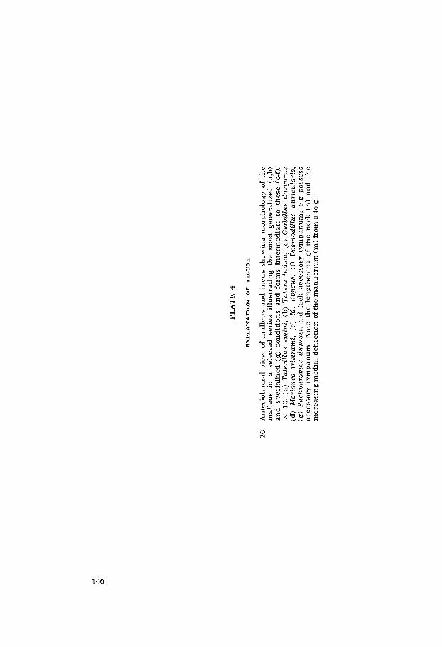
PL
AT
E 4
EX
PL
AN
AT
ION
O
F
FIG
UR
E
26
Ant
erio
late
ral
view
of
mal
leu
s an
d i
ncu
s sh
owin
g m
orph
olog
y of
th
e m
alle
us
in
a se
lect
ed
seri
es
illu
stra
ting
th
e m
ost
gene
rali
zed
(a,b
) an
d s
peci
aliz
ed
(g)
cond
itio
ns a
nd
for
ms
inte
rmed
iate
to
thes
e (c
-0.
X
10.
(a)
Tat
eril
lus
emin
i, (
b) T
ater
a in
dica
, (c
) G
erbi
Zlzr
s cl
asyu
nis
(d)
Mer
ione
s tr
istr
ami,
(e
) M
. Z
ibyc
us,
(0 D
esm
odil
lus
auri
cula
ris,
(g
) P
achy
wom
ys c
lupr
asi.
a-d
lac
k ac
cess
ory
tym
pan
um
; e-
g po
sses
s ac
cess
ory
tym
panu
m.
Not
e th
e le
ngth
enin
g of
th
e ne
ck (
n)
and
the
in
crea
sing
med
ial
defl
ecti
on o
f th
e m
anu
bri
um
(m
) fr
om a
to
g.

HE
AR
ING
OR
GA
NS
OF
GE
RB
ILL
INE
RO
DE
NT
S
Dou
glas
M.
Lay
P
LA
TE
4

PLATE 5
E X P L A N A T I O N O F FIGURES
Morphology of the middle ear ossicles of Pachyuromys cl?~lprasi. X 10.
27 Anterolateral view of malleus and incus. Malleus: (BR) processus brevis of manubrium; (L) lamina; (M) caput mallei; (MAN) manu- brium; (MU) processus muscularis; (N) neck. Incus: (B) processus brevis; (I) caput inci; (L) processus longus; (LE) processus lenticularis.
Stapes: (AC) anterior crus; (F) footplate; (H) head; (PC) posterior crus.
Morphology of the mastoid air chambers of Sekeetamys calurus. The outer walls of the mastoid have been removed; the ventral tympanic bulla remains intact. White bar in lower right corner, 1 mm.
29 Postero-dorsal view. (A) anterior semicircular canal; (AM) anterior mastoid chamber, (B) bony lamina within lateral extent of arc of lateral cansl, (EAM) external auditory meatus, (i) head of incus, (IL) posterior inferior lateral chamber, (IM) posterior inferior medial chamber, (L) lateral semicircular canal, (m) head of malleus, (P) pos- terior semicircular canal, (SL) posterior superior lateral chamber.
28
30 Antero-medial view. (AMI) accessory inferior medial chamber, (SA) subarcuate fossa, (SM) posterior superior medial chamber. Other abbreviations as in figure 29.
31 Posterior view. Arrowheads mark course of the posterior auricular nerve between laminae of the mastoid and tympanic bones.
102

HEARING ORGANS OF GERBILLINE RODENTS Douglas M. Lay
PLATE 5
103

PLATE 6
EXPLANATION OF FIGURES
The two primary components o f the auditory bulla in a specimen of Meriones unguiculatus.
32
33
Lateral view, (A) petromastoid, (B) tympanic.
Medial view. Abbreviations as in figure 32.
104

HEARING ORGANS OF GERBILLINE RODENTS Douglas M. Lay
PLATE 6
105

PLATE 7
EXPLANATION OF FIGURES
Internal mastoid morphology of Rattzrs rat tus . White bar in lower right corner, 1 mm.
34 Lateral view of bulla in which parts of lateral outer wall have been removed to display internal morphology of mastoid. Neither mastoid chamber opens into the tympanic chamber. Note that the large subar- cuate fossa fills virtually all of the posterior mastoid. (AM) anterior mastoid chamber, (EAM) external auditory meatus, (PM) posterior mastoid chamber.
35 Postero-lateral view. All of the lateral wall and spongy bone have been removed. (AC) anterior canal, (LC) lateral canal, (PC) posterior canal, (SA) subarcuate fossa.
Internal morphology of the posterior mastoid of Gerbillurus paeba. White bar, 1 mm.
36
37
38
Posterior view of mastoid with outer layer removed. An incipient posterior inferior chamber (PI) is present below the lateral canal but unconnected to the posterior superior chamber (PS). (SA) subarcuate fossa, (PC) posterior canal.
Postero-lateral view of same specimen. Note the medial extent of the posterior inferior chamber (PI). Anteriorly, the posterior superior chamber opens into the anterior chamber over the head of the incus (i). No septae separate these chambers.
Postero-superior view of anterior and posterior mastoid chambers of Desmodillus auricularis. Note the partial septum separating the two chambers. The superior part (S) is continuous with the anterior canal (A) and the inferior portion (I) lies upon the crista facialis (CF). (AM) anterior mastoid chamber, (L) lateral canal.
106

HEARING ORGANS OF GERBILLINE RODENTS Douglas M. Lay
PLATE 7
107

PLATE 8
EXPLANATION OF FIGURES
Fenestra between tympanic and common inferior chamber in Meriones crassus. The opening (horizontal white line) is of equal dimensions in populations from Iran and Egypt. Bulla volume of the Egyptian sample averages 2.1 X that of the Iranian sample. White bar, 1 mm.
39 Posterior view of Iranian specimen with outer wall removed. (PAN) posterior auricular nerve, (RW) round window of cochlea, black ar- rowheads denote mastoid component overlying tympanic component (white arrowheads) at tympanic-mastoid juncture.
Posterior view of Egyptian specimen with outer wall removed. Com- pare the width of the tympanic-mastoid juncture shelf (black line) with that typical of the Iranian specimens. This larger structure probably serves as a strengthening beam for the greatly enlarged bulla. (CI) common inferior posterior chamber, (TA) posterior arm of tympanic annulus.
40
Examples of generalized species with large and specialized species with small subarcuate fossae. White bar, 1 mm.
41 Antero-medial view of petrous temporal bone of Tatera indicu. (IAM) internal auditory meatus, (SA) subarcuate fossa, (TC) tentorium cere- belli, black arrowheads mark anterior canal.
Antero-medial view of petrous temporal bone of Rhombomys opimus. Compare with figure 41.
42
108

HEARING ORGANS OF GERBILLINE RODENTS Douglas M. Lay
PLATE 8
109

PL
AT
E 9
EX
PL
AN
AT
ION
O
F
FIG
UR
ES
Pne
umat
izat
ion
of t
he t
ympa
nic
ante
rior
to t
he e
xter
nal
audi
tory
mea
tus.
W
hite
bar
, 1
mm
.
43
44
45
Pac
hyur
omys
du
pras
i - g
reat
er
infl
atio
n do
rsal
ly
(D)
than
ve
n-
Rho
mbo
mys
op
imu
s - n
o in
flat
ion.
Des
mod
illu
s au
ricu
lari
s - li
ttle
infl
atio
n.
tral
ly (V
).
Mer
ione
s sa
cram
enti
- g
reat
er in
flat
ion
vent
rall
y (V
) th
an d
orsa
lly
(D).
4
6

HE
AR
ING
OR
GA
NS
OF
GE
RB
ILL
INE
RO
DE
NT
S
Dou
glas
M.
Lay
P
LA
TE
9

PLATE 10
EXPLANATION OF FIGURES
The brace of the tympanic annulus. White bar, 1 mm.
47 Lateral view of tympanic annulus of Meriones crassus as seen with lateral wall of tympanic cut away. (A) anterior arm of modified T brace, ( B ) base of modified T brace, (F) V-shaped floor of external auditory meatus, (P) posterior arm of modified T brace, (X) lamina separating tympanic and posterior mastoid chambers, arrowheads mark the tympanic annulus.
48 Medial view of annulus of M . shawi in bisected bulla. Abbreviations as in figure 47.
Second half-turn of organ of Corti of Pachyuromys duprasi . X 1720.
49 (C) Claudius’ cells, (D) Dieters’ cells, (H) Hensen cells, (HM) hyaline mass within pars pectinata of basilar membrane, (P) pillar cells, (R) Reisner’s membrane, (RL) reticular lamina, (SL) spiral ligament, (SM) scala media, (ST) scala tympani, (SV, black) scala vestibuli, (SV, white) stria vascularis, circles indicate outer hair cells, arrow- heads mark the basilar membrane.
112

HEARING ORGANS OF GERBILLINE RODENTS Douglas M. Lay
PLATE 10
113

PLATE 11
EXPLANATION OF FIGURES
Cochlear half turns one to six. X 600.
Tntera indica, hyaline mass absent. 50
51 Seheetnmys calurus, small hyaline mass present in half turns two to five.
114
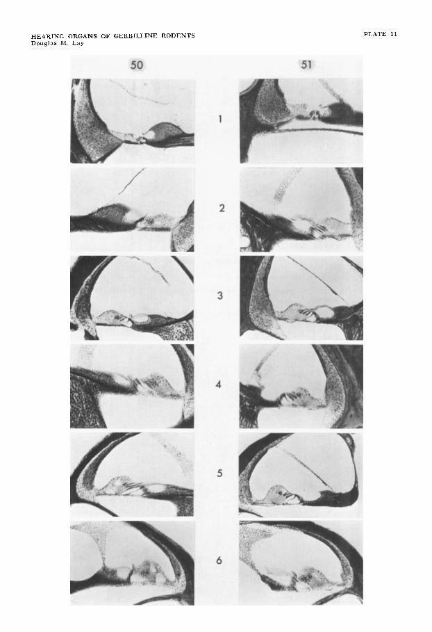
HEARING ORGANS OF GERBILLINE RODENTS Douglas M. Lay
PLATE 11

PLATE 12
EXPLANATION O F FIGURES
Cochlear half turns one to six. X 600.
52 Gerbillus yerbillus, small hyaline mass present in half turns two to four.
Meriones persicus, moderate hyaline mass present in half turns two to six.
53
116

HEARING ORGANS OF GERBILLINE RODENTS Douglas M. Lay
PLATE 12

PLATE 13
EXPLANATION O F FIGURES
54 Meriones tristrami, well developed hyaline mass present in half turns two to five. X 600.
Meriones crassus, well developed hyaline mass present in half turns two to six. X 600.
55
118

HEARING ORGANS OF GERBILLINE RODENTS Douglas M. Lay
PLATE 13

HEARING ORGANS OF GERBILLINE RODENTS Douglas M. Lay
PLATE 14
E X P L A N A T I O N OF FIGURE
56 Puchynromys dz ipras i , well developed hyaline ma55 present in half turns two to six. X 600.