term 1 lecture 6 heart and great vessels moodle version (1)
DESCRIPTION
inima si vasele de sangeTRANSCRIPT

B2050/2051: Human Anatomy and
Embryology
Lecture 6: Anatomy of the adult heart and
great vessels
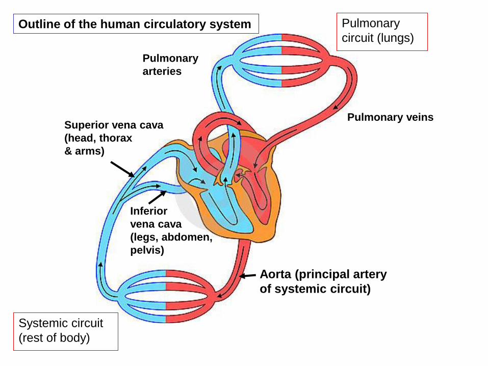
Pulmonary
circuit (lungs)
Systemic circuit
(rest of body)
Aorta (principal artery
of systemic circuit)
Superior vena cava
(head, thorax
& arms)
Inferior
vena cava
(legs, abdomen,
pelvis)
Pulmonary veins
Pulmonary
arteries
Outline of the human circulatory system

The heart lies within the chest cavity, to the left of the midline, protected by the
ribs and sternum
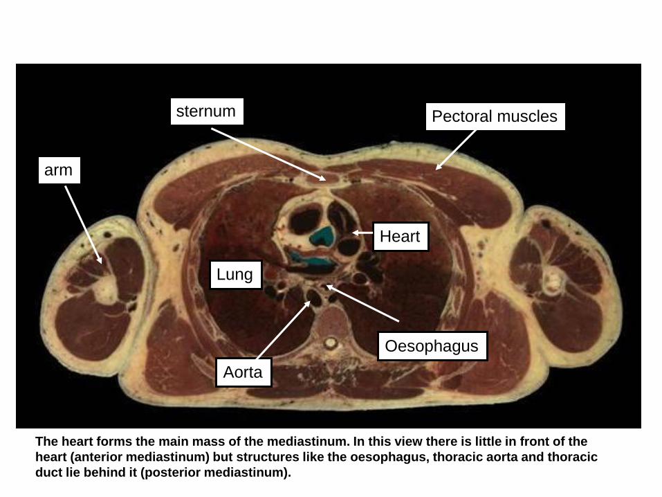
Pectoral muscles sternum
arm
Aorta
Oesophagus
Heart
Lung
The heart forms the main mass of the mediastinum. In this view there is little in front of the
heart (anterior mediastinum) but structures like the oesophagus, thoracic aorta and thoracic
duct lie behind it (posterior mediastinum).
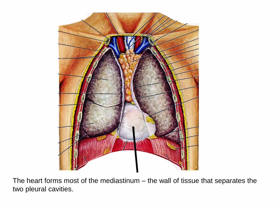
The heart forms most of the mediastinum – the wall of tissue that separates the
two pleural cavities.

Diagrammatic sagittal section of thorax
Heart
Superior mediastinum
(great vessels – aorta,
vena cava, trachea,
oesophagus)
Inferior mediastium
(heart)
+ thoracic aorta, oesophagus,
azygous vein, thoracic duct
Anterior Posterior
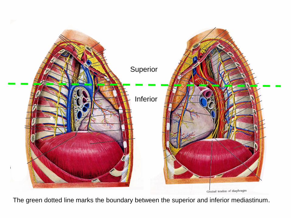
The green dotted line marks the boundary between the superior and inferior mediastinum.
Superior
Inferior

The heart is enclosed in a fibrous bag
- the fibrous pericardium – that is
fused inferiorly to the central
tendon of the diaphragm
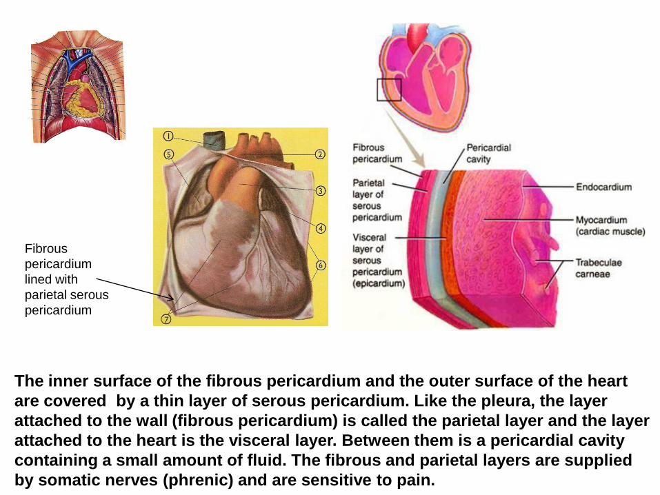
The inner surface of the fibrous pericardium and the outer surface of the heart
are covered by a thin layer of serous pericardium. Like the pleura, the layer
attached to the wall (fibrous pericardium) is called the parietal layer and the layer
attached to the heart is the visceral layer. Between them is a pericardial cavity
containing a small amount of fluid. The fibrous and parietal layers are supplied
by somatic nerves (phrenic) and are sensitive to pain.
Fibrous
pericardium
lined with
parietal serous
pericardium
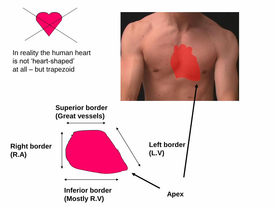
Apex
Right border
(R.A)
Inferior border
(Mostly R.V)
Left border
(L.V)
Superior border
(Great vessels)
In reality the human heart
is not ‘heart-shaped’
at all – but trapezoid

Apex
Right border
Inferior border
Left border
Superior
border

Normal chest Xray
Enlargement of RA
The four border of the heart can be seen on a normal chest X-ray.
Compare it with the X-ray on the right and you can see there how the right border
is more expanded – showing enlargement of the right atrium

Fibrous skeleton of the heart
Although most of the heart is cardiac
muscle, there is a fibrous skeleton,
roughly at the junction of the atria and
ventricles, that supports the valves

Right atrium Blood from:
Superior &
inferior vena
cava, coronary
sinus
Right ventricle
- To pulmonary trunk
Left atrium Blood from:
Pulmonary
veins
Left ventricle
- To aorta
Pulmonary arteries Aorta
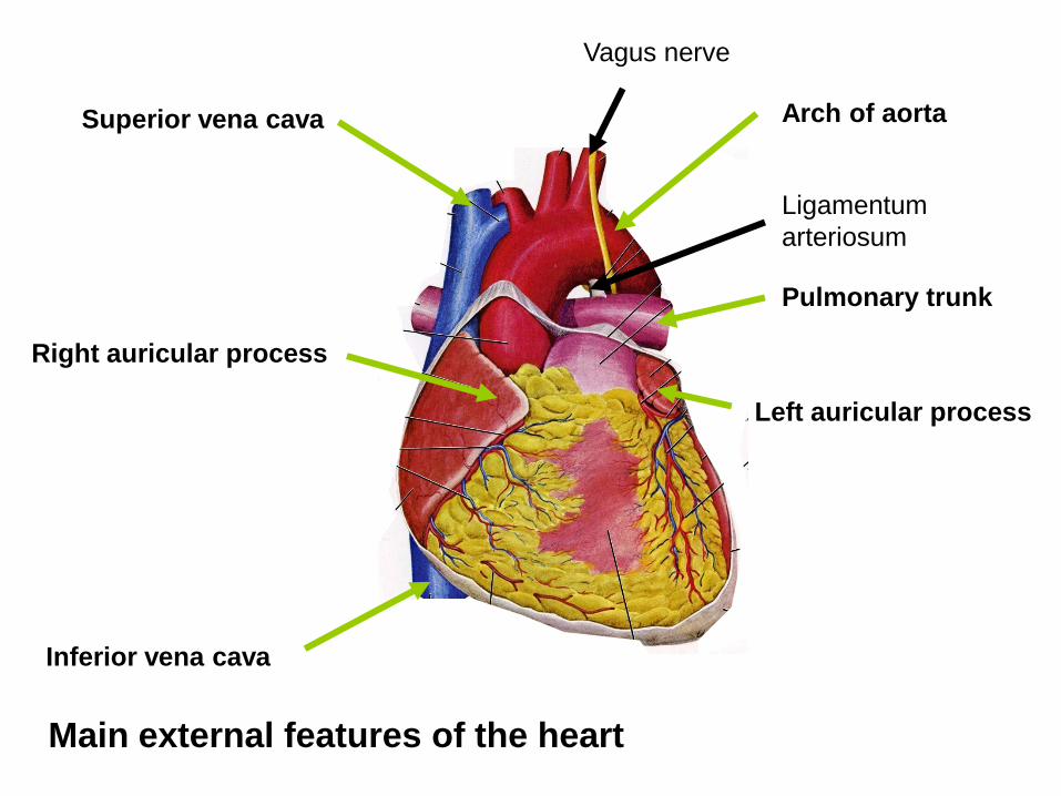
Superior vena cava
Inferior vena cava
Right auricular process
Arch of aorta
Pulmonary trunk
Left auricular process
Main external features of the heart
Ligamentum
arteriosum
Vagus nerve

Pectinate muscle
Crista terminalis (= boundary between
smooth and
rough walled parts
of the atrium)
Fossa ovalis Remnant of foramen
ovale in embryo)
Auricular
process
Opening of coronary sinus
Right atrium
SVC
Aorta
IVC
The atria are formed from two different regions in the embryo – the ‘true’ atrium and the
sinus venosus, a sac into which all the veins empty. The sinus becomes incorporated
into the atria, as do parts of the pulmonary vein on the left. The sinus/veins form the
smooth part of the atrial wall; the rough part is formed by the true atria.

Right atrium
Fossa ovalis
Ligamentum
arteriosum
These two structures are remnants of the foetal circulation:
a) The fossa ovalis is a remnant of the foramen ovale between the right and left
atria. In the foetus, this carries oxygenated blood from the placenta (coming via
the left umbilical vein and IVC) across to the left side for circulation to the growing
body and, especially, the brain.
b) The ligamentum arteriosum is a remnant of the ductus arteriosus which carries
blood from the pulmonary trunk into the aorta to bypass the lungs

Papillary muscles
Moderator band (Enlarged trabecula that carries
a branch of the conducting
system directly to the base of
the papillary muscles)
Pulmonary semi-lunar
valve
Chordae tendinae
Cusps of tricuspid
valve (3 cusps)
Right ventricle
(run from the edges of the
cusps to the papillary muscles)
(Contract before the ventricular wall
to ensure the valve does not flip ‘inside out’ and
let blood back into the atrium)
Vagus nerve

Atrioventricular valve in a fresh heart
Trabeculae carnae
Chordae tendinae
Papillary muscle
Valve cusplet

Pulmonary semi-lunar valve Aortic semi-lunar valve
Bicuspid (mitral) valve Tricuspid valve
View of heart from above with major vessels and atria removed –
showing the valves. Anterior is at the top.
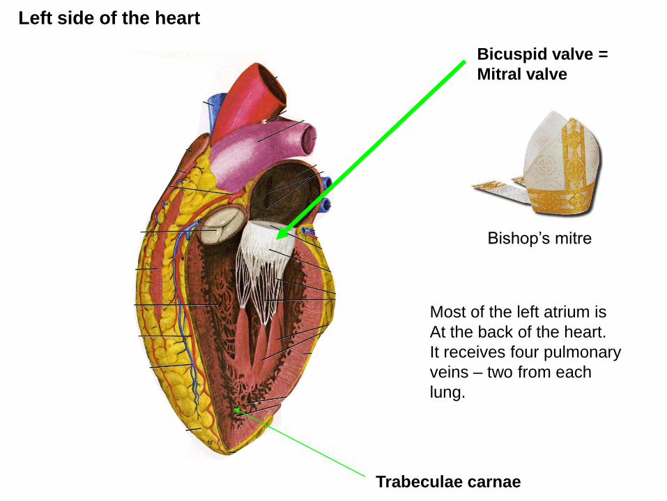
Left side of the heart
Trabeculae carnae
Bicuspid valve =
Mitral valve
Most of the left atrium is
At the back of the heart.
It receives four pulmonary
veins – two from each
lung.
Bishop’s mitre
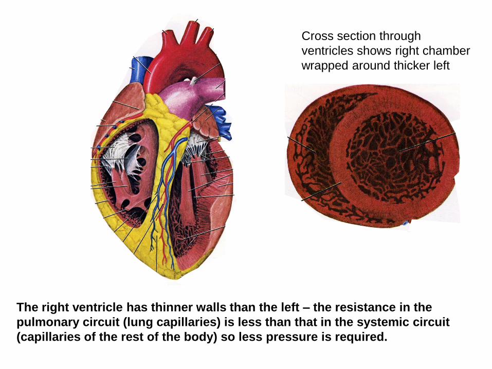
The right ventricle has thinner walls than the left – the resistance in the
pulmonary circuit (lung capillaries) is less than that in the systemic circuit
(capillaries of the rest of the body) so less pressure is required.
Cross section through
ventricles shows right chamber
wrapped around thicker left
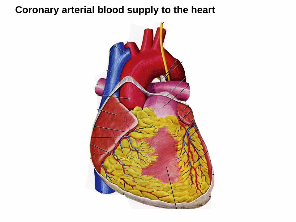
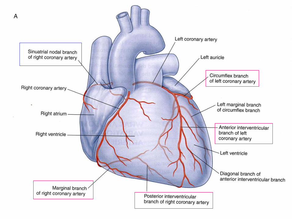
Coronary arterial blood supply to the heart

The coronary arteries branch from aorta immediately above the semi-lunar valves
and form a ‘crown’ (=coronet) around the top of the heart

Great cardiac vein
Middle cardiac vein
Anterior cardiac veins
Coronary sinus
Cardiac veins The veins of the heart do not form a ‘crown’ like the arteries and are therefore called cardiac
(not coronary) veins.
Pulmonary veins
(2 left, 2 right)

Pain travels with
sympathetic nerves:
Mainly T1,T2
Parasympathetic
(vagus)
The cardiac muscle has its own intrinsic beat but the speed and strength
of the beat is moderated by autonomic nerves

The beat is initiated at the
sino-atrial (SA) node which lies at
the junction of the superior vena
cava and the sinus part of the
heart.
The wave of contraction crosses
the atria and stimulates the atrio-
ventricular (AV node) which lies
close to the opening of the
coronary sinus in the wall of the
right atrium.
It then crosses the atrioventricular
junction in the AV bundle, which
then divides into left and right
branches that travel in the
interventricular septum to the
base of the heart.
One large branch from the right
(the septomarginal trabecula or
moderator band) passes to the
base of the right side papillary
muscles to ensure they contract
in time to tense the tricuspid valve

Placement of an artificial pacemaker where parts
of the conducting system or nodes have failed Conducting system
The major blood vessels of the thorax

The heart and great vessels
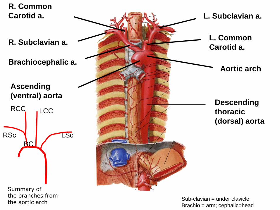
Descending
thoracic
(dorsal) aorta
Ascending
(ventral) aorta
Aortic arch Brachiocephalic a.
R. Subclavian a.
R. Common
Carotid a.
L. Common
Carotid a.
L. Subclavian a.
Summary of the branches from the aortic arch
RSc
RCC LCC
LSc
BC
Sub-clavian = under clavicle
Brachio = arm; cephalic=head

Vertebral a.
Intercostal a.s
(=thoracic
segmental a.)
Vertebral artery
Vertebral a. Branch of subclavian
artery.
Runs up within cervical
vertebrae, enters
skull, and joins with
opposite vertebral
artery to help supply the
brain .
In the skull – form
anastomoses with
internal carotid branches

Superior vena cava (SVC) and its tributaries (head, thorax, arms) Inferior vena cava (IVC) and its tributaries (abdomen, pelvis, legs)
Venous system
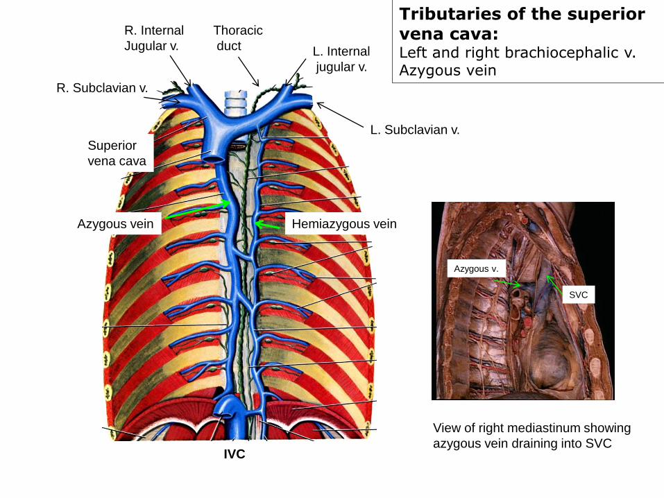
Tributaries of the superior vena cava: Left and right brachiocephalic v. Azygous vein
Superior
vena cava
R. Internal
Jugular v.
R. Subclavian v.
L. Subclavian v.
L. Internal
jugular v.
Hemiazygous vein Azygous vein
Thoracic
duct
IVC
View of right mediastinum showing
azygous vein draining into SVC
Azygous v.
SVC

Superior vena cava drainage
SVC Azygous v.
L. Brachiocephalic v. R.Brachiocephalic v.
Internal jugular v.
External jugular v.
External jugular vein
By the end of this lecture, you should be able to answer MCQ’s and
write short answers on:
a) The internal anatomy of the heart
b) The external anatomy of the heart and the coronary blood vessels
c) The conducting system of the heart
d) The position of the heart in the chest
e) The mediastinum
f) The arrangement of the major arteries and veins of the thorax
For those in the Dissecting Room Group this afternoon (L-Z):
You MUST bring a valid student photocard with you – you will not be
admitted without it.
Mobile phones may not be carried in the Dissecting Room –
even switched off. Put them into your bag and keep them there until
you have left the room.
Bags may be left at the back of the Dissecting Room with coats – don’t
leave them in the hallway outside