heart/neck vessels & peripheral vascular/lymphatics
TRANSCRIPT
Heart/Neck Vessels &Peripheral Vascular/Lymphatics
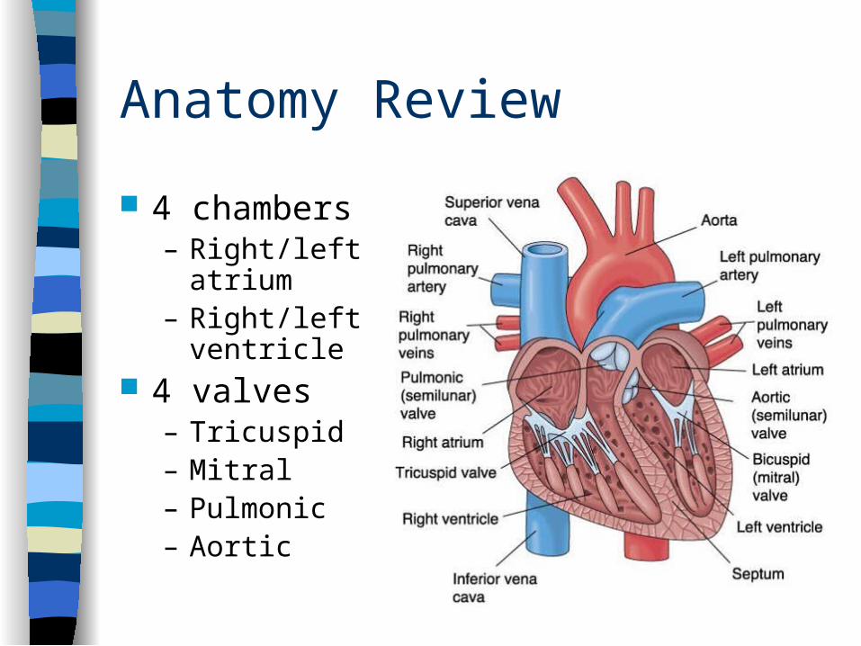
Anatomy Review
4 chambers– Right/left
atrium– Right/left
ventricle 4 valves
– Tricuspid– Mitral– Pulmonic– Aortic
Anatomy and Physiology
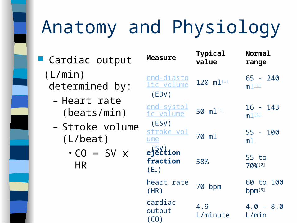
Cardiac output
(L/min) determined by:– Heart rate
(beats/min)– Stroke volume
(L/beat)• CO = SV x HR
Measure Typical value
Normal range
end-diastolic volume (EDV)
120 ml[1] 65 - 240 ml[1]
end-systolic volume (ESV)
50 ml[1] 16 - 143 ml[1]
stroke volume (SV)
70 ml 55 - 100 ml
ejection fraction (Ef)
58% 55 to 70%[2]
heart rate (HR)
70 bpm60 to 100 bpm[3]
cardiac output (CO)
4.9 L/minute4.0 - 8.0 L/min
Health History Chest pain
– Do you have any chest pain or discomfort?• OLDCART
– Do you do you use any recreational drugs?– Do you have any increased life stress/anxiety?
Dyspnea– Do you have any labored or difficulty breathing
(dyspnea)?• OLDCART• Related to exercise (exertional dyspnea)?
– Quantify: Have far can you walk before getting short of breath?
• Related to position/lying supine (orthopnea)? – How many pillows do you sleep on at night?
Health History
Palpitations– Ever have palpitations/or unpleasant awareness of
heartbeat? (“fluttering/ pounding”) Dizziness or Syncope
– Have you felt dizzy or ever lost consciousness/passed out (syncope)?
Fatigue– Do you seem to tire easily?
Cyanosis or pallor– Ever noted your facial skin turn blue or ashen
gray?
Health History
Cough– Any pink or blood tinged frothy sputum?
Edema– Do you have any swelling in your feet or legs?
Nocturia– Do you awaken at night with an urgent need to
urinate?
Health History
Past Cardiac History– CHF, angina, MI, murmurs, rheumatic fever,
congenital heart disease Assess for risk factors of coronary artery
disease– Hypertension, hyperlipidemia, diabetes, physical
inactivity, obesity, smoking, stress, increasing age. family history of CAD (especially in 1st degree relatives F<65, M<55)
– Additional for women: Menopause or use of oral contraceptives
What the History Can Tell You
Angina (pain resulting from ischemia)– Onset: Abrupt, often precipitated by event such as
emotion, exertion, cold or eating.
– Location: Substernal or retrosternal pain.
– Duration: Usually lasts a few minutes and then subsides.
– Characteristic: Described as squeezing or heavy pressure
– Radiation: May radiate to the neck, jaw, or arms
– Relieving Factors/Treatments Tried: Often relieved with sublingual nitroglycerin
What the History Can Tell You Myocardial Infarction
– Onset: Abrupt, often unrelated to precipitating event.
– Location: Substernal or over precordium.
– Duration: Prolonged
– Characteristic: Severe, described as viselike or crushing
– Associated Symptoms: dyspnea, dizziness, nausea, diaphoresis, palpitations, anxiety (sense of doom)
– Radiation: May radiate to neck, jaw, arms or hands.
– Treatments Tried: Sublingual nitroglycerin without relief
What the History Can Tell You Congestive Heart Failure
– Right-sided• Dependent Edema• Nocturia
– Left-sided• Coughing/Hemoptysis (pink frothy)• Orthopnea • Dyspnea with exertion• Cyanosis or ashen color• Cold, moist extremities• Oliguria• Restlessness/anxiety
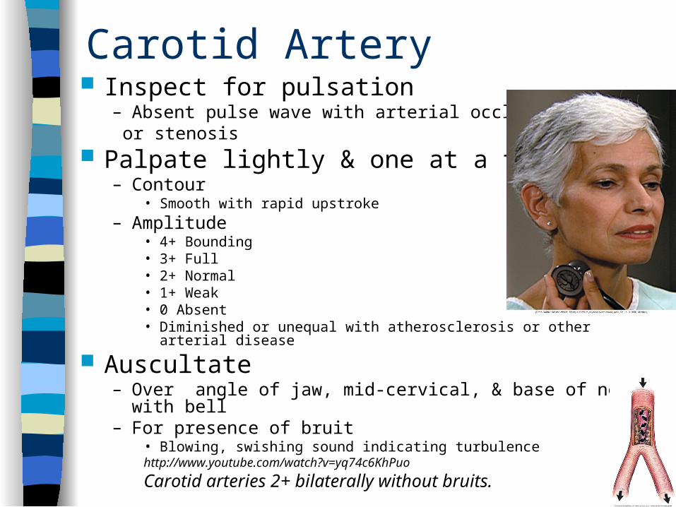
Carotid Artery Inspect for pulsation
– Absent pulse wave with arterial occlusion or stenosis
Palpate lightly & one at a time for:– Contour
• Smooth with rapid upstroke– Amplitude
• 4+ Bounding • 3+ Full• 2+ Normal• 1+ Weak• 0 Absent• Diminished or unequal with atherosclerosis or other arterial disease
Auscultate– Over angle of jaw, mid-cervical, & base of neck with bell– For presence of bruit
• Blowing, swishing sound indicating turbulencehttp://www.youtube.com/watch?v=yq74c6KhPuo
Carotid arteries 2+ bilaterally without bruits.
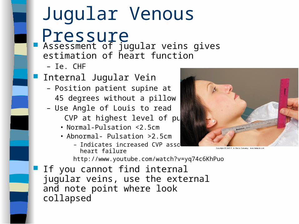
Jugular Venous Pressure Assessment of jugular veins gives
estimation of heart function– Ie. CHF
Internal Jugular Vein– Position patient supine at
45 degrees without a pillow– Use Angle of Louis to read CVP at highest level of pulsation
• Normal-Pulsation <2.5cm • Abnormal- Pulsation >2.5cm
– Indicates increased CVP associated with heart failure
http://www.youtube.com/watch?v=yq74c6KhPuo
If you cannot find internal jugular veins, use the external and note point where look collapsed
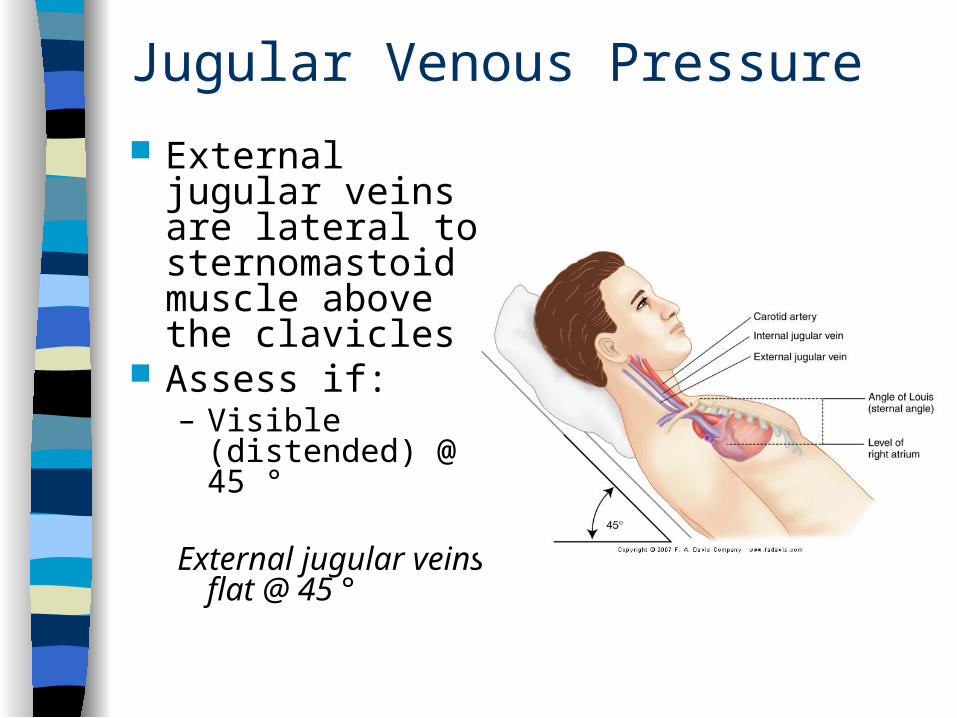
Jugular Venous Pressure
External jugular veins are lateral to sternomastoid muscle above the clavicles
Assess if:– Visible (distended)
@ 45 °
External jugular veins flat @ 45 °
Hepatojugular Reflux
Very sensitive in detecting right-sided heart failure
Elevate to 30 degrees Press firmly in right upper quadrant Observe neck for elevation in JVP
– Rise of >1cm is abnormalhttp://www.youtube.com/watch?v=X9fKPIe6nDQ
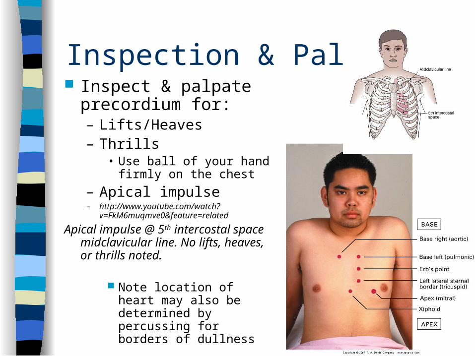
Inspection & Palpation Inspect & palpate
precordium for:– Lifts/Heaves– Thrills
• Use ball of your hand firmly on the chest
– Apical impulse– http://www.youtube.com/watch?
v=FkM6muqmve0&feature=related
Apical impulse @ 5th intercostal space midclavicular line. No lifts, heaves, or thrills noted.
Note location of heart may also be determined by percussing for borders of dullness
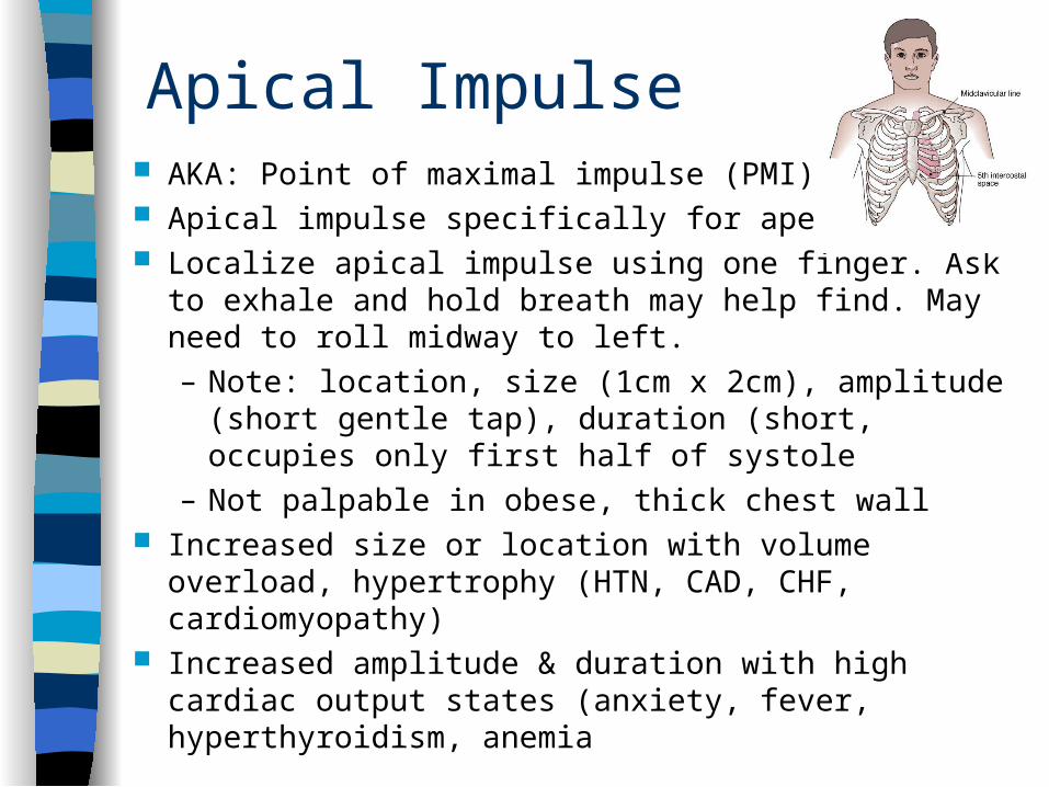
Apical Impulse AKA: Point of maximal impulse (PMI) Apical impulse specifically for apex beat. Localize apical impulse using one finger. Ask to exhale
and hold breath may help find. May need to roll midway to left.– Note: location, size (1cm x 2cm), amplitude (short
gentle tap), duration (short, occupies only first half of systole
– Not palpable in obese, thick chest wall Increased size or location with volume overload,
hypertrophy (HTN, CAD, CHF, cardiomyopathy) Increased amplitude & duration with high cardiac output
states (anxiety, fever, hyperthyroidism, anemia
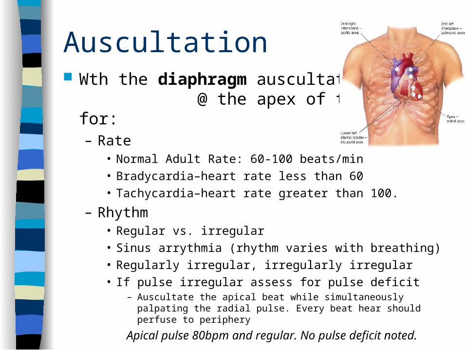
Auscultation Wth the diaphragm auscultate
@ the apex of the heart for:– Rate
• Normal Adult Rate: 60-100 beats/min• Bradycardia–heart rate less than 60• Tachycardia–heart rate greater than 100.
– Rhythm• Regular vs. irregular• Sinus arrythmia (rhythm varies with breathing)• Regularly irregular, irregularly irregular• If pulse irregular assess for pulse deficit
– Auscultate the apical beat while simultaneously palpating the radial pulse. Every beat hear should perfuse to periphery
Apical pulse 80bpm and regular. No pulse deficit noted.
Auscultation
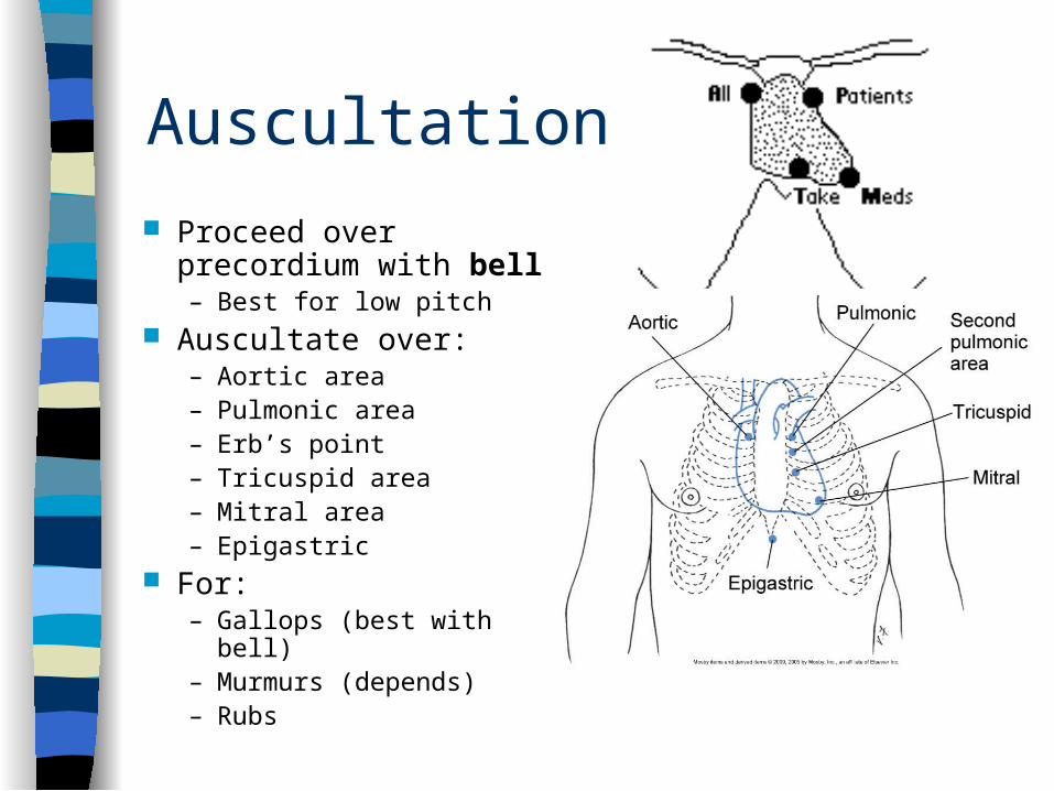
Proceed over precordium with bell– Best for low pitch
Auscultate over:– Aortic area– Pulmonic area– Erb’s point– Tricuspid area– Mitral area– Epigastric
For:– Gallops (best with bell)– Murmurs (depends)– Rubs
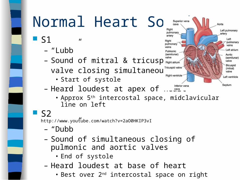
Normal Heart Sounds S1
– “Lubb”– Sound of mitral & tricuspid
valve closing simultaneously• Start of systole
– Heard loudest at apex of heart • Approx 5th intercostal space, midclavicular line on left
S2 http://www.youtube.com/watch?v=2aO0HKIP3vI
– “Dubb”– Sound of simultaneous closing of pulmonic and
aortic valves• End of systole
– Heard loudest at base of heart• Best over 2nd intercostal space on right
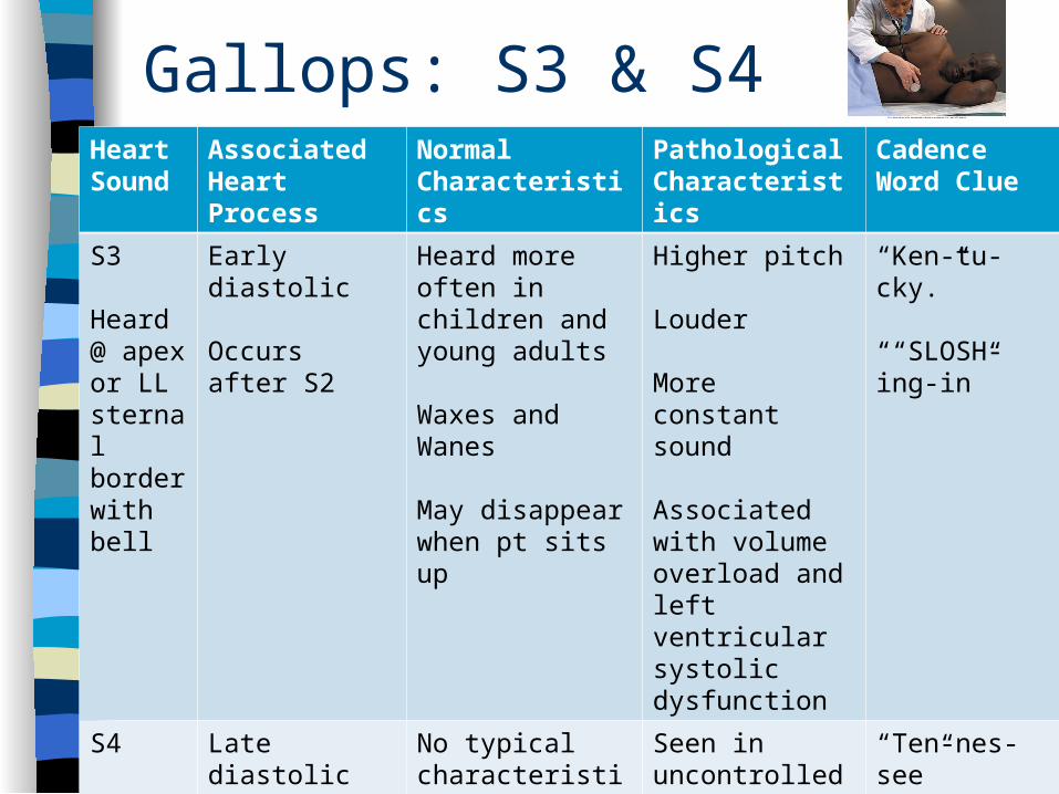
Gallops: S3 & S4Heart Sound
Associated Heart Process
Normal Characteristics
PathologicalCharacteristics
Cadence Word Clue
S3
Heard @ apex or LL sternal border with bell
Early diastolic
Occurs after S2
Heard more often in children and young adults
Waxes and Wanes
May disappear when pt sits up
Higher pitch
Louder
More constant sound
Associated with volume overload and left ventricular systolic dysfunction
“Ken-tu-cky.”
““SLOSH-ing-in”
S4
Heard @ apex with bell
Late diastolic (atrial filling)
Occurs before S1
No typical characteristics
Seen in uncontrolled hypertension
“Ten-nes-see”
“a-STIFF-wall”
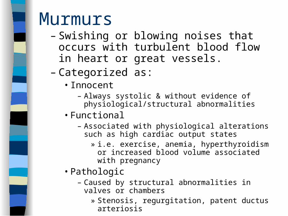
Murmurs– Swishing or blowing noises that occurs
with turbulent blood flow in heart or great vessels.
– Categorized as:• Innocent
– Always systolic & without evidence of physiological/structural abnormalities
• Functional– Associated with physiological alterations such as
high cardiac output states » i.e. exercise, anemia, hyperthyroidism or
increased blood volume associated with pregnancy
• Pathologic– Caused by structural abnormalities in valves or
chambers» Stenosis, regurgitation, patent ductus arteriosis
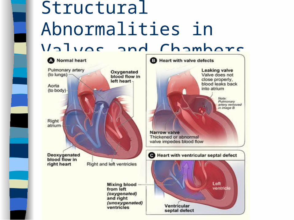
Structural Abnormalities in Valves and Chambers
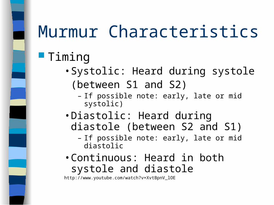
Murmur Characteristics Timing
• Systolic: Heard during systole (between S1 and S2)
– If possible note: early, late or mid systolic)
• Diastolic: Heard during diastole (between S2 and S1)
– If possible note: early, late or mid diastolic
• Continuous: Heard in both systole and diastole
http://www.youtube.com/watch?v=XvtBpnV_lOE
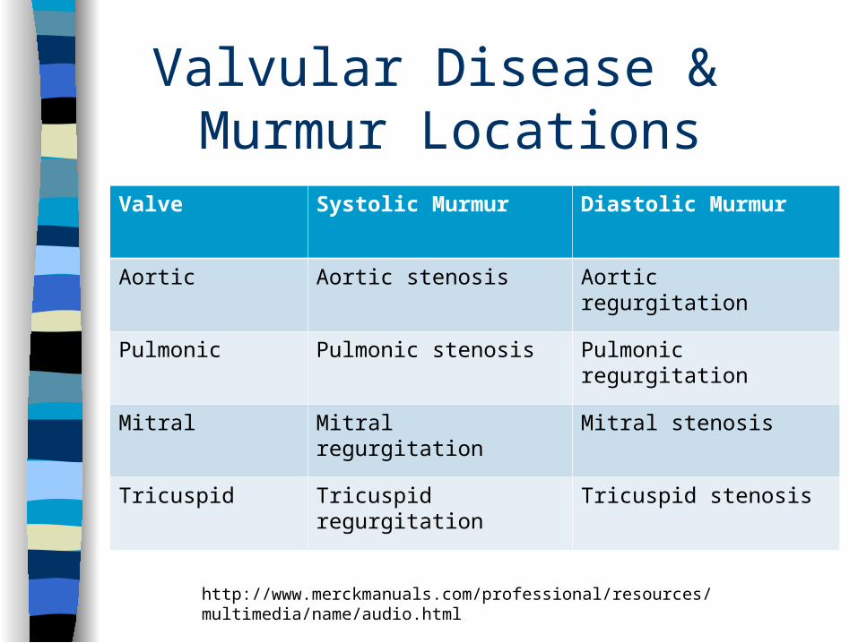
Valvular Disease & Murmur Locations
Valve Systolic Murmur Diastolic Murmur
Aortic Aortic stenosis Aortic regurgitation
Pulmonic Pulmonic stenosis Pulmonic regurgitation
Mitral Mitral regurgitation Mitral stenosis
Tricuspid Tricuspid regurgitation Tricuspid stenosis
http://www.merckmanuals.com/professional/resources/multimedia/name/audio.html
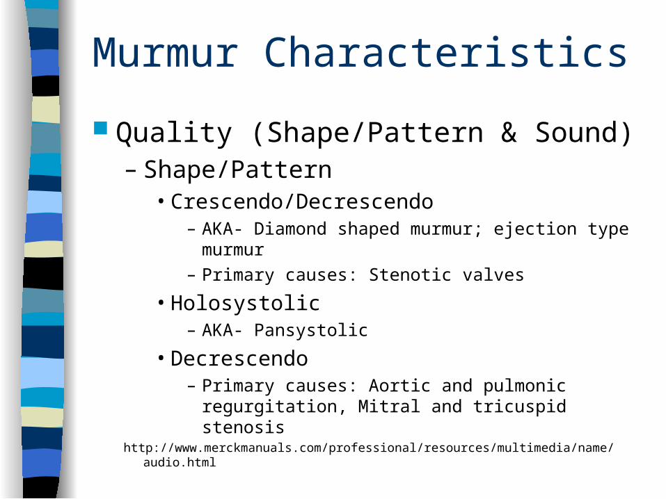
Murmur Characteristics Quality (Shape/Pattern & Sound)
– Shape/Pattern• Crescendo/Decrescendo
– AKA- Diamond shaped murmur; ejection type murmur
– Primary causes: Stenotic valves
• Holosystolic– AKA- Pansystolic
• Decrescendo– Primary causes: Aortic and pulmonic regurgitation,
Mitral and tricuspid stenosishttp://www.merckmanuals.com/professional/resources/multimedia/name/audio.html
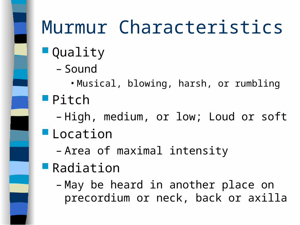
Murmur Characteristics Quality
– Sound• Musical, blowing, harsh, or rumbling
Pitch– High, medium, or low; Loud or soft
Location– Area of maximal intensity
Radiation– May be heard in another place on
precordium or neck, back or axilla
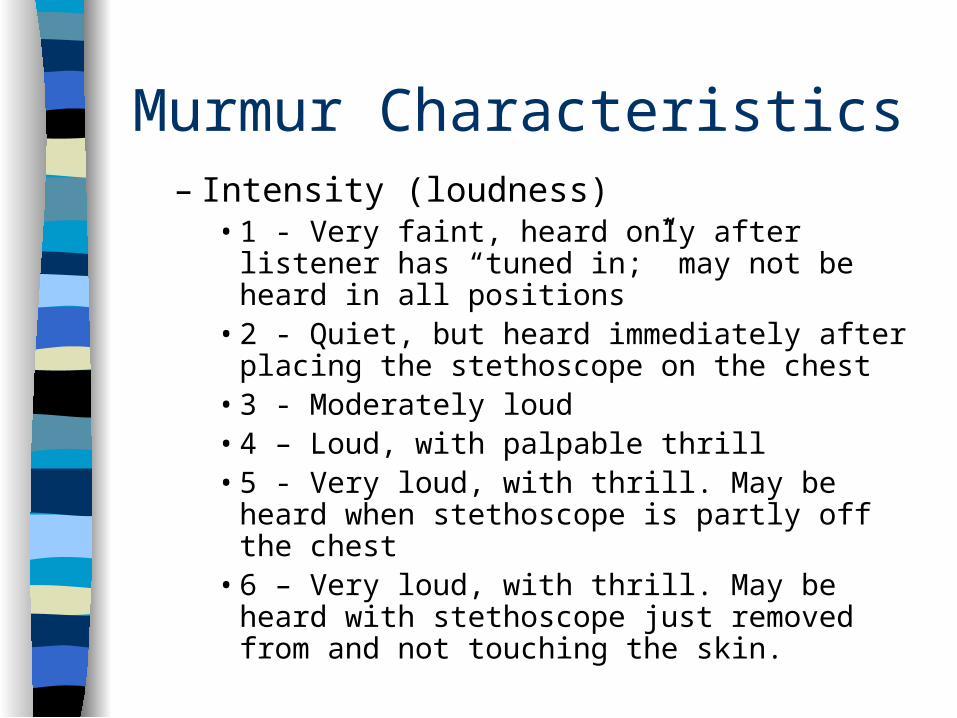
Murmur Characteristics– Intensity (loudness)
• 1 - Very faint, heard only after listener has “tuned in;” may not be heard in all positions
• 2 - Quiet, but heard immediately after placing the stethoscope on the chest
• 3 - Moderately loud• 4 – Loud, with palpable thrill• 5 - Very loud, with thrill. May be heard when
stethoscope is partly off the chest• 6 – Very loud, with thrill. May be heard with
stethoscope just removed from and not touching the skin.
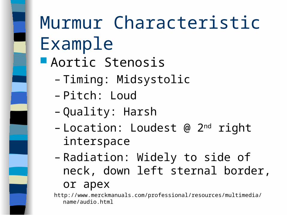
Murmur Characteristic Example
Aortic Stenosis– Timing: Midsystolic– Pitch: Loud– Quality: Harsh– Location: Loudest @ 2nd right interspace– Radiation: Widely to side of neck, down left
sternal border, or apexhttp://www.merckmanuals.com/professional/resources/multimedia/name/audio.html
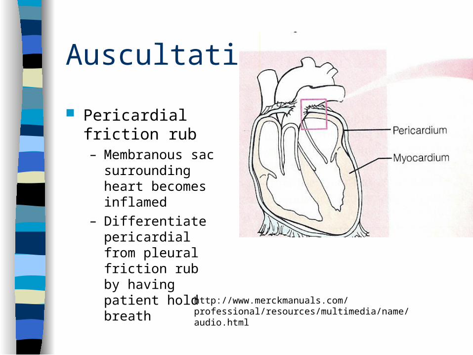
Auscultation
Pericardial friction rub– Membranous sac
surrounding heart becomes inflamed
– Differentiate pericardial from pleural friction rub by having patient hold breath
http://www.merckmanuals.com/professional/resources/multimedia/name/audio.html
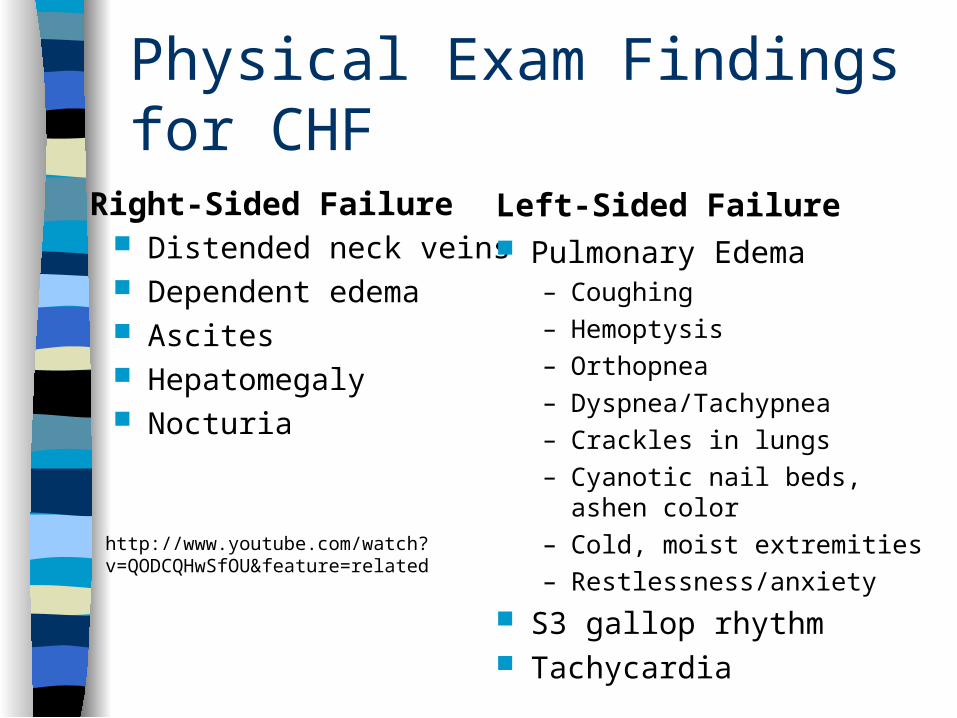
Physical Exam Findings for CHF
Right-Sided Failure Distended neck veins Dependent edema Ascites Hepatomegaly Nocturia
Left-Sided Failure Pulmonary Edema
– Coughing– Hemoptysis– Orthopnea– Dyspnea/Tachypnea– Crackles in lungs– Cyanotic nail beds, ashen
color– Cold, moist extremities– Restlessness/anxiety
S3 gallop rhythm Tachycardia
http://www.youtube.com/watch?v=QODCQHwSfOU&feature=related

Peripheral Vascular & Lymphatics
http://images.google.com
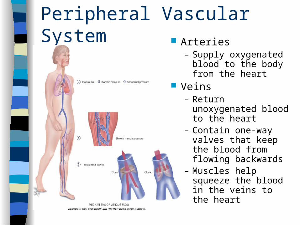
Peripheral Vascular System Arteries
– Supply oxygenated blood to the body from the heart
Veins– Return
unoxygenated blood to the heart
– Contain one-way valves that keep the blood from flowing backwards
– Muscles help squeeze the blood in the veins to the heart
Health History
Common or concerning symptoms– Pain in the arms or legs– Intermittent claudication: leg or arm pain that is exercise
induced– Cold, numbness, pallor in the legs; hair loss– Color change in fingertips or toes in cold weather– Swelling in calves, legs or feet– Swelling with redness or tenderness– High risk: Tobacco use, diabetes, HTN, Hyperlipidemia,
CV disease– Severity of peripheral vascular disease closely parallels
the risk for heart attack, stoke, and death from vascular causes
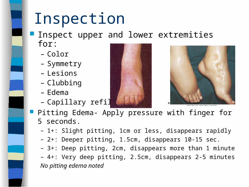
Inspection Inspect upper and lower extremities for:
– Color– Symmetry– Lesions– Clubbing– Edema– Capillary refill
Pitting Edema- Apply pressure with finger for 5 seconds.– 1+: Slight pitting, 1cm or less, disappears rapidly– 2+: Deeper pitting, 1.5cm, disappears 10-15 sec.– 3+: Deep pitting, 2cm, disappears more than 1 minute– 4+: Very deep pitting, 2.5cm, disappears 2-5 minutes
No pitting edema noted
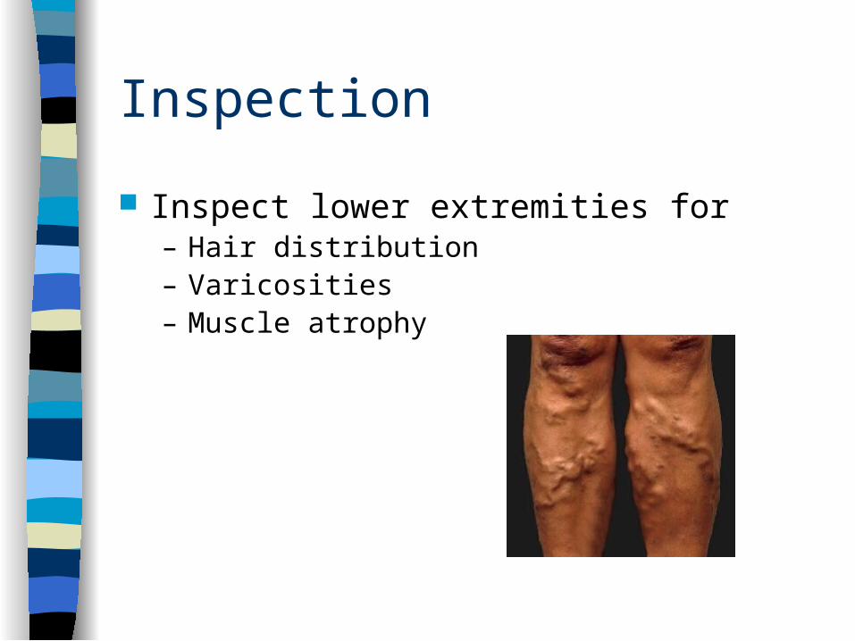
Inspection
Inspect lower extremities for– Hair distribution– Varicosities– Muscle atrophy
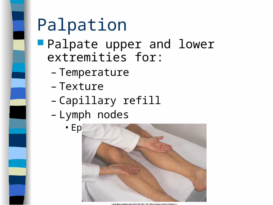
Palpation Palpate upper and lower extremities for:
– Temperature– Texture– Capillary refill– Lymph nodes
• Epitrochlear, Inguinal
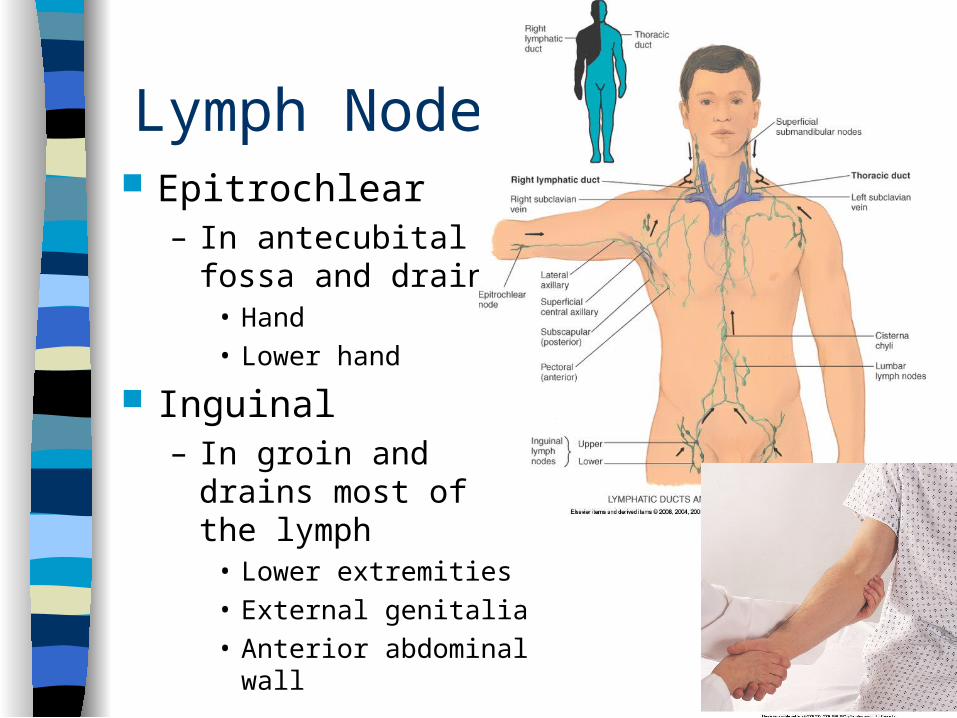
Lymph Nodes Epitrochlear
– In antecubital fossa and drains:
• Hand• Lower hand
Inguinal– In groin and drains
most of the lymph• Lower extremities• External genitalia• Anterior abdominal wall
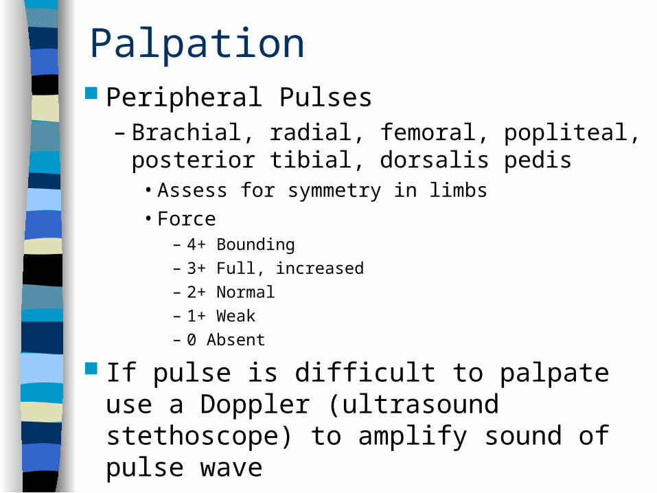
Palpation Peripheral Pulses
– Brachial, radial, femoral, popliteal, posterior tibial, dorsalis pedis
• Assess for symmetry in limbs• Force
– 4+ Bounding– 3+ Full, increased– 2+ Normal– 1+ Weak– 0 Absent
If pulse is difficult to palpate use a Doppler (ultrasound stethoscope) to amplify sound of pulse wave
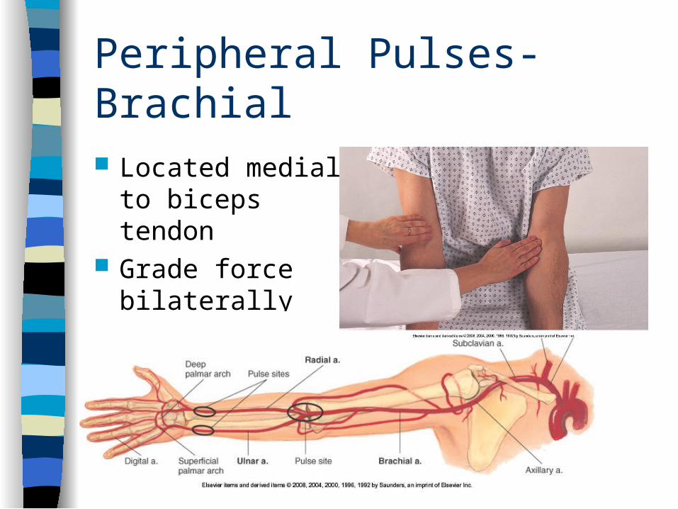
Peripheral Pulses- Brachial
Located medial to biceps tendon
Grade force bilaterally
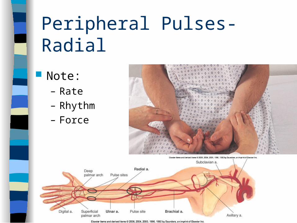
Peripheral Pulses-Radial
Note:– Rate– Rhythm– Force
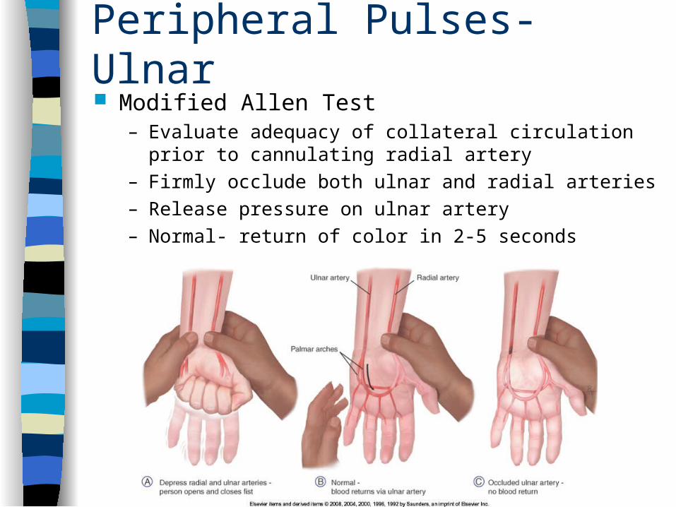
Peripheral Pulses-Ulnar Modified Allen Test
– Evaluate adequacy of collateral circulation prior to cannulating radial artery
– Firmly occlude both ulnar and radial arteries– Release pressure on ulnar artery– Normal- return of color in 2-5 seconds
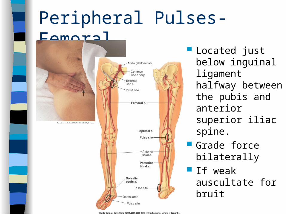
Peripheral Pulses-Femoral Located just
below inguinal ligament halfway between the pubis and anterior superior iliac spine.
Grade force bilaterally
If weak auscultate for bruit
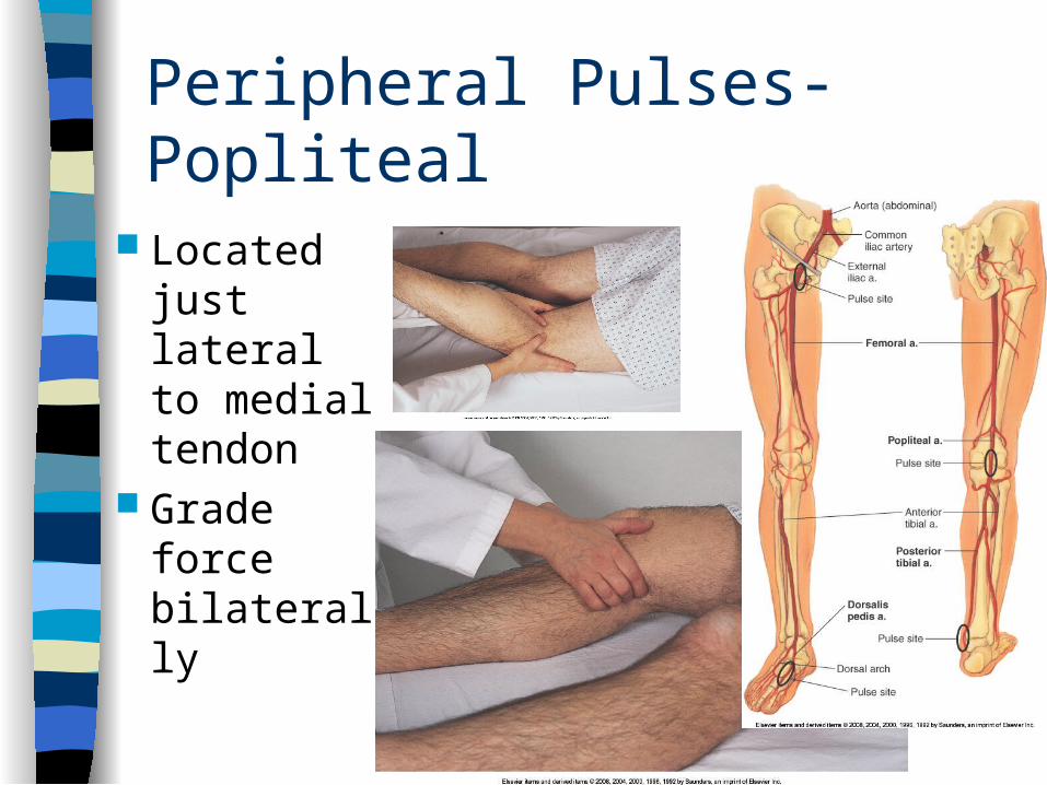
Peripheral Pulses-Popliteal
Located just lateral to medial tendon
Grade force bilaterally
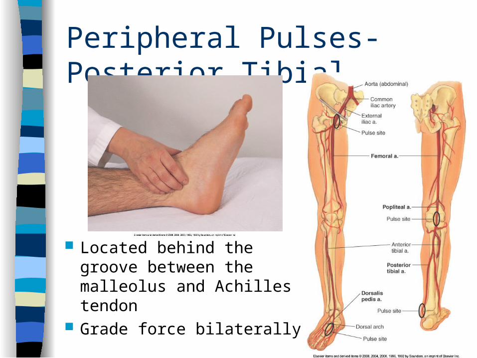
Peripheral Pulses-Posterior Tibial
Located behind the groove between the malleolus and Achilles tendon
Grade force bilaterally
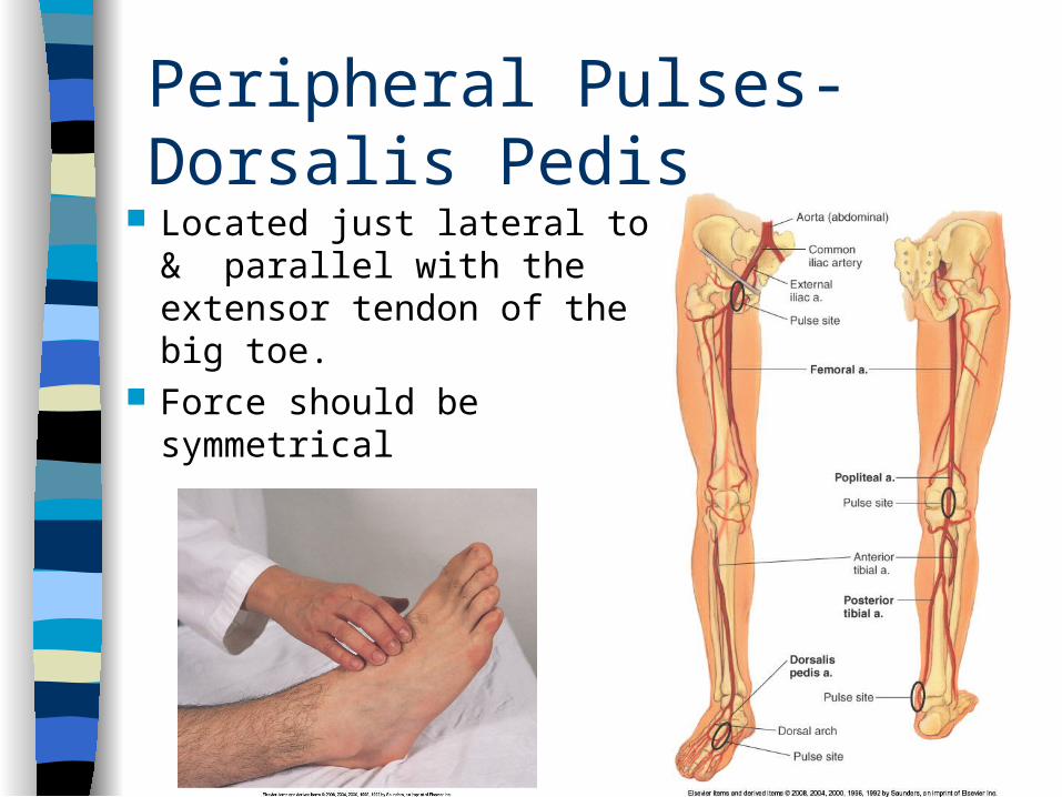
Peripheral Pulses-Dorsalis Pedis Located just lateral to &
parallel with the extensor tendon of the big toe.
Force should be symmetrical
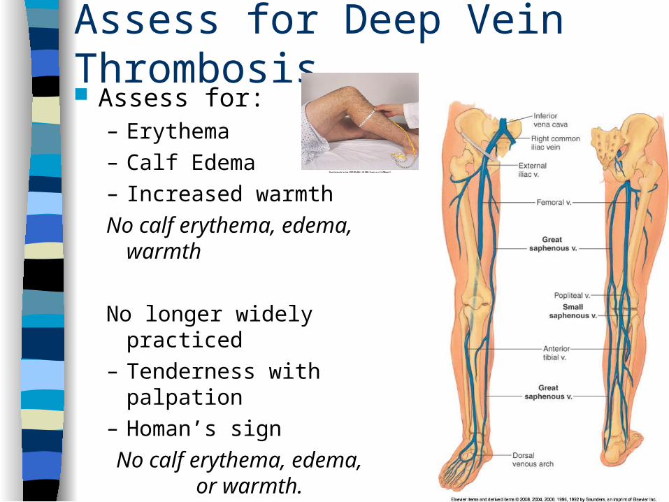
Assess for Deep Vein Thrombosis Assess for:
– Erythema– Calf Edema– Increased warmth
No calf erythema, edema, warmth
No longer widely practiced– Tenderness with
palpation– Homan’s sign
No calf erythema, edema, or warmth.
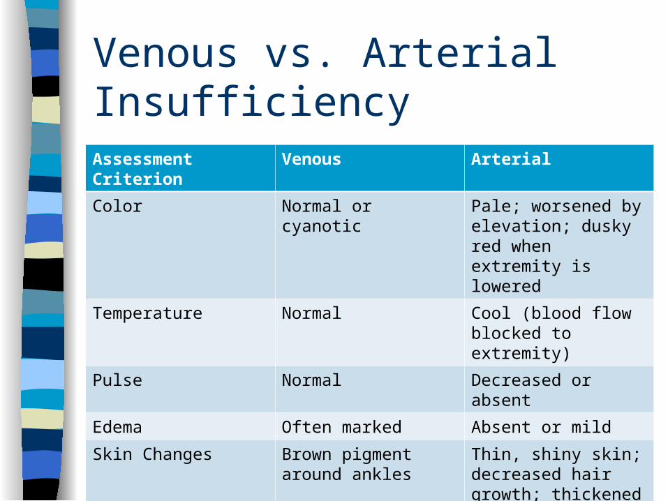
Venous vs. Arterial Insufficiency
Assessment Criterion Venous Arterial
Color Normal or cyanotic Pale; worsened by elevation; dusky red when extremity is lowered
Temperature Normal Cool (blood flow blocked to extremity)
Pulse Normal Decreased or absent
Edema Often marked Absent or mild
Skin Changes Brown pigment around ankles
Thin, shiny skin; decreased hair growth; thickened nails.
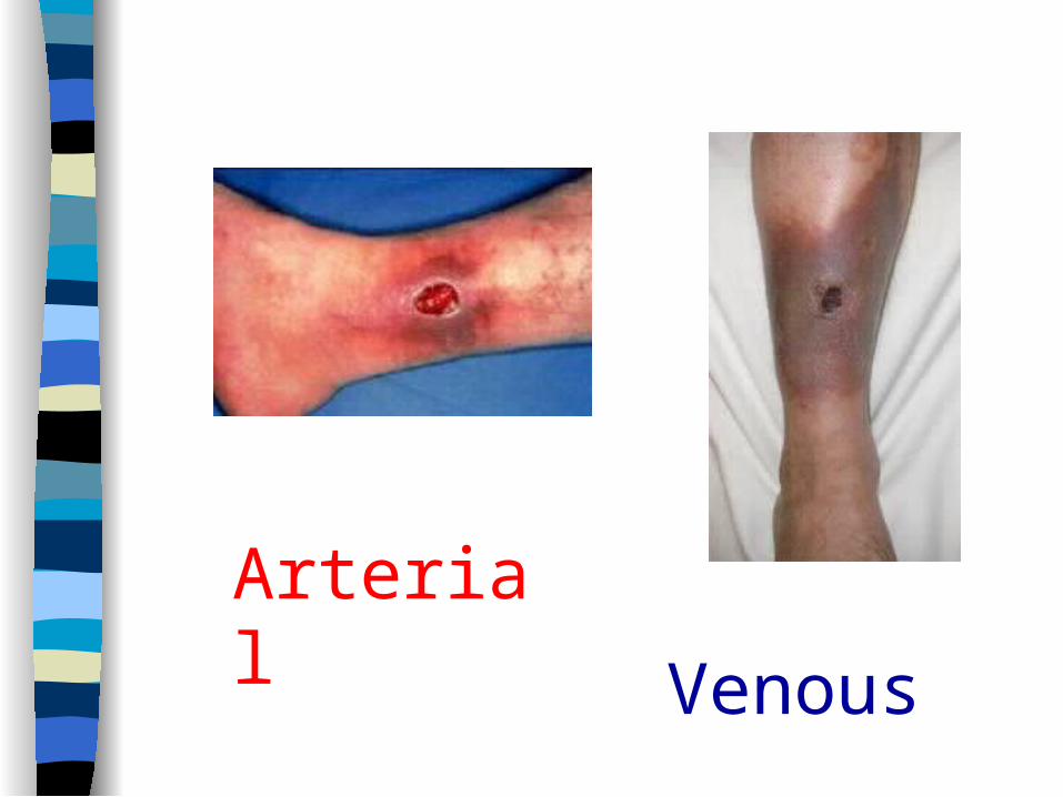
Arterial
Venous

Is that all?
MIDTERM 40 points all multiple choice