telomerase activity thesis - worcester polytechnic … · · 2002-04-30telomerase activity in...
TRANSCRIPT

Telomerase Activity in Human Umbilical Cord Cell Populations
Containing Hematopoietic Stem Cells
A THESIS
Submitted to the faculty
of the
WORCESTER POLYTECHNIC INSTITUTE
In partial fulfillment of the requirements for the
Degree of Master of Science
in
Biotechnology
By
____________________________
Vidya Murthy
May 1, 2002
APPROVED:
__________________ ___________________ ___________________
David S. Adams, Ph.D. Jill Rulfs, Ph.D. William Mackin, Ph.D. Major Advisor Committee Member Committee Member
WPI WPI ViaCell, Inc.

ii
ABSTRACT
Hematopoietic cell populations exhibiting detectable telomerase activity and
elongated telomere lengths display strong engraftment survivability in humans during
transplants. We investigated telomerase activity and telomere length in umbilical cord
blood hematopoietic cell populations obtained from ViaCell Inc. at various intervals of a
two-week ex vivo stem cell amplification process. Telomerase activity is increased with
time in ViaCell’s amplification process, perhaps in response to the removal of
differentiated cells and expansion of primitive hematopoietic stem cell populations in
tissue culture media containing a mixture of growth factors. Two of ViaCell’s cell
culture fractions were analyzed for telomere length using a TLA. Our results showed
relatively long telomere lengths for day-0 and day-14 cord populations, and that despite
an upregulation of telomerase activity in Day-14 samples, a loss of about 2 kb of
telomeric DNA occurs. Our data are consistent with a model in which the increase in
telomerase activity in day-14 ex vivo amplified cord blood hematopoietic cells relative to
fresh cord is sufficient to reduce, but not prevent, telomere shortening caused by cell
proliferation. Lastly, we investigated various culture conditions that could potentially
upregulate telomerase activity in the Day-14 amplified cells. However none of the
treatments tested altered telomerase activity. Our detection of increased telomerase
activity and relatively long telomere lengths in ViaCell’s Day-14 ex vivo amplified cord
blood stem cell fraction provides support for ViaCell’s Selective Clonogenic
AmplificationTM indicating a high engraftment potential for these cells.

iii
TABLE OF CONTENTS
ABSTRACT……………………………………………………………………………….2
TABLE OF CONTENTS………………………………………………………………….3
LIST OF FIGURES……………………………………………………………………….4
LIST OF TABLES………………………………………………………………………...5
ACKNOWLEDGEMENTS……………………………………………………………….6
BACKGROUND………………………………………………………………………….8
PROJECT PURPOSE……………………………………………………………………47
MATERIALS AND METHODS………………………………………………………...48
RESULTS………………………………………………………………………………..60
DISCUSSION……………………………………………………………………………79
BIBLIOGRAPHY………………………………………………………………………..87

iv
LIST OF FIGURES
Figure 1. Levels of stem cell differentiation………………………………………………8
Figure 2. A microscopic view of ES cells…………………………………………………9
Figure 3. Promise of stem cell research………………………………………………….11
Figure 4. Genetic manipulation of human ES cells……………………………………...12
Figure 5. Hematopoiesis…………………………………………………………………13
Figure 6. Electron micrograph of a hematopoietic stem cell…………………………….15
Figure 7. “Negative selection separation” process………………………………………27
Figure 8. Mitotic Clock…………………………………………………………………..32
Figure 9. Telomerase Function…………………………………………………………..33
Figure 10. Secondary structure of hTERC……………………………………………….35
Figure 11. Telomere Hypothesis…………………………………………………………39
Figure 12. Test of positive and negative controls of TRAP assay……………………….62
Figure 13. TRAP assay with descending protein mass…………………………………..63
Figure 14. TRAP assay on Cord-1, N=1…………………………………………………65
Figure 15. TRAP assay on Cord-1, N=2…………………………………………………67
Figure 16. Quantitation of Telomerase activity in Cord-1……………………………….67
Figure 17. TRAP assay on Cord-2, N=1…………………………………………………68
Figure 18. TRAP assay on Cord-2, N=2…………………………………………………69
Figure 19. Quantitation of Telomerase activity in Cord-2……………………………….69
Figure 20. TRAP assay on Cord-3, N=1…………………………………………………70
Figure 21. TRAP assay on Cord-3, N=2…………………………………………………71
Figure 22. Quantitation of Telomerase activity in Cord-3……………………………….71
Figure 23. Test of controls for Telomere length assay…………………………………..73
Figure 24. TLA on Cord samples, N=1………………………………………………….75
Figure 25. TLA on Cord samples, N=2………………………………………………….76
Figure 26. Pre-treatment-1 of cord samples……………………………………………...77
Figure 27. Pre-treatment-2 of cord samples……………………………………………...78

v
LIST OF TABLES
Table 1. Phenotypes of adult human hematopoietic stem and progenitor cells…………16
Table 2. ViaCell’s UCB amplification time course……………………………………...28
Table 3. Percent of CD34+/CD38- cells at each time point during the amplification
of the three cord samples………………………………………………………64
Table 4. Percent CD34+ content and telomerase quantitation for each cord…………….72
Table 4. FACS Analysis of Day-0 and Day-14 cells used for the TLA…………………74

vi
ACKOWLEDGEMENTS
I would like to thank Dr. David Adams, my major advisor, for encouraging and
guiding me throughout my Master’s program, and providing me with the opportunity to
work on this project. I would like to thank the members of my committee: Dr. Jill Rulfs
and Dr. William Mackin, for their advice in making this project focused and complete. I
thank ViaCell, Inc. (Worcester, MA) for providing all the cord blood samples and other
materials used in this project. Special thanks to Lizabeth Amaral, for her advice and
help, during this project. I wish to thank Ram, for always believing in me and supporting
me. I could not accomplish this without his help. Finally, I would like to thank my
family for their love and encouragement.

vii
For
Appa and Bhagu With
Love and gratitude

8
BACKGROUND Stem Cells
Stem cells have the ability to divide for indefinite periods in culture and to give
rise to specialized cells. They are characterized by their capacity for extensive
proliferation and differentiation (de Wynter et al., 1998). They are best described in the
context of normal human development. The fertilized egg is said to be totipotent (see fig.
1, upper diagram) because it has the potential to generate all the cells and tissues that
make up an embryo, including those that support the egg’s development in utero (like the
extra-embryonic tissues, placenta, and umbilical cord).
The term pluripotent (see fig. 1, middle diagram) is used to describe stem cells
that can give rise to cells derived from all three embryonic germ layers: mesoderm,
endoderm, and ectoderm. These three germ layers are the embryonic source of all cells
of the body. The embryonic stem (ES) cell is pluripotent (see fig. 2). The ES cell is
defined by its origin from one of the earliest stages of the development of the embryo, the
Figure 1. Levels of stem cell differentiation. (NIH 2000).

9
blastocyst. Specifically, ES cells are derived from the inner cell mass of the blastocyst at
a stage before it would implant in the uterine wall. The
defining properties of an ES cell include the following:
they are capable of long-term self-renewal (exhibit and
maintain a stable diploid, normal karyotype); are
clonogenic (a single cell can give rise to genetically
identical cells); can be induced to continue proliferating
or to differentiate (lack the G1 checkpoint in the cell
cycle and spend most of their time in the S phase of the cell cycle, during which they
synthesize DNA), show stable developmental potential to form derivatives of all three
embryonic germ layers even after prolonged culture (Smith, 2001; Thomson & Marshall,
1998).
The pluripotent stem cells undergo further specialization into stem cells that are
committed to give rise to cells that have a particular function. These more specialized
stem cells are called multipotent (see fig. 1, lower diagram). Examples of multipotent
cells include hematopoietic stem cells (HSC) that give rise to the various types of blood
cells.
Unipotent stem cells, a term that is usually applied to a stem cell in adult
organisms, means that these cells are usually capable of differentiating along only one
lineage. Adult stem cells in many differentiated tissues give rise to just one cell type
under normal conditions, in a process that allows a steady state self-renewal for the tissue
2
Figure 2. A microscopic view of ES cells (Newsweek, July 9, 2001).

10
(NIH, 2001). Recent data indicate that adult and multipotent stem cells may be capable
of more versatile differentiation than previously thought. For example, HSCs may have
the capacity to differentiate into neuronal cells (McGovern et al., 2001).
Potential Uses of Human Stem Cells
A new era in stem cell biology began in 1998 with the derivation of embryonic
stem (ES) cells from the inner cell mass of human blastocysts by Thomson et al (1998)
and from fetal tissue by John Gearhart (Shamblott et al., 1998). These breakthroughs
showed that ES cells provided a possible source of cells for cell based therapies for many
human diseases. The ES cells isolated from human blastocysts showed normal
karyotypes and expressed high levels of telomerase activity. These cells were grown
with mouse feeder fibroblasts, and after undifferentiated proliferation in vitro for 4-5
months, these cells still maintained the developmental potential to produce gut
epithelium, cartilage, bone, muscle, neural epithelium, embryonic ganglia, and stratified
squamous epithelium. When injected into SCID mice, these cells formed teratomas,
which are tumors containing a mix of differentiated human cell types (Thomson et al.,
1998). In another study, human primordial germ cells cultured in vitro retained their
karyotype while producing large, ES-like cell colonies capable of repeated passages
(Shamblott et al., 1998). Reports showed that specific media hold promise for a directed
differentiation of ES cells (Pittenger et al., 1999). In another key publication, eight
growth factors were tested on a human ES cell line expressing receptors for each growth
factor. Molecular markers identified all three germ layers, eleven tissues, and specific
gene expression in germ layers and tissues (Schuldiner et al., 2001). These studies show
11
that directed differentiation of stem cells into germ layers or specific tissues will become
possible in the near future.
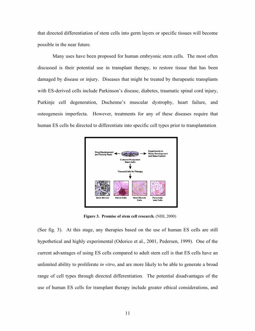
Many uses have been proposed for human embryonic stem cells. The most often
discussed is their potential use in transplant therapy, to restore tissue that has been
damaged by disease or injury. Diseases that might be treated by therapeutic transplants
with ES-derived cells include Parkinson’s disease, diabetes, traumatic spinal cord injury,
Purkinje cell degeneration, Duchenne’s muscular dystrophy, heart failure, and
osteogenesis imperfecta. However, treatments for any of these diseases require that
human ES cells be directed to differentiate into specific cell types prior to transplantation
(See fig. 3). At this stage, any therapies based on the use of human ES cells are still
hypothetical and highly experimental (Odorico et al., 2001, Pedersen, 1999). One of the
current advantages of using ES cells compared to adult stem cell is that ES cells have an
unlimited ability to proliferate in vitro, and are more likely to be able to generate a broad
range of cell types through directed differentiation. The potential disadvantages of the
use of human ES cells for transplant therapy include greater ethical considerations, and
Figure 3. Promise of stem cell research. (NIH, 2000)

12
the propensity of undifferentiated ES cells to induce the formation of tumors (teratomas),
which are typically benign (NIH, 2001). Because it is the undifferentiated ES cells rather
than their differentiated progeny that have been shown to induce teratomas, tumor
formation might be avoided by devising methods for removing any undifferentiated ES
cells prior to transplant (NIH, 2001). The potential immunological rejection of human
ES-derived cells might be avoided by using nuclear transfer technology to generate ES
cells that are genetically identical to the person who receives the transplant. It has been
suggested that this can be accomplished by using somatic cell nuclear transfer technology
or “therapeutic cloning” (Odorico et al., 2001) (see fig. 4).
Another potential use of ES cells that does not involve transplantation, is their use
in studying the early events in human development, for example to identify genetic,
molecular, and cellular events that lead to congenital birth defects, placental
abnormalities that lead to spontaneous abortions, and to identify methods for preventing
them (Rathjen et al., 1998). Human ES cells can also be used to test candidate
Figure 4. Genetic manipulation of human ES cells. (NIH, 2001).

13
therapeutic drugs because the ES- derived cells may be more likely to mimic the in vivo
response of the cells/tissues to drugs being tested, and so offer safer and potentially
cheaper models for drug screening. Human ES cells could also be employed to screen
potential toxins. However, to meet these objectives ES cells must be directed to
differentiate into specific cell types (NIH, 2001).
Hematopoiesis
Hematopoiesis (see fig. 5) is the formation of red and white blood cells from
hematopoietic stem cells (HSC). In human ontogeny, hematopoiesis begins in the yolk
sac and migrates to the fetal liver and then to the spleen. As the gestation continues, the
bone marrow becomes the major hematopoietic organ, and by the time of birth
hematopoiesis has ceased within the liver and spleen. Early in hematopoiesis, the
Figure 5. Hematopoiesis. Shows formation of red and white blood cells from hematopoietic stem cell (Tissue Therapeutics, 2000).

14
multipotent HSC differentiates along one of two pathways, giving rise to either a
lymphoid stem cell (upper pathway in the figure) or a myeloid stem cell (lower pathway
in the figure). The lymphoid stem cell generates T and B progenitor lymphocytes. The
myeloid stem cell generates progenitor cells for erythrocytes, neutrophils, eosinophils,
basophils, monocytes, mast cells, and platelets (Kuby, 1992). The earliest evidence
proving that the various cell lineages in bone marrow originate from HSCs came from
the classic experiment of Till and McCulloch (1961).
The complex orchestration of hematopoiesis through which the elaborate array of
blood cells is produced requires three physiologic components, each of which is essential:
1) the stem cell pool itself, 2) hematopoietic cytokines, which regulate hematopoiesis
through both endocrine and paracrine mechanisms, and 3) the hematopoietic inductive
microenvironment, which is made up of the bone marrow stroma and vasculature. The
unique microenvironment influences the growth and differentiation of hematopoietic
stem cells by providing a hematopoietic inducing microenvironment consisting of a
cellular matrix and either membrane bound or diffusible growth factors (Sullivan, 2000).
Hematopoietic Stem Cells
Hematopoietic stem cells (HSCs) (figure 6) are usually found in the bone marrow,
umbilical cord blood, and can be found in the peripheral blood if they are stimulated in
the bone marrow by factors such as granulocyte colony-stimulating factor (G-SCF) (t.
Breeders, 2000). An HSC can renew itself, can differentiate to a variety of specialized
cells, or can undergo apoptosis (NIH, 2001). HSCs are few in number, occurring with a
frequency of one stem cell per 104 bone marrow cells. Studies have revealed that there

15
appear to be two kinds of HSCs: 1) long-term stem cells that are capable of self-renewal
and can regenerate all the different types of cells, and 2) short term progenitor or
precursor cells which are relatively immature and are precursors to a fully differentiated
cell of the same tissue type. It is the long-term replicating HSCs that are most important
for developing HSC-based cell therapies (NIH, 2001).
Identification (Phenotype) of Hematopoietic Stem Cells
Both HSCs and lineage committed hematopoietic progenitor cells (HPC) express
the CD34 antigen (see Table 1). CD34+ cells constitute 1%-5% of cells in the adult bone
marrow, and 5%-10% of fetal bone marrow Cells (Krause et al., 1996) and 1% of all
nucleated cells in cord blood from full term deliveries (Civin and Gore, 1993). About 1
in every 100,000 cells in the marrow is a long term HSC (NIH, 2001). CD34 plays an
important role in the formation of progenitor cells in both fetal and adult hematopoiesis.
The CD34 antigen is an integral membrane glycoprotein of 90-120 kD and has been
Figure 6. Electron micrograph of a hematopoietic stem cell (McLaren, 2001).

16
suggested to function as a regulator of hematopoietic cell adhesion to stromal cells of the
hematopoietic microenvironment (Healy et al, 1995).
Stem Cells Progenitor Cells CD34+ CD34+ AC133+ AC133+ Lin- CD33+, CD54+, CD7+, CD19+, CD24+(3%-30%)
CD9+, CD18+, CD29+, CD31+, CD38+, CD44+ CD45-RAlo (>70%) CD45+ Thy-1+ Thy-1+ (5%-25%) HLA-DR- HLA-DR+ c-kit+ kit+ (70%-80%) Flk-2+ Flk-2+ (20%-50%) MDR1hi MDR1lo
Rhodaminedull Rhodaminebright
Primitive HSCs lack the differentiation antigens that are present on lineage-committed
progenitors and are thus CD38-, CD45lo, CD71lo (Craig et al., 1993). HLA-DR is absent
or is expressed at low levels on adult stem cells but is present on fetal and neonatal
hematopoietic stem cells (Lansdorp et al., 1993). Thy-1 antigen is present on all human
fetal and neonatal hematopoietic cells but is only expressed on a proportion of lineage-
committed progenitors in the adult (Craig et al., 1993). The MDR1 gene is strongly
expressed in HSCs and confers on them the ability to exclude the mitochondrial binding
dye rhodamine 123 (Chaudhary and Roninson, 1991). The receptors kit and flk-2, which
have intrinsic tyrosine kinase activity, are expressed on both stem cells and progenitors,
but some kit+ cells with stem cell function can be flk-2- (Zeigler et al., 1994). AC133 is
a recently described antigen whose function is unknown. The AC133 antibody selects a
subset of CD34+ cells which contain both short and long term repopulating cells and
therefore offers an alternative to the CD34 antigen for cell selection (Yin et al., 1997).
Table 1. Phenotypes of adult human hematopoietic stem andprogenitor cells (Stem cell biology and gene therapy, edited byQuesenberry PJ, Stein GS, Forget BG, and Sherman MW, 1998 Wiley-Liss, Inc.

17
Mayani and Lansdorp (1998) have reported that most primitive hematopoietic
stem/progenitor cells (HSPC) present in UCB are small mononuclear cells with the
following immunophenotype: CD34+CD38-CD45RAloCD71loThy-1+c-kit+Rhlow.
Sources of HSC
The classic source of HSCs is bone marrow. HSCs can be obtained from the bone
marrow, usually by puncturing the hipbone after anesthetizing the stem cell donor.
About 1 in every 100,000 cells in the marrow is a long term HSC (NIH, 2001). A small
number of stem and progenitor cells circulate in the peripheral blood. HSCs can also be
obtained from the peripheral blood by mobilizing the hematopoietic stem/progenitor cells
from the marrow by using a wide variety of cytokines and cytotoxics, alone or in
combination. Administration of antibodies to the adhesion factor VLA-4 results in rapid
mobilization (within 30 minutes) of progenitor cells (Papayannopoulou and Nakamoto,
1993). G-CSF treatment increases the numbers of CD34+ cells 4-62 fold in peripheral
blood (Pettengell and Testa, 1995).
In the late 1980s and early 1990s, physicians recognized that blood from the
human umbilical cord (UCB) and placenta was a rich source of HSCs. The presence of
relatively mature hematopoietic progenitor cells (HPC) in human UCB was demonstrated
by Knudtzon in 1974 (Knudtson, 1974). Later Ogawa and colleagues documented the
presence of primitive HPC in UCB (Nakahata and Ogawa, 1982). Studies by Broxmeyer
et al (1989) showed that the frequency of hematopoietic stem/progenitor cells in
umbilical cord blood equals or exceeds that of marrow, and greatly surpasses that of adult
blood (Broxmeyer et al., 1989). Since the first successful umbilical cord blood (UCB)

18
transplants in children with Fanconi anemia (Gluckman et al., 1989), the collection and
therapeutic use of these cells has grown quickly (NIH, 2001). Volume for volume,
human UCB is as rich a source of hematopoietic progenitor cells as bone marrow
(Broxmeyer and Carow, 1993). The proliferative potential of long-term culture-initiating
cells (LTC-IC) from UCB exceeds that of the adult bone marrow (Mayani and Lansdorp,
1995). This compensates in part for the lower number of cells that can be obtained from
a single donor compared with a conventional bone marrow harvest.
Sustained hematopoietic engraftment after myoablation has been obtained with as
few as 2 x 10⁴ LTC-IC from UCB (Wagner et al., 1996). Reports indicate that the UCB
provides sufficient transplantable HSCs for children with human leukocyte antigen
(HLA)-identical or single HLA antigen-disparate sibling donors, but whether this will
prove adequate for two or three HLA-antigen-disparate sibling donors and adults remains
to be determined. Recently, a successful engraftment was reported in three adult patients
of >50 kg transplanted with UCB (Kurtzberg et al., 1996). In reported UCB transplants,
the incidence of graft-versus-host-disease is low. An important source of HSCs in
research, but not in clinical use, is the developing blood-producing tissues of fetal
animals. Fetal hematopoietic progenitors have a greater growth potential than those in
UCB, adult bone marrow, or leukapheresis product (Lansdorp et al., 1993).
Therapeutic Applications of HSCs
Stem Cell Replacement Therapy
Among the first clinical uses of HSCs were the treatment of cancers of the blood
such as leukemia and lymphoma. Since HSCs are the source of all differentiated blood

19
cells, their destruction during cancer therapy is a major source of cancer treatment
morbidity and mortality. A key element of cancer treatment therefore is the replacement
of HSCs following radiation or chemotherapy, a procedure known as stem cell
replacement therapy. The HSCs used for replacement can be obtained from the patient’s
own bone marrow or peripheral blood (autologous) prior to chemotherapy, from the
peripheral circulation of an HLA-matched donor (allogeneic), or from UCB taken at birth
(allogeneic) (t. Breeders, 2000).
One of the problems with autologous HSC transplants in cancer therapy is that
cancer cells are sometimes inadvertently collected and reinfused back into the patient
along with the stem cells. Studies have shown that this can be prevented by purifying the
cells and preserving only the CD34+, Thy-1+ cells (Negrin et al., 2000). Since most
solid tumors do not express CD34, the selection of CD34+ cells has been used to reduce
tumor cell contamination of hematopoietic products used for autologous transplantation
for patients with these tumors. CD34+ cell selection can reduce tumor cell contamination
by a factor of 10-104 using immunomagnetic beads or biotin- avidin columns (Farley et
al., 1997).
One of the most exciting new uses of HSC transplantation is the graft-versus-
tumor treatment of cancer. A study by Joshi et al. (2000) shows that human UCB HSCs
show antitumor activity in the test tube against leukemia cells and breast cancer cells. In
recent years, researchers have contemplated hematopoietic stem cell therapy for
autoimmune diseases. Reports suggest that HSC replacement therapy may fundamentally
alter the patient’s immune system. Lupus patients, who underwent this therapy, remained

20
free from active lupus and improved continuously after transplantation, without the need
for immunosuppressive medications (Traynor et al., 2000).
UCB Transplantation
The first allogeneic UCB transplant (UCBT) was performed successfully in 1989,
to treat a child with Fanconi’s anemia; the UCB donor was his HLA-identical sister
(Gluckman et al., 1989). Several years post-transplant, this patient is doing well, with
full donor hematopoietic and lymphoid reconstitution. This first success opened the way
to an entire new field in the domain of allogeneic HSC transplant, as it showed that a
single UCB unit contained sufficient numbers of HSCs to reconstitute a child’s lympho-
hematopoietic compartment. It also showed that a UCB unit could be collected at birth
without any harm to the newborn infant, and that a UCB HSC graft could be
cryopreserved and transplanted to a myeloablated host after thawing without losing its
repopulating ability (Gluckman, 2000).
Since then a number of advantages of using UCB stem cells for transplantation
have become apparent. Simultaneously, UCB banks have been established for related or
unrelated UCBTs, with >30,000 units currently available, and >1,500 UCBTs having
been performed in children (and increasing numbers in adults) with malignant and
nonmalignant diseases (Rubinstein et al, 1998). The methods of UCB collection and
cryopreservation are easy and safe; as soon as the baby is delivered, the cord is clamped
and cut; cells are collected either by catherization of umbilical veins, by aspiration of
cord and placental vessels, by gravity or by flushing through catheters inserted into the
umbilical artery and vein. The mean volume obtained by these various methods is 100-

21
150 ml, with a mean total number of nucleated cells of 15 x 108 (Gluckman et al, 1993).
UCB cells have many advantages as grafts for stem cell transplantation because of the
immaturity of newborn cells. Hematopoietic progenitors from UCB are enriched for in
vivo long-term repopulating stem cells. Compared to adult stem cells, UCB HSCs
produce larger hematopoietic colonies in vitro, are able to expand in long-term culture in
vitro, engraft SCID-human mice in the absence of additional human growth factors, and
have longer telomeres (Noort and Falkenburg, 2000). These properties should
theoretically compensate for the relatively low number of cells contained in a single UCB
donation, and through rapid expansion reconstitute myeloablated adult patients. The
second advantage of UCB grafts relates to the immaturity of the immune system at birth.
This property should decrease the alloreactive potential of the lymphocytes within a cord
blood graft and consequently should reduce the incidence and severity of GVHD after
HLA-matched or HLA-mismatched transplants which are limitations of allogeneic bone
marrow transplants. Clinical analyses have shown that most UCBTs have been
performed with donors having one, two or three HLA antigen mismatches, compared to
unrelated BMTs where complete HLA identity for class I and class II antigens is required
(Gluckman, 2000).
The other practical advantages of using UCB as an alternative source of stem cells
are the relative ease of procurement, absence of risk to donors, reduced risk of
transmitting infection, and prompt availability of cryopreserved samples to transplant
centers. The other advantages of UCBT are the large donor pool, faster allocation
process and decreased risk of viral transmission. The most important factor in predicting
a positive outcome for transplant is that the number of nucleated cells infused be >3 X

22
107/kg (Gluckman et al., 1997). For the purposes of optimizing the chances of finding a
suitable UCB donor for the recipient, Netcord and Eurocord registries, which are a
cooperative network of large experienced UCB banks, were founded (Gluckman, 2000).
The major concern with UCBT has been engraftment, as all studies show delayed
neutrophil and platelet recovery, whereas long-term engraftment was similar after UCBT
and BMT. However, a cord blood nucleated cell dose >0.37 X 10⁸/kg increased the
speed and probability of engraftment (Wagner et al., 1996).
Stem Cell Based Gene Therapy
The multipotent HSCs form an ideal candidate for gene therapy because they are
a self-renewing population of cells and thus may reduce or eliminate the need for
repeated administrations of the gene therapy. Several investigators have reported on the
successful introduction of particular genes into primitive hematopoietic cells from BM,
and similar approaches are being used with UCB cells (Mayani and Lansdorp, 1998).
HSCs have been a delivery cell of choice for several reasons: 1) although small in
number, they are readily isolated from the body from the circulating blood, bone marrow,
or UCB, 2) they give rise to many different types of blood cells, and once the engineered
stem cells differentiate, the therapeutic transgene will reside in all the different types of
blood cells, 3) HSCs ‘home’ in to a number of different spots in the body- bone marrow,
liver, spleen and lymph nodes. These may be strategic locations for localized gene
delivery of therapeutic agents. The only type of human stem cell used in gene therapy
trials so far is the HSC (NIH, 2001). Generally however, gene therapies using HSCs
have encountered a phenomenon known as ‘gene silencing’ where over time, the

23
therapeutic transgene gets turned off due to cellular mechanisms that alter the structure of
the chromosome where the transgene is inserted (Challita and Kohn, 1994). Stem cell
gene therapy could also allow the development of novel methods for immune modulation
in autoimmune diseases. The goal is to modify the aberrant, inflammatory immune
response that is characteristic of autoimmune diseases. Studies in a lupus mouse model
have shown that genetic modification of HSCs with a ‘decoy’ receptor for the
inflammatory cytokine interferon gamma, arrested disease progression (Lawson et al.,
2000). Long-term in vivo gene transfer studies in mice have shown that recombinant
murine retroviruses are able to infect murine HSCs with high efficiency. Because of the
success in murine studies, it was believed that gene therapy would soon be applicable to
treat a wide variety of congenital or acquired human diseases associated with the
hematopoietic system. Human congenital diseases which are manifested predominantly
in one or more of the blood lineages are, in principle, target diseases for stem cell gene
therapy, since all blood cells are derived from a common ancestor, the HSC. There are,
however, some limitations. First, the precise genetic defect causing the disease must be
known. Second the defect should not be dominant. In general, those diseases that can be
treated by allogeneic bone marrow transplantation are candidates for stem cell gene
therapy. The aberrant gene in the HSC can be replaced by a correct copy in a process
known as homologous recombination, or correct copies of the gene can be inserted into
the host genome using viral delivery (Havenga et al., 1997). Recently genetically
manipulated CD34+ UCB cells have been used in the treatment of patients with SCID
(Kohn et al., 1995).

24
Ex-vivo HSC Amplification
The use of UCB as a source of marrow repopulating cells for the treatment of
pediatric malignancies is well established. However, the major potential limitation to the
widespread use of UCB as a source of HSCs for marrow replacement and gene therapy is
that the ability to engraft an adult might require ex vivo manipulations (Piacibello et al.,
1997). The proliferation potential of hematopoietic stem/progenitor cells as well as their
expansion potential appear to be biologic features that depend upon intrinsic factors.
These are related to whether the cell is already committed to a particular lineage of
differentiation and, if so, the specific hematopoietic lineage to which it specifically
belongs and its stage of maturation. However, the ability of a cell to exhibit such
potentials depends on extrinsic factors that include different cell types and cytokines that
form part of the microenvironment in which the cell develops (Mayani et al., 1992). In
vitro proliferation and expansion of hematopoietic stem/progenitor cells (HSPC) also
depend on variables such as type of medium, medium change schedule, temperature,
presence or absence of serum, number of cells plated per culture, etc. Several groups
have assessed the in vivo expansion and proliferation of UCB progenitors using either
total CD34+ cells, or CD34+ cell subsets. In general, it is clear that primitive
subpopulations of CD34+ cells possess greater expansion potential than their more
mature counterparts (Mayani and Lansdorp, 1998).
The ability of HSPCs to express expansion and proliferation potential in vitro
depends predominantly on the cytokines present in culture. In terms of HSPC expansion,
the best results have been obtained when cytokines are used in combinations that include
early acting factors, such as SCF, flt-3 ligand (FL), and Tpo. The greatest expansion of

25
UCB-derived CD34+ cells reported to date (146,000-fold expansion in CD34+ cell
numbers, and 2 x 10⁶ fold expansion in CFC numbers) was achieved using both FL and
Tpo (Piacibello et al., 1997). Reports have shown that in a simpler medium, with two
cytokines, flt-3 (FL) and thrombopoietin/c-mpl ligand (TPO/ML), significant expansion
of HSC populations was observed, including LTC-IC that could be maintained long-term
(up to six months) (Gilmore et al., 2000). Addition of late acting factors, such as Epo,
usually contribute to the production of large numbers of mature cells, however they do
not seem to have an effect on HSPC expansion (Mayani et al., 1993). In contrast to
cytokines, hematopoietic inhibitors, such as transforming growth factor-β, tumor necrosis
factor-α, and macrophage inflammatory protein-1α have been shown to significantly
reduce both expansion and proliferation of CD34+ cell populations from UCB (Mayani et
al., 1995). Some investigators have used an antitransforming growth factor-β
monoclonal antibody, together with stimulatory cytokines, to achieve a significant
expansion of primitive progenitor cells (Cardoso et al., 1993). Investigators have shown
that UCB-derived HSPC possess higher expansion and proliferation potentials than their
BM counterparts (Hows et al., 1992).
Currently, there are several unresolved issues about the ex vivo expansion and
transplantation of HSPC. The first question surrounds the problem of defining the cells
responsible for short and/or long term hematopoietic recovery after transplantation. The
most controversial and important issue regarding the clinical use of ex vivo manipulated
cells is whether on eventual exhaustion of stem cells might result from prolonged growth
factor stimulation ex vivo (Brugger et al., 2000).

26
Two kinds of enrichment methods are currently used for the purification of
CD34+ stem cells from UCB. Purification of HSCs is performed either with a
combination of monoclonal antibodies to remove unwanted differentiated cells (negative
selections) using the Stem Sep method (discussed in the Viacell section), or with a
positive cell selection based on their surface CD34 antigens using the Mini Macs system
(Pafumi et al., 2001).
ViaCell, Inc.
ViaCell, Inc. is a new cellular medicine company merged from two companies:
Viacord, Inc. (Boston, MA) and t. Breeders, Inc. (Worcester, MA). The goal of the new
combined biotechnology company is to use its high quality cord blood banking service
and patented stem cell expansion technique to develop a premier cellular pharmaceutical
company providing the highest quality products and services for the treatment of diseases
using stem cells (ViaCell, Inc. Annual Report 2000). In June 2001, ViaCell filed an
investigational New Drug (IND) application for approval from the FDA for a phase I/II
clinical trial for its proprietary selective amplification technology, which involves
expansion of rare hematopoietic stem cells and other rare primary cell types. The Phase 1
study involving one patient every 3 months is currently underway. The targeted
population for therapy currently includes myoablative therapy patients, patients with
genetic diseases, patients with hematological malignancies, and those with neurological
disorders (Craig, 2000). ViaCell will still continue to offer cord blood banking to their
clients under the name of Viacord, and will continue research and development of
expanded stem cell products under the name of t. Breeders (Stringer, 2000).

27
Figure 7. ‘Negative Selection Separation’ Process. (Zimmerman, 1998)
ViaCell’s patented method of expansion called Selective Clonogenic
AmplificationTM enables simultaneous selection and amplification of stem cells from bone
marrow, mobilized peripheral blood, or cord blood through the use of highly specific
markers on stem cells and amplifying these cells under culture conditions that foster the
outgrowth of stem cells. In the strict sense, Selective Clonogenic AmplificationTM is a
process for “breeding” cells, i.e., selecting preferred events of biological fission to
produce target populations from among a variety of irrelevant derivative populations (t.
Breeders, 2000). The salient features of the Selective Clonogenic AmplificationTM
process include: removal of differentiated cells and their by-products during cell culture
production of highly defined target populations, active purging of co-isolated cancer cells
(which do not carry the CD
34+ antigen), and efficient,
cost-effective production
(t.Breeders, 2000).
The Selective
Clonogenic AmplificationTM
utilizes the ‘negative
selection separation’ of
hematopoietic stem/progenitor
cells. This separation
technique, as shown in fig 7, works by immunomagnetically labeling and removing the
unwanted cells in the column. Cells are labeled by the use of unmodified colloidal
magnetic dextran iron (orange in the figure) and non-covalent bispecific antibody cross-

28
linking reagents called tetrameric antibody complexes. The tetrameric antibody complex
is comprised of two murine IgG monoclonal antibodies (orange and blue in the figure),
held in a tetrameric array by two rat anti-mouse IgG monoclonal antibody molecules
(yellow in the figure). One murine antibody molecule recognizes the differentiated cell
surface antigen (orange) and the other (blue) recognizes the dextran on the magnetic
particle. The cell suspension is passed through a column and the unwanted differentiated
magnetically labeled cells bind to the column, while the unlabeled cells (containing CD
34+ cells) pass through (StemCell Technologies, 2001). A cocktail of monoclonal
antibodies against differentiation surface markers is used to weed out the differentiated
cells from the UCB cell population. The above separation process results in the
separation of a subpopulation of hematopoietic stem/progenitor cells characterized as
CD34+/CD38-/Lin- cells. Cells that lack 13-14 different mature blood-lineage markers
including: CD2, CD3, CD14, CD16, CD19, CD24, CD56, CD66b, and glycophorin A
(collectively referred to as Lin- cells). These markers are expressed on the surface of
mature red blood cells, monocytes, natural killer cells, and T-cells. ViaCell’s Selective
Clonogenic AmplificationTM is a 14-day long amplification process and includes the
following steps, as shown in table 2.
Time- Point Fraction Total Cells
%CD34+/CD38-
Day-o Whole Cord Blood ~6.5x10⁸ 0.3% Freeze Thaw Pre-Sep-1 ~5.2x10⁷ 0.2%
Day-0.5 Post-Sep-1 ~2.7x10⁶ 3% Day-7 Pre-Sep-2 ~2.1x10⁷ 31%
Day-7.5 Post-Sep-2 ~6.2x10⁶ 33% Day-14 Post Culture ~3.0x10⁷ 11%
Table 2. ViaCell’s UCB amplification time course.

29
Telomerase
Structure and Function of Telomeres
Telomeres are nucleoprotein structures located at the ends of eukaryotic
chromosomes that contain protein-bound, simple repeat units of a nucleotide sequence
(Rhyu, 1995). Telomeres protect chromosomes from shortening and unraveling during
each replication cycle. It has been suggested that telomeres protect chromosome ends,
because damaged chromosomes lacking telomeres undergo fusion, re-arrangement and
translocation (Blackburn, 1991). Telomeres play an essential role in the stable
maintenance of the eukaryotic chromosome within a cell by specifically binding to
structural proteins. These proteins cap the ends of linear chromosomes, preventing
nucleolytic degradation, end-to-end fusion, irregular recombination and other specific
events that are normally lethal to a cell. Additionally telomeres are involved in nuclear
architecture, and interact with other proteins to repress the expression of adjacent genes
(Blackburn, 1991).
Telomeres have been studied in a variety of eukaryotic organisms. For example,
Tetrahymena contains up to 40,000 telomere repeats per DNA macromolecule, each
containing the repeat sequence GGGGTT (Blackburn and Gall, 1978). Telomeres of
many insects and Lepidopteran species contain the pentanucleotide repeat sequence
TTAGG (Sasaki and Fujiwara, 2000). In the diploid human cell, there are 46
chromosomes, each containing two telomeres, and each telomere contains the nucleotide
repeat sequence TTAGGG, which may repeated up to 15 Kb per telomere (Moyzis et al.,
1998). The telomere repeats in most species tends to be G-C rich, with a strand bias so
that the G-rich strand is oriented with its 3′ end towards the end of the DNA (Kurenova

30
and Mason, 1997). In humans the 3′ -terminal G-rich strand is about 200 nucleotides
longer than the C-rich strand, leaving a 3′ overhang (Wright et al., 1997).
The functional telomere is organized into a special chromatin structure, the
‘telosome’ (Wright at al., 1992), which contains telomeric DNA complexed with
sequence-specific telomere binding proteins such as TRF1, TRF2 and more loosely with
proteins such as tankyrase (Broccoli et al., 1997). The single stranded, G-rich 3’
extension is not only hidden by association with numerous telomere binding proteins, it is
folded back and entangled in internal double stranded telomeric DNA and thus forms the
telomeric t-loop (Griffith et al., 1999). The 200 bp G-rich, 3’ terminal, single stranded
extensions are required for binding of TRF2, and failure to do so results in genome
instability by chromosomal end-to-end fusions or, depending on the cell type, in
apoptotic cell death (van Steensel et al., 1998).
End Replication Problem
In somatic cells, telomere length is progressively shortened with each cell division
both in vivo and in vitro (Harley et al., 1990; Lindsey et al., 1991), due to the inability of
the DNA polymerase complex to replicate the very 5’ end of the lagging strand (Watson,
1972; Olovnikov, 1973). DNA replication in the S-phase of the cell cycle starts by
extending small RNA primers by DNA polymerases, which are unable to start de novo
synthesis. After generation of the new DNA strand, the RNA primers are removed and
all internal gaps are filled with DNA. The primers can be replaced everywhere except at
the extreme 5’ end which makes this new strand slightly shorter than the parental strand.
This phenomenon is the molecular basis of the ‘end replication problem’, which was
31
described long before the structure of the chromosomal ends was known (Olovnikov,
1973). Although the chromosomal loss is potentially very small, this loss will occur
every cell division, and must eventually compromise cell or chromosomal viability
following the removal of essential DNA sequences, either functional genes or telomeric
sequences required for an essential end protective function (Kipling, 2001). Human
telomeres are programmed to undergo gradual shortening by about 100 bp per cell
division, and when several kilobases of the telomeric DNA are lost, cells stop dividing
and senesce (De Lange, 1998).
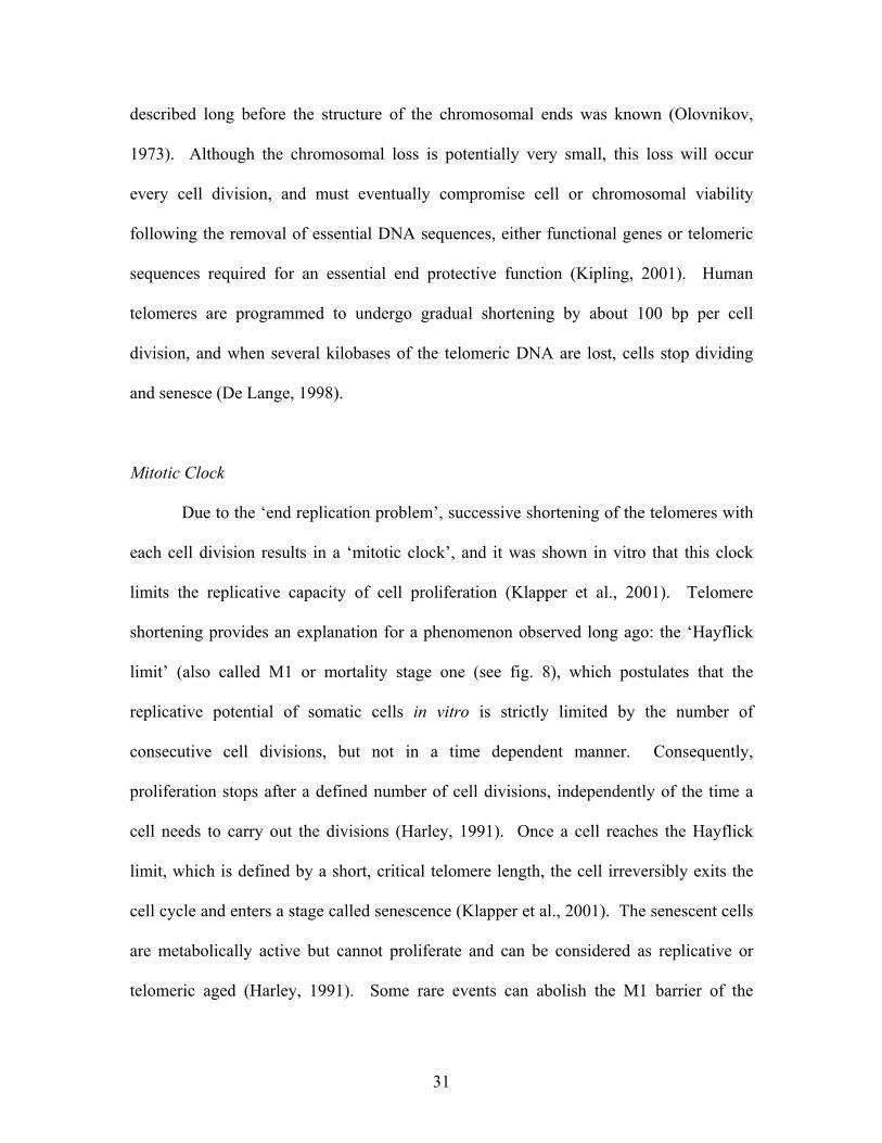
Mitotic Clock
Due to the ‘end replication problem’, successive shortening of the telomeres with
each cell division results in a ‘mitotic clock’, and it was shown in vitro that this clock
limits the replicative capacity of cell proliferation (Klapper et al., 2001). Telomere
shortening provides an explanation for a phenomenon observed long ago: the ‘Hayflick
limit’ (also called M1 or mortality stage one (see fig. 8), which postulates that the
replicative potential of somatic cells in vitro is strictly limited by the number of
consecutive cell divisions, but not in a time dependent manner. Consequently,
proliferation stops after a defined number of cell divisions, independently of the time a
cell needs to carry out the divisions (Harley, 1991). Once a cell reaches the Hayflick
limit, which is defined by a short, critical telomere length, the cell irreversibly exits the
cell cycle and enters a stage called senescence (Klapper et al., 2001). The senescent cells
are metabolically active but cannot proliferate and can be considered as replicative or
telomeric aged (Harley, 1991). Some rare events can abolish the M1 barrier of the

32
proliferation; the best-studied alterations are the expression of viral oncogenes that
inactivate p53 and retinoblastoma (Rb) (Shay et al., 1991; 1993). But infrequent
accumulation of these genetic aberrations leaves only a few cells that proliferate beyond
the Hayflick limit (Harley, 1991), resulting in further telomere shortening.
A second checkpoint is reached at a critical telomere length called crisis
(mortality stage two or M2).
At this stage, almost all cells die due to extensive chromosomal aberrations, caused
by short and dysfunctional telomeres; however, very rarely some immortal cells
arise. To overcome crisis (M2) and become immortal, the cell activates
telomerase activity (Harley, 1991; Klapper et al., 2001).
Figure 8. Mitotic Clock (Klapper et al., 2001) Shows that telomeres in somaticcells shorten with each cell division and enter senescence, while in telomerasepositive germ line and stem cells, the telomere lengths are kept constant.
33
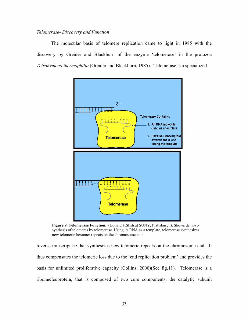
Telomerase- Discovery and Function
The molecular basis of telomere replication came to light in 1985 with the
discovery by Greider and Blackburn of the enzyme ‘telomerase’ in the protozoa
Tetrahymena thermophilia (Greider and Blackburn, 1985). Telomerase is a specialized
reverse transcriptase that synthesizes new telomeric repeats on the chromosome end. It
thus compensates the telomeric loss due to the ‘end replication problem’ and provides the
basis for unlimited proliferative capacity (Collins, 2000)(See fig.11). Telomerase is a
ribonucleoprotein, that is composed of two core components, the catalytic subunit
Figure 9. Telomerase Function. (Donald.F.Slish at SUNY, Plattsburgh). Shows de novo synthesis of telomeres by telomerase. Using its RNA as a template, telomerase synthesizes new telomeric hexamer repeats on the chromosome end.

34
hTERT and the RNA component hTR. Using its RNA component as the template, it
synthesizes and directs telomeric repeats onto the 3’ end of existing telomeres. In this
respect, telomerase is acting as a reverse transcriptase, insofar as it is synthesizing DNA
based upon an RNA template (Greider and Blackburn, 1989; Morin, 1989). In vitro
synthesized hTERT and hTR can assemble to form catalytically active telomerase
holoenzyme, thus demonstrating that these two components can form a minimal core
enzyme (Weinrich et al., 1997).
Telomerase Structure
The telomerase complex represents a specialized terminal reverse transcriptase
with an estimated molecular mass of ~1000 kDa (Dhaene et al., 2000). The telomerase
RNA component was first cloned in Tetrahymena thermophila. Later, the homologous
genes were identified in ciliates such as Oxytrichia and Euplotes, in yeast S. cerevisiae
(TLC1), and in mammals such as mouse (mTR) and human (hTR, currently referred to as
hTERC for human telomerase RNA component) (Feng et al., 1995). hTERC is a single
copy gene present on chromosome 3 (3q26.3). In humans, the length of the mature
hTERC gene transcript is 451 nucleotides and lacks polyadenylation. In all organisms
analyzed to date, a ‘template’ region complementary to the sequence of the telomere
repeats is embedded in the integrated telomerase RNA sequence. For humans, the
hTERC template consists of 11 nucleotides: 5’CUAACCCUAAC 3’. Mammalian
telomerase RNAs resemble small nucleolar RNAs (snoRNAs)- an RNA family required
for pseudouridine modification and precursor processing of rRNA – because of the
presence of an H/ACA box in their 3’ domain (Mitchell et al., 1999). The primary

35
structure of the RNA component has evolved rapidly between species, but there seems to
be a secondary structure core that is highly conserved even between distant groups
(Blackburn, 2000). Four conserved structural elements are universally present in the
predicted secondary structure of
RNA: these are the pseudoknot
domain, the CR4-CR5 domain, the
H/ACA box, and the CR7 domain
(see fig 10).
Telomerase reverse transcriptase is a
special class of reverse transcriptases
that functions as the rate limiting step
in telomerase activity. It has been
identified in yeast (Sc-Est2p), the
ciliate E. aediculatus (Ea-p123),
Tetrahymena thermophila (Tt-
TERT/p133), and in mammals such as mouse (mTERT) and human
(hTRT/hEst2/hTCS1/TP2, currently referred to as hTERT). hTERT contains a
telomerase specific amino acid motif (T motif) and seven conserved reverse transcriptase
motifs (RT motifs), making it phylogenetically related to RTs (Dhaene et al., 2000;
Nakamura et al., 1997). Substitution of conserved amino acid residues in the RT domain
of hTERT completely abolishes telomerase activity. The 40-kB single copy hTERT
gene, located on chromosome 5 (5p15.33), codes for a 127-kDa protein of 1132 amino
Figure 10. Secondary structure of hTERC (Chen et al.,2000). Shows the RNA template sequence (nucleotides 46-53) and its 5’ and 3’ ends.

36
acids contained in 6 exons (Meyerson et al., 1997). The human telomerase reverse
transcriptase subunit (hTERT) has been cloned by Nakamura et al., (1997).
Another telomerase-associated protein includes the mammalian p80 homologue
identified in rat, mouse and human (TP1/TLP1, currently referred to as hTEP1 for human
telomerase-associated protein1), but similar to hTERC, the expression of this protein
does not correlate with telomerase activity in cells and tissues. It has been suggested that
hTEP1 may play a role in some aspect of ribonucleoprotein structure, function or
assembly (Harrington et al., 1997).
Telomerase vs. Cancer
In the mid-1990s, the hypothesis emerged that the upregulation or re-expression
of telomerase is a critical event responsible for continuous tumor cell growth. In contrast
to normal cells, in which a gradual mitosis-related erosion of telomeres eventually limits
replicative life span, tumor cells have telomerase activity and show no loss of
chromosomal ends. It was thus suggested that telomere stabilization might be required
for cells to escape replicative senescence and to proliferate indefinitely (Dheane et al.,
2000). But a key debate emerged on whether telomerase upregulation by itself induce a
malignant phenotype, i.e. does telomerase act as an oncogene. And if so, then how does
this relate to the debatable levels of tolerance in HSCs.
One point is clear; telomerase activity has been demonstrated in the vast majority
of tumor biopsies (85%) (Kim et al., 1994). Moreover, cell lines immortalized either
spontaneously or after transformation by oncogenic viruses (such as simian virus 40 or
human papillomavirus types 16 or 18) are usually telomerase-positive (Belair et al.,

37
1997). Such observations lead to the current hypothesis that telomerase is activated
during immortalization in vitro and tumorigenesis in vivo (De lange, 1994). However,
telomerase activity is not always detectable in immortal cell lines (Bryan et al., 1995).
Most results have shown that normal somatic cells are telomerase negative,
whereas germ cells and stem cells in renewable tissues are telomerase positive (Belair et
al., 1997). It has been suggested that normal cells contain an inhibitor of telomerase,
possibly on chromosome 3, whose deletion or inactivation is required for immortalization
and tumorigenic transformation (Seachrist, 1995). Telomerase activity has also been
demonstrated in highly proliferative non-cancerous tissues such as the basal layer of the
epidermis, endometrial tissue during the proliferative phase of the menstrual cycle, and
oral mucosa (Belair et al., 1997). These latter studies are not consistent with a model in
which activation of telomerase occurs during tumorigenic transformation. Instead, they
suggest that telomerase activity may more directly be associated with cell proliferation.
Belair et al. (1997) demonstrated using both normal and tumorous human
uroepithelial tissues that telomerase activity is a marker for cell proliferation, not
malignant transformation. They showed that normal cells do have the capability to
express telomerase activity given their proliferative conditions in vitro. Uncultured
normal human uroepithelial cells (HUCs) were telomerase negative. However, the same
cells, when established as proliferating cultures in vitro, showed telomerase activity but at
lower levels that in tumorous cells. Here they attribute the relatively high telomerase
activity in tumor biopsies, in part to their high proliferative ability. These results support
a model in which the detection of telomerase in tumor biopsies, but not in uncultured
normal cells, reflects differences in proliferation between tumor and normal cells in vivo

38
(Belair et al., 1997). hTERT transfection experiments have convincingly shown that
hTERT is rate limiting for telomere elongation (Nakayama et al., 1998). Most somatic
human cells do not express the reverse transcriptase subunit of telomerase but contain all
other components of the enzyme so that expression of the missing hTERT component
leads to reconstitution of enzyme activity (Weinrich et al., 1997). Transfection of pre-
senescent cultures of telomerase-negative retinal pigment epithelial cells, human vascular
endothelial cells and young/midlife and old fibroblasts (Bodnar et al., 1998; Vaziri &
Benchimol, 1998; Yang et al., 1999) as well as pre-crisis cells, with hTERT gene resulted
in an increase in telomerase activity, elongation of telomeres and indefinite replicative
growth, thus establishing a causal relationship between telomere shortening and in vitro
cellular senescence. While sufficient for immortalization, this ectopic expression of
telomerase did not result in changes typically associated with malignant transformation,
such as increased growth rate, loss of contact inhibition, acquisition of serum-
independent growth, disturbances in the pRB and p53-mediated cell cycle checkpoints,
and cytogenetic abnormalities, indicating that telomerase expression per se is not
oncogenic (Jiang et al., 1999).
Most recently, studies conducted with mice doubly null for mTR and p53 (mTR-/-
p53-/-mice) or INK4a/ARF (mTR-/-INK4a-/-mice) showed that telomerase may play a
paradoxical role, either promoting or inhibiting tumor formation depending on the
genetic context of the would be cancer cell (Chin et al., 1999; Greenberg et al., 1999).
Progressive telomere shortening occurs with the division of primary human cells and can
39
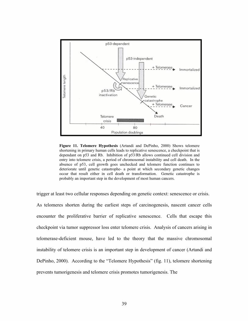
trigger at least two cellular responses depending on genetic context: senescence or crisis.
As telomeres shorten during the earliest steps of carcinogenesis, nascent cancer cells
encounter the proliferative barrier of replicative senescence. Cells that escape this
checkpoint via tumor suppressor loss enter telomere crisis. Analysis of cancers arising in
telomerase-deficient mouse, have led to the theory that the massive chromosomal
instability of telomere crisis is an important step in development of cancer (Artandi and
DePinho, 2000). According to the “Telomere Hypothesis” (fig. 11), telomere shortening
prevents tumorigenesis and telomere crisis promotes tumorigenesis. The
Figure 11. Telomere Hypothesis (Artandi and DePinho, 2000) Shows telomereshortening in primary human cells leads to replicative senescence, a checkpoint that isdependant on p53 and Rb. Inhibition of p53/Rb allows continued cell division andentry into telomere crisis, a period of chromosomal instability and cell death. In theabsence of p53, cell growth goes unchecked and telomere function continues todeteriorate until genetic catastrophe- a point at which secondary genetic changesoccur that result either in cell death or transformation. Genetic catastrophe isprobably an important step in the development of most human cancers.

40
telomere hypothesis was formulated to explain the important role of telomeres in
senescence, the observation that telomerase is reactivated in 80-90% of human cancers,
and the observation that telomeres in tumor lines are often shorter than in primary
somatic cells. The model states that in a developing cancer cell both senescence and
crisis represent barriers to continued tumor growth (Artandi and DePinho, 2000).
It has recently been shown that transcription of the hTERT gene is regulated
directly by the immortalizing oncoprotein Myc, whose upregulation is an obligate feature
of all cancers (Greenberg et al, 1999). Inhibition of telomerase or experimental
interference with telomere function arrests and often kills cells even if they are
transformed (van Steensel et al., 1998). Thus telomerase activity appears to make an
important contribution to the viability of transformed cells, but its action does not fit the
usual roles ascribed to oncogenes and tumor suppressors (de Lange & DePinho, 1999).
Telomerase and Aging (Telomerase- the immortality enzyme?)
Is telomerase really all that is needed for cellular immortalization? Will enforced
somatic expression of telomerase lead to a cancer-prone condition? Definitive answers to
these questions have yet to emerge. However, the first major advance was provided with
the finding that ectopic expression of hTERT in primary human cells could confer
endless growth in culture (De Lange & DePinho, 1999). The cloning of the cDNA
encoding the catalytic subunit of telomerase (hTERT) (Meyerson et al., 1997), made it
possible to test the telomere hypothesis. Two telomerase-negative somatic human cell
types, retinal pigment epithelial cells and foreskin fibroblasts, were transfected with
hTERT. The telomerase-expressing clones had elongated telomeres, divided vigorously,

41
and showed reduced staining for Senescence-associated β-galactosidase (SA-β-Gal), a
biomarker for senescence. These cells also showed a normal karyotype and exceeded
their normal life span by at least 20 doublings, thus establishing a causal relationship
between telomere shortening and in vitro cellular senescence (Bodnar et al., 1998).
These reports also indicate that, a very low level of telomerase activity is insufficient to
prevent telomere shortening. This is consistent with the observation that hematopoietic
stem cells have low but detectable telomerase activity; yet continue to exhibit shortening
of their telomeres throughout life. Thus it appears that a threshold level of telomerase
activity is required for actual life-span extension (Bodnar et al., 1998). Similar findings
were observed in a similar study in which Vaziri & Benchimol (1998) expressed hTERT
in normal fibroblasts, which lack telomerase activity. Similar results were also reported
with endothelial cells (Yang et al., 1999). Other cell types like keratinocytes and
mammary epithelial cells may need, in addition to hTERT expression, additional genetic
changes to extend their life span beyond crisis. These cells arrest prematurely as a result
of accumulation of p16INK4A, a critical inhibitor of the RB pathway and key mortality
gene (Kiyono et al., 1998). These cells are immortal but do not show any changes
associated with the transformed phenotype. The ability of telomerase to prevent the
senescence of primary human cells without causing any overt change to a more cancerous
phenotype has created great excitement in the gerontological community as a potential
route to therapeutic intervention in human aging (Kipling, 2001).
Telomere based barriers to unlimited cell division can be imposed in several ways
(Holt et al., 1996). One is via the triggering of replicative senescence, as is seen in
normal fibroblasts (Bodnar et al., 1998). The second is the triggering of apoptosis, as has

42
been described following telomerase repression and subsequent telomere erosion on
several human cancer cell lines (Hahn et al., 1999). The third is the ultimate loss of
telomere protective function and the triggering of non-specific “genome crisis”
(Halvorsen et al., 1999). All three outcomes can be prevented by telomerase (Kipling,
2001).
All pathological syndromes associated with accelerated aging show alterations in
telomere biology. Telomere defects in Werner syndrome, Bloom syndrome, Hutchinson-
Gilford progeria, Down syndrome, Dyskeratosis congenital, and Ataxia telengiectasia
have been reported (Klapper et al., 2001). Forced expression of hTERT in primary
fibroblasts isolated from Werner syndrome patients confers detectable telomerase activity
and leads to extension of cellular life span. These studies indicate a potential route to
therapeutic intervention in a human ageing syndrome (Kipling, 2001). Cellular
senescence is believed to contribute to multiple conditions in the elderly, and could in
principle be remedied by cell life span expansion in situ (Bodnar et al., 1998). Expansion
of normal cells in vitro, followed by reimplantation might be a future form of cell based
therapy for several aging related diseases that are based on loss of irreplaceable cells.
Attempts to use telomerase-immortalized cells for in vitro tissue engineering of adrenal,
vascular, skin, pancreatic or muscle tissue are already underway (Yang et al., 1999).
Telomerase and Stem Cells
In most somatic cells, telomerase activity is lacking. However, primitive
hematopoietic cells have shown to exhibit low but detectable telomerase activity (Hiyama
43
et al., 1995; Broccoli et al; 1995; Chiu et al., 1996). But despite having detectable
telomerase activity, telomere shortening is observed in blood leukocytes with age, and in
vivo hematopoietic progenitor cultures (Vaziri et al., 1994). In their study, telomerase
activity in human BM and PB was assigned to the hematopoietic progenitor cell fraction
expressing the CD34 antigen. CD34+ cells lacking co-expression of CD33 demonstrated
higher levels of telomerase than myeloid committed CD34+/CD33+ cells. The presence
of growth factors inducing differentiation resulted in a decrease of telomerase activity. In
addition, telomerase activity increased in PB during cytokine-induced mobilization of
hematopoietic progenitor cells. Based on these results, it has been suggested that at least
a portion of the hematopoietic stem/progenitor cell fraction expresses telomerase, and
downregulates its expression during differentiation (Hohaus et al., 1997)
Overall, the observed levels of telomerase activity in stem cells appear to be
related to the mitotic or cycling state of the cell population. Reports indicate that
telomerase is generally present in rapidly expanding cells, upregulated at cell cycle entry
as cells progress through S-phase, and repressed in quiescent Go cells (Holt et al., 1996;
Engelhardt et al., 1997). Telomerase activity in CD34+/CD38+ cells (non-quiescent),
from bone marrow (BM), Peripheral blood (PB), cord blood (CB) and fetal liver (FL),
exceeded levels in CD34+/CD38-, CD34- (quiescent), and mononuclear cells (Engelhardt
et al., 1997). Telomerase activity was reduced in noncycling FL and CB CD34+ cells
compared to more actively cycling PB CD34+ and BM CD34+ cells (Engelhardt et al.,
1997). Recent studies have established the role of hematopoietic cytokines in ex-vivo
expansion systems (Moore & Hoskins, 1994). Stem cell self-renewal, as measured by
increases in the numbers of long-term culture initiating cells, can be achieved in

44
particular with KL and Flk-L cytokine combinations. Cytokine synergistic growth
promoting interactions have been reported on CD34+ cells from different sources such as
CB, PB and BM (Petzer et al., 1996). In the absence of growth factors, CD34+ cells
undergo apoptosis. Single cytokines preserve cells in expansion cultures and block
apoptotic death, but do not induce noncycling progenitors into cycle, whereas cytokine
combinations result in the progression of cells into DNA synthesis and induction of cell
cycle proteins (Moore & Hoskins, 1994). In vitro culture of CD34+ cells derived from
BM, PB, CB, FL in the presence of a cytokine combination (KL, IL-3, IL-6,
erythropoitin, granulocyte colony-stimulating factor) showed upregulation of telomerase
activity which peaked after 1 week of culture, and decreased to baseline levels or below
detection after 3-4 weeks. In contrast, stimulation of CD34+ cells with single cytokines
resulted in no (or minor) telomerase upregulation (Engelhardt et al., 1997).
It has been shown that telomerase activity is low in CB derived CD34+CD38- and
CD34+c-kit- cells compared to CD38+ or c-kit (high or low) cells, suggesting that
CD34+CD38- or c-kit- cells are likely to be more quiescent. These results suggest that
the CD34+CD38- and CD34+c-kit- cell populations are primitive stem/progenitor cells,
and that the telomerase activity of these cells correlates with their proliferative capacity
as well as their stage of differentiation (Sakabe et al., 1998). Telomerase activity has
been attributed more to actively dividing mature subsets (CD34+71+45+) than to more
primitive progenitors with a CD34+71low45low phenotype or to CD34- cells (Chiu et al.,
1996). Telomerase was found in actively cycling CD34+/CD38+ cells exceeding the
levels found in CD34- cells and in quiescent CD34+/CD38- cells. Non-expanding
CD34+ cells showed a low or undetectable telomerase activity. Secondary CD34+ cells,

45
however, showed a reduced ability to upregulate telomerase activity and to proliferate
after 1 week of expansion compared with primary CD34+ cells, which suggests that
CD34+ cells lose telomerase activity and may undergo replicative aging on cell
proliferation. The secondary CD34+ cells refer to primary CD34+ cells that were
harvested from a delta culture and selected for CD34+ for a second time using
immunomagnetic beads (Engelhardt et al., 1997). Elevated telomerase activity is found
in BM progenitor stem cells and activated lymphocytes in vitro as well as in vivo,
indicating that cells with high growth requirements can readily upregulate telomerase
(Norrback & Roos, 1997). The reason for elevated telomerase activity in lymphocytes
may be that the repeated expansion of individual clones during antigen exposure
throughout their life span requires telomerase to slow down the rate of telomere erosion
that normally occurs in normal somatic cells without telomerase activity (Holt et al.,
1997).
Cell expansion analyses have shown that telomerase is highly expressed in
populations where the greatest proliferation and cell expansion takes place. But,
telomerase decreases with the reduction of cell renewal and expansion potential
(Engelhardt et al., 1997). A “cell cycle” model has been suggested, which postulates that
telomerase is repressed in quiescent stem cells (CD34+CD38-), is activated on cell
proliferation, expansion, cell cycle entry, and progression into progenitor compartment
(CD34+/CD38+), and is repressed again on terminal cell differentiation (CD34-).
From these reports it can be concluded that telomerase is upregulated in response
to multi-cytokine-induced proliferation and cell cycle activation in primitive

46
hematopoietic cells, and that induction of a differentiation program downregulates
telomerase activity.

47
PROJECT PURPOSE
Hematopoietic cell populations showing elevated CD34+/CD38- cells (HSCs),
detectable telomerase activity and elongated telomere lengths display increased graft
survivability in humans during transplants. The goal of this project was to investigate
telomerase activity and telomere length in umbilical cord blood cell populations enriched
for HSCs during ViaCell’s amplification process. The first aim of this study was to assay
telomerase activity in each of ViaCell’s amplification fractions comprising cord cell
samples obtained at various stages of a two-week ex vivo stem cell amplification process.
The second aim was to determine the average telomere length of these fractions. The
third aim was to investigate various culture conditions that could potentially upregulate
telomerase activity and thus elongate the telomere length of the final cell fraction slated
for perfusion into the patient to improve engraftability.
48
MATERIALS AND METHODS
Cord Blood Samples
Human umbilical cord blood samples were provided by ViaCell Inc. (Worcester,
MA). The cord blood samples were donated to ViaCell from UMass Memorial Hospital.
For the TRAP assay, 105 CD45+ cells were provided at various time points during
ViaCell’s stem cell amplification process. Cord cell samples from three different donors
were tested using this assay. For the telomerase length assay (TLA), 107 CD45+ cells
from three pooled donors were required for genomic DNA isolation. Cells were cultured
in Stem Span Medium (Stem Cell, Vancouver B.C., Cat#09650) supplemented with
chemically defined lipid (0.2% final concentration) (Gibco, Cat#11905-031) and
gentamycin (0.1% final concentration) (Mediatech, Cat#30-005-CR). Before being
transported to WPI, the cultured cells were left in an aliquot of original culture media or
PBS on ice.
TRAP (Telomerase Repeat Amplification Protocol) Assay
Cell Extract/Lysate Preparation
Cord blood whole cell extract was prepared using 1X CHAPS lysis buffer (10
mM Tris-HCl, pH 7.5, 1 mM MgCl2, 1 mM EGTA, 0.1 mM Benzamidine, 5 mM β-
mercaptoethanol, 0.5% CHAPS, 10% Glycerol) supplied with the TRAPeze telomerase
detection kit (Intergen, #S7700). Cord blood cell samples containing 105 CD45+ cells
were microfuged for 15 sec at room temperature to pellet the cells. The supernatant was
discarded. This centrifugation was performed twice to thoroughly remove all the media

49
or PBS that the cells were suspended in. Cell pellets from 105 cells were resuspended in
20 µl 1X CHAPS lysis buffer by pipetting up and down. For 106 cells, 200 µl of 1X
CHAPS lysis buffer was used. The suspension was incubated on ice for 30 min. The
lysate was then spun in a microcentrifuge at 10,000 xg for 20 min at 4oC to pellet cell
debris. The supernatant was aliquoted and stored at –80oC. One of the aliquots of each
of the samples was heat inactivated by incubating at 85oC for 10 min, to serve as a
negative control in the assay. 5 µl of supernatant of each sample was transferred into a
fresh eppendorf tube to determine the protein concentration.
Determination of Protein Concentration
Protein concentration was determined for whole cell lysates using a Coomassie
assay (Pierce) and a BSA standard curve. BSA standard dilutions were prepared at the
following concentrations: 1.25 µg/ml, 2.50 µg/ml, 5 µg/ml, 10 µg/ml, 20 µg/ml, and 40
µg/ml. In the first tube, 500 µl distilled water was added. In the second tube, 5 µl of cell
extract was diluted with 495 µl of distilled water. In the remaining tubes 500 µl of each
of the BSA standard dilutions were added. To equalize the temperature, all the tubes
were incubated at 37oC for 1 min. 0.5 ml of Coomassie protein assay reagent (Pierce)
was added to each tube. Samples were mixed, then the OD was read at 595 nm relative
to the tube containing only distilled water.
TS Primer Kination
End labeling of the TS primer was performed according to Intergen’s TRAPeze
Telomerase detection protocol (#S7700). The TS primer (5’-

50
AATCCGTCGAGCAGAGTT-3’) was 5’ end labeled with [γ-32P] ATP (ICN
Pharmaceuticals) using T4 polynucleotide kinase (Ambion). All the reagents were
thawed and kept on ice. The following reagents were combined in a 0.5 ml eppendorf
tube to make a 20 ul reaction: 10 µl of TS primer, 2.5 µl of [γ-32P] ATP (3,000 Ci/mmol),
2 µl of 10X kinase buffer, 0.5 µl T4 polynucleotide kinase (10 units/µl) (Ambion, #2310)
and 5 µl of PCR grade water. These reagents were then mixed and spun briefly in a
microcentrifuge. The reagent mix was then incubated for 20 min at 37oC, then for 5 min
at 85oC to inactivate the kinase. The kinased samples were stored at –20oC. 2 µl of
kinased TS primer was used per TRAP assay reaction.
Telomerase Reaction and PCR
‘Master Mix’ preparation for the PCR amplification was performed according to
Intergen’s TRAPeze Telomerase detection protocol (#S7700). The master mix was
prepared by combining all of the following reagents in a 1.5 ml eppendorf tube. All
reagents were thawed and kept on ice. The amount of reagents used for each assay was
as follows: 5 µl of 10X TRAP reaction buffer (200 mM Tris-HCl, pH 8.3, 15 mM MgCl2,
630 mM KCl, 0.5% Tween 20, 10 mM EGTA), 1 µl of 50X dNTP mix (2.5 mM each
dATP, dTTP, dGTP, dCTP), 2 µl 32P-labeled TS primer, 1 µl TRAP primer mix (RP
primer, K1 primer, TSK1 template), 0.4 µl of Taq polymerase (5 units/µl, Amersham
Pharmacia Biotech, #27-0799-01), and 38.6 µl of PCR grade water. The tubes were
vortexed and spun briefly in a microcentrifuge. For each assay, 48 µl of the ‘Master
Mix’ was aliquoted into a 0.5 ml eppendorf tube. Any one of the following sample cell
extracts or controls were added to the master mix aliquoted in each tube: 2 µl of CHAPS
51
lysis buffer (primer-dimer/PCR contamination control), 2 µl of heat inactivated extract
(negative control), 2 µl of cancer cell positive control, or a volume of cord cell extract
containing 1 µg of protein (usually 0.5-2 µl). The tubes were then mixed and spun
briefly in a microcentrifuge. The tubes were placed in a thermocycler and incubated at
30oC for 30 min to allow ladder extension of the TS primer. A 2-step PCR was then
performed at 94oC/30 sec, and 59oC/30 sec for 27 cycles. Following PCR, the samples
were stored at 4oC, or the PCR products were analyzed on a 10% non-denaturing
polyacrylamide gel.
TRAP Gel Electrophoresis
The TRAP reaction products were analyzed on a 10% non-denaturing
polyacrylamide gel containing 0.5x TBE. First, the BRL V-16 glass plates were set up
using 0.8 mm thick spacers and comb. A narrow toothed comb was used to analyze more
samples. 30 ml of acrylamide gel solution was prepared by mixing 10 ml of 30%
polyacrimide / bisacrylamide, 1.5 ml of 10X TBE, 3 ml of 5% ammonium persulfate (to
make 0.1%), dH2O to make 30 ml, and 30 µl TEMED to make a 0.8 mm thick, 7 inches
long, 10% gel. The gel was left to polymerize for 30 min, then the comb and lower
spacer were removed. The gel was mounted into the electrophoresis unit, and the upper
and lower reservoirs were filled with 0.5X TBE buffer. Before loading the samples, the
gel was pre-electrophoresed at 287 V for 15 min. 5 µl of 10X loading dye-containing
bromophenol blue and xylene cyanol (0.05%) and 10% glycerol was added to each PCR
reaction tube. The tubes were then vortexed and spun. 5 µl from each of the reaction
tubes was loaded per lane. The remaining reaction mixes were stored at 4oC. The gel was

52
then electrophoresed at 287 V for 1 hour and 30 min, until the xylene cyanol ran 70-75%
of the gel length.
Gel Drying and Autoradiography
After electrophoresis, the radioactive electrode buffer was discarded in the isotope
sink and the PAGE unit was dismounted. The gel was separated from the glass plates,
and the lower right corner of the gel was marked for orientation. The gel was then
carefully spread out on 2 layers of 3 MM filter paper and was covered with saran wrap.
The gel covered with saran wrap was placed in the gel drier and dried for 1 hour at 80°C.
The telomerase reaction products on the dry gel were then visualized by autoradiography
using Kodak X-OMAT AR X-ray film.
TRAP Assay Quantitation
The telomerase products were quantitated using a Dupont Benchtop Radioisotope
Counter. Radioactive India ink was used to orient the gel with the X-Ray film. Then the
portion of the gel corresponding to the telomerase reaction products (i.e. all bands ≥ 50 –
mer) was carefully cut out from the gel, squished into an eppendorf tube, and placed in
the counter. The radioactive signal was read as counts per minute (CPM).
Telomere Length Assay
Cord Blood Samples
Human umbilical cord blood samples containing 107 CD45+ cells were obtained
from 2-3 pooled donors at two time points (Day-0 and Day-14) during ViaCell’s stem

53
cell amplification process. For the purposes of the Telomere Length Assay (TLA), 107
cells were required to obtain a good yield of genomic DNA. Before being transported to
WPI, the cultured cells were left in an aliquot of the original culture media. The cells
were then transported to WPI on ice.
Isolation of Genomic DNA
Genomic DNA isolation based on magnetic bead technology, was performed at
room temperature according to Roche’s DNA isolation protocol (Roche, #2032805).
This method utilizes the ability of nucleic acids to adsorb to silica (glass) in the presence
of a chaotropic salt. The volume of reagents used for DNA extraction was taken from
Roche’s chart for 1 X 10⁷ cells. All the reagents used were supplied in the DNA isolation
kit for Blood/Bone Marrow/Tissue (Roche, #2032805). First the media containing the
cells was split into 4 eppendorf tubes. Cord blood cells were pelleted in an eppendorf
tube by centrifugation at 2000-3000 rpm for 2-3 min. The following reagents were
pipetted into a fresh 15 ml plastic tube to prepare the lysis buffer solution: 2 ml of lysis
buffer, 2 ml of distilled water. The contents of the tube were then mixed. The 4 ml of
diluted lysis buffer solution was added to the pelleted cells split into 4 eppendorf tubes
and the tubes were vortexed gently. The cell solution was mixed with 200 µl of
proteinase K (50 µl per each of the 4 eppendorf tubes) and vortexed twice for 10 sec.
This treatment helps ensure cell lysis and inactivation of nucleases. Then 10 Magnetic
Glass Particles (MGP) tablets (approx. 3 tablets per eppendorf tube) were added to the
lysate to immobilize the DNA by binding to it. The lysate with the beads was vortexed
for 10 sec, causing the beads to break into a powdered form to bind DNA more

54
efficiently. The lysate was incubated for 5 min at room temperature on a rotating mixer.
Next, the MGP beads were separated by placing the eppendorf tubes in a magnetic
particle separator (Roche # 1641794) for 2 min, and the supernatant was discarded. In a
separate tube, washing buffer solution containing RNAse was prepared by mixing 10 µl
RNAse solution with 5 ml of washing buffer. The separated MGP pellet was suspended
in the RNase mixture (1.25 ml for each of the 4 eppendorf tubes) and incubated for 5 min
at 37oC. This treatment with RNAse was done to remove minor contaminations of the
DNA sample with RNA. The MGP pellet was again separated in a magnetic particle
separator and the supernatant was removed. Next, the MGP pellet was washed by
repeated steps of separation and resuspension. The MGP pellet was washed twice using
washing buffer solution without RNAse, as follows: the separated MGP was suspended
by pipetting in 5 ml (1.25 ml for each of the 4 eppendorf tubes) of washing buffer, and
separated by placing the tube in a magnetic particle separator for 2 min. The wash
supernatants were completely removed and discarded. Finally the DNA was eluted from
the MGP pellet in the following manner: the MGP containing the DNA was resuspended
in 1 ml (0.25 ml per eppendorf tube) of elution buffer, and incubated for 5 min at 70oC on
a heating block with intermittent vortexing. This was followed by microcentrifugation
for 4 min at 13000 rpm. The supernatant containing the DNA was then aliquoted and
stored at –20oC.
Digestion of Genomic DNA
The digestion of genomic DNA isolated from cord blood cells was performed
according to Roche’s TeloTAGGG Telomere Length Assay protocol (#2209136). Per

55
sample, 1 µg of extracted genomic DNA was diluted with nuclease free water (supplied
in the TeloTAGGG Kit) to a final volume of 17µl. Handling of all solutions and pipeting
was done on ice. The following reagents were mixed in a 0.5 eppendorf tube to make a
20 µl reaction: 2 µl of 10X digestion buffer, 1 µl of Hinf 1 (40 U/µl), 1 µl of Rsa 1 (40
U/µl). Depending on the assay, 1 µg genomic DNA (high molecular weight control DNA
(high molecular weight telomeres, 100 ng/µl), low molecular weight control DNA (low
molecular weight telomeres, 100 ng/µl) or cord sample) in 16 µl volume was added. The
above reaction mixture was then incubated for 2 hours at 37oC. Before loading onto the
gel, 5 µl of 5X loading buffer was added to each 20 µl reaction mix to make a final
volume of 25 µl.
Genomic DNA Electrophoresis
Digested genomic DNA was separated by agarose gel electrophoresis. A 0.8%
horizontal agarose gel was prepared as follows: 0.8 g highly pure nucleic acid grade
agarose (International Biotechnologies Inc.) was added to 100 ml 1X TAE buffer in an
Erlenmeyer flask. The solution was microwaved for 2-3 min until the agarose was fully
dissolved. The hot agarose solution was then poured into an 8 cm x 10 cm
electrophoresis tray, and left to solidify at room temperature for 45 min. Once the gel
solidified, the gel comb was removed and the electrophoresis unit was filled with 1X
TAE running buffer. The DIG molecular weight maker reaction mix was prepared just
before loading the samples onto the gel, the following reagents were mixed in a 0.5 ml
eppendorf tube: 4 µl of DIG molecular weight marker, 12 µl of nuclease free water, 4 µl
of 5X loading buffer. This 20 µl marker sample was microfuged briefly and incubated at
56
65oC for 10 min. 25 µl of each cord sample was loaded per lane and 10 µl of the DIG
labeled molecular weight marker was loaded on each side of the gel. The gel was
electrophoresed at 22 V for 5 hours until the Bromophenol blue tracking dye had traveled
approx. ¾ the length of the gel.
Southern Blotting
Southern transfer of the digested genomic DNA was done by high salt capillary
transfer to nitrocellulose membrane using a 20X SSC (Sodium Saline Citrate) transfer
buffer. After electrophoresis, a small piece from the lower right corner of the gel was cut
for orientation purposes. All the gel-washing steps were performed with gentle agitation
on a gyrotory shaker at 25°C in a tupperware dish. The gel was first submerged in for 5-
10 min in HCl solution (0.25 M HCl) until the BPB went yellow. This step was done to
depurinate the DNA. The gel was rinsed 2 times with distilled water, then was denatured
by submerging 2 times for 15 min in Denaturation solution (0.5 M NaOH, 1.5 M NaCl).
This was followed by rinsing the gel 2 times with distilled water, and neutralization by
submerging it 2 times for 15 min in Neutralization solution (0.5 M Tris-HCl pH 7.5, 3 M
NaCl,). All washes were decanted to waste.
Nitrocellulose membrane (BA-45, 0.45 µm pore size) and two 3MM filter papers
cut to the size of the gel were pre-soaked in 2X SSC buffer for 30 min before blotting the
gel to the membrane. This was done to decrease the chance of bubble formation and to
facilitate the transfer of the DNA. The digested DNA from the gel was blotted to the
nitrocellulose membrane by capillary transfer at 25°C using 20X SSC (3 M NaCl, 0.3 M
Sodium Citrate, pH 7.0) as a transfer buffer. The southern blot transfer was performed as

57
follows: a tupperware dish was used as the transfer unit, and a piece of dry 3MM filter
paper served as a wick in the transfer unit. The tupperware dish was then filled with 20X
SSC buffer and the ends of the wick were submerged in the buffer. Extra buffer was
poured over the wick, and all the air bubbles were removed by smoothing out the wick
using a gloved hand. One of the pre-moistened 3MM filter paper squares was then
placed on top of the wick. The gel was placed on the 3MM sheet and all air bubbles were
removed. The pre-moistened nitrocellulose membrane was then placed over the gel, and
its corner corresponding to the gel was also cut, and all air bubbles were removed.
Another pre-moistened 3MM filter paper was then layered over the membrane. Next, a
sheet of saran wrap was placed over the whole unit and the center of the saran wrap
corresponding to the size of the gel was cut out. The saran wrap was then overlayered
with a piece of dry 3MM paper, which in turn was overlayered with several layers of dry
paper towels to make a stack about 10 cm thick. The paper towels were placed in such
way that they did not directly touch the SSC buffer in the tupperware dish, as this would
short-circuit the flow of buffer through the gel. The paper towels were covered with a
glass plate, and a big book was placed on top of the plate. The blot was allowed to sit
overnight for maximum sensitivity and reproducibility of transfer.
After blotting, the membrane was washed in 2X SSC solution. The membrane
was then placed between 2 sheets of dry 3MM filter paper cut to the size of the
membrane, and baked at 120oC in a glassware drying oven for 2 hours. If not used
immediately for hybridization and chemiluminescence detection, the membrane was
wrapped in a foil and stored at 4oC.

58
DNA Hybridization
The hybridization and chemiluminescence detection steps were performed
according to Roche’s TeloTAGGG Telomere Length Assay protocol (Roche, #2209136).
The hybridization and wash temperatures were precisely controlled for maximum
sensitivity and reproducibility of results. The hybridization was performed as follows:
the DIG hybridization solution was pre-warmed to 42oC. For pre-hybridization, the
membrane was submerged in 10 ml of pre-warmed DIG hybridization solution in a
hybridization bag, and incubated for 30-60 min at 42oC on a gyrotory shaker.
Hybridization solution was prepared by adding 1 µl of telomere probe (DIG labeled
telomere specific hybridization probe, Roche, #2209136) to 5 ml pre-warmed hyb-
solution, and mixed. After pre-hyb incubation of membrane, the pre-hyb solution was
discarded and the 5 ml Hybridization solution containing the telomere probe was
immediately added. The membrane was incubated in a hybridization bag for 3 hours at
42oC on a gyrotory shaker.
After hybridization, the Hybridization solution was discarded, and the membrane
was washed 2 times with 100 ml stringent wash buffer-I (2X SSC, 0.1 SDS) for 5 min at
25oC with gentle agitation. The membrane was then washed 2 times with pre-warmed
stringent wash buffer-II (0.2X SSC, 0.1 SDS) at 50oC with gentle agitation.
DIG Antibody Binding
These washes were followed by rinsing the membrane in washing buffer-1X
(supplied with the Roche kit # 2209136) for 1-5 min at 25oC on a gyrotory. The
membrane was then incubated in freshly prepared Blocking solution for 30 min on a

59
gyrotory at 25oC. The antibody solution was prepared as follows: The vial containing the
Anti-DIG –AP antibody (0.75 U/µl, Fab fragments of a polyclonal antibody from sheep,
conjugated to alkaline phosphatase (AP), Roche, #2209136) was microfuged at 13, 000
rpm for 5 min. This was done to remove particulates to reduce background by aggregated
antibody. The antibody was then diluted 1:10,000 with fresh blocking solution by adding
5 µl antibody to 50 ml blocking solution. The membrane was incubated in this solution
for 30 min at 25oC on a gyrotory. This was followed by washing the membrane 2 times
with 100 ml washing buffer-1X at 25oC on a gyrotory.
TLA Chemiluminescence Detection
The membrane was then incubated in 100 ml detection buffer-1X for 2-5 min at
25oC on a gyrotory. The membrane with the DNA side up was then placed on a dry
3MM filter paper, placed on top of a clear plastic sheet, so that the membrane did not dry
completely. 3 ml of substrate solution (containing CDP-Star, a highly sensitive
chemiluminescence substrate) was applied immediately. A second plastic sheet was
immediately used to cover the membrane so that the substrate solution spread evenly. All
bubbles over the membrane were removed, and the membrane was incubated for 5 min at
25oC. Excess substrate solution was squeezed out from the plastic sheets, and the
membrane was exposed to X-ray film for 1 hour at 25oC. Luminescence continued for
24 hours allowing multiple exposures. The signal intensity increased during the first few
hours, so weak initial exposures were strengthened by waiting 1-2 hrs.

60
RESULTS
The goal of this project was to investigate telomerase activity in umbilical cord
blood cell populations during ViaCell’s hematopoietic stem cell (HSC) amplification
process. A TRAP assay was used for this purpose. Detection of this activity in the day-
14 fraction could serve as a new means for validating ViaCell’s product. Second, a TLA
assay was used to investigate telomere length in these cell populations. Third, because
cell populations elevated in telomerase have previously been shown to contain elevated
engraftment potential, different HSC culture conditions that could potentially upregulate
telomerase activity were also investigated.
TRAP Assay
A TRAP (Telomerase Repeat Amplification Protocol) assay was used to measure
telomerase activity. The TRAPeze telomerase detection kit (Intergen, # S7700) was
chosen because this kit features several improvements over the original method described
by Kim et al., (1994), such as inclusion of a modified reverse primer sequence which
eliminates the need for a wax barrier PCR hot start, reduces amplification artifacts, and
permits better quantitation of telomerase activity. Each reaction mixture also contains an
additional primer (TK) and a template (TSK1) for amplification of a 36 bp internal PCR
control. Incorporation of this control makes it possible to identify false-negative samples
that contain Taq polymerase inhibitors. The TRAP assay is a highly sensitive in vitro
assay system for detecting telomerase activity in as little as 0.5 µg of total cell lysate. The
technique is based on the ability of telomerase to recognize and elongate in vitro an 18-
mer artificial oligonucleotide substrate TS, 5’-AATCCGTCGAGCAGAGTT-3’. In the

61
first step of the reaction, telomerase adds a number of telomeric repeats (TTAGGG) onto
the 3’ end of a substrate oligonucleotide (TS). In the second step, the resulting hexamer-
extension products are amplified via PCR using as primers the original TS
oligonucleotide and a reverse primer, a 14-mer oligonucleotide, RP. For a telomerase-
extended product to be amplified by TS and RP primers, it must have at least 3 telomeric
repeats. Therefore, the shortest band on the “telomere ladder” is a 50-mer (18
nucleotides of TS, 14 of RP and 18 of the 3 telomeric repeats). A ladder spanning a
range from 50, 56, 62, 68, 74 etc. is expected in telomerase positive samples.
Telomerase Activity in Cord Cell Fractions Test of Controls
First a test of positive and negative controls was performed, as shown in fig 12.
Cancer cell (HeLa) extract lane 1, a rich source of telomerase, was used as the positive
control. The presence of the 36 bp internal control indicates no inhibition of the PCR
reaction. Heat inactivation of the cancer sample (lane 2) is a negative control: telomerase
is a heat sensitive enzyme. The sample was heat treated by incubating at 85oC for 10
min. Only the 36 bp internal PCR control is observed in this assay (lane 2). A Primer-
Dimer/ PCR contamination control (lane 3), in which cell extract was substituted with
CHAPS lysis buffer indicated no telomerase activity, only the 36 bp internal PCR control
was observed as expected.

62
Optimizing the Protein Concentration in Cord Cell Samples
In order to determine the optimum cord cell lysate protein concentration to use for
the assay, it was performed with decreasing amounts of protein (fig 13). The optimum
protein concentration was found to be 0.5 to 1 µg, because at this concentration, the
telomerase ladder of products for cord samples extends higher and darker than the other
protein concentrations tested with no inhibition of the internal PCR standard. Note that
the telomerase activity exhibited by the optimized 1 µg cord cell samples appears to be
equal to 1 µg of cancer cell extract positive control. Also note that when the protein mass
Figure 12. Test of positive and negative controls of theTRAP Assay. Lanes show cancer cell extract positivecontrol (lane 1), heat inactivation negative control (lane 2),cell extract substituted with CHAPS lysis buffer-(negativecontrol) (lane 3)

63
is too high, amplification of the 36 bp internal PCR control is diminished, and when the
protein mass is too low, the amplification of the telomerase ladder is diminished.
Time Course Experiments
The first phase of this project was to investigate telomerase activity in Viacell’s
cord blood populations. These populations are variously enriched in CD34+
Hematopoietic Stem Cells, and some fractions are amplified by growth in a rich medium
containing a mixture of cytokines known to stimulate HSC growth. Table 3, shows the
percentage of CD34+/CD38- at various time points during the amplification process. For
this project, I conducted time course experiments on three different sets of cord blood
samples obtained from three different donors. For the time course experiments, 105
CD45+ cells were provided at various time-points in ViaCell’s amplification process.
Figure 13. TRAP Assay with descending protein mass. Shows the effect of different amounts of protein on telomerase activity.
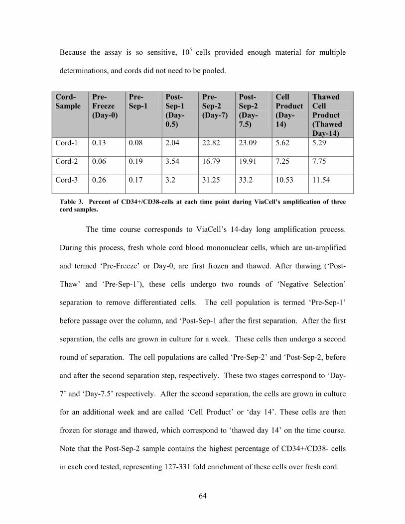
64
Because the assay is so sensitive, 105 cells provided enough material for multiple
determinations, and cords did not need to be pooled.
Cord- Sample
Pre- Freeze (Day-0)
Pre- Sep-1
Post- Sep-1 (Day-0.5)
Pre- Sep-2 (Day-7)
Post- Sep-2 (Day-7.5)
Cell Product (Day-14)
Thawed Cell Product (Thawed Day-14)
Cord-1 0.13 0.08 2.04 22.82 23.09 5.62 5.29
Cord-2 0.06 0.19 3.54 16.79 19.91 7.25 7.75
Cord-3 0.26 0.17 3.2 31.25 33.2 10.53 11.54
Table 3. Percent of CD34+/CD38-cells at each time point during ViaCell’s amplification of three cord samples.
The time course corresponds to ViaCell’s 14-day long amplification process.
During this process, fresh whole cord blood mononuclear cells, which are un-amplified
and termed ‘Pre-Freeze’ or Day-0, are first frozen and thawed. After thawing (‘Post-
Thaw’ and ‘Pre-Sep-1’), these cells undergo two rounds of ‘Negative Selection’
separation to remove differentiated cells. The cell population is termed ‘Pre-Sep-1’
before passage over the column, and ‘Post-Sep-1 after the first separation. After the first
separation, the cells are grown in culture for a week. These cells then undergo a second
round of separation. The cell populations are called ‘Pre-Sep-2’ and ‘Post-Sep-2, before
and after the second separation step, respectively. These two stages correspond to ‘Day-
7’ and ‘Day-7.5’ respectively. After the second separation, the cells are grown in culture
for an additional week and are called ‘Cell Product’ or ‘day 14’. These cells are then
frozen for storage and thawed, which correspond to ‘thawed day 14’ on the time course.
Note that the Post-Sep-2 sample contains the highest percentage of CD34+/CD38- cells
in each cord tested, representing 127-331 fold enrichment of these cells over fresh cord.

65
Telomerase Activity in Cord-1 Time Course
An ascending telomerase activity profile was observed during the time course
experiment on cord-1 (sample# EXPO91001A) as shown in figure 14. As expected, the
cancer cell extract positive control (lane 1) showed high telomerase activity. 1 µg cord
cell lysate protein load was used for the time course experiments in accordance with the
optimization experiments. The same ‘Master Mix’ for the PCR amplification was used to
assay all the samples, which proved to be critical for obtaining an even amplification of
the PCR control.
Figure 14. TRAP Assay on Cord-1, N=1

66
Telomerase activity was undetected early in ViaCell’s process, in ‘pre-freeze’
(Day-0), ‘Post thaw’ and ‘Pre-Sep 1’ time points (lanes 1-3). Telomerase activity was
low but detectable in ‘Post-Sep 1’ (lane 4), was high in ‘Pre- Sep 2’ (lane 5), and peaked
at ‘Post-Sep 2’ (lane 6). Telomerase activity at ‘Post-Sep 2’ (Day-7) was comparable to
the cancer cell extract positive control (lane 1). At ‘Day 14’, however, a dip in
telomerase activity was observed. Surprisingly, there was resurgence in telomerase
activity in ‘thawed-day 14’ cells which only differ from the ‘day 14’ cells by a single
round of freeze/thaw. The 36 bp internal PCR control was observed in all the lanes,
which indicates no sample contained an unusual amount of Taq Polymerase inhibitor.
To determine the reproducibility of the results obtained in cord 1 and to assay the
intra-sample variability, a trial 2 of the time course experiment on cord 1 was conducted
(figure 15). This second trial showed the same trends as trial 1. Because the 32P for trial-
2 was fresh, it proved sufficient to quantitate the telomerase bands cut out of the gel (fig
16). The histoplot determined by counting 32P corresponds with the telomerase activity
estimated by eye from the x-ray films. The Post-Sep-2 sample contained the highest
activity at 5.7x the fresh cord level.

67
Telomerase Activity in Cord-2 Time Course
Telomerase activity in cord-2 (Sample # EXPO91001B) reflected the same
pattern as in cord-1 under conditions in which the internal control was equally amplified
Figure 16. Quantitation of Telomerase Activity in Cord-1. The Y-axis shows values as percent relative to the cancer positive control. The counts per minute are shown on the histobars.
Figure 15. TRAP Assay on Cord-1, N=2

68
(figure 17). This particular analysis provided the most extended ladders of this entire
thesis.
Similar results were obtained in Trial 2 for Cord-2 (figure 18), except for the appearance
of an unusual band in the Pre-Sep-1 sample (lane 2). Because trial-2 for cord-2 used
fresh 32P, quantitation was performed (fig 19), except on the unusual Pre-Sep-1 sample.
The trends reflect what was seen earlier in cord-1. The Post-Sep-2 sample showed a 12x
or an 80% increase over fresh cord.
Figure 17. TRAP Assay on Cord-2, N=1

69
Figure 18. TRAP Assay on Cord-2, N=2
Figure 19. Quantitation of Telomerase activity in Cord-2. The Y-axis shows values as percent relative to cancer positive control.

70
Telomerase Activity in Cord-3 Time Course
Telomerase activity in cord-3 (figure 20) (sample # EXPO91001C) exhibited an
identical trend as observed in cord-1 and cord-2 samples. The assay continued to show
low intra-sample variability (fig 21), and the quantitation for cord-3 (fig 22) indicated an
8.0x or a 120% increase in telomerase activity for Post-Sep-2 relative to fresh sample.
The above TRAP results show that there is little intra-sample variability in the
assay. Although differences were observed between cords regarding the fold-increase in
activity, the main trend of telomerase activity observed in the three time courses was
identical. Table 4, shows percent CD34+ content and telomerase quantitation of each of
the cords tested. Although for each cord tested the highest telomerase activity occurred
for fraction containing the highest percent CD34+ cells, a direct correlation was not
always observed.
Figure 20. TRAP Assay on cord-3, N=1

71
Figure 21. TRAP Assay on cord-3, N=2
Figure 22. Quantitation of Telomerase activity in Cord-3. The Y-axis shows values as percent relative to cancer positive control.

72
Fraction CORD-1 CORD-2 CORD-3
%CD34+ Percent Telomerase Quantitation
Ratio (%CD34/ %Quant)
%CD34+ Percent Telomerase Quantitation
Ratio %CD34+ Percent Telomerase Quantitation
Ratio
Pre-Freeze 0.13 23 0.005 0.06 4 0.015 0.26 17 0.015 Pre-Sep-1 0.08 30 0.002 0.19 0.17 30 0.005 Post-Sep-1 2.04 33 0.061 3.54 16 0.221 3.2 37 0.086 Pre-Sep-2 22.82 59 0.386 16.79 51 0.329 31.25 85 0.367 Post-Sep-2 23.09 123 0.187 19.91 84 0.237 33.2 133 0.249
Day-14 5.62 73 0.076 7.25 29 0.250 10.53 63 0.167 ThawedDay-
14 5.29 100 0.052 7.75 43 0.180 11.54 97 0.118
Telomere Length Assay
In the second phase of this project, the telomere lengths of two of Viacell’s cord
populations were investigated via a telomere length assay (TLA). Various methods have
been described to detect telomeres and to measure telomere length (Harley, 1995;
Lansdorp et al., 1996). The TeloTAGGG Telomere Length Assay (Roche, # 2209136)
was chosen as the as the commercial source. This method utilizes Southern blot analysis
of terminal restriction fragments (TRF) obtained by digestion of genomic DNA using
frequently cutting restriction enzymes such as Rsa 1 and Hinf 1. The specificity of the
enzymes is such that the telomeric DNA (TTAGGG)n is not cut. After digestion, the
DNA fragments are separated by gel electrophoresis, blotted and the TRFs are visualized
by hybridization with DIG-labeled telomere-specific probe. Finally, after exposure of the
blot to an X-ray film, an estimate of the mean TRF length was obtained by visually
comparing the mean size of the smear to the DIG-labeled molecular weight marker.
Telomere length of human cell samples may range over one order of magnitude. Even
within a population of cell lines and on a single cell level, considerable heterogeneity of
Table 4. Shows %CD34+ content and Telomerase Quantitation for each cord.

73
telomere length is observed. Therefore, analyzing a population of cells provides the
average telomere length of the telomeres in the sample, indicated by a smear whose
average size is compared to the molecular weight marker. TRFs comprise not only the
variable terminal telomeres but also a short sub-telomeric region. In addition to a
molecular weight marker, two positive control DNAs (Control-DNA-low and Control-
DNA-high) obtained from immortal cell lines and supplied with the TeloTAGGG kit
were used to compare the mean TRF length of each sample. The mean TRF length of
these positive control cell lines has been estimated at 3.9 kb and 10.2 kb respectively.
After several false starts with this tricky assay, the controls (fig. 23) produced their
expected profiles.
Figure 23. Test of Controls for Telomere length Assay. Shows the DIG-labeled molecularweight marker and two positive control DNAs. The mean TRF length of control-DNA-High andcontrol-DNA-Low has been estimated at 10.2 kbp and 3.9 kbp respectively.
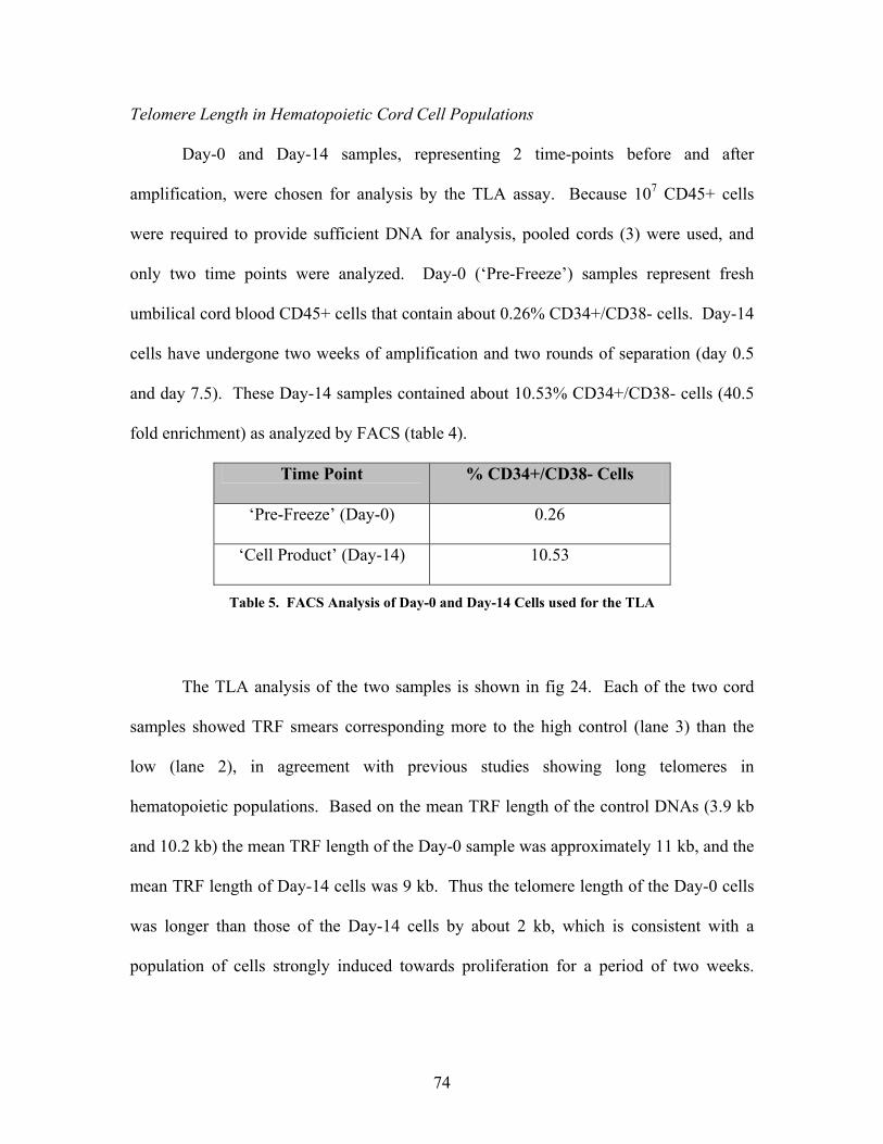
74
Telomere Length in Hematopoietic Cord Cell Populations
Day-0 and Day-14 samples, representing 2 time-points before and after
amplification, were chosen for analysis by the TLA assay. Because 107 CD45+ cells
were required to provide sufficient DNA for analysis, pooled cords (3) were used, and
only two time points were analyzed. Day-0 (‘Pre-Freeze’) samples represent fresh
umbilical cord blood CD45+ cells that contain about 0.26% CD34+/CD38- cells. Day-14
cells have undergone two weeks of amplification and two rounds of separation (day 0.5
and day 7.5). These Day-14 samples contained about 10.53% CD34+/CD38- cells (40.5
fold enrichment) as analyzed by FACS (table 4).
Time Point % CD34+/CD38- Cells
‘Pre-Freeze’ (Day-0) 0.26
‘Cell Product’ (Day-14) 10.53
Table 5. FACS Analysis of Day-0 and Day-14 Cells used for the TLA
The TLA analysis of the two samples is shown in fig 24. Each of the two cord
samples showed TRF smears corresponding more to the high control (lane 3) than the
low (lane 2), in agreement with previous studies showing long telomeres in
hematopoietic populations. Based on the mean TRF length of the control DNAs (3.9 kb
and 10.2 kb) the mean TRF length of the Day-0 sample was approximately 11 kb, and the
mean TRF length of Day-14 cells was 9 kb. Thus the telomere length of the Day-0 cells
was longer than those of the Day-14 cells by about 2 kb, which is consistent with a
population of cells strongly induced towards proliferation for a period of two weeks.

75
Trial 2 of the TLA analysis of the two samples showed that the results obtained were
reproducible (fig. 25).
Thus, despite our detection of telomerase activity at Day-14 compared to none
detected at Day-0, an average telomere loss of about 2 kb occurs after the two-week
amplification. So, the activity increase is not sufficient for fully maintaining telomere
length in the cells pushed towards proliferation. Therefore, the above results show that
the presence of telomerase activity does not necessarily correspond to longer telomeres.
Figure 24. TLA on Cord Samples, N=1. Lane 1 shows DIG labeled molecularweight marker, lane 2 shows control DNA-low, lane 3 shows control DNA-high,lane 4 shows Day 0 telomeric DNA, and lane 5 shows Day 14 telomeric DNA.

76
Initial Investigation of Culture Conditions that Could Potentially Alter Telomerase Activity
Based on previous studies showing that cell populations with elevated telomere
lengths and detectable telomerase activity show higher engraftment survival, ViaCell
may eventually be interested in exploring various culture conditions that can increase
telomerase activity further in their product and therefore compensate for the 2 kb
telomere loss observed during the amplification. Two preliminary experiments were
performed here. In the first set of treatments (fig. 26), Day-14 cord blood cells were
treated as follows A) ½ cord treated with Annexin (to rid apoptotic cells), B) whole cord
treated with Annexin, C) ¼ cord treated with 30% BSA (Bovine Serum Albumin) and D)
Figure 25. TLA on Cord Samples, N=2. Lane 1 shows the DIG labeledmolecular weight marker, lane 2 shows control DNA-low, lane 3 shows controlDNA-high, lane 4 shows Day 0 telomeric DNA, and lane 5 shows Day 14telomeric DNA.

77
¼ cord treated with 20% HSA gradients (to remove non-viable cells), then were analyzed
for telomerase activity. All four culture treatments (lanes A-D) showed equal telomerase
activity, which was comparable to the cancer cell extract positive control.
In the second set of treatments (fig. 27), day-14 cord cells were first put through a density
centrifugation with 20% HSA (to remove non-viable cells). Samples A and C were ½
cord cells treated with HSA coated plastics, and samples B and D were ½ cord cells
treated with uncoated plastics. These samples were then analyzed for telomerase activity.
As shown in fig 27, both the uncoated and coated plastic pre-treatments showed equal
telomerase activity.
Figure 26. Treatment of Day-14 Cord Cells with Annexin, BSA and HSA. Lane 1 shows cancercell extract positive control. A=1/2 cord treated with Annexin, B= whole cord treated withAnnexin, C= ¼ cord treated with 30% BSA, D= ¼ cord treated with 20% HSA.

78
Thus no cord treatment was identified in this preliminary analysis that altered the
telomerase activity of the day-14 cell population.
Figure 27. Day-14 cord cells have undergone density centrifugation with 20%HSA. Coating refers to a brief pre-treatment of the plastics with 5% HSA.
A= with HSA coat of plastics, B= without coat, C= with HSA coat of plastics,D= without coat. These samples were also heat inactivated to serve as negativecontrols. However, samples B and C were not completely heat inactivated andtherefore show faint telomerase activity.

79
DISCUSSION
Hematopoietic cell populations showing elevated telomerase activity (Morrison et
al., 1996) and elongated telomere lengths (Kobari et al., 2000; Lansdorp et al., 1997;
Vaziri et al., 1994; Notaro et al., 1997; Wynn et al., 1998;) display strong engraftment
survivability and higher replicative potential in humans during bone marrow transplants.
Thus the telomerase activity and telomere length are important parameters to analyze in
hematopoietic cell fractions slated for transplant into patients. In this study, we assessed
telomerase activity and telomere length in selectively amplified umbilical cord blood
(UCB) cell populations prepared using ViaCell’s patented stem cell amplification
process. The first aim of this project was to assay telomerase activity throughout
ViaCell’s entire ex vivo cell amplification process. The second aim was to analyze
telomere length in two of their samples. The third aim was to investigate various culture
conditions that could potentially upregulate telomerase activity in ViaCell’s final day-14
cell fraction slated for perfusion into a patient.
Telomerase Activity Assay
We have demonstrated using a PCR-based TRAP assay that telomerase activity is
undetectable early on in Day-0 (‘Pre-Freeze’), ‘Post-Thaw’ and ‘Pre-Sep-1’ (Day-0.5)
samples), increases following removal of differentiated cells in Post-Sep-1 samples, and
increase by as much as 120-fold after 1 week of ex-vivo expansion (as observed in ‘Post-
Sep-2’ (Day-7) samples). Because telomerase activity was relatively high in all fractions
induced for proliferation (Pre-Sep-2, Post-Sep-2, Day-14, Thawed Day-14) compared to
fresh cord, Post-Thaw or Pre-sep-1, our results agree with others (Engelhardt et al., 1997;
80
Holt et al., 1996; Zhu et al., 1996) indicating that telomerase is present in rapidly
expanding cells. It has also been previously shown that the expression of human
telomerase reverse transcriptase (hTERT) was low in freshly isolated cord blood cells,
and was significantly increased when these cells were cultured in vitro along with
optimal cytokines (Ma and Zou, 2001). Reports have previously shown that telomerase
is upregulated at cell cycle entry as cells progress through S-phase, and repressed in
quiescent Go cells Sakabe et al., (1998). Our low activity in the unamplified, unselected,
cell population may simply reflect the low abundance of HSCs in this population, or the
quiescent primitive nature of these stem/progenitor cells. Telomerase activity increased
(in all 3 cords tested) in Post-Sep-1 samples compared to Pre-Sep-1. This increase may
simply reflect the removal of Lin+ cells known to be low in telomerase activity.
After 2 weeks of ex vivo expansion, telomerase activity showed a slight decline in
all ‘Day-14’ samples relative to Day-7 (although it was still above fresh cord, Post-Thaw,
and Pre-sep-1). This decline may be due to the differentiation of a subset of
hematopoietic stem/progenitor cells in the Post-Sep-2 samples into more mature
telomerase-low blood cells during the weeklong growth (indicated by the decrease from
33.2% to 10.53% of CD34+/CD38- cells in the Day-14 sample). These results agree
with previous studies showing that the induction of a differentiation program decreases
telomerase activity in the hematopoietic sample (Engelhardt et al., 1997).
Surprisingly the telomerase activity was again upregulated after the ‘Day-14’
samples were frozen for storage and thawed. The reason for this is unknown. However,
this observation could be explained in part by an increase in the survival of telomerase-
rich cells following freezing, stimulation of an unknown telomerase activator, or

81
denaturation of a telomerase inhibitor. The increase is not likely due to an increase in
percent CD34+ cells in this fraction because the CD34 count goes up by only 1% by
FACS analysis.
Telomere Length Assay
In concordance with previous studies (Vaziri et al., 1994; Chiu et al., 1996), we
report telomere shortening of hematopoietic cells on proliferation despite the presence of
detectable levels of telomerase activity. Southern blot analysis of telomere length in the
total nucleated cell population obtained at 2 different time points (Day-0 and Day-14),
demonstrated relatively long telomeres in the these hematopoietic fractions compared to
the DNA from the differentiated cell controls, and a 2 kb loss of telomeric DNA in these
cells after 14 days of amplification. These results are consistent with a model in which
the upregulation of telomerase activity in the Day-14 sample (compared to fresh cord) is
insufficient to maintain telomere lengths following cell amplification. These results
agree with previous studies by Engelhardt et al., 1997; Chiu et al., 1996, showing
telomere shortening in amplified hematopoietic populations. Direct analysis of telomeres
in HSCs by in situ hybridization during serial transplantation of murine HSCs also
revealed a reduction in telomere size (Allsopp et al., 2001). Telomerase activity is
upregulated in primitive hematopoietic cells following their entry into cell cycle, which is
sufficient to reduce (not to completely prevent) telomere loss when bulk cell turnover,
cell expansion, and massive cell proliferation takes place (Engelhardt et al., 1997).
It has been suggested that most primitive hematopoietic cells lose telomeric DNA
at a rate that is roughly comparable to other somatic cells (50-100 bp per doubling)

82
(Allsopp et al., 1992; Vaziri et al., 1993). From our data it can be inferred that the
hematopoietic cell population has undergone around 20 doublings, assuming a constant
rate of loss of 100 bp telomeric DNA per cycle. A large body of evidence on telomere
length in somatic cells in vitro and in vivo indicates that telomere length serves as a
biomarker of the replicative history of cells (Harley et al 1990; Vaziri et al., 1994). It has
been suggested that the replicative senescence within a hematopoietic lineage may be
causally linked to functional differences such as a decrease in the production of CD34+
cells, and a decreased proliferation rate of CD34+ cells and those cells responding to a
mixture of hematopoietic cytokines (Lansdorp et al., 1993).
The proliferative lifespan of stem cells to sustain hematopoiesis throughout life is
not known. Lansdorp et al (1997) propose that HSCs like other somatic cells may have
only a limited replicative potential (<100 divisions). This hypothesis is supported by the
consideration that, in theory, 55 divisions can yield 4 x 10¹⁶ cells, which is about the
same as the estimated number of blood cells produced over a lifetime. Lansdorp et al.,
(1996) have shown that the proliferative potential of most, if not all, HSCs is limited,
decreases with age, and correlates directly with telomere length.
Telomerase studies have widespread implications for hematopoietic
transplantations, as well as gene therapy. Reports have shown that the proliferative
potential of HSCs is decreased after hematopoietic reconstitution of myelo-ablated
patients. The mean TRF length was shown to be consistently shorter in the bone marrow
transpant (BMT) recipient than in the respective donor. One interpretation of this finding
is that the fewer the HSCs transferred to a recipient, the more cell divisions are needed
for reconstitution of hematopoiesis. Consequently, a greater consumption of telomeres

83
takes place. The donor stem cells must presumably undergo a larger number of telomere
shortening rounds in the engrafting recipient than have naturally occurred in the donor
(Notaro et al., 1997). Similar conclusions were obtained for autologous peripheral blood
stem cell transplantation (Lee et al., 1999). Direct analysis of telomeres in HSCs by in
situ hybridization during serial transplantation of murine HSCs also revealed a reduction
in telomere size (Allsopp et al., 2001).
The fate of telomeres may also be crucial for the outcome of gene therapy
protocols in which one or few stem cells are expected to repopulate the bone marrow
(Notaro et al., 1997). Another factor affecting gene therapy is that hematopoietic
engraftment imposes replicative stress on stem cells, resulting in aging effect, which
would carry the risk of an increased frequency of clonal hematopoietic disorders during
later life. This is particularly important in young recipients with a lifetime of
hematopoietic demand before them (Wynn et al., 1998). In this regard, cord blood cells
would be a better source for allogeneic transplantation. Studies have shown significant
functional differences between UCB and adult bone marrow (BM) and peripheral blood
(PB) cells. The UCB cells have longer telomeres compared with PB and BM cells. This
suggests that CB has higher replicative potential than adult PB or BM cells, which
combined with their greater expansion potential, would support the use of such cells for
allogeneic transplantation (Mayani and Lansdorp, 1998).
In conclusion, our TLA and TRAP data support the prevailing hypothesis that
telomerase activity in hematopoietic cells reduces rather than completely eliminates
telomere loss on proliferation, and may thus help extend the proliferative life span of
hematopoietic cells. Reports have shown that the developmental characteristic most

84
consistently associated with telomerase expression is self-renewal potential (Morrison et
al., 1996). Therefore, the relatively high telomerase activity and telomere lengths in the
Day-14 samples is encouraging because it provides a new way to validate ViaCell’s
clonogenic amplification protocol for the cell populations that will be perfused into an
immunosuppressed patient and indicates a high self-renewal potential for these cells.
Treatments to Elevate Telomerase Activity
Different culture conditions and treatments that could potentially elevate
telomerase activity were investigated. Day-14 cord blood fractions were treated with
annexin, 30% BSA (Bovine Serum Albumin), 20% HSA, or first put through a density
centrifugation with 20% HSA and treated with HSA coated plastics. These treatments
were performed to remove apoptotic cells or non-viable cells from the population, which
are known to be low in telomerase activity. Unfortunately, none of the treatments altered
telomerase activity, so perhaps these unwanted cells only constituted a small percent of
the population. Viability studies should be performed to ascertain the abundance of these
unwanted cells in the population. Therefore in our analysis, no cord blood was identified
that could potentially elevate telomerase activity.
Future Investigations
In future, telomere length should be assessed in all the different time points of
ViaCell’s amplification process, especially Post-Sep-1 (unamplified, but selected) and
Post-Sep-2 (amplified and selected). Alternative methods of telomere length
measurement that are less labor intensive than a TLA could be tested, such as a “telomere

85
amount and length assay” (TALA) (Gan et al., 2001). TALA is based on solution
hybridization and does not require blotting, pre-hybridization and washing. Compared to
the TLA, one lab claims TALA shows a 4-fold greater sensitivity, >2 fold-higher
reproducibility and 4-fold less time requirement (Gan et al., 2001). However at this
moment this assay is not commercially available. Other methods such as flow cytometry-
based fluorescent in situ hybridization (FISH) can also be used for measuring telomere
length in situ, in single cells. A TelBam8 probe that is unique for the subtelomeric region
of the long arm of chromosome 7 can also be used to measure the telomere length of one
end of a single chromosome pair. This method reduces the variation size of the telomeric
length that is seen in blots hybridized to a (TTAGGG)n telomeric repeat probe (Notaro et
al., 1997).
The telomerase experiments could be expanded by conducting Northern blots or
RT-PCR for telomerase RNA, or alternatively by performing western blots using
antibodies against the reverse transcriptase subunit. Such antibodies have recently
become commercially available following the cloning of the hTERT gene by Nakamura
et al. (1997). In vitro studies with telomerase inhibitors can be conducted to further
understand the specific role of telomerase in telomere maintenance in hematopoietic stem
cells. Recent studies indicate the existence of an alternative “lengthening of telomeres”
(ALT) system in which telomere maintenance occurs in the absence of telomerase
activity (Bryan et al., 1997). Previous studies have shown the presence of ALT in yeast,
and in a subset of tumors and tumor-derived cell lines (Bryan et al., 1997). This could be
investigated by treatment of CB derived cell populations with telomerase inhibitors such
as TRF1 and TRF2, differentiation inducing agent “all-trans retinoic acid” (ATRA), and

86
other putative telomerase inhibitors such as alterperylenol (a fungus metabolite) (Togashi
et al., 1998) or a bis-dimethylaminoethyl derivative of quindoline (an alkaloid from the
west African shrub Cryptolepis sanguinolenta), which stabilizes the folded G-quadruplex
structures and thus inhibit telomerase activity (Caprio et al., 2000). Such studies could
shed new light on the means whereby ALT is repressed in normal hematopoietic cells and
HSCs.
Finally, since the level of telomerase activity is insufficient to fully maintain
telomere lengths in hematopoietic stem cells, perhaps its activity could be increased by
transfecting these cells with plasmids encoding the hTERT gene. The expression of the
hTERT gene parallels telomerase activity, while the RNA component is ubiquitously
expressed in all cells, therefore, such gene therapy could be successful with only the
hTERT gene transfection. Perhaps such treated cells would show further elevated
telomerase activity which would fully maintain telomere length in spite of the
proliferation, and would show increased survivability of the graft in the host.

87
BIBLIOGRAPHY
Artandi SE, DePinho RA (2000) A critical role for telomerase in suppressing and facilitating carcinogenesis. Curr Opin Genet Dev 10: 39-46. Allsopp RC, Vaziri H, Patterson C, Goldstein S, Young-lai EV, Futcher AB, Greider CW, Harley CB (1992) Telomere length predicts replicative capacity of human fibroblasts. Proc. Natl. Acad. Sci.USA 89: 10114-10118. Allsopp RC, Cheshier S, Weissman IL (2001) Telomere shortening accompanies increased cell cycle activity during serial transplantation of hematopoietic stem cells. J Exp Med 193: 917-924. Bachand F & Autexier C (2001) Functional regions of human telomerase reverse transcriptase and human telomerase RNA required for telomerase activity and RNA- protein interactions. Molecular and Cellular Biology 21: 1888-1897. Belair CD, Yeager RT, Lopez PM, Reznikoff CA (1997) Telomerase activity: A biomarker of cell proliferation, not malignant transformation. Proc. Natl. Acad. Sci. USA 94: 13677-13682. Blackburn EH and Gall JG (1978) A tandemly repeated sequence at the termini of the extrachromosomal ribosomal RNA genes in Tetrahymena. J.Mol.Biol. 120: 33-53. Blackburn EH (1991) Structure and function of telomeres. Nature 350: 569-573. Blackburn EH (2000) The end of the (DNA) line. Nat Struct Biol 7: 847-50. Blasco MA, Lee HW, Hande MP, Samper E, Lansdorp PM, DePinho RA, Greider CW (1997) Telomere shortening and tumour formation by mouse lacking telomerase RNA. Cell 91: 25-34. Bodnar AG, Ouellette M, Frolkis M, Holt SE, Chiu CP, Morin GB, Harley CB, Shay JW, Lichtsteiner S, Wright WE (1998) Extension of life-span by introduction of telomerase into normal human cells. Science 279: 349-352. Broccoli D, Young JW, De Lange T (1995) Telomerase activity in normal and malignant hematopoietic cells. Proc Natl Acad Sci USA 92: 9082-9086 Broccoli D, Smorgorzewska A, Chong L, de Lange T (1997) Human telomeres contain two distinct Myb-related proteins, TRF1 and TRF2. Nat Genet 17: 231-235. Broxmeyer HE, Douglas GW, Hangoc G, Cooper S, Bard J, English D, Arny M, Thomas L (1989) Human umbilical cord blood as a potential source of transplantable hematopoietic stem/progenitor cells. Proc. Natl. Acad. Sci. USA. 86: 3828-3832.

88
Broxmeyer HE and Carrow CE (1993) Characterization of cord blood stem/progenitor cells. J Hematother 2: 197-199. Brugger W, Scheding S, Zeigler B, Buhring HJ, Kanz L (2000) Ex vivo manipulation of hematopoietic stem and progenitor cells. Seminars in Hematology 37: 42-49. Bryan TM, Englezou A, Gupta J, Bacchetti S & Reddel RR (1995) Telomere elongation in immortal human cells without detectable telomerase activity. EMBO J. 14: 4240-4248. Bryan TM, Englezou A, Dalla-Pozza L, Dunham MA, Reddel RR (1997) Evidence for an alternative mechanism for maintaining telomere length in human tumours and tumour-derived cell lines. Nat Med 3: 1271-1274. Caprio V, Guyen B, Opoku-Boahen Y, Mann J, Gowan SM, Kelland LM, Read MA, Neidle S (2000) A novel inhibitor of human telomerase derived from 10H-indolo[3,2-b] quinoline. Bioorg Med Chem Lett 10: 2063-2066. Cardoso A, Li M-L, Betard P, Hatzfeld A, Brown EL, Levesque JP, Sookdeo H, Panterne B, Sansilvestri P, Clark SC (1993) Release from quiescence of CD34+ CD38 human umbilical cord blood cells reveals their potentiality to engraft adults. Proc. Natl. Acad. Sci USA 90: 8707-8711. Chaudhary PM and Roninson IB (1991) Expression and activity of P-glycoprotein, a multidrug efflux pump, in human hematopoietic stem cells. Cells 66: 85-94. Challita PM and Kohn DB (1994) Lack of expression from a retroviral vector after transduction of murine hematopoietic stem cells is associated with methylation in vivo. Proc. Natl. Acad. Sci USA 91: 2567-2571. Chin L, Artandi SE, Shen Q, Tam A, Lee SL, Gottlieb, GJ, Greider, CW, DePinho RA (1999) p53 deficiency rescues the adverse effects of telomere loss and cooperates with telomere dysfunction to accelerate carcinogenesis. Cell 97: 527-538. Chiu CP, Dragowska W, Kim NW, Vaziri H, Yui J, Thomas T E, Harley CB, Lansdorp PM (1996) Differential expression of telomerase activity in hematopoietic progenitors from adult bone marrow. Stem Cells 14: 239-248. Civin CI and Gore SD (1993) Antigenic analysis of hematopoiesis: a review. J Hematother 2: 137-144. Collins K (2000) Mammalian telomeres and telomerase. Curr. Opin. Cell Biol. 12: 378-383. Craig W, Kay R, Cutler RL, Lansdorp PM (1993) Expression of Thy-1 on human hematopoietic progenitor cells. J Exp Med 177: 1331-1342.

89
Craig J (2000) ViaCord, Inc. and t. Breeders, Inc. Merge to from ViaCell, Inc., ViaCell-Press Release De Lange T (1994) Activation of telomerase in a human tumor. Proc. Natl. Acad. Sci. USA 91: 2882-2885. De Lange T (1998) Telomeres and senescence: Ending the debate. Science 279: 3349-54. De Lange T & DePinho RA (1999) Unlimited mileage from telomerase? Science 283: 947-949. De Wynter EA, Buck D, Hart C, Heywood R, Coutinho LH, Clayton A, Rafferty JA, Burt D, Guenechea G, Bueren JA, Gagen D, Fairburn LJ, Lord BI, and Testa NG (1998) CD34+ AC133+ Cells isolated from cord blood are highly enriched in long-term culture-initiating cells, NOD/SCID-repopulating cells and dendritic cell progenitors. Stem Cells 16: 387-396. Dheane K, Marck EV, Parweresch R (2000) Telomeres, telomerase and cancer: an up-date. Virchows Arch 437: 1-16. Engelhardt M, Kumar R, Albanell J, Pettengell R, Han W, Moore MA (1997) Telomerase regulation, cell cycle, and telomere stability in primitive hematopoietic cells. Blood 90: 182-193. Farley TJ, Ahmed T, Fitzgerald M, Preti RA (1997) Optimization of CD34+ cell selection using immunomagnetic beads: Implications for use in cryopreserved peripheral blood stem cell collections. J Hematother 6: 53-60. Feng J, Funk WD, Wang SS, Weinrich SL, Avilon AA, Chiu CP, Adams RR, Chang E, Allsopp RC, Yu J, Le S, West MD, Harley CB, Andrews WH, Greider C, Villeponteau B (1995) The RNA component of human telomerase. Science 269: 1236-1241. Gan YB, Engelke KJ, Brown CA, Au JLS (2001) Telomere amount and length assay. Pharmaceutical Research. 18: 1655-1659. Gilmore GL, DePasquale DK, Lister J, Shadduck RK (2000) Ex vivo expansion of human umbilical cord blood and peripheral blood CD34+ hematopoietic stem cells. Exp Hematol 28: 1297-1305. Gluckman E (2000) Current status of umbilical cord blood hematopoietic stem cell transplantation. Exp Hematol. 11: 1197-205. Gluckman E, Broxmeyer H, Auerbach AD, Friedman HS, Douglas GW, Devergie A, Esperou H, Thierry D, Socie G, Lehn P, Cooper S, English D, Kurtzberg J, Bard J, Boyse

90
EA (1989) Hematopoietic reconstitution in a patient with Fanconi’s anemia by means of umbilical cord blood from an HLA-identical sibling. N Engl J Med 321: 1174-1178. Gluckman E, Wagner J, Hows J, Kernan N, Bradley B, Broxmeyer HE (1993) Cord blood banking for hematopoietic stem cell transplantation: an international cord blood transplant registry. Bone Marrow Transplantation 11: 199-200. Gluckman E, Rocha V, Boyer Chammard A, Locatelli F, Arcese W, Pasquini R, Ortega J, Souillet G, Ferreira E, Laporte JP, Fernandez M, Chastang C (1997) Outcome of cord blood transplantation from related and unrelated donors. Eurocord Transplant Group and the European Blood and Marrow Transplantation Group. N Engl J Med 337: 373-381. Greenberg RA, Chin L, Femino A, Lee KH, Gottlieb GJ, Singer RH, Greider CW, DePinho RA, (1999) Short dysfunctional telomeres impair tumorigenesis in the INKa(∆2/3) cancer-prone mouse. Cell 97: 515-525. Greider CW and Blackburn EH (1985) Identification of a specific telomere terminal transferase activity in Tetrahymena extracts. Cell 43: 405-413. Greider CW and Blackburn EH (1989) A Telomeric sequence in the RNA of Tetrahymena telomerase required for telomere repeats synthesis. Nature 337: 331-337. Greider CW & Blackburn EH (1996) Telomeres, telomerase and cancer. Sci. Am. 274: 92-97. Griffith JD, Comeau L, Rosenfield R, Stansel M, Bianchi A, Moss H, and De Lange T (1999) Mammalian telomeres end in a large duplex loop. Cell 97: 503-514. Hahn WC, Stewart SA, Brooks MW, York SG, Eaton E, Kurachi A, Beijersbergen RL, Knoll JH, Meyerson M, Weinberg RA (1999) Inhibition of telomerase limits the growth of human cancer cells. Nat. Med. 10: 1164-1170. Halvorsen TL, Leibowitz G, Levine F (1999) Telomerase activity is sufficient to allow transformed cells to escape from crisis. Mol Cell Biol 19: 1864-70. Harley CB, Futcher AB and Greider CW (1990) Telomeres shorten during aging of human fibroblasts. Nature 345: 458-460. Harley CB (1991) Telomere loss: mitotic clock or genetic time bomb? Mutat Res. 256: 271-282. Harley CB (1995) Telomeres and aging. In Telomeres. Cold spring Harbour Laboratory, New York.

91
Harrington L, McPhail T, Mar V, Zhou W, Oulton R, Amgen EST Program, Bass M B, Arruda I, Robinson MO (1997) A mammalian telomerase associated protein. Science 275: 973-977. Havenga M, Hoogerbrugge P, Valerio D, Helmuth HG (1997) Retroviral stem cell gene therapy. AlphaMed. Press 15: 162-179. Healy L, May G, Gale K, Grosveld F, Greaves M, Enver T (1995) The stem cell antigen CD34 functions as a regulator of hematopoietic cell adhesion. Proc Natl Acad Sci USA 92: 12240-12244. Hiyama K, Hirai Y, Kyoizumi S, Akiyama M, Hiyama E, Piatzsek MA, Shay JW, Ishoka S, Yamakido M (1995) Activation of telomerase in human lymphocytes and hematopoietic progenitor cells. J Immunol 155: 3711-3715 Holt SE, Shay JW, Wright WE (1996) Refining the telomere-telomerase hypothesis of ageing and cancer. Nat Biotech 14: 836-9. Holt SE, Wright WE, Shay JW (1996) Regulation of telomerase activity in immortal cell lines. Mol Cell Biol 16: 2932-2939. Holt SE, Wright WE, Shay JW (1997) Multiple pathways for the regulation of telomerase activity. Eur J Cancer 5: 761-766. Hows JM, Bradley BA, Marsh JC, Luft T, Coutinho L, Testa NG, Dexter TM (1992) Growth of human umbilical cord blood in long-term hematopoietic cultures. Lancet 340: 73-76. Hohaus S, Voso MT, Barbera OL, Cavallo S, Bellacosa A, Rutella S, Rumi C, Genuardi M, Neri G, Leone G (1997) Telomerase activity in human hematopoietic progenitor cells. Haematologica 82: 262-268. Jiang XR, Jimenez G, Chang E, Frolkis M, Kusler B, Sage M, Beeche M, Bodnar AG, Wahl GM, Tlsty TD, Chiu CP (1999) Telomerase expression in human somatic cells does not induce changes associated with a transformed phenotype. Nat Genet 21: 111-114. Joshi SS, Tarantolo SR, Kuszynski CA, Kessinger A (2000) Antitumor therapeutic potential of activated human umbilical cord blood cells against leukemia and breast cancer. Clin. Cancer Res 6: 4351-4358. Kim NW, Piatyszek MA, Prowse KR, Harley CB, West MD, Ho PL, Coviello GM, Wright WE, Weinrich SL, Shay JW (1994) Specific association of human telomerase activity with immortal cells and cancer. Science 266: 2011-2015. Kipling D (2001) Telomeres, replicative senescence and human ageing. Maturitas 38: 25-38.

92
Kiyono T, Foster SA, Koop JI, McDougall JK, Galloway D, Klingelhutz AJ (1998) Both Rb/p16INK4a inactivation and telomerase activity are required to immortalize human epithelial cells. Nature 396: 84-88. Klapper W, Parwaresch R, Krupp G (2001) Telomere biology in human aging and aging syndromes. Mechanisms of Ageing and Development. 122: 695-712. Knudtzon S (1974) In vitro growth of granulocyte colonies from circulating cells in human cord blood. Blood 43: 357-361. Kobari L, Pflumio F, Giarratana M, Li X, Titeux M, Izac B, Leteurtre F, Coulombel L, Douay L (2000) In vitro and in vivo evidence for the long-term multilineage (myeloid, B, NK, and T) reconstitution capacity of ex vivo expanded human CD34(+) cord blood cells Exp Hematol 28:1470-80 Kohn DB, Weinberg KI, Nolta JA, Heiss LN, Lenarsky C, Crooks GM, Hanley ME, Annett G, Brooks JS, el-Khoureiy A, et al. (1995) Engraftment of gene-modified umbilical cord blood cells in neonates with adenosine deaminase deficiency. Nat Med 1: 1017-1023. Krause DS, Fackler MJ, Civin CI, May WS (1996) CD34: structure, biology and clinical utility. Blood 87: 1-3. Kuby J.1992 Immunology. W.H.Freeman and Company. New York, N.Y; 1992, pp 40-47. Kurenova EV and Mason JM (1997) Telomere Functions. A Review. Biochemistry (Mosc) 62:1242-1253. Kurtzberg J, Laughlin M, Graham ML, Smith C, Olson JF, Halperin EC, Ciocci G, Carrier C, Stevens CE, Rubinstein P (1996) Placental blood as a source of hematopoietic stem cells for transplantation into unrelated recipients. N Eng J Med 335: 157-166. Lansdorp PM, Dragowska W, Mayani H (1993) Ontogeny related changes in proliferative potential of human hematopoietic cells. J Exp Med 178: 787-791. Lansdorp PM, Verwoerd NP, Van de Rijke FM, Dragowska V, Little MT, Dirks RW, Raap AK, and Tanke HJ (1996) Heterogeneity in telomere length of human chromosomes. Hum. Mol. Genet. 5: 685-691. Lansdorp PM, Poon S, Chavez E, Dragowska V, Zijlmans M, Bryan T, Reddel R, Egholm M, Bacchetti S, Martens U (1997) Telomeres in the hematopoietic system. Ciba Found Symp. 211: 219-222.

93
Lawson BR, Prud’homme GJ, Chang Y, Gardner HA, Kuan J, Kono DH, Theofilopoulos AN (2000) Treatment of murine lupus with cDNA encoding IFN-gammaR/Fc. J Clin. Invest. 106: 207-215. Lee J, Kook H, Chung I, Kim H, Park M, Kim C, Nah J, Hwang T (1999) Telomere length changes in patients undergoing hematopoietic stem cell transplantation. Bone Marrow Transplant 24: 411-415. Lindsey J, McGill NI, Lindsey LA, Green DK and Cooke HJ (1991) In vivo loss of telomeric repeats with age in humans. Mutat. Res. 256: 45-48. Lundblad V & Wright WE (1996) Telomeres and telomerase: a simple picture becomes complex. Cell 87: 369-375. Ma Y and Zou P (2001) The expression of human telomerase reverse transcriptase gene in cord blood hematopoietic stem cells and its significance. Zhonghua Nei Ke Za Zhi. 40: 657-659. (Article in Chinese). Mayani H, Dragowska W, Lansdorp PM (1993) Characterization of functionally distinct subpopulations of CD34+ cord blood cells in serum free long-term cultures supplemented with hematopoietic cytokines. Blood 82: 2664-2672. Mayani H, Guilbert LJ, Janowska-Wieczorek A (1992) Biology of the hematopoietic microenvironment. Eur J Haematol 49: 225-233. Mayani H, Little M-T, Dragowska W, Thornbury G, Lansdorp PM (1995) Differential effects of the hematopoietic inhibitors MIP-1α, TGF-β, and TNF-α on cytokine –induced proliferation of subpopulations of CD34 cells purified from cord blood and fetal liver. Exp Hematol 23: 422-427. Mayani H and Lansdorp PM (1995) Proliferation of individual hematopoietic progenitors purified from umbilical cord blood. Exp Hematol 23: 1453-1462. Mayani H and Lansdorp PM (1998) Biology of human umbilical cord blood-derived hematopoietic stem/progenitor cells. Stem Cells 16: 152-165. McGovern MF, Michalenka LM, Parsons MK (2001) Selective amplification of cryopreserved human umbilical cord blood stem cells. WPI MQP, April, 2001. Meyerson M, Counter CM, Eaton EN, Ellisen LW, Steiner P, Caddle SD, Ziagura L, Beijersbergen RL, Davidoff MJ, Liu QY, Bacchetti S, Haber DA, Weinberg RA (1997) hEST2, the putative human telomerase catalytic subunit gene, is upregulated in tumor cells and during immortalization. Cell 90: 785-795. Mitchell JR, Cheng J and Collins K (1999) Abox H/ACA small nucleolar RNA-like domain at the human telomerase RNA 3’ end. Mol Cell Biol 19: 567-576.
94
Moore MAS and Hoskins J (1994) Ex vivo expansion of cord blood-derived stem cells and progenitors. Blood Cells 20: 468. Morin GB (1989) The human telomere terminal transferase enzyme is a ribonucleoprotein that synthesizes TTAGGG repeats. Cell 59: 521-529. Morrison SJ, Prowse KR, Ho P, Weissman IL (1996) Telomerase activity in hematopoietic cells is associated with self-renewal potential. Immunity 3: 207-216. Moyzis RK, Buckingham JM, Cram LS, Dani M, Deaven LL, Jones MD, Meyne J, Ratliff RL, Wu JR (1988) A highly conserved repetitive DNA sequence, (TTAGGG)n, present at the telomeres of human chromosomes. Proc. Natl. Acad. Sci. USA 85: 6622-6626. Nakahata T and Ogawa M (1982) Hematopoietic colony-forming cells in umbilical cord blood with extensive capability to generate mono- and multipotential hematopoietic progenitors. J Clin Invest 70: 1324-1328. Nakamura TM, Morin GB, Chapman KB (1997) Telomerase catalytic subunit homologs from fission yeast and human. Science 277: 955-959. Nakayama J, Tahar H, Tahara E, Saito M, Ito K, Nakamura H, Nakanishi T, Ide T, Ishikawa F (1998) Telomerase activity by hTERT in human normal fibroblasts and hepatocellular carcinomas. Nat Genet 18: 65-68. Negrin RS, Atkinson K, Leemhuis T, Hanania E, Juttner C, Tierney K, Hu WW, Johnston LJ, Goldstein KE, Blume KG, Weissman IL, Bower S, Karanes C, Peters W, and Klein J (2000) Transplantation of highly purified CD34+ Thy-1+ hematopoietic stem cells in patients with metastatic breast cancer. Biol. Blood Marrow Transplant. 6: 262-271. NIH (2001) Stem Cells: Scientific Progress and future Research Directions. http://www.nih.gov/news/stem cell. Noort WA, Falkenburg JHF (2000) Hematopoietic content of cord blood. London: Martin Dunitz, pp 13-37. Norrback KF & Roos G (1997) Telomeres and telomerase in normal and malignant hematopoietic cells. Eur J Cancer 33: 774-80. Notaro R, Cimmino A, Tabarini D, Rotoli B, and Luzzatto L (1997) In vivo telomere dynamics of human hematopoietic stem cells. Proc. Natl. Acad. USA 94: 13782-13785. Odorico JS, Kaufman DS, and Thomson JA, (2001) Multilineage differentiation from human embryonic stem cell lines. Stem Cells 19: 193-204.

95
Olovnikov AM (1973) A theory of marginotomy. The incomplete copying of template margin in enzymic synthesis of polynucleotide and biological significance of the phenomenon. J. Theor. Biol. 41: 181-190. Pafumi C, Zizza G, Pernicone G, Bandiera S, Farina M, Russo A, Iraci M, Cianci A (2001) Two enrichment methods to obtain CD34+ stem cells from umbilical cord blood. Bratisl Lek Listy 102: 183-186. Papayannopoulou T and Nakamoto B (1993) Peripheralization of hematopoietic progenitors in primates treated with anti-VLA4 integrin. Proc. Natl. Acad Sci USA 90: 9374-7378. Pedersen RA (1999) Embryonic stem cells for medicine. Sci. Am. 280: 68-73. Pettengell R and Testa NG (1995) Biology of blood progenitor cells used in transplantation. Int J Hematol 61: 1-15. Petzer A L, Zandstra PW, Piret JM, Eaves CJ (1996) Differential cytokine effects on primitive (CD34+CD38-) human hematopoietic cells: Novel responses to Flt-3 ligand and thrombopoietin. J Exp Med 183: 2551-2558. Piacibello W, Sanavio F, Garetto L, Severino A, Bergandi D, Ferrario J, Fagioli F, Berger M, and Aglietta M (1997) Extensive amplification and self-renewal of human primitive hematopoietic stem cells from cord blood. Blood 89: 2644-2653. Pittenger MF, Mackay AM, Beck SC, Jaiswal RK, Douglas R, Mosca JD, Moorman MA, Simonetti DW, Craig S, Marshak DR (1999) Multilineage potential of adult human mesenchymal stem cells. Science 284: 143-147. Rathjen PD, Lake J, Whyatt LM, Bettess MD, and Rathjen J (1998) Properties and uses of embryonic stem cells: prospects for application to human biology and gene therapy. Reprod. Fertil. Dev.10: 31-47. Rhyu MS (1995) Telomeres, telomerase and immortality. Natl.Cancer Inst. 87: 884-894. Rubinstein P, Carrier C, Scaradavou A, Kurtzberg J, Adamson J, Migliaccio AR, Berkowitz RL, Cabbad M, Dobrila NL, Taylor PE, Rosenfield RE, Stevens CE (1998) Outcomes among 562 recipients of placental blood transplants from unrelated donors. N Engl J Med 339: 1565-1577. Sakabe H, Yahata N, Kimura T, Zeng ZZ, Minamiguchi H, Kaneko H, Mori KJ, Ohyashiki K, Ohyashiki JH, Toyama K, Abe T, Sonoda Y (1998) Human cord blood-derived primitive progenitors are enriched in CD34+c-kit- cells: correlation between long-term culture-initiating cells and telomerase expression. Leukemia. 12: 728-34.

96
Sasaki T, Fujiwara H (2000) Detection and distribution patterns of telomerase activity in insects. Eur.J.Biochem. 267: 3025-3031. Seachrist L (1995) Telomeres draw a crowd at Toronto cancer meeting. Science 268: 29-30. Shay JW, Pereira-Smith OM, Wright WE (1991) A role for both RB and p53 in the regulation of human cellular senescence. Exp. Cell Res. 196: 33-39. Shay JW, Wright WE, Brasiskyte D, Van der Haegen BA (1993) E6 of human papillomavirus type 16 can overcome the M1 stage of immortalization in human mammary epithelial cells but not in human fibroblasts. Oncogene 8: 1407-1413. Shay JW (1995) Aging and cancer: are telomeres and telomerase the connection? Mol. Med. Today 1: 378-384. Shay J W (1999) Toward identifying a cellular determinant of telomerase repression. J Natl Cancer Inst. 91: 4-6. Shamblott MJ, Axwlman J, Wang S, Bugg EM, Littlefield JW, Donovan PJ, Blumenthal PD, Huggins GR, and Gearhart JD. (1998) Derivation of pluripotent stem cells from cultured human primordial germ cells. Proc. Natl. Acad. Sci.USA 95: 13726-13731. Smith AG (2001) Origins and properties of mouse embryonic stem cells. Annu. Rev. Cell. Dev. Biol. 53: 275-287. Smith S and De Lange T (2000) Tankyrase promotes telomere elongation in human cells. Curr Biol 10: 1299-1302. Smogorzewska A, Van Steensel B, Bianchi A, Oelmann S, Schaefer M R, Schnapp G, De Lange T (2000) Control of human telomere length by TRF1 and TRF2. Mol and Cell Biol 20: 1659-1668. Stem Cell Technologies, Inc. StemSepTM Cell Separation Manual: Negative Cell Selection, Version 5.0.2. 2001, StemCell Technologies, Inc. Vancouver, Canada. Strahl C, Blackburn EH (1996) Effects of reverse transcriptase inhibitors on telomere length and telomerase activity in two immortalized human cell lines. Mol Cell Biol 16: 53-65. Stringer J (2000) ViaCell Born from Stem Cells. Mass High Tech: The Journal of New England Technology, Vol 18 Issue 17. Sullivan FR (2000) Lecture notes: Hematopoiesis. http://www.bcm.tmc.edu/medicine/hema-onco/lectures/Hematopoiesis.html

97
Szydlo R, Goldman JM, Klein JP, Gale RP, Ash RC, Bach FH, Bradley BA, Casper JT, Flomenberg N, Gajewski JL, Gluckman E, Henslee-Downey PJ, Hows JM, Jacobsen N, Kolb HJ, Lowenberg B, Masaoka T, Rowlings PA, Sondel PM, van Bekkum DW, van Rood JJ, Vowels MR, Zhang MJ, Horowitz MM (1997) Results of allogeneic bone marrow transplants for leukemia using donors other than HLA-identical siblings. J Clin Oncol 15: 1767-1777. t. Breeders, Inc. (2000) Amplifying opportunities in cellular therapy. Corporate Overview. Thomson JA, Eldor JI, Shapiro SS, Waknitz MA, Swiergiel JJ, Marshall VS, Jones JM (1998) Embryonic stem cell lines derived from human blastocysts. Science 282:1145-1147. Thomson JA and Marshall VS (1998) Primate embryonic stem cells. Curr.Top. Dev. Biol. 38: 133-165. Togashi K, Kakeya H, Morishita M, Song YX, Osada H (1998) Inhibition of human telomerase activity by alterperylenol. Oncol Res. 10: 449-453. Traynor AE, Schroeder J, Rosa RM, Cheng D, Stefka J, Mujas S, Baker S and Burt RK (2000) Treatment of systemic lupus erythematosus with high-dose chemotherapy and hematopoietic stem cell transplantation: a phase I study. Lancet. 356: 701-707. Van Steensel B, Smogorzewska A, deLange T (1998) TRF2 protects human telomeres from end to end fusions. Cell 92: 401-413. Vaziri H, Schachter F, Uchida I, Wei L, Zhu X, Effros R, Cohen D, Harley C B (1993) Loss of telomeric DNA during aging of normal and trisomy 21 human lymphocytes. Am. J. Hum. Genet. 52: 661-667. Vaziri H, Dragowska W, Allsopp RC, Thomas TE, Harley CB, Lansdorp P M (1994) Evidence for a mitotic clock in human hematopoietic stem cells: Loss of telomeric DNA with age. Proc Natl Acad Sci USA. 91: 9857-9860. Vaziri H, Benchimol S (1998) Reconstitution of telomerase activity in normal human cells leads to elongation of telomeres and extended replicative life span. Curr Biol 8: 279-282. ViaCell, Inc. Annual Report. 2000. Wagner JE, Rosenthal J, Sweetman R, Shu XO, Davies SM, Ramsay NK, McGlave PB, Sender L, Cairo MS (1996) Successful transplantation of HLA matched and HLA mismatched umbilical cord blood from unrelated donors: Analyses of engraftment and acute graft versus host disease. Blood 88: 795-802.

98
Watson JD (1972) Origin of concatemeric T7 DNA. Nature New Biol. 239: 197-201. Weinrich SL, Pruzan R, Ma LB (1997) Reconstitution of human telomerase with the template RNA component hTR and the catalytic protein subunit hTRT. Nat Genet 17: 498-502. Weissman IL, Baum CM, Tsukamoto AS, Buckle AM, Perault B (1992) Isolation of a candidate human hematopoietic stem-cell population. Proc. Natl. Acad. Sci. USA 89: 2804-2808. Wright WE, Tesmer VM, Huffman KE, Levene SD, Shay JW (1997) Normal human chromosomes have long G-rich telomeric overhangs at one end. Genes Dev 11: 2801-2809. Wright JH, Gottschling DE, Zakian VA (1992) Saccharomyces telomeres assume a non-nucleosomal chromatin structure. Genes Dev. 6: 197-210. Wu KJ, Grandori C, Amacker M, Simon-Vermont N, Polack A, Lingner J, Dalla-Favera R (1999) Direct activation of TERT transcription by c-myc. Nat Genet 21: 220-224. Wynn RF, Cross MA, Hatton C, Will AM, Lashford LS, Dexter TM, Testa NG (1998) Accelerated telomere shortening in young recipients of allogeneic bone-marrow transplants. Lancet 351: 178-181. Yang JY, Chang E, Cherry AM, Bangs CD, Oei Y, Bodnar AG, Bronstein A, Chiu CP, Herron GS (1999) Human endothelial cell life extension by telomerase expression. J Biol Chem 274: 26141-26148. Yin HA, Miraglia S, Zanjani ED, Almeida-Porada G, Ogawa M, Leary AG, Olwaus J, Kearny J, Buck DW (1997) AC133, a novel marker for human hematopoietic stem and progenitor cells. Blood 90: 5002-5012. Zhu X, Kumar R, Mandal M, Sharma N, Sharma HW, Dhingra U, Sokoloski JA, Hsiao R, Narayanan R (1996) Cell cycle-dependent modulation of telomerase activity in tumor cells. Proc. Natl. Acad. Sci. USA 93: 6091-6095.