pharmacological regulation of telomerase
TRANSCRIPT

1
PHARMACOLOGICAL REGULATION OF
TELOMERASE: INDUCTION OF TELOMERASE
DEPENDENT CELL DEATH USING
CHIMERIC OLIGONUCLEOTIDES
OR ALL-TRANS-RETINOIC-ACID/ARSENIC-TRIOXIDE
COMBINATION TREATMENT
ILONA TARKANYI
DOCTORAL (Ph.D.) THESIS
University of Debrecen, Medical and Health Science Center,
Department of Biochemistry and Molecular Biology
&
Universite Paris XI,
Faculte de Medecine Paris-Sud

2
CONTENTS
INTRODUCTION 3
TELOMERES 4
TELOMERE MAINTAINING MECHANISMS 5
TELOMERIC PROTEINS 6
STRUCTURE, REGULATION AND FUNCTION OF TELOMERASE 9
STRUCTURE OF THE TELOMERASE COMPLEX 9
REGULATION OF TELOMERASE 10
ROLE OF TELOMERASE 18
TELOMERASE IN CLININCS 18
TELOMERASE AND SENESCENCE 18
TELOMERASE AND CANCER 19
TELOMERES AND NON-MALIGNANT DISEASES 21
TELOMERASE BASED THERAPY 22
TUMOR SPECIFIC IMMUNOTHERAPY 22
TELOMERASE-MEDIATED TUMOR SPECIFIC GENE THERAPY 22
TELOMERASE INHIBITORS 22
TELOMERASE ACTIVATORS 29
ACUTE PROMYELOCYTIC LEUKEMIA 30
OBJECTIVES 35
MATERIALS AND METHODS 37
RESULTS I. 47
(Chimeric oligonucleotides as telomerase inhibitors)
DISCUSSION I. 59
RESULTS II. 63
(Telomerase dependent cell death using ATRA/AS2O3)
DISCUSSION II. 73
CONCLUDING REMARKS 80
SUMMARY 83
ACKNOWLEDGEMENT 84
ABBREVIATIONS 85
REFERENCES 86
APPENDIX

3
INTRODUCTION
It was more than 30 years ago when first questions were raised about the special
problem concerning the replication of the linear chromosomes’ end. Incomplete
replication of the 5’-end, thus progressive loss of chromosomal sequence seemed to be
the theoretical barrier of unlimited proliferation. The answer followed in 1984, when an
enzyme responsible for the replication of the lagging strand’s end was discovered. For a
period of time we thought to have the answer to all our questions: apparently we had the
key to unlock all the secrets of immortality, a basic feature of unlimited growth, thus
neoplastic diseases. The new enzyme, termed telomerase, has been announced as a
universal cancer target, and the solution for treatment of malignant proliferative diseases
seemed to be just as simple as finding the appropriate inhibitor. The following decades
passed with changing enthusiasm and disappointment. Fact is, that even today, our
knowledge about regulation, molecular interactions and functions of telomerase is very
limited and as a consequence, inhibitors and inhibition strategies did not progress to
clinical application. Research of the pharmacological regulation of telomerase has
become a sober minded field, with more moderate expectations. The challenge,
however, is big: basic research, as well as pharmaceutical industry are seeking for a way
to have the control over life span of living cells.

4
TELOMERES
It has been clear since the early decades of modern cell biology that living cells
have to be able to distinguish between the physiological end of a linear chromosome
and a double strand break created by an injury. Special genetic structures at the end of
the chromosomes, termed “telomere” from the Greek words “telos”, meaning “end” and
“meros” meaning “part”, were described long before their significance and function was
discovered.
Telomeres are terminal protein-DNA complexes that stabilize chromosomal ends
and prevent them from aberrant recombination and being recognized as DNA double-
strand breaks (1). Besides the classical function to protect the chromosome end, they
have an important role in chromatin organization and cell proliferation control. They
consist of repetitive DNA sequences, in mammals 5'-TTAGGG-3', and specific protein
complexes. Telomere length is species specific: human telomeres usually extend to 5-15
kb length. Modest in-species variability might exist, as well as variation due to the
nature of the tissues, the age and the normal or pathological status of the cells. The 3’G-
rich extends beyond the double helix and forms a single-stranded overhang with about
150-200 nucleotides (2).
Human telomeric DNA loops back on itself forming a t-loop (telomere-loop),
with the G-rich tail invading the duplex and forming a D-(displacement) loop. This
structure, stabilized by telomeric proteins, seems to exist to protect against nuclease
degradation and recombination. Moreover, telomeres in this folded structure seem to be
prevented from elongation by telomerase. A recent hypothesis suggests that before
telomerase was developed by nature, t-loop was alone responsible for telomere
maintenance (3).

5
Another possibility to eliminate G-tail function of the telomeres and regulate
telomerase is the alteration of the overhang structure. DNA sequences that contain
stretches of guanines can form four-stranded structures called G-quadruplexes. This
type of telomere folding was demonstrated first in vitro, but now also found in vivo in
Stylonychia lemnae macronuclei (4). There is yet no demonstration about their
existence in human, and the physiological role has also not been proven.
TELOMERE MAINTAINING MECHANISMS
DNA-polymerases replicate DNA only in the 5' to 3' direction and cannot initiate
synthesis of the DNA chain de novo, just with a small RNA stretch as primer. Normal
lagging-strand DNA replication fails to copy the 5’-end of the chromosome, thus
leaving a gap between the final RNA priming event and the terminus. This leads, in the
absence of a compensatory mechanism, to progressive telomere shortening at each cell
division with 50-200 basepairs. This phenomenon has been termed as “end-replication
problem”. It is clear that progressive loss of the genetic material is incompatible with
life, thus organisms must have developed methods to reconstitute their telomeres. The
repetitive structure already suggests the possible mechanisms: a processive DNA-
polymerase that adds de novo repeats or homologous recombination.
Telomerase
Telomerase is a ribonucleoprotein complex that includes an RNA template
(hTR) and a catalytic subunit (hTERT) with reverse transcriptase activity. Telomerase
binds the 3’ overhang of the telomeric DNA, and extends it by copying its RNA-

6
template that is complementary to the hexameric unit of the DNA telomeric repeat
sequence (5). Telomerase has been shown to add repeats to the telomeres in a processive
manner (6): the 3’ end of the telomere binds to the template region of hTR and is
elongated by the addition of bases complementary to the template via the catalytic
subunit, the complex then translocates and repeats the elongation step.
Alternative Lengthening of Telomeres (ALT)
Telomere maintenance was long time associated exclusively with telomerase. In
1994 the existence of a telomerase-independent maintenance of mammalian telomeres
was described, and termed "Alternative Lengthening of Telomeres" (ALT). Telomeres
in ALT cells are maintained via homologous recombination: DNA strand from one
telomere anneals with the complementary strand of another telomere, thereby priming
the synthesis of new telomeric DNA using the complementary strand as template (7).
ALT-positive cells are characterized by telomeres very heterogeneous in length (ranging
from very short to long) as well as the so-called associated PML (Promyelocytic
leukemia) bodies (APB). ABPs are nuclear structures containing telomere DNA,
telomere binding proteins (TRF1 and TRF2), a variety of proteins involved in DNA
repair, recombination, replication and PML (8). New findings show that telomere
recombination exists as well without the above characteristics (9)
Majority of epithelial tumors as well as normal cells with replicative capacity rely on
telomerase, ALT is more often present in cell lines and tumors of mesenchymal origin.
Coexistence of ALT and telomerase seems to be possible but was not yet clearly
demonstrated in vivo. Recent findings suggest the possibility that human tumors may be
able to reversibly convert their telomere maintenance phenotype from a telomerase-
dependent to a telomerase-independent mechanism (10). A predicted consequence of the

7
existence of ALT in some human tumors is that telomerase inhibitors will be ineffective
in this tumor subset, and that use of telomerase inhibitors for cancer treatment will
provide a selective advantage to cells which activate an ALT mechanism.
REGULATION OF TELOMERE LENGTH AND STRUCTURE:
TELOMERIC PROTEINS
Organization of telomeric DNA is different from the rest of the chromosome,
thus DNA-associated proteins are as well special ones. Telomeric proteins play
important roles in regulating telomere length, integrity and function. Main factor in
telomere length regulation is Telomeric Repeat binding Factor 1 (TRF1), which binds as
dimer to the double-stranded TTAGGG sequence. The crude model of its action is the
following: long telomeres recruit a large number of TRF1 proteins, which inhibits
telomerase in adding more repeats, whereas short telomeres with less TRF1 have a
higher chance to be elongated. Overexpression of wild-type TRF1 or the expression of
the dominant-negative mutant form of TRF1 was shown to induce telomere shortening
or elongation , respectively, until the setting of a new equilibrium (11).
Binding of TRF1 to the telomeres can be inhibited by Tankyrase 1 (TRF1-interacting
ankyrin-related poly-(ADP-ribose)-polymerase 1) and Tankyrase 2. ADP-ribosylation
of TRF1 by tankyrases enables telomerase to access the telomeres and elongate them
(12). TIN2 (TRF1-interacting nuclear protein 2) stabilises TRF1-tankyrase interaction,
and acts as negative regulator of telomere length by causing conformational changes in
TRF1 protecting it from being modified by tankyrase (13).

8
hPOT1 (Protection of telomere 1), a single-stranded DNA binding protein with great
sequence specificity for telomeric repeats, plays also a critical role in maintaining
telomere integrity. This protein transduces the information about telomere length to the
terminus where telomerase exerts its activity. TRF1 complex recruits POT1 to the
telomeric chromatin, and POT1 inhibits telomerase directly or indirectly trough the
sequestration of the 3’ terminus (14). New findings, however, suggest a more komplex
role of hPOT1 in telomere maintenance: to disrupt G-quadruplex structures in telomeric
DNA, thereby allowing proper elongation by telomerase (15).
Telomeric Repeat binding Factor 2 (TRF2) is found in the double-stranded part
of the T-loop and in the loop-tail junction, and stabilizes the specific two-loop structure
formed be the sequestered 3’ overhang. Inhibition of TRF2 results in loss of the single
stranded termini and induction of the DNA damage pathway finally leading to apoptosis
(16). TRF2 associates with several proteins involved in DNA damage and repair
responses, notably RAD50/MER11/NBS1 complex, a key component of the
homologous recombination and non-homologous end-joining pathways, the
ERCC1/XPF nucleotide excision repair endonuclease, as well as ATM kinase, the WRN
(Werner protein) and BLM (Bloom protein) helicases (reviewed in (17)). Recent results
demonstrated that POT1 and TRF2 share in part the same pathway for telomere capping
and suggested that POT1 binds to the telomeric single-stranded DNA in the D-loop and
cooperates with TRF2 in t-loop maintenance (18).
9
STRUCTURE, REGULATION AND FUNCTION
OF THE TELOMERASE HOLOENZYME
Essential components of the telomerase holoenzyme are the human telomerase
RNA (hTR) with a well defined secondary structure, and the catalytic subunit, the
human telomerase reverse transcriptase (hTERT).
STRUCTURE OF THE TELOMERASE COMPLEX
hTERT (human Telomerase Reverse Transcriptase)
The catalytic component of the telomerase enzyme, located at the end of the
chromosome 5 (5p15.33), is a DNA polymerase with reverse transcriptase activity. The
amino-terminal moiety of the 127 kDa protein is essential for the nucleolar localization,
and multimerisation. The COOH-terminal region is involved in the processivity of the
enzyme and is indispensable for in vivo activity (19). The central region contains the
motifs characteristic of reverse transcriptase proteins (1, 2, A, B', C, D, and E) and a
conserved RNA-binding domain, required for specific binding of hTR by the hTERT
subunit (20). Domains attributed to the classical functions of telomerase are relatively
well established, but protein parts corresponding to novel functions, like anti-apoptotic
activity or nuclease activity (21) serving a probable proofreading role, are almost
unknown.
hTR (human Telomerase RNA)
The human telomerase RNA (hTR) extends on 451 nucleotides and contains the
11 nucleotide long template sequence for telomeric repeat synthesis. The half-life of this
RNA, transcribed from 3q21-q28 locus by RNA polymerase II, varies from 4 to 32 days

10
(22), and is the longest ever reported for a eukaryotic RNA. Processing and stability of
hTR require binding of nucleolar RNA binding proteins, for e.g. dyskerin. Although
sequence homology between different organisms is limited, secondary structure seems
to be conserved within families (23). Conserved regions in vertebrates include H/ACA
type domain (CR6-8) at its 3’ end and other domains, like the template, pseudoknot
(CR2-3), CR4-5, CR7, which are involved in hTR stabilization, its accumulation, its
assembly with other components and its cellular localization (Fig.1).
Figure 1. :Structure model of
telomerase RNA. The vertebrate
telomerase RNA consists of four
universally conserved structural
elements: the pseudoknot domain,
CR4-CR5 (conserved region 4
and conserved region 5), box
H/ACA and CR7 (conserved
region 7) domain.
The interaction of hTR with hTERT determines the catalytic activity, processivity,
telomere binding and has a role in proper ribonucleoprotein folding and assembly as
well. Like other snoRNP complexes, telomerase assembly occurs in the nucleolus.
REGULATION OF TELOMERASE
Telomerase is active during embryonic development, then gradually repressed
during differentiation in the majority of somatic cells. In adult, it is detected only in
certain stem cells of renewable tissues. Telomerase repression is thought to act like a

11
tumor suppressor mechanism. In normal fibroblasts low telomerase activity is present in
S phase, most probably to maintain telomere structure, thereby preventing premature
ageing due to telomere dysfunction (24).
Since deficiency as well as overexpressiom are pathogenic, telomerase has to be fine
regulated. Although both essential telomere components are targeted by several
regulatory mechanisms, hTERT seems to be the rate-limiting factor in the regulation of
telomerase activity.
Transcriptional regulation of hTERT
Continuous telomere maintenance is predominantly due to the transcriptional
upregulation of hTERT expression, which is governed by complicated regulatory
pathways. Most important regulatory mechanisms influencing hTERT transcription
include nuclear hormones, transcription factor families, like E-box binding proteins and
ETS proteins, viral enhancers and several tumor suppressor proteins (Fig. 2).
Figure 2.: 5' regulatory region of hTERT summarizing some of the binding sites of
transcription factors. TIS = transcription initiation site,
ATG = translation initiation codon.

12
Estrogen activates telomerase in several cell types, first by interacting with the
estrogen response element in the distal promoter, second by a stimulatory sequence near
the recognition site of Sp1 transcription factor (25). According to this, Tamoxifen, a
known anti-estrogen has been shown to downregulate telomerase in some cells, for e.g.
in cancer derived from mammary epithelium (26). Progesterone has also been shown to
activate telomerase, although molecular backgrounds are still unclear (27) similar to
androgens where the delayed response is likely due to an indirect effect (28).
Repressory activity has been proven for the combination of vitamin D3 and 9-cis-RA,
which inhibited telomerase activity through direct interaction of the heterodimer of
vitamin D receptor (VDR) and RXR with the specific response element in the hTERT
promoter (29).
Transactivation and repression by Myc/Max/Mad network of transcription
factors is mediated by binding to the two E-boxes within the hTERT promoter and
seems to have a crucial role in the regulation of telomerase. Max can form heterodimers
with Myc and Mad , resulting in gene activation (Myc/Max) or repression (Mad/Max).
Reciprocal occupation of the E-boxes by the different heterodimers is due to inversely
regulated expression levels of the family members. c-Myc is known to be activated in
proliferating and many neoplastic cells. Switching from Myc/Max to Mad/Max at the
promoter is accompanied by hTERT downregulation (30), whereas the reverse switch
has been noted with acquisition of telomerase activity. The amount of c-Myc can be
regulated at gene level by several cytokines for e.g. the transforming growth factor-!
(TGF-!) or by factors binding the protein, like the tumor suppressor BRCA1 (31).
Core promoter of hTERT contains besides the E–box, several canonical and degenerate
binding sites for the transcription factor Sp1 (32). The role of Sp1 in endogenous

13
hTERT regulation is not clear since its function to activate or repress seems to depend
on the promoter context (33).
ETS binding sites (EBS) are highly conserved between the human and mouse hTERT
promoters. Despite of this, the significance of Ets transcription factors is not clear yet.
One of the strongest evidences for their role in telomerase expression was found in
Ewing's sarcoma, where the fusion oncoprotein EWS/ETS was shown to activate the
transcription of hTERT (34).
Activation of hTERT has also been observed in oncogenic viral infections. As a
good example, the HPV E6 protein can also associate with c-Myc and thereby activate
hTERT transcription (35).
In addition to these inducers of telomerase activity, negative regulators of
hTERT transcription have also been described, including factors already mentioned like,
Mad1 and the TGF-β regulated transcription factor SIP1 (Smad-interacting protein), as
well as known tumor suppressors as the Wilms' tumor 1 protein (WT1) myeloid specific
zinc finger protein 2 (MZF-2) (36) and Menin (product of MEN1 tumor suppressor,
mutated in multiple endocrine neoplasia). More general repressor candidates are p53,
pRB and E2F (reviewed in (37)).
Obviously, regulation of hTERT transcription is quite complex, and in many details still
obscure. Making it even more complicated we should not forget that the chromatin
context as well plays an important role in the regulation of telomerase.
Epigenetic regulation of hTERT promoter
Epigenetic mechanisms, like DNA-methylation and histone postranslational
modification, thus changes in the chromatin structure represent an important level of
gene expression, and emerging evidence suggests their implication in hTERT

14
regulation. Histone modifications are likely to control the structure and/or function of
the chromatin fiber, with different modifications yielding distinct functional
consequences. Site-specific combinations of histone modifications were shown to
correlate well with particular biological functions. In case of the telomerase catalytic
subunit the importance of chromatin remodeling has been shown on several models.
In normal cycling fibroblasts, transitorial induction of hTERT was linked to assembly
and disassembly of E2F-pocket protein-histone deacetylase complex (38).
During differentiation, as shown in HL-60 cells, the switch of c-Myc to Mad1 can
induce active deacetylation at the hTERT promoter, leading to rapid decrease in hTERT
mRNA. (30)
Histone modifications have been implicated in telomerase repression in ALT cells: lack
of expression of hTR and hTERT seems to be associated with histone H3 and H4
hypoacetylation and methylation of Lys9 on histone H3 (39).
The presence of CpG islands on hTERT promoter suggested that methylation
can play an important role in the regulation of hTERT transcription in normal and
cancer cells. However, experiments to discover the relationship between hTERT
promoter methylation and telomerase expression lead to contradictory results.
Surprising first findings demonstrating a positive correlation between hypermethylation
of the hTERT promoter and hTERT mRNA expression (40), initiated the systematic
evaluation of the methyl-cytosines in the CpG island surrounding the hTERT promoter.
Promoter region from -500 to +1 (including core promoter region, see Fig. 2) revealed a
clonal hypermethylated pattern in tumor tissues and cell lines, and a totally
unmethylated pattern in normal tissues. In this region of the hTERT promoter, a
correlation between methylation and gene expression was defined (41). A decrease of
hypermethylation was observed in the first hundred bases of the proximal exonic region

15
in telomerase-positive cells, followed by a complete methylation. Indeed, reporter
constructs showed already before, that exon1, plays an important role in the regulation
of hTERT. What seems to be clear now, is that hTERT might teach us new lessons in
epigenetics.
Alternate splicing
Alternate splicing of hTERT transcripts seems to be another regulatory
mechanism for telomerase. Growing knowledge of telomerase biology identified several
processes where the amount of catalitically active protein was regulated by splicing.
Hypoxic conditions for example, involve hTERT regulation by alternative splicing,
which involves a switch in the splice pattern in favor of the active variant (42). TGF-ß1
was able to modulate the splicing pattern of a skin keratinocyte line, by shifting its
expression from the full-length active form to the inactive smaller ß-variant (43).
Regulation trough splicing is not restricted to pathological conditions, like tumors:
splicing during human development was shown to occur in tissue and gestational age
specific patterns (44). Systematic evaluation of telomerase positive cell lines showed
that potentially functional form accounts only for 5% of the total message (45).
. Figure 3.: hTERT splice variants: ! and ∀ deletion

16
Besides the potential active form, several deletion-type splice variants and a number of
insertion-type molecules have been reported for hTERT. The variants with ∀ and !
deletions are the best characterized. Both of them remove regions of the conserved
reverse transcriptase domain of hTERT, thus none of them encode a catalitically active
protein (Fig. 3)
hTERT postranslational modifications
As in case of many enzymes, postranslational modifications are used in some
cell types to control hTERT activity. hTERT phosphorylation by serin/threonin or
tyrosin-kinases has been shown to play a role in several pathological and physiological
processes.
During megakaryocytic differentiation, which is uniquely accompanied by DNA
replication, a transient increase in TA can be observed in cell culture models. This is
caused by hTERT-phosphorylation, can be inhibited by PKC inhibitors, and is
suggested to have an importance in lineage specific differentiation (46).
In T lymphocytes, that express hTERT constitutively, activation causes
phosphorylation and nuclear translocation. This process seems to be important for the
telomerase activation that accompanies clonal expansion (47).
Besides protein kinase C and B dependent activation, c-Abl dependent inhibition
seems to play a role in telomerase biology. The latter could be responsible for the low
telomerase activity found in chronic myeloid leukemia harboring a constitutively active
kinase due to Bcr-Abl fusion (48) .

17
Regulation of hTR
Although hTR does not seem to be the rate limiting factor for telomerase,
the difference found in its levels between cell types indicates that acquisition of
telomerase activity is associated with an active regulation of the RNA component as
well, which results in an higher steady-state level of hTR in telomerase-positive cells.
Modest information gained about hTR regulation shows that its level is determined by
transcriptional and posttranscriptional mechanisms. Immortalization of cells, causing
telomerase derepression, resulted in upregulation of hTR transcription, which could not
be reproduced by the introduction of exogenous hTERT, suggesting the existence of a
regulatory mechanism that coordinates the expression of the telomerase holoenzyme
subunits (22). Another mechanism, that proved to account for the higher levels of hTR
in telomerase positive cells is the prolonged half-life in the presence of the protein
subunit (22). Our logic suggests that there must be a molecular machinery that
synchronizes the expression of the protein and RNA component, like for other enzymes
with prosthetic groups, but coordinating factors are not elucidated yet.
Regulation of telomerase by chaperones
The assembly of the holoenzyme and the formation of the active telomerase is
controlled by several chaperone proteins (49). While the interaction with Hsp70 protein
is transitory, the Hsp90 and p23 proteins remain permanently associated to the
telomerase complex affecting its activity.

18
ROLE OF TELOMERASE
Classical role of telomerase is to maintain telomeres, by copying the template
sequence of its RNA moiety. This process is regulated by the fine interaction of the
telomeric proteins and telomerase itself. Primitive hematopoietic cells show progressive
telomere shortening with a low telomerase expression (50), thus it has been suggested
that the low telomerase activity is insufficient to maintain telomeres Experimental
overexpression of hTERT showed in contrast, that elevated telomerase activity alone
can not stabilize the telomere length (51). This indicates that telomere biology is more
than regulation of the telomerase- but it is also clear now that telomerase knows more
than just synthesizing telomeric sequences.
RT domains are essential for the telomere maintaining function, but the large
hTERT protein contains conserved domains as well outside this regions. Furthermore
immortal cells prefer telomerase over alternative mechanisms. This indirect evidences
suggested additional functions (often referred as “extracurricular activities”) long before
the first observations that telomerase could contribute to install an immortal phenotype
by preventing apoptosis. This new function of telomerase can be mediated by its action
on the regulation of anti-apoptotic and growth controlling genes (52, 53).
TELOMERASE IN CLININCS
TELOMERASE AND SENESCENCE
Proliferation of telomerase negative cells results in progressive telomere
shortening. When telomeres reach a critical length, proliferation will be irreversibly
arrested in G1 phase of the cell cycle, and cells acquire a characteristic enlarged
morphology accompanied by biochemical changes, including the activation of the ß-

19
galactosidase active at acidic pH. This process, termed replicative senescence, is
believed to be signaled by a DNA damage repair signaling program. However, telomere
lengths in most senescent cells have been reported to be 5–10 kb long, thus telomere
length alone seems not to induce the senescent phenotype. On the other hand, recent
observations in senescent fibroblasts include speculations that erosion of the single-
strand overhang plays a role in replicative senescence (54). In vitro cultured cells escape
the initial growth arrest and divide until they enter crisis, when telomeres become
extremely short, and apoptosis occurs. Furthermore, it has been shown that the shortest
telomere is critical for cell viability (55).
Cellular senescence is thought to serve as a protecting mechanism against cancer
promoted by immortalization, but consequent telomere dysfunction will be involved in
tumorigenesis late in life.
TELOMERASE AND CANCER
Telomere hypothesis of cancer
Telomerase is activated in more than 85% of malignant tumors (56). In contrast,
telomerase activity is usually not detectable in normal somatic tissues. Loss of telomere
capping, thus telomere dysfunction results in genomic instability observed in early
malignant lesions. Loss of genomic integrity leads to dysregulation of genes involved in
growth control. Genetic alterations must occur in multiple pathways to convert normal
cells to cancer: telomere maintenance can be one of them, since it provides a clear
advantage in survival.

20
Telomerase as tumor marker
Using the difference in telomerase expression between tumor and normal cells,
the detection of telomerase activity or telomerase components may be useful as
potential novel diagnostic marker for a wide range of cancers (57). Experience of the
past years shows that telomerase, despite of being a quite common feature of malignant
proliferative diseases from different histological origins, will not be able to replace other
tumor markers, thus the telomerase-based diagnostic tests should be used in association
with other well-established methods. Telomerase measurements also have potential as a
prognostic tool in patients with cancer. Detection of either telomerase activity or protein
components in tumor tissues usually means a less favorable prognosis. This could be
explained by the fact that telomerase confers a proliferative advantage to malignant
cells, and was shown to be responsible for chemo resistance, because of its antiapoptotic
role. Positive correlation of telomerase with poor prognosis and/or therapy
refractoriness has been demonstrated for solid tumors like breast cancers (58), or
hematological malignancies for e.g. AML (59). Measurement of telomerase activity in
easily obtained fluids might be as well a useful tool for diagnosis and monitoring (60).
As a good example, TRAP-assay from urine probes seems to be a good screening test
for urinary bladder cancer in high risk populations (61).
Using telomerase as tumor marker raises some technical problems. Attention
should be paid to false negative results due to the instability of the enzyme, the hTERT
mRNA, the existence of polymerase chain reaction inhibitors (as most of the activity
detection methods use PCR), and the specificity of hTERT antibodies (in immuno-
histochemistry methods). False-positive results can occur in pathologic specimens with
significant lymphocytic infiltrations (62).

21
TELOMERES AND NON-MALIGNANT DISEASES
Dyskeratosis congenita was the first primary telomere maintenance disorder
identified in human. Its characterized by the mucocutaneous triad of skin
hyperpigmentation, oral leukoplakia and nail dystrophy as well as progressive aplastic
anemia. Autosomal dominant cases result from mutations in the H/ACA domain of
hTR, while the X-linked forms result from mutations in dyskerin which impair
telomerase assembly. Both of these mutations lead to defective telomere maintenance in
stem cells with proliferative activity. Recently a null mutation in motif D of the reverse
transcriptase domain of the protein component of telomerase was associated with the
autosomal dominant form, which underlines the significance of telomerase function in
this disease (63).
Mammalian telomeres are also bound by nucleosome arrays, containing histones
that have undergone specific modifications that are characteristic of constitutive
heterochromatin. Telomeric heterochromatin can influence the transcriptional silencing
of the neighboring genes: this phenomenon is known as telomere position effect (TPE).
TPE has been made responsible for some human diseases like facioscapulohumeral
muscular dystrophy (64).
Mutations in TRF2-interacting telomeric proteins, leading to chromosomal
instability, are usually characterized by premature ageing syndromes. It is likely that the
presence or critically short telomeres is an important part of the disease presentation in
cases of Ataxia telangiectasia (ATM), Werner syndrome (WRN );Bloom syndrome
(BLM), Nijmegen breakage syndrome (NBS) and ataxia telangiectasia-like disorder
(MRE11) (17).
22
TELOMERASE BASED THERAPY
The relative specificity and high frequency of telomerase expression in wide
variety of cancers highlight the potential of telomerase and telomeric structure as targets
of anti-cancer therapies. Strategies under development include drugs that directly target
either telomerase and telomeres or the telomerase-associated regulatory mechanisms,
approaches targeting telomerase-positive cells as telomerase immunotherapy and
telomerase promoter driven gene therapy.
TUMOR SPECIFIC IMMUNOTHERAPY
By immunizing patients against hTERT T-lymphocyte mediated killing of tumor cells
can be provoked. hTERT-specific immune response has been generated by priming
dendritic cells with telomerase RNA, and strong cellular immune response could be
obtained without serious side effects (65).
TELOMERASE-MEDIATED TUMOR SPECIFIC GENE THERAPY
Genes placed under the control of hTERT promoter suggested to be
expressed in malignant cells but not in normal cells. The hTERT proximal promoter was
used to produce tumor specific expression of therapeutic genes, such as tumor necrosis
factor-related apoptosis-inducing ligand (TRAIL), caspase-6/8 or Fas-associated protein
with death domain (FADD) (reviewed in (66)). Using these constructs, the growth of
tumors in mouse models was significantly suppressed. This strategy has also been used
to express enzymes capable of activating prodrugs into cytotoxic drugs in malignant
cells. Administration of the prodrug leads to selective killing of cancer cells.
Interestingly, suicide constructs using hTR promoter were found in some cases stronger
in efficacy and equal in specificity compared with those using hTERT (67).

23
TELOMERASE INHIBITORS
hTR targeting
hTR was a quite obvious target for telomerase inhibition strategies, as its
function as template makes it accessible also for inhibitory strategies. Further insights
into telomerase biology, exploring the tumor promoting action of the catalytic subunit
independent from telomeric repeat synthesis, raised first doubts for the rationality of
such approaches. Recent findings, however, bring anti-hTR treatments back to the
frontline, as the RNA component seems to be indispensable to mediate the tumor
promoting effect of hTERT overexpression (68).
Although the interest in oligonucleotide-based anti-cancer treatments shows a
decreasing trend, the number of the members of the anti-telomerase/hTR oligomers,
referred as “antisenses” or “antagonists”, is continuously growing (Table 1). Most
oligonucleotide type inhibitor-constructions are complementary to the hTR template
site, but research of the past years proved the biological activity of particular non-
template directed sequences as well. The advantage compared with conventional
antisense approaches is, that in contrast to mRNA-s, hTR-interacting molecules can
function as enzyme-inhibitors, by impairing hTR template function and disrupting
telomerase holoenzyme integrity, and these effects are independent from RNase H-
mediated degradation or the translational machinery. The unfavorable chemical
properties of classical phospodiesther DNA-oligonucleotides were a challenge to
develop chemically altered oligomers. Chemical modifications of telomerase inhibiting
oligonucleotides meant usually altered backbones and were mainly addressed to
improve inhibitory parameters, stability, internalization and subcellular delivery. Trials
were made with partially and fully phosphorothioate-DNA (DNAPS) (69), peptide

24
nucleic acid (PNA) (70,71), 2’-O-alkyl (methyl or 2-methoxyethyl) RNA (2-Ome/2'-
OHOE-RNA) (70,72), N3’#P5’ phosphoramidate (NP) or thio-phosphoramidate (NPS)
(73,74) oligomers. Other efforts to enhance stability of antisenses are focused on
oligonucleotide structure. Ribbon antisenses (RiAS) consist of two identical antisense
loops at both ends and a stem interconnecting the loops. The covalently closed structure
resulted in an enhanced stability of hTR-RiAs, and was shown to cause degradation of
hTR and subsequent cell death (75).
It has been shown that some of the above types of oligomers are able to cause decrease
in telomerase activity, progressive telomere shortening and cell death only in particular
conditions, while they fail to be biologically active in others. One of the problems, still
without a breakingthrough solution, is the cellular uptake of the compounds, raising
attention to the fact that IC50s from cell-free experiments might not be informative for
cellular efficacy. Several augmenting methods were tested for oligonucleotides in term
of successful telomerase inhibition: various liposomal transfection agents(70,76,77,78),
DNA-Tat peptide-lipid complex (DPL-complex) (75) electroporation (71), and for PNA
molecules, that represent a special problem having uncharged backbones,
photochemical internalization (79), and Antennapedia peptide conjugation (80) as well.
hTR template antagonist siRNA (76,81) needed a lentiviral delivering/expressing
system to be effective. Despite of all these difficulties, new molecules are developed,
for e.g. containing a palmitoyl lipid, conjugated covalently to an aminoglycerol
thiophosphate linker (both developed by Geron, Menlo Park, California), that were
shown to be potent and well-tolerable anti-cancer agents in vivo tumor models. First
clinical trials for these compounds are in preparation.
Among the hTR antagonist molecules, not directed against the template region,
the most effective ones are those targeting regions of hTR that are responsible for

25
holoenzyme complex assembly. 2’-O-methyl RNAs (2-OMe-RNA) complementary to
the pseudoknot domain at the P3/P1 intersection and the CR4-5 domain at the P6.1
stem-loop proved to be inhibitory on telomerase with IC50s in nanomolar range when
added prior to the assemblage (82). A successful construct against non-template hTR
was a 2’-5’ oligoadenylate-antisense (2-5A-anti-hTR) designed to activate RNase L
mediated degradation of the target. This oligomer caused decreased cell viability and
apoptosis in vitro, and was effective in vivo against several subcutaneous tumor models
and intracranial gliomas in nude mice as well (83). 2-5A-anti-hTR seems to stimulate
the apoptotic pathways regardless of telomere length.
Ribozymes, catalytic RNA molecules capable of site-specific cleavage of
target RNAs, designed against the RNA component, were also able to induce telomere
shortening and apoptosis in certain in vitro cell cultures (84).
Targeting the catalytic subunit
Taking its activity as reverse transcriptase, first inhibitors used to target hTERT
were nucleoside analogs that were successfully applied to inhibit viral reverse
transcriptases. AZT (3'-azido-2',3'-dideoxythymidine), AZT-TP (AZT-5'-triphosphate),
and ddG (2'-3'-dideoxyguanosine) decreased telomerase activity and caused telomere
shortening, but it was not sufficient to cause cell death (85) and lack of selectivity for
telomerase has limited this approach. The new nucleotide analogs, 6-methoxy-7-deaza-
2∃-deoxyguanosine 5∃-triphosphate (OMDG-TP) and 6-thio-7-deaza-2∃-deoxyguanosine
5∃-triphosphate (TDG-TP) inhibited telomerase in cell-free assays with an IC50 in the
nanomolar range and a quite good selectivity (86)
Specific small-molecule selective enzyme inhibitors, the ideal way to block
telomerase activity, do not really exist. The most promising candidate was the non-

26
nucleosidic, non-competitive inhibitor 2-[(E)-3-naphtalen-2-yl-but-2-enoylamino]-
benzoic acid, termed BIBR1532, which was shown to cause telomere shortening and
senescence both in vitro and in vivo in mouse xenograft model (87). Recent evaluation
of the pharmacological effects showed anyhow, that this kind of compounds exert a
direct cytotoxicity by the damage of the telomere structure, thus by causing telomere
dysfunction (88). Interestingly the inhibitor did not affect normal cells, which might be
a result of a different telomeric structure, or a consequence of the fact that transformed
cells possess already shortened telomeres.
Classical antisenses, molecules directed against hTERT mRNA or pre-mRNA,
are less established, although emerging knowledge about hTERT function in promoting
cancer cell survival independent from telomere elongating activity makes the protein
component a promising target. Selected regions are sequences near the 5’-end (77,78),
splice sites (89) or computational predicted single-stranded parts (77). Effective
ribozyme-constructs targeted the 5’-end (90) or the conserved T-motif (91). siRNAs
were also capable to decrease hTERT mRNA and, as a consequence, the protein levels
(92). Cell death was often an early event and occurred without telomere shortening
(77,89,91) but treatments leading to progressive telomere erosion were as well
demonstrated in the literature (93). The reasons for the differences observed in these
responses are still unclear. New results, from hTR knock-down experiments using
siRNAs (94), indicate that the decrease of telomerase protein level, either through
hTERT antisense or through hTR diminution, causes changes in global gene expression,
leading subsequently to a rapid cellular response, in contrast to oligomer-based
approaches which result in inhibition of telomerase activity and a telomere length
dependent delayed effect.

27
hTERT mRNA
pre-mRNA
� antisense oligonucleotides
(DNAPD/PS) (DNAPS)
(2-OMe-RNAPS)
(PNA)
� ribozyme
� siRNA
(77) (78)
(89)
(79)
(90) 91)
(92)
hTR � „antisense” oligonucleotides
� template region
(DNAPS)
(2-OMe-RNAPS/PD) (2'-OMOE RNAPS)
(PNA)
(NP) (NPS)
(siRNA)
� non-template region
(2-OMe-RNA)
(2-5A-anti-hTR)
(RiAS)
� ribozyme
(69)
(70) (72)
(70) (71)
(73) (74)
(81) (76)
(82)
(83)
(75)
(84)
Telomere � G-strand overhang antagonists (PNA)
� MT-hTR (95)
(81)
Table 1.: Oligonucleotide type telomere and telomerase inhibitors
Targeting telomeres
Single-strand overhang provides also a target for sequence-specific strategies.
Small, 13-mer, PNAs complementary to the G-rich telomeric DNA-strand could induce
telomere shortening, cell growth arrest, and apoptosis in AT-SV1 transformed human
fibroblasts (95).
Development of G-quadruplex interactive compounds was initiated by the fact
that the RNA template region of telomerase requires a linear, non folded telomeric DNA
as primer for telomere extension. According to this, presence of telomeric G-quadruplex
structure would result in telomerase inhibition. Classes of G-quadruplex inhibitors
described to date include anthraquinones, fluorenones, acridines (like BRACO19,
RHPS4) cationic porphyrines (e.g.TMPyP4), triazines (such as 12459), 2,6-pyridine-
dicarboxamide derivatives (96), indolo-quinolines and the microbial product

28
telomestatin (SOT-095). The development of these molecules was severely impeded by
the relatively poor selectivity for binding to quadruplex versus duplex DNA, however
new generation of derivates has improved potency for telomerase inhibition. G-
quadruplex interactive compounds confer their biological activity through multiple
mechanisms: first, by interaction with the telomeric G-rich single-strand overhang and
thereby affecting telomere capping leading to rapid senescence or apoptosis, and second
by inhibiting the access of telomerase to telomeres. Main advantage of a rapid telomere
dysfunction is that the onset of the antitumor effect is generally more rapid, than with
agents solely inhibiting telomerase activity. Detailed evaluation of the agents’ cellular
effects showed anyhow, that the explanations might be even more complicated. In cells,
treated with 12459 the decrease of telomerase activity was caused by an alteration of the
hTERT splicing pattern: an almost complete disappearance of the active (∀ß+) transcript
and an overexpression of the inactive ß-transcript, caused by the stabilization of
quadruplexes located in the hTERT intron 6 (97). Despite of many existing compounds,
only a few like BRACO19 (98) could progress into in vivo tests. These agents could
theoretically target the telomeres of ALT and normal cells as well.
An interesting approach, using the knowledge about the sequence specificity of
telomere function, is the introduction of mutant template telomerase RNA (MT-hTR).
Since terminal mutant telomere repeats impair the binding of telomeric proteins and
disrupt normal telomeric structure, MT-hTR incorporation into telomerase holoenzyme
could cause cell death independent from telomere shortening, so from initial telomere
length, overriding the problem of the late onset of the therapeutic effect. Using lentiviral
delivery and expression, MT-hTR could be overexpressed in tumor cell lines, enough to
compete efficiently with the exceptionally stable endogenous hTR for incorporation into
the ribonucleoprotein complex, and cause consequent growth inhibition and apoptosis in

29
vitro and in mouse xenografts (99).
Telomere homeostasis can be influenced as well by targeting the members of
the telomere binding protein complexes. As a good example inhibition of tankyrase
using PARP-inhibitors has been shown to be effective in concomitant treatment with
telomerase inhibitors (100).
Pharmacological regulation of other regulation steps is less evaluated. Trials
were made to influence transcription of hTERT by interfering with the hormonal
regulation patways or by using biological response modifiers known to induce
differentiation (retinoids/ vitaminD3), thus hTERT downregulation.
Inhibitors of Protein kinase C or chaperone Hsp90 were also tested to inhibit correct
posttranslational processing of telomerase holoenzyme.
TELOMERASE ACTIVATORS
For a very long time research focused just on the strategies to inhibit telomerase.
Longevity in the western-type populations turned the attention on chronic diseases and
their possible therapy. Activation of telomerase has a potential for the treatment of a
broad range of degenerative diseases or chronic conditions in which cellular aging plays
a role. New molecules patented by Geron, GRN139951 and GRN140665, were shown
to stimulate T-cell proliferation and IFN(gamma) production. These compounds are
promising for the future integration into the clinical protocols for diseases associated
with immune aging, for e.g. AIDS.
30
ACUTE PROMYELOCYTIC LEUKEMIA
Acute promyelocyic leukemia (APL), a subtype of acute myeloid leukemia
(FAB classification: M3) is characterized by the accumulation of abnormal
promyelocytes, disseminated intravascular coagulation and the chromosomal
translocation t(15;17) which fuses retinoid receptor-alpha (RAR∀, located on
chromosome 17) to the promyelocyic leukemia gene (PML) on chromosome 15 and
leads to the expression of the PML-RAR∀ fusion protein (101).
Role of retinoids and their receptors in APL
RAR∀, a nuclear hormone receptor, binds as RAR/RXR (retinoid X receptor)
heterodimer to specific retinoic acid responsive elements (RARE) in the gene promoters
and regulate gene expression via transcriptional coactivator and corepressor complexes.
In the absence of the ligands the heterodimer binds corepressors, like N-CoR (nuclear
hormone receptor-corepressor) and SMRT (silencing mediator of retinoid and thyroid
hormone receptors), which recruit histone deacetylases (HDAC) and make inhibitory
contacts that interfere with transcriptional initiation. In the presence of ligands, the
receptor will release the corepressor-komplex and bind coactivators, like histone
covalent modifiers, including acetylases and methylases, ATP-dependent chromatin-
remodeling complexes and components of the mediator complexes (like TRAP/DRIP)
that assist in the preinitiation complex assembly. RAR∀ can bind all-trans and 9-cis
retinoic acids, but only the all-trans form can induce corepressor release (102).

31
The PML-RAR∀ aberrant transcription factor retains the DNA-binding domain
and the ligand binding domain, which has an affinity to ATRA comparable to the wild
type (103). It binds to the target genes in the form of oligomers or in complexes with
RXR (104), recruits N-CoR-HDAC (105) and DNA-methyltransferases, leading to
deregulation of retinoic acid target genes, that play critical roles in myeloid
differentiation (Fig. 4). As a result maturation is blocked in the promyelocytic stage.
Aside from recruiting chromatin modifiers, PML-RAR∀ seems to exert its oncogenic
trascription control by multiple repressive pathways since the unliganded PML-RAR∀
was shown to interfere with the formation of preiniciation complex (106) even if
chromatin dependent repression is released by using HDAC-inhibitors. Affected
pathways due to altered transcription will include those using AP1 transcription factor
or interferon regulatory factors (107). Beside the above effects it will sequester RXR
and PML, latter leading to nuclear body reorganization (108) and decrease of apoptosis
(109). Physiological doses of retinoids are insufficient to trigger the release of the N-
CoR-HDAC, but pharmacological doses (10-6
M) can induce the coactivator-corepressor
change, although the recruitment of coactivators and transcriptional apparatus will be
still impaired. PML-RAR∀-RXR oligomers can be functional targeted by cAMP, which
boosts transcriptional activation by ATRA, and restores ATRA triggered differentiation
in retinoid-resistant APL cells (110).

32
TRANSCRIPTION
Figure 4.: Leukemogenic effect of PML-RAR!
In the absence of ligand, RAR!/RXR heterodimers recruit transcription
corepressors (CoR), which mediate transcriptional silencing by mechanisms that
include direct inhibition of the basal transcription machinery and recruitment of
chromatin-modifying enzymes. In the presence of physiological concentrations
(#0-8M) of ATRA, the transcription corepressor is released and the coactivator is
recruited to the RAR!/RXR heterodimer, resulting in histone acetylation (Ac) and
overcoming of the transcription blockage.
PML-RAR! fusion protein binds to target genes either on its own or with RXR and
then recruits corepressors, leading to transcriptional repression and myeloid
differentiation inhibition.
ATRA-based therapy for acute promyelocytic leukemia
The introduction of ATRA-based differentiation therapy changed not only the
prognosis of acute promyelocytic leukemia: it represents the success of biological
response modifiers in cancer therapy. Disadvantage of ATRA monotherapy is the
relative quick development of resistance. Mechanisms responsible for acquired ATRA
33
resistance are increased activity of the cytochrome P450 (CYP) enzymes, leading to
increased metabolism, upregulation of small cellular RA-binding proteins causing
sequestration of ATRA and alterations in the cellular signal acceptor structures, like
mutations in PML-RAR∀ (111). The most dangerous side effects during ATRA
induction therapy is the so-called “Retinoic Acid-syndrome” (RAS) in 5-20% of the
patients. This syndrome is characterized by respiratory distress, pleural effusions,
pericarditis, pulmonary infiltrates, fluid retention, hypotension and fever, typically
starting at day 10 of the treatment. Proposed reasons are the release of cytokines,
including interleukin-1ß, IL-6, IL-8, and TNF∀, by APL cells undergoing differentiation
and cell-endothel adhesion followed by extravasation. ATRA also leads
to aggregation
of APL cells, which appears to be mediated by ATRA induced expression of leukocyte
function-associated antigen-1 (LFA-1) and its ligands on the cell surface like ICAM-2.
Recommended treatment and prevention of RAS is administration of dexamethasone
from the earliest sign until the complete disappearance of the symptoms (112).
Arsenic trioxide in hematologic malignancies
Arsenic trioxide has become a center of attention as it turned out to be the active
component in the traditional Chinese receipt Ailing-1, that has been successfully used to
induce complete remission in APL patients. First studies showed efficacy of As2O3 as
single-agent therapy in relapsed or refractory APL after ATRA and anthracycline
treatment- the initial multicenter trial, followed by several others, reported a 85% CR
rate and molecular remission in most of the patients (113). The use of arsenic trioxide in
patients with newly diagnosed disease yielded results similar to those observed in
patients with relapsed disease (114). Considering that ATRA and arsenic showed
synergistic effect in vitro (115) and no cross resistance in clinics, made the idea of

34
combinational treatment attractive. First experiments confirmed, that combinational
induction therapy results in higher rate of molecular remission (116) without increasing
toxicity. Despite of encouraging trials, the success of the ATRA plus chemotherapy
based induction pushes other protocols into the second line (117). Only patients with
comorbid conditions, like sever organ failure or anticoagulant therapy are induced with
ATRA/As2O3 combination. Side effect profile of arsenic treatment is favorable:
gastrointestinal dyscomfort, reversible neuropathy, transient liver dysfunction and QT-
prolongation leading in few cases to torsade de pointes ventricular arrhythmia (118).
Several molecular mechanisms have been implicated in arsenic action. One of
the most intensively studied effects is the induction of reactive oxygen species, which
could oxidize the cellular proteins leading to cell death. In multiple myeloma,
mechanisms involve inhibition of NF-%B, in chronic myeloid leukemia downregulation
of disease-specific Bcr-Abl tyrosine kinase was observed, whereas in solid tumors
disruption of angiogenesis as made responsible for the favorable effects.
The palette of mechanisms underlying the cooperative action of ATRA and As2O3 on
molecular level contains several members. ATRA was shown to induce cleavage of the
PML-RAR∀ fusion protein by caspases (119), as well as proteosomal degradation (120).
In contrast to ATRA, which targets the RAR∀ moiety, arsenic targets the PML moiety
of PML-RAR∀, through a still unclear mechanism, and causes PML to localize to the
nuclear matrix and become sumoylated. Sumoylation is necessary for proteasome
recruitment and arsenic-trioxide-induced degradation. Another mechanism underlying
the convergence is the phosphorylation of RXR by JNK activated by As2O3 (121).
Increased phosphorylation of SMRT and its dissociation from the fusion protein was
also observed after arsenic treatment (122). Importantly, these effects are not restricted
to cells bearing PML-RAR∀.

35
OBJECTIVES
Previous studies have shown that anti-template phosphorothioate-modified
oligonucleotides show competitive behavior regarding primer-substrates, suggesting
their interaction with a protein motif, termed primer binding site, which normally binds
to DNA to be elongated (123)
We intended to design, synthesize and study the in vitro activity of oligonucleotide-
based telomerase inhibitors which contain, beside the well established anti-template
sequence, a chemically modified oligonucleotide moiety known to interact with reverse
transcriptases, thus presumably also with the catalytic subunit of telomerase.
1) We planed to synthesize antisense (13mer) oligonucleotides carrying (s4dU)n (n
= 4, 8, 16, 24) at its 3’ or 5’ end, determine the IC50s of these inhibitors on partially
purified human telomerase and examine their specificity by measuring their inhibitory
activity on HIV reverse transcriptase.
2) We wanted to determine the cellular and nuclear uptake of these telomerase
inhibitors with confocal laser scanning microscopy studies using fluorescently labeled
derivatives of the inhibitors with the most favorable pharmacological properties.
3) With the most effective internalization method found during the uptake studies,
we would have liked to test the chimeric oligonucleotides on immortal cell lines, to
investigate whether they are effective in causing telomerase dependent cell death.

36
Clinical treatment of acute promyelocytic leukemia (APL) is based on retinoids.
All-trans-retinoic acid (ATRA) has also been shown to downregulate telomerase
independently from its activity to induce differentiation, thus our interest is turned to
potential combinations of ATRA with other types of telomerase inhibitors. Experiments
were focused on putative cooperating effect of As2O3 and ATRA, as the benefits of
sequential or combinational therapy has already been shown.
1) We wanted to demonstrate in vitro that the success ATRA/As2O3 treatment in
APL pathology can be explained at least in part by a synergistic effect of these two
drugs in triggering downregulation of telomerase.
2) Our aim was to investigate whether the decrease is efficient enough to cause
telomere shortening and subsequent cell death in several ATRA-maturation resistant
APL cells.
3) Previous investigations restricted the mechanism of the synergism of
ATRA/As2O3 to the modulation and/or degradation of PML-RARα oncoprotein through
distinct pathways. Since these events seem not to be important in case of telomerase,
determination of the factors and the mechanisms responsible for the repression of the
telomerase by the retinoids would be important, to find other points of possible
convergence.

37
MATERIALS AND METHODS
Partial purification of human telomerase
The partially purified human telomerase was prepared from S100 extract of HL-
60 cells with slight modification of the previously described protocol (124). Cells were
collected and washed in ice cold PBS. The pellet was resuspended in 2.3x Hypo buffer
[1x Hypo buffer: 10 mM Hepes-KOH (pH 8.0), 3 mM KCl, 1 mM MgCl2, 1 mM
dithiothreitol, 0.1 mM PMSF, 10 U/ml RNasin (Promega)], incubated on ice for 10 min,
then homogenized in a glass homogenizer. After further 30 minutes on ice followed by a
centrifugation (10 min, 16 000g) the supernatant was supplemented with one fifteenth
volume of 5 M NaCl and centrifuged for 1h at 100 000g at 4ºC. This extract was
precipitated with 40% ammonium sulfate and the precipitate was collected by
centrifugation (10 min, 16 000g). Pellet was resuspended in 1x Hypo buffer
supplemented with 10% glycerol, and the solution was applied to DEAE–Sepharose
column (Pharmacia). The column was washed with 1x Hypo buffer containing 0.35 M
NaCl. Active fractions were pooled and dialyzed against 1x Hypo buffer. Following
dialysis, extract was reapplied to DEAE-Sepharose column and eluated as previously.
Telomerase inhibition studies with the pooled extract after the first anion-exchange
chromatography step or active fractions after final purification gave the same results.
Protein concentration was determined by the Bio-Rad protein assay (Bio-Rad
Laboratories).
38
Oligonucleotides
Chemically modified oligonucleotides used for inhibition studies, Cy5-labeled
molecules and primers used in TP-TRAP assay were synthesized and purified in our
laboratory (124,125). Briefly, oligonucleotides were synthesized by standard
phosphoramidite chemistry on a Gene Assembler Plus (Pharmacia) oligonucleotide
synthesizer, and then purified on an &KTA Purifier (Amersham-Pharmacia) using
Resource Q anion exchange column with 5 mM Tris-HClO4 (pH 8.2) buffered NaClO4
eluent gradient (125). For the preparation of chimeras the starting material was an
oligonucleotide in which the (s4dU)n part was replaced with (dC)n then converted to
(s4dU)n by H2S treatment (10 days at 55ºC) (126). The same thiolation method was used
to prepare (s4dU)n from (dC)n.
3’-blocked (s4dU)8AS was synthesized using 3’-Spacer C3 CPG (Glen Research,
Sterling, Virginia).
Telomerase activity measurement
In the case of kinetic inhibition studies, telomerase activity was measured using
a modification of the TP-TRAP (Two Primer-Telomere Repeat Amplification Protocol)
assay (124). Briefly, inhibitor oligonucleotides were preincubated at 25ºC with the
partially purified telomerase enzyme in TRAP-buffer [20 mM Tris-HCl (pH 8.3), 1.5
mM MgCl2, 63 mM KCl, 0.005% Tween 20, 1 mM EGTA, 0.1 mg/ml BSA], then the
enzyme reaction was initiated by the addition of the MTS
[AGCATCCGTCGAGCAGAGTT] substrate primer (25 pmol) as well as dATP, dCTP,
dGTP (50 µM), dTTP (2 µM), 0.5 µCi [Me-3H]dTTP (Amersham), 2.25 U JumpStart
Taq (Sigma) and reverse primers [TAGAGCACAGCCTGTCCGTG and
TAGAGCACAGCCTGTCCGTG(CTAACC)3].

39
For telomerase mediated extension of MTS (Modified Telomerase Substrate) primer,
reaction mixtures (50 µl each) were incubated at 30ºC for 10 minutes, then heated to
95ºC for 2 min to denaturate telomerase and activate the JumpStart Taq for hot-start
PCR in order to amplify extension products (cycles 1-2: 94ºC/30s, 50ºC/60s, 72ºC/90s;
cycles 3-27: 94ºC/30s, 63ºC/30s, 72ºC/30s). After PCR, the products were precipitated
with 50 µl 10% TCA containing 0.1 M sodium pyrophosphate, kept on ice for 10 min,
then collected by filtration on Whatman GF/C filters. Filters were washed with 5% TCA
followed by ethanol, then air dried. 3H-labeled nucleotide incorporation was quantified
by liquid scintillation counting. The oligonucleotides for inhibition studies were
prepared in various concentrations at a range between 2 nM to 4 µM (final
concentrations). Measurements were performed at least as duplicates. Activity was
plotted as a function of concentration of the added inhibitor using GraphPad Prism 2.01
software, and IC50 parameters were determined graphically from these graphs, by the
software.
Experiments with protein extracts of the oligomer and ATRA/As2O3 treated cell cultures
were performed with TRAPeze Elisa telomerase detection kit (Qbiogene, Illkirch,
France) according to the manufacturer’s instruction. The main principle of this enzyme
linked immunosorbent assay is similar to that of the TP-TRAP-assay introduced above,
as it is based on the PCR-amplification of the telomeric extension products, and the
subsequent detection of the labeled DNA pieces. The telomerase mediated extension
and PCR-amplification is performed with a biotinylated substrate primer and
dinitrophenyl(DNP)-labelled dCTP. The TRAP-products, tagged with biotin and DNP
residues, are immobilized onto streptavidin-coated microtiter plates via biotin-
streptavidin interaction, and then detected by anti-DNP-antibody, conjugated to
horseradish peroxidase. By detecting HRP activity, using substrates 3,3’,5,5’ -

40
tetramethylbenzidine and subsequent color development, we can determine the quantity
of the products, thus the relative activity of the telomerase.
The above two methods, used to detect telomerase activity changes after treating
partially purified telomerase or cell cultures with pharmacons, give similar results, thus
can be equally used to monitor inhibitors.
Reverse Transcriptase-assay
Measurement of reverse transcriptase activity was performed as described earlier
(126). Inhibitors were preincubated at 25ºC for 10 min with 0.1 U HIV reverse
transcriptase (Amersham) in a reaction mixture containing 100 mM Tris-HCl (pH 8.0),
50 mM KCl, 6 mM Mg2Cl, 100µg/ml bovine serum albumin, 5 mM dithiothreitol and
10µM [Me-3H]dTTP. Reverse transcriptase action was primed by adding the template
primer [poly(A)·(dT)16], followed by incubation at 37oC for 1 h and terminated with 100
µl 10% TCA containing 0.1 M sodium pyrophosphate. Radioactive products were
separated and measured as in the TP-TRAP-assay.
Oligonucleotide uptake studies
A431 human planocellular carcinoma and 293T human emrional kidney cells
were cultured in Dulbecco’s modified Eagle’s medium (DMEM) supplemented with
10% fetal calf serum (FCS, from PAA laboratories), 2 mM glutamine (Sigma) and 20
U/ml penicillin - 20 µg/ml streptomycin (Sigma). Cells were plated in 8-well slide
chambers and were allowed to adhere overnight, then washed with DMEM without
serum and antibiotics, and transfected with Oligofectamine Reagent (Invitrogen) and
Cy5-labeled oligonucleotides [Cy5-X4AS and Cy5-X8AS] according to the
manufacturer’s directions. The final concentration of the oligonucleotides in the

41
transfection mixture was 200 nM. After 4h of incubation, transfection medium was
removed and cells were washed with PBS. Following a fixation/permeabilisation step
using 4% formaldehyde and 0.2% Triton X-100 in PBS, cells were incubated (15 min at
room temperature) with 30 µg/ml Alexa Fluor 488-tagged 528 monoclonal antibodies
against epidermal growth factor receptors (EGFR). Cells were then washed and stained
with ethidium bromide (0.5 µg/ml) followed by another washing step with PBS. The
cellular distribution of Cy5-labeled oligonucleotides in A431 cells was studied by
confocal laser scanning microscopy (Zeiss LSM 510). The contours of the plasma
membrane and the nucleus were visualized by fluorescent anti-EGFR antibodies and
ethidium bromide as described above. For the excitation of Alexa Fluor 488 the 488 nm
line of an Ar ion laser, for ethidium a 543 nm HeNe laser and for Cy5 a 633 nm HeNe
laser were used. Fluorescence emission was detected through 500-500 nm and 560-610
nm bandpass and 650 nm longpass filters, respectively. With a Plan-Apochromat 63×
oil immersion objective (NA 1.4, Zeiss) image stacks of 0.6 µm thick optical sections
were collected.
Long-term treatment of cell cultures with chimeric oligonucleotides
293T, human embryonic kidney, cells carrying SV40 T antigen, were treated
with 1-3 µM backbone-modified X8AS, containing two terminal phosphorothioate
internucleotide links [X8AS(2PS)] in the presence of Oligofectamine. Oligofections were
performed as in case of uptake studies, but after 4h of transfection DMEM with 20%
FCS was added to have a culture medium with 10% FCS end-concentration without the
removal of the inhibitor. Cultures were counted (using Coulter counter, Beckman),
replated and transfected every second or third day. Parallel passages were performed on
cells receiving only X8AS(2PS) or Oligofectamine treatments. Cell proliferation was

42
expressed as population doubling. Telomerase activity during treatment was monitored
from samples collected after 24 and 72h. To control the specificity of X8AS(2PS) on the
telomerase activity of the cell cultures, X8SCR(2PS), containing a scrambled sequence
instead of the hTR antisense, was similarly transfected with our without Oligofectamine,
and cells collected after 24h were subjected to TRAP-assay.
Cytochemical staining for senescence activated ß-galatosidase
Cells were washed in PBS, fixed for 3-5 min in 2% formaldehyde/0.2%
glutaraldehyde, washed, and incubated at 37°C (no CO2) overnight with fresh
senescence-associated-galactosidase stain solution: 1 mg of X-Gal DMSO, 40 mM citric
acid/sodium phosphate (pH 6.0), 5 mM potassium ferrocyanide, 5 mM potassium
ferricyanide, 150 mM NaCl, 2 mM MgCl2. To detect lysosomal ß-galactosidase citric
acid/sodium phosphate (pH 4.0) was used (127).
Acute promyelocytic leukemia cell-lines
The maturation-resistant human promyelocytic leukemia cell lines NB4-LR1 and
NB4-LR2 (128,129) the selected subline NB4-LR1SFD (Saved From Death)
(130), the
retrovirally infected NB4-LR1/hTERT-GFP (131) and the NB4-LR2/hTERT-GFP
sublines expressing both hTERT protein and the green fluorescent protein (GFP)
reporter from the same transcript, the NB4-LR1/GFP and the NB4-LR2/GFP sublines
expressing only the GFP control vector were cultured in RPMI 1640 medium
supplemented with 10% fetal calf serum (FCS, from PAA laboratories), 2 mM
glutamine (Gibco) and 50 U/ml penicillin - 50 µg/ml streptomycin (Gibco) in a
humidified 5% CO2 atmosphere. We verified that expression of GFP alone did not
modify the proliferation and molecular responses of the parental cells to treatment.

43
In long-term treatments, cells were seeded at 1x10⁵/ml density, and drugs were added to
yield the appropriate end-concentration: 1µM for ATRA (Sigma), 0.2 µM for As2O3
(Sigma) respectively. Every second or third day, cells were counted (Coulter Counter),
and reseeded in cell culture medium containing the respective pharmacons.
Quantitative RT-PCR to detect mRNA yielding functionally active telomerase
We detected hTERT transcripts, that give rise to active hTERT protein, with the
help of LightCycler TeloTTAGGG hTERT Quantification kit (Roche Diagnostics)
using LightCycler Instrument. We amplify a 198 bp fragment with the help of
hybridisation probes, labeled with flurescein or LightCycler Red 640. As a reference
and control for RT-PCR performance porphobilinogen deaminase is processed.
Measurement of Terminal Restriction Fragment length
Average telomere length of the treated cells was measured with the Terminal
Restriction Fragment (TRF) method using the chemiluminescent TeloTTAGGG
Telomere Length Assay (Roche Diagnostics). Genomic DNA was purified with salting
out method (132). Cells were resuspended TE-buffer (10 mM Tris-HCl, 1 mM EDTA,
pH 8) with 2% SDS and digested with with RNAse A (Sigma) for 30 min at room
temperature. Extracts were complemented with NaCl (to end-concentration 0.4 M) and
digested overnight with proteinase K (Sigma). After digestion was complete, saturated
NaCl was added, the precipitated proteins were separated by centrifugation and the
supernatant containing the DNA was transferred to another tube and precipitated with 2
volumes of absolute ethanol. The precipitated strands were removed with a pipette,
dried and resuspended in TE. 3µg of the purified DNA was digested with the restriction
enzymes Hinf I and Rsa I. These frequently cutting enzymes have no recognition site in

44
the telomeric sequences, thus leave them intact in contrast to the non-telomeric DNA,
which is degraded in small fragments. Digested DNA was then separated by gel
electrophoresis, and transferred to positively charged nylon membrane (Roche) by
traditional capillary Southern blot. Blotted sequences are hybridized with digoxigenin-
labeled probe complementary to telomeric repeats, incubated with an anti-digoxigenin
antibody coupled to alkaline phosphatase, and visualised by using a chemiluminescent
substrate of alkaline phosphatase. A population of cells provides the average telomere
length of the sample, indicated by a smear and we can estimate the TRF-length by
comparing the mean size of the smear to a molecular weight marker.
Real-time quantitative PCR for hTERT expression after AzaC and ATRA
NB4-LR1 cells were plated and treated with 0.25-0.5 µM 5-azacythidine
(AzaC). On day 6 the cultures were divided, and half of the cells were treated with 1 uM
ATRA in addition, while the other half was cultured just as before. The samples were
collected on day 4 of the ATRA treatment. RNA was isolated with TRIzol Reagent
(Invitrogen), according to the manufacturer’s instruction. The purified RNA was
digested with RQ1 RNase-Free Dnase (Promega) to eliminate DNA contamination.
Reverse transcription was performed at 42°C for 30 min from 100 ng of total RNA
using Moloney Murine Leukemia Virus Reverse Transcriptase (M-MLV RT, Promega).
Quantitative PCR was performed using real-time PCR (ABI PRISM 7900, Applied
Biosystems), 40 cycles of 95°C for 12 s and 60°C for 1 min using Taqman assays. All
PCR reactions were done in triplicate with one control reaction containing no RT
enzyme. Absolute standard curve method was used to quantify transcripts and normalize
for cyclophilin. Taqman-assay for hTERT was performed with the primers 5’-
TTCCTGCACTGGCTGATGAG-3’ (fwd),

45
5’-TCTTTTGAAACGTGGTCTCCG-3’(rew) and FAM-TAMRA–labeled probe
5’-ACGTCGTCGAGCTGCTCAGGTCTTTC-3’, corresponding to a region included in
all splice variants (FAST DB, 133).
For hCyclophilin we used the following primers and probes:
5’-ACGGCGAGCCCTTGG-3’(fwd), 5’-TTTCTGCTGTCTTTGGGACCT-3’(rew),
5’-CGCGTCTCCTTTGAGCTGTTTGCA-3’(probe).
Chromatin Immunoprecipitation Assay (ChIP)
ChIP assays were performed using the Chromatin Immunoprecipitation (ChIP)
Assay Kit from Upstate, and following the instructions of the kit supplier. Briefly, APL
cells (∋1x10⁷) receiving ATRA (1 µM), As2O3 (0.2 µM), 8-(4-chlorophenyl-
thio)adenosine cyclic 3’5’-monophosphate (CPT-cAMP, 200 µM) treatment in different
combinations were fixed with formaldehyde (final concentration 1% v/v) in culture
medium at 37°C for 10 min. Fixed cells were pelleted by centrifugation and washed
twice in ice cold PBS. The cells were then resuspended in SDS lysis buffer
supplemented with Protease Inhibitor Cocktail (Sigma), kept on ice for 20 min and were
sonicated (Branson sonicator,five times with 10 sec pulses and 50 sec rest between each
pulse) to make soluble chromatin. After centrifugation, samples were diluted 10-fold
dilution with ChIP dilution buffer; aliquots of total chromatin were taken at this point to
use as a positive control in the PCR. The cell lysates were precleared by incubation with
Salmon Sperm DNA/Protein A agarose Slurry (50%) and then immunoprecipitated with
anti-AcH3 or anti-AcH4 (Upstate Biotechnology) antibodies at 4°C overnight.
Antibody/histone complexes were collected by incubation with Salmon Sperm
DNA/Protein A agarose Slurry. Beads were subjected to several rounds of washing
using the following buffers:

46
Low Salt Immune Complex Wash Buffer (0.1% SDS, 1% Triton X-100, 2 mM EDTA,
20 mM Tris-HCl, pH 8.1, 150 mM NaCl),
High Salt Immune Complex Wash Buffer, (0.1% SDS, 1% Triton X-100, 2mM EDTA,
20mM Tris-HCl, pH 8.1, 500 mM NaCl),
LiCl Immune Complex Wash Buffer, (0.25 M LiCl, 0.5% NP-40, 0.5% deoxycholic acid,
20 mM Tris, pH 8.1) and
TE Buffer (10 mM Tris-HCl, 1 mM EDTA, pH 8).
Bound DNA-protein complexes were eluted from the antibodies with two incubations in
elution buffer (0.1 M NaHCO3,1% SDS) at room temperature for 15 min. Cross-links
were reversed by incubation at 65°C for 4h and the sequential proteinase K-digestion
(45°C, 1h). DNA was extracted with QIAquick PCR Purification Kit (Qiagen). Products
from the ChIP assay were amplified using the primers 5’-
AACAGATTTGGGGTGGTTTG-3’ (sense), 5’-CTGGCCTGATCCGGAGAC-3’
(antisense) flanking the region -712/-435 of the hTERT promoter. PCR cycles used:
94°C/10min , then 33 cycles - 95°C/30sec, 54°C/45sec, 72°C/45sec and finally
72°C/5min. As a positive control a fragment of the RARß gene promoter was amplified
using the primers 5’-TCCTGGGAGTTGGTGATGTCAG-3’ (sense) and 5’-
AAACCCTGCTCGGATCGCTC-3’ (antisense). PCR conditions were as follows:
94°C/5min for denaturation, followed by 33 cycles - 95°C/30sec, 58°C/1min,
72°C/1min. PCR products were analyzed on 2% agarose gels stained with ethidium
bromide. An aliquot of total chromatin was subjected to PCR analysis as input control,
as well as no antibody controls to check if background is present. Oligonucleotides were
all purchased from Proligo France, PCR was performed with DyNAzyme EXT™
(Finnzymes) with 5% DMSO as additive in case of the GC-rich hTERT sequences.

47
RESULTS I.
TELOMERASE DEPENDENT CELL DEATH USING CHIMERIC
OLIGONUCLEOTIDES AS TELOMERASE INHIBITORS
Effect of 3' or 5' attached (s4dU)n on the telomerase inhibiting potential of a 13mer
antisense oligonucleotide
The (s4dU)35 (abbreviated as X35; X represents a 4-thio-deoxyuridylate unit,
structure shown on Fig. 5) was a potent competitive inhibitor of HIV reverse
transcriptase and human telomerase with respect to the functioning RT template-primer
and telomerase substrate primer (124, 126).
Figure 5.
Structure of the monomer unit of
(s4dU)n (4-thio-deoxyuridylate).
The inhibitory features of X35 prompted us to develop chimera oligonucleotides,
presumably specific inhibitors of telomerase, composed of an antisense sequence and a
3' or 5' Xn (n = 4, 8, 16, 24) moiety, which is thought to interact with the protein subunit
of human telomerase. This type of chimeric molecules would fulfill the predicted
requirements of targeting both the catalytic and the RNA-component of the enzyme.
NH
N
O
S
O
O
OH
P
O
O
O

48
First we determined the IC50 values for the inhibitors on partially purified telomerase
using the TwoPrimer-TRAP assay.
The telomerase inhibiting potential of X35 was significantly affected by preincubation of
the inhibitor with the enzyme (124). Since inhibitory parameters of chimeric molecules
were also likely to be affected by the duration of preincubation, first we tested the effect
of various preincubation times on inhibition (Fig. 6).
We chose for this experiment the oligonucleotide X16AS in 40 nM concentration, as we
knew from preliminary studies that it generates analyzable data in contrast to the
utilization of a much weaker or a much stronger inhibitor. Measuring the telomerase
activity after enzyme exposure to the inhibitor for 0 to 30 minutes, we could verify that
preincubation time is a significant parameter that influences the effect of chimeric
molecules. For the IC50 studies we chose 10 min preincubation, because this period was
measured to be obligatory and sufficient for the chimeras to exhibit maximal inhibitory
potential. Considering the fact that oligonucleotides are relatively large molecules that
need to hybridize to the target sequence to act as inhibitors this is not an unusual
phenomenon, underlined also by the experimental layout for telomerase inhibition
studies of other oligonucleotides (72, 134).

49
0.0
50.0
100.0
0 10 20 30
preincubation time (min)
telo
mer
ase
act
ivit
y (
%)
Figure 6.
Effect of preincubation time on the
inhibitory activity of X16AS. Black
diamonds indicate the telomerase activity
measured with TP-TRAP assay in the
presence of 40 nM X#6AS. Empty circle
represents telomerase control without
inhibitor.
The 13mer phosphodiesther antisense oligonucleotide (AS), designed to target hTR
template region, inhibited the partially purified telomerase with an IC50 of 174.2±14.1
nM (Table 2).
Sequence specificity of AS-mediated inhibition was proven by scrambled control
(IC50SCR > 4000 nM; Table 2).
The chimeras were prepared by thiolation of the (dC)n-extended AS. Although
thiolation procedure was not expected to cause chemical modification (135) to ensure
that the antisense (AS) component remains unaltered, we thiolated a sample of AS, too,
according to the standard procedure. It must be noted that the AS 13mer (free or in
chimeras) does not contain cytidilates, which would be sensitive to H2S (for sequences
see Table 2). Comparison of the UV-spectra and the inhibition parameters before or
after the thiolation showed no difference, in good agreement with the literature (135).

50
Antisense oligonucleotides with 5’ (s4dU)ns were more potent inhibitors either than the
antisense alone or the 3’ modified ones (Table 2 and Fig. 7 as an example of a dose-
response curve).
Figure 7.
Dose-response curve for inhibition of
telomerase with X8AS (filled circles) or
ASX8 (empty circles). Enzyme activity
(presented as DPM values) was measured by
TP-TRAP assay. The square represents
control experiments for JumpStart Taq using
X8AS or ASX8 at 200 nM, added after
reaction and before the PCR step.
Increasing length of the thiolated moiety increased the telomerase inhibitory potential
up to IC50 = 24 nM, when 16 or 24 modified bases were attached to the 5’-end.
However, the inhibitory activity of the 3’ X4 and X8 modified oligonucleotides were less
than that of the 13mer antisense. A further increase of the length of the 3’ modified
moiety increased the inhibition close to the same extent than the 5’ modified
counterparts (IC50 [ASX24] = 31.32 nM).
0
20000
40000
60000
80000
100000
120000
140000
0 200 400 600 800 1000
inhibitor concentration (nM)
rad
ioa
ctiv
ity
(D
PM
)

51
Name Sequence IC50±SD(nM) Name Sequence IC50±SD(nM )
AS AGTTAGGGTTAGA 174.2 ± 14.1 SCR GTGATGATGATGA >4000
(X)4AS (s4dU)4AGTTAGGGTTAGA 134.2 ± 16.9 SCR(X)4 GTGATGATGATGA(s
4dU)4 2127.1 ± 509.9
(X)8AS (s4dU)8AGTTAGGGTTAGA 38.2 ± 12.4 SCR(X)8 GTGATGATGATGA(s
4dU)8 154.4 ± 19.6
(X)16AS (s4dU)16AGTTAGGGTTAGA 24.0 ± 3.6 SCR(X)16 GTGATGATGATGA(s
4dU)16 65.7 ± 9.2
(X)24AS (s4dU)24AGTTAGGGTTAGA 24.3 ± 3.6 SCR(X)24 GTGATGATGATGA(s
4dU)24 25.3 ± 11.3
AS(X)4 AGTTAGGGTTAGA(s4dU)4 292.4 ± 10.6 (C)4AS
(C)4AGTTAGGGTTAGA 2760.1 ± 617.4
AS(X)8 AGTTAGGGTTAGA(s4dU)8 308.1 ± 13.7 (C)8AS
(C)8AGTTAGGGTTAGA >4000
AS(X)16 AGTTAGGGTTAGA(s4dU)16 112.4 ± 25.5 (C)16AS
(C)16AGTTAGGGTTAGA ***
AS(X)24 AGTTAGGGTTAGA(s4dU)24 31.3 ± 13.5 (C)24AS
(C)24AGTTAGGGTTAGA 417.1 ± 66.2
(X)4 (s4dU)4 >4000
(X)8 (s4dU)8 2974.1 ± 365.6 (X)8ASsp
28.3 ± 5.1
(X)16 (s4dU)16 310.9 ± 76.0
(s4dU)8AGTTAGGGTTAGA
spacer
(X)24 (s4dU)24 116.6 ± 8.1
***telomerase activity was elevated by C16AS
Table 2. Inhibition of telomerase by oligonucleotides. Shown are synthesized inhibitors with 3’ or 5’ (s4dU)n and respective controls.

52
Control experiments for chimeric molecules included oligonucleotides comprising just
the chemically modified component [Xn], scrambled molecules with 5’ (s4dU)ns to prove
sequence specifity [(X)nSCR] and parent compounds for chemically modified chimeras
[(C)nAS] to evaluate the role of the thiolated moiety. Xn homooligomeres inhibited
telomerase in chain length dependent manner, similarly as observed on HIV RT (126),
where 25-40mers were shown to be active. Derivates containing scrambled sequences
showed week inhibition when the modified part was short, but the IC50 for X24SCR
reached the level of X24AS. 5’ cytidilates decreased, almost abolished, anti-telomerase
activity of the AS sequence. A surprising phenomenon was observed with C16AS. We
found that it substantially increased the telomerase activity. (Experiments were repeated
several times with oligonucleotides of different origin.) As this augmentation was only
present in the case of C16AS, we suggest that it might be the result of an aptamer-like
effect, underlined by the fact that base pairing between cytidilates and the G-rich AS-
part is very likely.
Effect of blocking the 3’-OH of XnAS
The (X)nAS oligonucleotide inhibitors carry telomeric sequences at their 3’-end,
therefore they can be utilized as telomerase substrates elongated by telomeric repeats.
During extension steps the 5’ (X)n moiety could insure the high affinity attachment of
the inhibitor substrate to the catalytic subunit.
To evaluate if the extension of the inhibitors has a role in their function, we used
oligonucleotides carrying 3’ propyl-group as blocker of the 3’-function [(X)8ASsp]. As
demonstrated in Table 2, we did not see a difference between the IC50 of (X)8AS and
(X)8ASsp (IC50 [X8AS]: 38.2 nM and IC50 [X8ASsp]: 28.3 nM ± 5.1).

53
Specificity of chimeric oligonucleotides for telomerase
Caveats for PCR-based assays like TRAP always include cautiousness in aspect
to DNA-polymerase inhibitors, as they could inhibit Taq-polymerase, too. To exclude
this possibility control experiments were run with 200 nM of the appropriate inhibitors,
which were added in this case after the telomerase reaction, but before the PCR.
Amplification was barely affected by the presence of the inhibitors (Fig. 7 and data not
shown), indicating that the effects observed in the TP-TRAP-assay were due to the
inhibition of telomerase.
Specificity of the oligonucleotides was also proven on HIV reverse transcriptase, which
shares common motifs with hTERT (136). We tested all synthesized oligonucleotides in
reverse transcriptase assays using them at concentrations that corresponded to the IC90s
for telomerase. (4 µM, the maximal tested oligonucleotide concentration in the TP-
TRAP-assay, was used in cases when calculated IC90 would have been higher than this
concentration.) The conditions were the same as in the TRAP-assay. Chimeras with
long modified moieties turned out not to be specific for telomerase (Fig. 8), as
suggested by the 90% inhibition of RT at their IC90 for telomerase. In contrast, AS and
AS with 4 or 8 (X)s were weak inhibitors of the viral reverse transcriptase.

54
0.0
20.0
40.0
60.0
80.0
100.0
120.0
AS
X8
AS
X2
4A
S
X4
X1
6
X4
SC
R
X1
6S
CR
AS
X8
AS
X2
4
C4
AS
C1
6A
S
HIV
RT
-act
ivit
y (
%)
Figure 8. Inhibition of HIV reverse transcriptase by anti-telomerase oligonucleotides.
Applied concentrations correspond to the IC90 (dashed line indicates #0%
activity) of the inhibitor for partially purified telomerase or 4 µM, the maximal
tested concentration in the TP-TRAP assay (in cases when calculated IC90 would
have been higher than this concentration). RT-activity assays were performed in
duplicate, reported values are averages, error bars correspond calculated
standard deviations (±).
Cellular uptake of oligonucleotide inhibitors
The efficiency of antisense strategies is highly dependent on the cellular delivery
of oligonucleotide agents. It was found in our laboratory that X35 does not penetrate into
the cell (137), suggesting that we may face similar problem with X-extended anti-
telomerase oligonucleotides. We examined therefore the cellular uptake of the inhibitors
with confocal laser scanning microscopy using Cy5-labeled oligonucleotides. Chosen
candidates for uptake studies were X4AS and X8AS, since their inhibition characteristics
seem to be the most attractive for anti-telomerase strategies in cellular systems.

55
X8AS showed significantly improved inhibitory activity on telomerase (IC50 [X8AS]: 38.2
nM compared to IC50 [AS]: 174.2 nM), sequence specificity (IC50 [X8AS]/IC50 [X8SCR]: 38.2
nM/154.4 nM) and a good selectivity (200 nM corresponding to the IC90 for telomerase
inhibited RT just by 22.5%). X4AS is less effective as a telomerase inhibitor
(IC50 [X4AS]: 134.2 nM) but it is favorable because of its high sequence specificity (IC50
[X4SCR]: 2127.1 nM). Their length and the number of chemically modified nucleotides
also supported the possibility of the cellular uptake. We treated A431 human
planocellular carcinoma cells with 200 nM Cy5-X4AS or Cy5-X8AS and
Oligofectamine, a lipid-type carrier. After 4 hours of transfection cells were fixed and
stained. Fluorescent-labeled antibody against epidermal growth factor receptor (EGFR)
was used to visualize the plasma membrane allowed by the fact that A431 are known to
express EGFR in high number (138). Nuclei were stained with ethidium bromide.
Inhibitor-transfected cultures showed no morphological changes and no signs of acute
toxicity with light microscopy compared with the control samples treated only with
Oligofectamine.
Pictures obtained with the confocal microscope clearly showed the internalization of the
chemically modified oligonucleotides (Fig. 9). Labeled inhibitors were found in the
cytosol and in the nucleus as well, with a characteristic perinuclear enhancement.
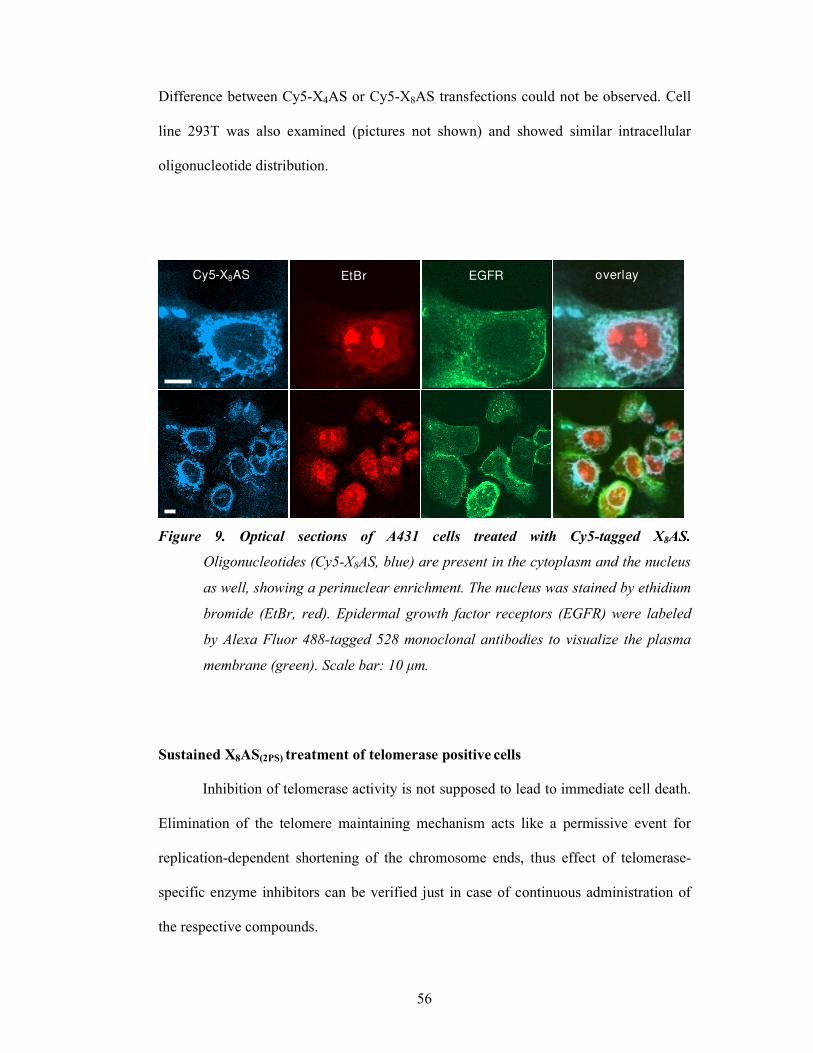
56
Difference between Cy5-X4AS or Cy5-X8AS transfections could not be observed. Cell
line 293T was also examined (pictures not shown) and showed similar intracellular
oligonucleotide distribution.
Figure 9. Optical sections of A431 cells treated with Cy5-tagged X8AS.
Oligonucleotides (Cy5-X8AS, blue) are present in the cytoplasm and the nucleus
as well, showing a perinuclear enrichment. The nucleus was stained by ethidium
bromide (EtBr, red). Epidermal growth factor receptors (EGFR) were labeled
by Alexa Fluor 488-tagged 528 monoclonal antibodies to visualize the plasma
membrane (green). Scale bar: #0 µm.
Sustained X8AS(2PS) treatment of telomerase positive cells
Inhibition of telomerase activity is not supposed to lead to immediate cell death.
Elimination of the telomere maintaining mechanism acts like a permissive event for
replication-dependent shortening of the chromosome ends, thus effect of telomerase-
specific enzyme inhibitors can be verified just in case of continuous administration of
the respective compounds.
EGFREtBr Cy5-X8AS overlay

57
We treated a short-telomere variant of 293T cells with 1-3 µM X8AS(2PS), since X8AS
bearing the higher inhibitory potential could be equally internalized as X4AS. X8AS(2PS)
differs from X8AS, used in the kinetic studies, in two 3’ terminal internucleotide bounds
which have been changed from phosphodiester to phosphorothioate. This modification
has been introduced to enhance inhibitor stability in the cellular environment, without
increasing aspecific association known to be a property of fully phosphorothioate
oligonucleotides. 24h after treatment telomerase activity was measured to verify the
specific action of the inhibitor, also in comparison with X8SCR(2PS). Examined cultures
included treatments with oligonucleotides and Oligofectamine Reagent, “oligo only”, as
well as untreated controls and samples just with the transfection agent.
0
20
40
60
80
100
120
140
Un
trea
ted c
ontr
ol
Oli
gofe
cta
min
e™
co
ntr
ol
3 µ
M X
8SC
R
Oli
gofe
cta
min
e™
1 µ
M X
8A
S
Oli
gofe
cta
min
e™
3 µ
M X
8A
S
Oli
gofe
cta
min
e™
3 µ
M X
8A
S
3 µ
M X
8S
CR
TA
(x)/
TA
(con
t)
Figure 10. Telomerase activity of oligonucleotide treated 293T cultures 24h (white
bars) and 72h (grey bars) after transfection measured with TRAPeze Elisa.
Measurements were performed as triplicates, reported are averages and
standard deviations. Cell population marked with asterisk underwent cell
death after long-term treatment.

58
Telomerase activity in cultures treated with the antisense containing chimera and its
scrambled pair was unchanged. The transfection agent alone caused a diminution of
10%, most probably as a consequence of the nonspecific toxicity the liposomes
observed also in decrease of the cell growth and change of the microscopic morphology.
Transfected inhibitors could decrease telomerase activity by 75% after 24h, when
applied at a 3 µM concentration. After 72h without re-transfection TA increased to 50%
of the untreated control. X8SCR(2PS) in similar conditions had just a slight effect: it
caused a 10% decrease in addition to Oligofectamine.
Figure 11.
Proliferation of 293T cells
during long-term treatment
with X8AS(2PS). Empty circle,
marked with asterisk represents
dying cell population treated
with 3 µM X8AS(2PS) and
Oligofectamine. Black triangle
shows cultures transfected with
# µM inhibitor. Controls were
untreated cells (cross), 3 µM X8AS(2PS) “oligo-only”(black square) and cultures treated
with Oligofectamine empty diamonds).
Cultures treated with 3 µM X8AS(2PS) in the presence of Oligofectamine died after 20-25
days (Fig 11). The transfection agent alone caused a slight decrease in cell growth
compared to the untreated population. A diminution in the same extent could be
observed in case of 1 µM treatment with lipids. Cells with just oligos did not show any
kind of change in growth kinetics, as it was predictable after unchanged TA.
0
5
10
15
20
25
30
35
0 5 10 15 20 25 30 35 40 45
Days
Po
pu
lati
on
Do
ub
lin
gs

59
DISCUSSION I.
Characteristics of telomerase oligonucleotide interactions
The inhibitory potential of the above presented chimeric oligonucleotides seem
to support the dual targeting hypothesis, namely that AS sequence of XnAS inhibitors
forms double helix with the template region of hTR, while Xn interacts with the protein
part (Fig. 12) and the two interactions enhance each other. However, structural proves
will need detailed experiments. Evaluation of primer-enzyme interactions proved that
beside the interaction with the 3’ of the primer at the catalytic site, the upstream region
of the DNA also has a sequence-specific contact with the protein, which seems to
influence binding affinity as well as rate of polymerization (139). It seems to be possible
that the protein surface that normally interacts with the 5’ of its primer-DNA has a high
affinity for thiolated oligonucleotides too, and their binding impairs the catalytic activity
or inhibits the interaction of the telomerase with the natural DNA-substrate. The fact
that blocking of the 3’ end has no effect on inhibition suggests that extension of the
chimeric molecules has no crucial role in the mechanism. Reported experiments using
phosphorothioate modified telomeric repeats with 3’ deoxy-nucleotides gave the same
results (113). Of course we cannot exclude that non-blocked inhibitors might be
elongated.
Figure 12.
Putative mechanism of
XnAS inhibitors. AS
forms a double helix with
the template region of
hTR, and Xn interacts
with the protein part
responsible for primer (telomerase substrate) binding
3’5’
hTR
hTERT
I I II II I I I I I
I
3’
5’
Primer binding site
3’5’
I I II II I I I I I
I
3’
Antisense component(s dU) component
4
n
hTR template region

60
In case of ASXn type of inhibitors, the hybridization to the RNA and the binding of the
chemically modified part to the protein (presumably to other sites than 5’-extended
ones) is also possible. The “antisense”-contact to the RNA is slightly weakened when
short thiolated moiety is attached to the 3’-end, indicating that they may disturb the
interaction of the AS with the target sequence. The similar inhibitory activity of X24
modified antisenses (3’ or 5’) indicates that these long inhibitors may not follow the
proposed mode of action. This conclusion was also supported by the potent inhibitory
potential of the (X)24SCR which is not able to form base pairs with the hTR template
sequence (IC50s of (X)24AS, AS(X)24 and (X)24SCR did not differ essentially). Moreover
these derivates are not specific, since they inhibited HIV RT with similar parameters
(see Fig. 8). These findings can be explained by the property of the chemically modified
oligonucleotides to inhibit DNA-polymerases in a sequence nonspecific manner, since
37mer chimeras (X24 modified antisenses and scramble) reach the length reported to
have maximal inhibitory activity [(30 to 40mers) (126)]. In contrast, X24 (as shown by
its IC50 in Table 1) is too short to exert full sequence nonspecific inhibition.
Considering is the physico-chemical nature of the interaction of thiolated
oligonucleotides with the protein, there are two likely modes: (a) hydrophobic
interaction, due to the hydrophobic character of the 4-thiono group, or (b) disulfide bond
formation between properly localized cysteinyl side chains on hTERT surface and
reactive –SH, formed on the (s4dU) base due to tautomeric conversion. The latter,
covalent interaction between proteins and (s4dU) containing oligonucleotides is
chemically feasible and was proven (137).

61
Cellular uptake of oligonuleotides
An important factor, proved by the Cy5 labeled chimeras, was the observed
nuclear uptake, since telomerase exerts its function as a DNA-polymerase obligatory in
the nucleus. The perinuclear portion might correspond to the dissociating lipid-
oligonucleotide complexes (140). Experiments to evaluate hTERT subcellular
localization in immortal cell lines showed that hTERT is immunologically detectable
not only in nuclei but also in the cytoplasm (141). Strong Cy5 signals detected in the
cytoplasm may also correspond, at least to some degree, to oligonucleotides bound to
the cytoplasmic telomerase. Whether this interaction has a pharmacological
significance, needs further investigations.
Chimeric oligonucleotide telomerase inhibitors in immortal cell cultures
Chimeric oligomeres were able to decrease the telomerase activity in a dose
dependent manner which was sufficient to cause the death of the treated cell population
after continuous long-term administration. The delayed onset of the phenotypic effect
can be explained by the fact that these inhibitors suppose to decrease the activity of the
telomerase, but not the protein levels, thus cell death results from critical telomere
shortening after several cell divisions. Although precise nature of the achieved cell
death was not evaluated, senescence seems to be excluded, since dying cell population
neither showed senescent phenotype nor displayed enhanced senescence activated ß-
galactosidase activity. T antigen is known to bind p53 protein and cause a perturbation
in its function, thus cell carrying this viral oncogene are considered to have a
functionally inactive p53 (142). Although opinions are contradictory, it seems that
senescence triggered by telomere shortening involves p53 (143), and that p53 status is
one of the determining factors in the choice between senescence and apoptotic cell

62
response in vivo (144).The above factors could explain the absence of senescence as a
response on telomere dysfunction in 293T cells.
As clearly seen on the growth curves, cell death occurred just in case the telomerase
activity could be kept below a certain level. Since 1 µM, yielding a 50-60% TA had no
effect on cell proliferation, threshold for telomere maintenance seems to be below that
level. This reinforces the method of re-transfection after 48-72h, since also in cultures
effectively treated [ 3µM X8AS(2PS) + Oligofectamine] activity of telomerase after 72h
was as high as 50%, due to oligonucleotide degradation and dilution of intracellular
concentration as a consequence of cell division. X8AS(2PS) and X8SCR(2PS) without
liposomal agents could not enter the cells as proven by their telomerase activity. Despite
of this, they could affect the cellular response, since their binding to proteins localized
in or on the membrane is possible, similar to X35 (137). Although we can not exclude
this possibility, we could not observe signs of neither acute nor chronic toxicity.
In conclusion, the properties of the novel family of chimeric oligonucleotides indicate
that further development of such molecules might be a favorable anti-telomerase
strategy, considering additional improvement in inhibitory parameters and intracellular
properties as well (for e.g. by introducing various backbone modifications). Further tests
on immortal, telomerase positive cell lines of different cellular origin are needed to
evaluate susceptible cell lines, possible limiting factors, and pharmacological
combination possibilities. Experiments have to be made to validate intracellular
molecular events and telomerase-chimera interaction as well. Following the idea of dual
interaction, using the presented chemically modified chimeras as lead compounds, could
provide new data in the biology of telomerase inhibition.

63
RESULTS II.
TELOMERASE DEPENDENT CELL DEATH USING ATRA AND As2O3
Effect of the sustained ATRA/As2O3 combinational treatment on the viability of
maturation resistant promyelocytic leukemia cell lines
Majority of therapy-naive acute promyelocytic leukemia cells as well as NB4
APL cell line enters granulocytic differentiation upon ATRA treatment (145). As shown
previously, cell death can be achieved also in some cell lines resistant to ATRA-induced
differentiation (NB4-LR1), since long-term treatment with pharmacological doses is
able to repress telomerase catalytic subunit expression, thus cause death due to telomere
shortening (131). Repression of telomerase with subsequent cell death can also be
obtained using synthetic RAR∀ and RXR selective agonists not only in NB4-LR1 but
also in “ATRA-unresponsive” NB4-LR2 cells, harbouring a dominant negative,
truncated PML/RAR∀ (146). Subline NB4-LR1SFD
retains a functional pathway of
telomerase downregulation associated with maturation, but its hTERT expression fails
to respond to ATRA or selective agonists (130). Cell death of long-term ATRA-treated
NB4-LR1 cells is caused by telomere shortening due to the downregulation of hTERT,
however NB4-LR2 and NB4-LR1SFD
cells resist to this treatment. Since ATRA as well
as As2O3 inhibits hTERT expression, we wondered whether this combination, proven to
be effective in clinic, could affect synergistically ATRA-resistant cells. It has been
previously shown that treatment of NB4-LR1 and NB4-LR2 cells with high
concentrations (1 µM) of As2O3 alone or in combination initiates massive apoptosis
(115). In these conditions the assessment of a molecular synergy of long-term treatment
with ATRA would be difficult. Furthermore, one critical factor that limits the utility of

64
As2O3, especially in case of extended treatments, will be its cytotoxicity to normal
tissues, thus, one of our objectives was the substantial reduction of its dose. For this
reason NB4-LR1, NB4-LR1SFD
, and NB4-LR2 cells were treated with As2O3 at low
concentration (0.2 µM) in the presence or absence of ATRA (1 µM).
Long-term treatment of NB4-LR1 cells with 1 µM ATRA induced cell growth
arrest followed by cell death at day 75, similar to previous reports (Fig. 13A). NB4-
LR1SFD
and the NB4-LR2 cells continued to proliferate even after 3 or 4 months of
continuous treatment with ATRA. 0.2 µM As2O3 alone had no effect either on
proliferation or viability in none of the cell lines included in the study. Combinational
treatments, using both drugs, accelerated cell death in NB4-LR1 compared to the
population treated with ATRA alone, and reduced cell growth and viability in both
NB4-LR2 cells and NB4-LR1SFD
.
Changes in the telomerase homeostasis in the APL cell lines receiving
combinational treatment
As previously shown, growth arrest in NB4-LR1 after long-term exposition to
ATRA is a consequence of telomerase catalytic subunit downregulation, leading to a
marked decrease in telomerase activity, and finally to telomere shortening (131). To
evaluate how telomerase is affected by the combinational treatment we measured
hTERT expression and TA, as well as changes in telomere length in all three cell lines
after extended treatment with ATRA and As2O3 alone or as co-treatment. In the ATRA-
prone population of each cell line we could observe a remarkable reduction in hTERT
mRNA level (Fig. 13B), measured by quantitative RT-PCR. However, only in the NB4-
LR1 cells was this decrease (95%) associated with an efficient diminution of TA (Fig.
13C) and telomere shortening (Fig. 14A) resulting in cell death (as previously seen on

65
Fig. 13A). In these cases, no telomere shortening was observed (Figure 14A and B). In
all the three cell lines, As2O3 alone induced much less modifications in hTERT
expression, which yielded a telomerase activity above 50% of the control without any
consequences on either telomere length or cell proliferation. In sharp contrast, the
combination of the two compounds reduced hTERT mRNA and TA below the levels
needed to prevent the reduction of telomere length and cell death.
Effect of ectopic hTERT-expression on the viability of retinoid maturation-
resistant cells during ATRA/As2O3- treatments
Altogether, our results suggested that, after long-term treatment of maturation-
resistant cells with ATRA plus As2O3, loss of telomerase activity and telomere
shortening causes cell death. To substantiate this result, we investigated whether ectopic
expression of hTERT could rescue NB4-LR1 and NB4-LR2 cells from cell death. NB4-
LR1/hTERT-GFP and NB4-LR2/hTERT-GFP continued to proliferate without any
change on cell viability, not only during long-term treatment with ATRA or As2O3, but
also in case of co-treatments (Fig. 15)

66
A
B
NB4-LR1
P.D
.
days
0
20
40
60
80
100
120
ATRA As2O3ATRA+
As2O3
hT
ER
T/P
BG
D (
%)
0 4 9 14 46 0 4 9 14 46 0 4 9 14 46days
0
20
40
60
80
100
120
ATRA As2O3ATRA+
As2O3
0 9 14 23 44 0 9 14 23 44 0 9 14 23 44days
T.A
. (%
)
C
0
20
40
60
80
0 10 20 30 40 50 60 70
*
NB4-LR2
0
20
40
60
80
100
120
0 4 9 38 0 4 9 38 0 4 9 38
ATRA As2O3ATRA+
As2O3
0
20
40
60
80
100
120
ATRA As2O3ATRA+
As2O3
0 4 9 21 38 0 4 9 21 38 0 4 9 21 38
NB4-LR1SFD
0
20
40
60
80
100
120
ATRA As2O3ATRA+
As2O3
0 4 9 14 46 0 4 9 14 46 0 4 9 14 46
ATRA As2O3ATRA+
As2O3
0
20
40
60
80
100
120
0 9 14 23 44 0 9 14 23 44 0 9 14 23 44
0
20
40
60
80
0 10 20 30 40 50 60 700
20
40
60
80
0 10 20 30 40
*
*
*
Figure 13:
ATRA and As2O
3synergize to induce
growth arrest, hTERT
downregulation, inhibition of
telomerase activity and
subsequent cell death.
A. Proliferation of cells cultured in
the presence of medium alone
(empty square),in the continuous
presence of ATRA (# µM; cross),
As2O3 (0.2 µM; black diamond),
or the combination of both drugs at
these concentrations (empty
circle). Cell proliferation was
assessed as population doublings
(PD). Asterisk indicates cultures
that died after prolonged treatment
with the indicated compounds.
B. At the indicated time, hTERT
mRNA expression was quantified
by fluorescence-real-time RT-PCR
using LightCycler TeloTAGGG
hTERT Kit. hTERT level was
normalized to the expression of the
housekeeping gene
phosphobilinogen deaminase
(PBGD).
C. Telomerase activity (TA) was
measured with TRAPeze Elisa
telomerase detection kit, and
expressed as a percentage of that
detected in untreated cells.

67
Figure 15:
Proliferation of ATRA and/or
As2O treated cells expressing
ectopic hTERT. NB4-
LR#/hTERT and NB4-
LR2/hTERT cells were cultured
in in the presence of medium
alone (empty square), the
continuous presence of ATRA
(# µM; cross), As2O3 (0.2 µM;
black diamond), or the
combination of both drugs at
these concentrations (empty
circle). Cell proliferation was
assessed as population
doublings (PD).
kbp
ATRA (1 µM)
As2O3 (0.2 µM)
Day 32
* kbp
8.6
6.1
5.0
2.7
- - + -
+ + - +
- - +-
++ -+
-- +-
++ -+
LR1 LR1SFD
LR1/hTERT
8.6
6.1
5.0
2.7
Day 51
-- +-
+ - -- + -
+ + - +
- - +-
++ -+
LR1 LR1SFD
LR1/hTERT
- + + - - ++ -LR2 LR2/hTERT
ATRA (1 µM)
As2O3 (0.2 µM)
A
Day 24
5.0
3.5
2.7
1.9
1.1
kb
- + - + - +- +
B
Figure 14:
Average telomere length during long-term
treatment of APL cell lines with ATRA/As2O3.
Terminal restriction fragment lengths, measured
using a non-radioactive chemiluminescent assay
TeloTAGGG Telomere Length Assay (Roche
Diagnostics) were determined after 32 days and
5# days (NB4-LR# and NB4-LR#SFD cells, A) and
after 24 days of treatment (NB4-LR2 cells, B).
P.D.
P.D.
days
0 20 40 60 80
0 10 20 30 40 50 60 70
NB4-LR1/hTERT
0
20
40
60
80
0 10 20 30 40
NB4-LR2/hTERT

68
Role of DNA-methylation in the ATRA-induced hTERT downregulation
Increased methylation of the promoter has been implicated in hTERT silencing
during ATRA-induced differentiation in myeloid (147) and non-myeloid cell lines
(148). Although literature of nature and significance of epigenetic changes caused by
therapeutic arsenic is quite limited, several studies showed that methylation of
appropriate genes is an important factor in arsenic carcinogenesis (149). Epigenetic
silencing trough DNA methylation could represent the point of convergence in
telomerase downregulaton observed after ATRA treatment. To test this hypothesis, we
cultured NB4-LR1 cells in the presence of 250-500 nM of the methylation inhibiting
agent 5’-azacytidine during 6 days. Concentrations higher than 500 nM caused massive
cell death thus were not applicable. 1 µM ATRA was added to the pretreated cultures,
which were grown for additional 4 days, parallel to cells without ATRA. hTERT
expression was determined with quantitative RT-PCR from total RNA of the harvested
cells. Levels of telomerase catalytic subunit mRNA did not change upon AzaC
treatment, but decreased significantly after receiving ATRA irrespective of pretreatment
with the methylation inhibitor (Fig. 16).
Figure 16: Effect of the DNA-methylation inhibitor (AzaC) on hTERT expression.
NB4-LR# cells were grown in AzaC containg medium, then treated with #µM ATRA. hTERT expression was analysed by real-time RT-PCR using a Taqman assay and normalized to the mRNA level of cyclophilin. AzaC pretreatment had no effect on ATRA-induced downregulation of hTERT.
0,000
0,005
0,010
0,015
0,020
0,025
R1 cont R1 AzaC
250nM
R1 AzaC
500nM
R1 ATRA R1 AzaC
250nM +
ATRA
R1 AzaC
500nM +
ATRA
[hT
ER
T(x
)/cy
c(x
)]

69
Role of histone acetylation ATRA/As2O3-induced hTERT repression
Histone modification is another universal mechanism employed by eukaryotes
for gene regulation, and plays a central role in retinoid-induced trans-activation. We
tested therefore the changes in acetylation of histones 3 and 4 (H3 and H4) associated
with the hTERT proximal promoter (-712/-435) with chromatin immunoprecipitation
(ChIP). Samples were collected from day 2 and day 9 of ATRA (1 µM)/ As2O3 (0.2 µM)
treatment. RARß, being a direct RAR∀ target gene in NB4 cells (150), could be
followed as positive control of the treatment. Parental cell line NB4 underwent
differentiation upon ATRA treatment, which was accompanied by decreased H3 and H4
acetylation. Changes seen after 2 days were already present after 4h (Fig. 17).
Figure 17 :Changes in histone acetylation during maturation
of NB4 cells
NB4 cell were differentiated with # µM ATRA, and histone
acetylation of hTERT promoter was assayed with ChIP.
Acetylation of the hTERT associated histones decreased
visibly as soon as 4h after adding ATRA.

70
In the maturation resistant NB4-LR1 cells, harvested at day 2, acetylation status was
unchanged. At day 9, however, acetylation of both H3 and H4 decreased in ATRA-
treated cells, and diminished even more in cultures receiving combinational treatment of
ATRA and As2O3. As2O3 alone did not cause any changes (Fig 18A).
A)
B)
C)
Figure 18.: Changes in histone acetylation during treatment with ATRA (1µM) and
As2O3 (0.2 µM)
ChIP assay was performed with cells collected at the indicated days using anti-AcH3
and anti Ach4 antbodies. PCR following immunoprecipitation amplified hTERT
promoter or RARß promoter respectively. Treated cell lines were A) NB4-LR#,
B) NB4-LR2
C) NB4-LR#SFD

71
RARß promoter used as control showed that decrease of acetylation is not a general
phenomenon- liganded RAR∀ activated RARß and caused histone acetylation, in good
agreement with the literature. It is worth to note, that co-treatment was more effective,
than ATRA alone, also in the transcriptional activation.
In treated NB4-LR2 populations (Fig. 18B) the histones in the control gene promoter
presented similar changes to those seen in NB4-LR1, but hTERT associated H3 and H4
acetylation was not decreased. Histones on telomerase catalytic subunit promoter in
NB4-R1SFD
(Fig. 18C) were also not altered after 9 days of combinational treatment.
Interestingly, in these cells histones at the RARß promoters were refractory to changes
when treated with ATRA and/or arsenic.
As manifest changes in the acetylation status could not even be induced with those
treatments, that were efficient to downregulate hTERT in NB4-R2 and NB4-R1SFD
cells,
we asked whether differentiation is able to cause the same hypoacetylation as in NB4
cells. NB4-LR1SFD
is resistant to ATRA induced maturation, but combination of 1 µM
ATRA and 200 µM CPT-cAMP (a membrane-permeable cAMP analogue) is able to
differentiate these cells.

72
After 2 days of treatment, hTERT expression decreased as predictable, but decrease in
acetylation of it’s promoter could not be observed. RARß AcH3 in these samples was
elevated.
Figure 19.: Changes in histone acetylation during treatment of NB4-LR1SFD
Comparison of changes in histone acetylationa after ATRA/As2O3 versus differentiation
with ATRA/CPT [8-(4-chlorophenyl-thio)adenosine cyclic 3’5’-monophosphate]

73
DISCUSSION II.
Effect of ATRA and As2O3 on telomerase in maturation resistant APL cells
The mechanisms underlying the chemopreventive and chemotherapeutic
properties of ATRA, as well as the improved efficacy of ATRA/As2O3 compared to
single drug treatments among APL patients are still unknown. Our findings provide
direct evidence that telomerase targeting can represent a likely new mechanism by
which ATRA/As2O3 therapy can exert its action. We knew previously that telomerase-
dependent cell death can be obtained in NB4-LR1 cell line, resistant to ATRA-induced
maturation, by long-term treatment with ATRA (131). In NB4-LR2 similar effect was
seen with the combination of specific ligands for RAR∀ and RXR (146). We
demonstrated here, that only the combination is effective to induce a cellular death
response in all the three maturation-resistant cell lines (NB4-LR1, NB4-LR2, NB4-
LR1SFD
, Fig. 20) even though ATRA alone was able to regulate telomerase expression
to some extent at a molecular level.
Although As2O3 alone in higher concentration has been shown to be enough to induce
apoptosis (115), and literature data suggest that micromolar doses could decrease
hTERT in a biological significant degree (151), our low-dose (0.2 µM) application
proved to be insufficient to influence long-term cell viability. It should be noted that
modulation of TA and induction of cell death by ATRA/AS2O3 in NB4-LR1 cells
occurred faster than in cells treated with ATRA alone, although both treatments were
able to eliminate the entire cell population.

74
Figure 20.: Cellular response of
acute promyelocytic leukemia cell
lines to natural and synthetic
retinoid ligands and their
combinations.
The shorter period of time required to induce cell death in NB4-LR2 cells could be
explained by the shorter telomere of these cells, compared to the two others (see TRF
comparatively in Fig. 14 A and B). As a significant portion of the telomeres of the NB4-
LR2 cells is expected to be at critical stage of shortening, even a small erosion
(compared to what observed in NB4-LR1 and NB4-LR1SFD
using TRF assay) could be
enough to trigger crisis and death of these cells. These results indicate that cell death
induced by ATRA/As2O3 combination is mainly due to the decrease in TA, which is
itself the consequence of hTERT downregulation. Importantly, we demonstrate that
ectopic hTERT expression is sufficient to prevent cell death induced by ATRA/As2O3
combination, further supporting the contention that the therapeutic action of this
combination of drugs relies on a telomerase and telomere dependent mechanism.

75
Interestingly, NB4-LR1 cells with ATRA alone proliferate at day 32 with telomeres as
short as those of the cells receiving combination treatment and have already a decreased
viability at this time point. It has been recently shown that not only the overall telomere
length but also the length of the telomeric 3' single-strand overhang are important
factors in triggering cell death. Since the TRF-method used in our experiments gives no
information on the integrity of the 3'-overhang, it is not excluded that As2O3 in
combination with ATRA induces some kind of telomere dysfunction. As a matter of
fact, higher doses of arsenic have been shown to enhance the formation of tetra-stranded
t-loop junction, which has been suggested to be a result of single-strand overhang loss
(152). The telomerase-independent mechanism of the combination is also indicated by
the reduced cell proliferation upon 40 days in NB4-LR1/hTERT-GFP cells (Fig. 15).
Furthermore, we have to keep in mind that telomerase is endowed with survival
functions independent of telomere maintenance, thus, the diminution in hTERT protein
level, by eliminating it as a surviving factor, can also contribute to the effect observed in
the parental cells without excessive amount of hTERT due to ectopic expression.
However, the duration of the lag period before the induction of cell death suggest that
telomere shortening is actually an important factor in the observed effect.
Converging mechanisms of ATRA/As2O3 to influence hTERT transcription
Molecular network involved in the ATRA/Arsenic induced downregulation of
hTERT may have several members. Approaches like high throughput genomic and
proteomic assays, can provide a first list of interplayers. Systemic evaluation has
already been conducted, but just focused on changes during differentiation of NB4 cells
(153) and not on the effect of chronic treatment of maturation resistant cells.

76
Degradation of PML-RAR!
Mechanisms responsible for ATRA-induced telomerase downregulation in the
absence of differentiation could not be deciphered yet. Although causal relation between
degradation of PML-RAR∀ and changes in gene expression followed by ATRA/As2O3
administration cannot be doubted, it is clear that it can not solely account for the
presented downregulation of hTERT, since the breakdown of the chimeric protein
following ATRA treatment alone is already rapid and occurs to the same extent in NB4-
LR1 and NB4-LR1SFD
cells, despite of different biological response (cell death or
survival, respectively (130).
Transrepression
Classical consensus sequence for the heterodimeric RAR∀/RXR receptor (5’-
AGGTCA-3’) is not present in the hTERT promoter, but in light of the recent findings
describing a new cis-acting element for retinoids found in the promoter of the Burkitt’s
lymphoma receptor 1 gene of the myeloid cell line HL-60 (154), we have to realize that
other binding sequence motif with novel binding complexes may exist. Little is known
anyhow about ligand induced gene-repression at the molecular level. A nice example of
transrepression by nuclear receptors has been demonstrated for vitamin D dependent 1∀-
hydroxilase repression (155). In that case a sequence specific activator was able to bind
liganded VDR/RXR, which caused the switch from co-activators to HDAC co-repressor
complex. A similar molecular structure could also be imaginable for ATRA-dependent
hTERT downregulation.
Anti-AP1 acivity
Retinoids modulate gene expression by two different ways. First by the binding
of RAR/RXR heterodimers to response elements, followed by transactivation. Secondly,
as transrepressive factors that negatively affect proliferation-associated genes via their

77
ability to functionally interact with transcriptional factor AP1 (156). The AP1
transcription complex consists of homodimers and heterodimers of members from the
fos and jun families and activates target genes by binding with high affinity to particular
DNA cis-element,. The regulation of AP1 activity is complex and occurs at different
levels. First, through differential expression of individual members from jun and fos
families, which will determine the composition of AP1 dimer (157) Second, AP1
activity is regulated at posttranslational levels by several external stimuli mainly
involving mitogen-activated protein kinase (MAPK) cascades. Mechanisms by which
retinoids exert anti-AP1 activity is still unclear. Proposed mechanisms included
inhibition of jun N-terminal kinase or triggering the assembly of distinct AP1
complexes, different from those in unstimulated cells (158), and sequestration .of c-
jun/c-fos sequestration through physical interactions with RAR (159). Although anti-
AP1 effect in telomerase downregulation can not be excluded and would be an
interesting question to be investigated, there some main concerns that make it
improbable. One of them is the report about NB4 cells treated with anti-AP1 retinoids
(GALDERMA CD2409), which had just a modest effect on cell growth and
differentiation. It should be noted though, that treatments were just conducted on short
term (160). Next contradictory fact is the requirement of liganded RXR to repress
hTERT (146), since anti AP1 effects do not require the heterodimeric nuclear receptor
and RAR is enough to produce full effect (161). Interestingly, most recent findings
suggest that in an unusual manner, AP1, a traditionally positive regulator of
proliferation has a repressional function on hTERT (162).
Epigenetic modifications
Treatment of several telomerase positive cell types with methylation-inhibiting
agents led to a decrease of the catalytic subunit, while others reported an increased

78
expression. In NB4-LR1 we did not observe any kind of effect. Retinoids are known to
decrease methylation by inhibiting the expression of methyltransferases (163).
Realizing, that ATRA hardly acts directly, other transcription factors, affected by
retinoid signaling, could be as well involved in changes modifying the chromatin.
Inhibition of DNA-methylation by pretreating NB4-LR1 with AzaC, proved neither
preventive from ATRA-induced transcriptional repression, nor enhanced the effect of it.
Role of DNA-methylation in hTERT biology seems to differ from cell to cell. In our cell
lines no evidence was found for its role in telomerase regulation.
Acetylation of promoter proximal histones is associated with gene expression,
while transcriptional silencing, seen for hTERT in normal human somatic and ALT
cells, can be induced by transcription factors that recruit HDAC. Early downregulation
of hTERT during differentiation is a well known phenomenon, and has also been shown
to be associated with histone deacetylation previously (30). Parental NB4 cells treated
with ATRA during two days, demonstrated the same pattern. In NB4-R1 cells ATRA
and arsenic act cooperatively in increasing histone acetylation of RARß promoter and
the presence of the same synergism can be observed when the two agents decrease
acetylation associated with hTERT. It is worth to keep in mind that the first is a direct
RAR∀ target gene while the latter is supposed to be effected indirectly. PML-RAR∀ of
NB4-LR2 harbors a mutation, generating an in-phase stop codon, thus truncated form of
PML-RAR∀ which has lost its C-terminal end. Fusion mutant exerts a dominant
negative effect on wild-type PML, PML-RAR∀ and RAR∀ transcription activity (164).
Its likely that the altered fusion protein is less effective in the formation of activating
nucleoprotein complexes and as a consequence improper HDAC activity can be
observed after ATRA and combinational treatment. NB4-LR1SFD
is quite unique, as
hyperacetylation of RARß promoter was just present in cell undergoing differentiation.

79
There might be several explanations. Taking the fact that genetic background of the
altered phenotype is not found yet, we can just guess, that the reason of the observed
deviations might be a defect in the retinoid signaling pathway, not only affecting
hTERT. Preexisting histone modifications, like preacetylation, were shown to interfere
with chromatin methylation (165). Since we had not the opportunity yet for the
quantitative comparison of the cell lines, we can just guess, that preexisting epigenetic
changes could account for retinoid resistance.
Although we could not show clear correlation between histone acetylation state and
biological response to the treatment, it is important to keep in mind that epigenetic
regulation functions trough a “histone-code”, thus a combination of different non-
independent histone modifications, what highlights the future importance of working
with a palette of modifications. It is as well undoubted that using non quantitative
methods, like our experimental layout, makes us impossible to speak about kinetics and
amplitude of changes, we are just able to see a kind of endpoint, and make statements
about manifest changes.
Taking together, we are unable to give a clear explanation how ATRA regulates
hTERT, and how the converging pathways with As2O3 are able to cooperate. The main
concern about making any suggestion is the time course of our observation: even in case
of a secondary effect, a retarded response like seen in NB4-LR1SFD
can not be explained
with our today’s knowledge.
.

80
CONCLUDING REMARKS
Our observations made with ATRA/As2O3 and chimeric oligonuleotides
highlight a few important points for telomerase as future cancer drug target.
Although there are already studies focusing at minimum levels of telomerase
activity for tumorigenesis (166), the notion of thresholds necessary for telomere
shortening and cell death are stated rarely. The first threshold features to which extent
hTERT mRNA expression has to be decreased to obtain an efficient reduction of TA.
The second one identifies the level of enzyme activity for which telomerase inhibition
resulted in a sufficient shortening of telomeres to cause cell death. The biological
significance of these two distinct thresholds has to be taken into account for the
development of telomerase inhibitors targeting either hTERT mRNA expression or
telomerase activity itself. In fact, focusing solely on a relative inhibition of telomerase
may overestimate the therapeutic efficacy of telomerase inhibitors. Indeed, tumor cells
can be responsive to a drug, in terms of downregulation of hTERT and decrease in TA,
without demonstrating any modification in proliferation and viability. Importantly, it is
also clear that these thresholds, as well as the outcome of telomerase inhibition, may
vary depending on the physiology of the targeted cancer cells.
A number of oligonucleotides were tested as telomerase inhibitors mostly
interacting with the template region of hTR. The (s4dU)nAS type inhibitors, developed
by us, have a quite unique feature; they interact with the protein subunit of the
telomerase enzyme, too. Although, such telomerase inhibitors were also developed by
others (123), our family of inhibitors is the only one, which is able to form covalent
linkage with proteins. The further development of (s4dU)nAS type type-inhibitors is
possible because the antisense stretch offers an excellent target for further modifications
increasing the stability of the inhibitor against nucleases as well as enhancing the

81
stability of the double helix formed with hTR. Molecules, tightly bound to telomerase
could also be used to map three dimensional organizations of functional domains, since
structure of the enzyme is not fully evaluated yet.
Considering the therapeutic utility of oligonucleotide-type telomerase inhibitors,
including ours, the low toxicity of these agents allows a treatment may be applied for a
long period of time. Considering problems of stability and penetration systemic
application might not be appropriate for oligonucleotides, but local administration as a
part of the post-surgical treatment of should be considered for certain malignancies.
Our findings provide evidence that telomerase targeting can represent a likely
new mechanism by which ATRA/As2O3 therapy can exert its action in APL patients.
The synergism between these two agents highlights important aspects of their possible
integration into the future clinical protocols (118). Although benefit of arsenic trioxide
was shown first in APL patients in the first or subsequent relapse after ATRA and
chemotherapy, a recent study showed (116) the clear benefit of arsenic-ATRA
combination in newly diagnosed APL in induction and maintenance therapy. We
suggest that the success of the latter trial can be also explained by the fact, that the
administration of the combination was sustained over the period required for complete
remission, which is around 30-40 days in general. In light of our results, presented here,
the time span of induction therapy seems to be not always enough to obtain critical
telomere shortening, thus resistant clones could escape, while sustaining the therapy
with the combination would also allow the eradication of multiple kinds of resistant
cells. It seems, that long-term low-dose combinational treatments could lead to a
favorable outcome in APL patients, with the enhanced benefit of minimal toxicity, as
As2O3 concentration used in our study (0.2 µM) is much lower than the therapeutic
serum levels of arsenic during standard and alternative therapeutic protocols, which are

82
settled to yield a concentration around 1 µM, with peak concentrations of 3-6 µM (167).
As arsenic trioxide as postremission therapy seems to be favorable in patients receiving
this drug after relapse, our experiments indicate, that it might be used as well in low
dose in combination with ATRA. The fact, that deregulation of hTERT expression is a
mechanism that is not restricted to APL, clearly indicates, that the eradication of the
residual cells will likely require pharmacological treatment targeting telomerase.
Combinational treatments raise the idea of a more general concept to investigate
other combinations, in order to know, whether their extended administration,
influencing telomerase in a convergent manner, could not be of therapeutic benefit. This
type of strategies might involve combinations of drugs targeting telomere
accessibility/function and hTR/hTERT component of the telomerase holoenzyme (71,
81, 100), co-treatments against hTR and hTERT (78), combinations that influence
regulation of hTERT on different levels, as well as the usage of molecules that exert
synergy by targeting the same regulation event by different mechanisms Especially
attractive would be the applications that include clinically approved drugs, which have
not been used before in extended protocols, but have already the permission to be used
for human therapy. Since malignant cells with elevated telomerase activity can be
resistant to anti-cancer agents, and this resistance seems to be reversed by concomitant
treatment with telomerase inhibitors (168) several types of drug resistant cancers could
represent a rational target.
Although the complexity of telomerase regulation and malignant transformation,
as well as the individuality in the clinical response show clearly that treatment
protocols, similar to our experimental layouts may not be effective in all cases, and not
applicable as first and only treatment, our data encourage their integration into extended
trials performed in vitro and in the clinic.

83
SUMMARY
Immortalization is an indispensable step in oncogenesis. Most of the human
malignant cells attain their immortality through stabilization of their telomere length by
reactivating telomerase enzyme, a specialized reverse transcriptase. I presented in my
thesis two promising approaches to target telomerase in immortal human cell lines using
combinational strategies.
I. TELOMERASE DEPENDENT CELL DEATH USING CHIMERIC
OLIGONUCLEOTIDES AS TELOMERASE INHIBITORS
1. We designed an oligonucleotide family, composed of a 13mer antisense
sequence against the hTR template part and a 3' or 5' (s4dU)n moiety, possibly
interacting with the protein subunit of human telomerase and we characterized
the oligonucleotides by meaning of their inhibition parameters.
2. We proved, that the derivate (s4dU)8AS(PS), shown to be specific and effective
as well, can enter human cells.
3. Long-term treatment with (s4dU)8AS(PS) caused the death of the entire treated
immortal cell population.
II. TELOMERASE DEPENDENT CELL DEATH USING ATRA AND As2O3
1. We proved that all-trans-retinoic acid and As2O3 is able to downregulate hTERT
expression even retinoid resistant cell lines. The decrease in expression
diminished telomerase activity and caused telomeres to shorten.
2. Long term low dose combinational treatment was sufficient to cause cell death.
3. Synergistic effect of all-trans-retinoic acid/As2O3 treatment could not be
exclusively explained by the modification of the chromatin structure.

84
ACKNOWLEDGEMENTS
I am especially grateful for the help of my two tutors Dr. Evelyne Segal-
Bendirdjian and Dr. Janos Aradi for the scientific guidance and personal friendship.
I would like to thank Dr. Laszlo Fesüs, head of the Department of Biochemistry and
Molecular Biology of the University of Debrecen, and Dr. Michel Lanotte, head of
INSERM U-685 in Paris, for the opportunity to work in their prestigious institutes.
I thank Andras Horvath for the synthesis and purification of the oligonucleotide
inhibitors, Helga Eizert for the preparation of the partially purified telomerase, György
Vamosi and Sandor Damjanovich for the shots with the laser scanning microscope and
Istvan Szatmari for introducing me the methods to measure telomerase activity.
I also thank Josette Hillion, Frederic Pendino and Charles Dudognon for their help
concerning the investigations with APL cells.
I would like to acknowledge all members of the Department of Biochemistry and
Molecular Biology of the University of Debrecen, especially Klara Katona and Iren
Mez∀ and members of INSERM U-685 for their help and patience.
This work was supported by OTKA T-038163 (Hungary) research grant, BIO-
00032/2001 Biotechnology Grant, F-1/03 French-Hungarian Bilateral
Intergovernmental S&T Cooperation and Association pour la Recherche contre le
Cancer (ARC). I. T. was a Short Term Fellowship holder of EMBO, Société Française
du Cancer and Société Française d'Hématologie.
Chimeric oligoncleotides are patented by Aradi, Tarkanyi, Horvath, Szatmari,
Segal-Bendirdjian: [„Oligonucleotide chimeras as reverse transcriptase or telomerase
inhibitors.”, Hungarian patent application No.: P05 00154]

85
ABBREVIATIONS
ALT - Alternative Lengthening of Telomeres
APL -Acute Promyelocytic Leukemia
ATRA -All-Trans Retinoic Acid
AzaC -5’-azacytidine
ChIP -Chromatin ImmunoPrecipitation
CR -Complete Remission
DNP -Dinitrophenyl
FCS -Fetal Calf Serum
MTS -Modified Telomerase Substrate
PBS -Phosphate Buffered Salina
PCR -Polymerase Chain Reaction
PKC -Protein Kinase C
PML -Promyelocytic Leukemia Protein
RAR! - Retinoic Acid Receptor alpha
RXR -Retinoid X Receptor
TRAP -Telomeric Repeat Amplification Protocol
TP-TRAP -Two-Primer Telomeric Repeat Amplification Protocol
TRF -Terminal Restriction Fragment

86
REFERENCES
1. Blackburn, E.H. (2001) Switching and signaling at the telomere. Cell, 106, 661-673.
2. McElligott, R., Wellinger, R.J. (1997) The terminal DNA structure of mammalian
chromosomes. Embo J, 16, 3705-3714.
3. de Lange, T. (2004) T-loops and the origin of telomeres. Nat Rev Mol Cell Biol, 5,
323-329.
4. Schaffitzel, C.; Berger, I.; Postberg, J.; Hanes, J.; Lipps, H.J.;Pluckthun, A. (2001) In
vitro generated antibodies specific for telomeric guanine-quadruplex DNA react with
Stylonychia lemnae macronuclei. Proc Natl Acad Sci U S A, 98,8572-8577.
5. Morin, G.B. (1989) The human telomere terminal transferase enzyme is a
ribonucleoprotein that synthesizes TTAGGG repeats. Cell, 59, 521-529.
6. Greider, C.V. (1991) Telomerase is processive. Mol Cell Biol, 11, 4472-4580.
7. Dunham, M.A., Neumann, A.A., Fasching, C.L. and Reddel, R.R. (2000) Telomere
maintenance by recombination in human cells. Nat Genet, 26, 447-450.
8. Yeager, T.R., Neumann, A.A., Englezou, A., Huschtscha, L.I., Noble, J.R., Reddel,
R.R. (1999) Telomerase-negative immortalized human cells contain a novel type of
promyelocytic leukemia (PML) body. Cancer Res, 59, 4175-4179.
9. Marciniak, R.A., Cavazos, D., Montellano, R., Chen, Q., Guarente, L. and Johnson,
F.B. (2005) A novel telomere structure in a human alternative lengthening of telomeres
cell line. Cancer Res, 65, 2730-2737.
10. Kumakura, S., Tsutsui, T.W., Yagisawa, J., Barrett, J.C. and Tsutsui, T. (2005)
Reversible conversion of immortal human cells from telomerase-positive to telomerase-
negative cells. Cancer Res, 65, 2778-2786.

87
11. Smogorzewska, A. and de Lange, T. (2004) Regulation of telomerase by telomeric
proteins. Annu Rev Biochem, 73, 177-208.
12. Smith, S., de Lange, T. (2000)Tankyrase promotes telomere elongation in human
cells. Curr Biol, #0, 1299-1302.
13. Ye, J.Z. and de Lange, T. (2004) TIN2 is a tankyrase 1 PARP modulator in the
TRF1 telomere length control complex. Nat Genet, 36, 618-623.
14. Loayza, D., de Lange, T. (2003) POT1 as a terminal transducer of TRF1 telomere
length control. Nature , 423, 1013-1018.
15. Zaug, A.J., Podell, E.R. and Cech, T.R. (2005) Human POT1 disrupts telomeric G-
quadruplexes allowing telomerase extension in vitro. Proc Natl Acad Sci U S A, 102,
10864-10869.
16. Karlseder, J., Broccoli, D., Dai, Y., Hardy, S., de Lange T. (1999) p53 and ATM –
dependent apoptosis induced by telomeres lacking TRF2. Science, 283, 1321-1325.
17. Blasco, M.A. (2005) Telomeres and human disease: ageing, cancer and beyond. Nat
Rev Genet, 6, 611-622.
18. Yang, Q., Zheng, Y.L. and Harris, C.C. (2005) POT1 and TRF2 cooperate to
maintain telomeric integrity. Mol Cell Biol, 25, 1070-1080.
19. Banik, S.S., Guo, C., Smith, A.C., Margolis, S.S., Richardson, D.A., Tirado, C.A.
and Counter, C.M. (2002) C-terminal regions of the human telomerase catalytic subunit
essential for in vivo enzyme activity. Mol Cell Biol, 22, 6234-6246.
20. Kelleher, C., Teixeira, M.T., Forstemann, K. and Lingner, J. (2002) Telomerase:
biochemical considerations for enzyme and substrate. Trends Biochem Sci, 27, 572-579.
21. Huard, S. and Autexier, C. (2004) Human telomerase catalyzes nucleolytic primer
cleavage. Nucleic Acids Res, 32, 2171-2180.
88
22. Yi, X., Tesmer, V.M., Savre-Train, I., Shay, J.W., Wright, W.E. (1999) Both
transcriptional and posttranscriptional mechanisms regulate human telomerase template
RNA levels. Mol Cell Biol, 19, 3989-3997.
23. Chen, J.L.; Blasco, M.A.;Greider, C.W. (2000) Secondary structure of vertebrate
telomerase RNA. Cell, 100, 503-514.
24. Masutomi, K., Yu, E.Y., Khurts, S., Ben-Porath, I., Currier, J.L., Metz, G.B.;
Brooks, M.W., Kaneko, S., Murakami, S., DeCaprio, J.A., et al. (2003)Telomerase
maintains telomere structure in normal human cells. Cell, 114 ,241-253.
25. Kyo, S., Takakura, M., Kanaya, T., Zhuo, W., Fujimoto, K., Nishio, Y., Orimo, A.
and Inoue, M. (1999) Estrogen activates telomerase. Cancer Res, 59, 5917-5921.
26. Wang, Z., Kyo, S., Maida, Y., Takakura, M., Tanaka, M., Yatabe, N., Kanaya, T.,
Nakamura, M., Koike, K., Hisamoto, K., Ohmichi, M. and Inoue, M. (2002) Tamoxifen
regulates human telomerase reverse transcriptase (hTERT) gene expression differently
in breast and endometrial cancer cells. Oncogene, 21, 3517-3524.
27. Wang, Z., Kyo, S., Takakura, M., Tanaka, M., Yatabe, N., Maida, Y., Fujiwara, M.,
Hayakawa, J., Ohmichi, M., Koike, K. and Inoue, M. (2000) Progesterone regulates
human telomerase reverse transcriptase gene expression via activation of mitogen-
activated protein kinase signaling pathway. Cancer Res, 60, 5376-5381.
28. Guo, C., Armbruster, B.N., Price, D.T., Counter, C.M. (2003) In vivo regulation of
hTERT expression and telomerase activity by androgen. J Urol, 170, 615-618.
29. Ikeda, N., Uemura, H., Ishiguro, H., Hori, M., Hosaka, M., Kyo, S., Miyamoto, K.,
Takeda, E. and Kubota, Y. (2003) Combination treatment with 1alpha,25-
dihydroxyvitamin D3 and 9-cis-retinoic acid directly inhibits human telomerase reverse
transcriptase transcription in prostate cancer cells. Mol Cancer Ther, 2, 739-746.

89
30. Xu, D., Popov, N., Hou, M., Wang, Q., Bjorkholm, M., Gruber, A., Menkel, A.R.,
Henriksson, M. (2001) Switch from Myc/Max to Mad1/Max binding and decrease in
histone acetylation at the telomerase reverse transcriptase promoter during
differentiation of HL60 cells. Proc Natl Acad Sci U S A, 98, 3826-3831.
31. Xiong, J., Fan, S., Meng, Q., Schramm, L., Wang, C., Bouzahza, B., Zhou, J.,
Zafonte, B., Goldberg, I.D., Haddad, B.R., Pestell, R.G. and Rosen, E.M. (2003)
BRCA1 inhibition of telomerase activity in cultured cells. Mol Cell Biol, 23, 8668-
8690.
32. Takakura, M., Kyo, S., Kanaya, T., Hirano, H., Takeda, J., Yutsudo, M. and Inoue,
M. (1999) Cloning of human telomerase catalytic subunit (hTERT) gene promoter and
identification of proximal core promoter sequences essential for transcriptional
activation in immortalized and cancer cells. Cancer Res, 59, 551-557.
33. Won, J., Yim, J. and Kim, T.K. (2002) Sp1 and Sp3 recruit histone deacetylase to
repress transcription of human telomerase reverse transcriptase (hTERT) promoter in
normal human somatic cells. J Biol Chem, 277, 38230-38238.
34. Shindoh, M., Higashino, F. and Kohgo, T. (2004) E1AF, an ets-oncogene family
transcription factor. Cancer Lett, 216, 1-8.
35. McMurray, H.R. and McCance, D.J. (2003) Human papillomavirus type 16 E6
activates TERT gene transcription through induction of c-Myc and release of USF-
mediated repression. J Virol, 77, 9852-9861.
36. Fujimoto, K., Kyo, S., Takakura, M., Kanaya, T., Kitagawa, Y., Itoh, H., Takahashi,
M. and Inoue, M. (2000) Identification and characterization of negative regulatory
elements of the human telomerase catalytic subunit (hTERT) gene promoter: possible
role of MZF-2 in transcriptional repression of hTERT. Nucleic Acids Res, 28, 2557-
2562.

90
37. Liu, L., Lai, S., Andrews, L.G. and Tollefsbol, T.O. (2004) Genetic and epigenetic
modulation of telomerase activity in development and disease. Gene, 340, 1-10.
38. Won, J., Chang, S., Oh, S. and Kim, T.K. (2004) Small-molecule-based
identification of dynamic assembly of E2F-pocket protein-histone deacetylase complex
for telomerase regulation in human cells. Proc Natl Acad Sci U S A, 101, 11328-11333.
39. Atkinson, S.P., Hoare, S.F., Glasspool, R.M. and Keith, W.N. (2005) Lack of
telomerase gene expression in alternative lengthening of telomere cells is associated
with chromatin remodeling of the hTR and hTERT gene promoters. Cancer Res, 65,
7585-7590.
40. Guilleret, I. and Benhattar, J. (2003) Demethylation of the human telomerase
catalytic subunit (hTERT) gene promoter reduced hTERT expression and telomerase
activity and shortened telomeres. Exp Cell Res, 289, 326-334.
41. Guilleret, I. and Benhattar, J. (2004) Unusual distribution of DNA methylation
within the hTERT CpG island in tissues and cell lines. Biochem Biophys Res Commun,
325, 1037-1043.
42. Anderson, C.J., Hoare, S.F., Ashcroft, M., Bilsland, A.E. and Keith, W.N. (2005)
Hypoxic regulation of telomerase gene expression by transcriptional and post-
transcriptional mechanisms. Oncogene.
43. Cerezo, A., Kalthoff, H., Schuermann, M., Schafer, B. and Boukamp, P. (2002)
Dual regulation of telomerase activity through c-Myc-dependent inhibition and
alternative splicing of hTERT. J Cell Sci, 115, 1305-1312.
44. Ulaner, G.A., Hu, J.F., Vu, T.H., Giudice, L.C. and Hoffman, A.R. (2001) Tissue-
specific alternate splicing of human telomerase reverse transcriptase (hTERT)
influences telomere lengths during human development. Int J Cancer, 91, 644-649.

91
45. Yi, X., Shay, J.W. and Wright, W.E. (2001) Quantitation of telomerase components
and hTERT mRNA splicing patterns in immortal human cells. Nucleic Acids Res, 29,
4818-4825.
46. Nakatake, M., Sasaki, N., Murakami-Murofushi, K. and Yamada, O. (2004)
Transient posttranslational up-regulation of telomerase activity during megakaryocytic
differentiation of K562 cells. Biochem Biophys Res Commun, 314, 1080-1085.
47. Liu, K., Hodes, R.J., Weng, N. (2001) Cutting edge: telomerase activation in human
T lymphocytes does not require increase in telomerase reverse transcriptase (hTERT)
protein but is associated with hTERT phosphorylation and nuclear translocation. J
Immunol, 166, 4826-4830.
48. Bakalova, R., Ohba, H., Zhelev, Z., Ishikawa, M., Shinohara, Y. and Baba, Y.
(2003) Cross-talk between Bcr-Abl tyrosine kinase, protein kinase C and telomerase-a
potential reason for resistance to Glivec in chronic myelogenous leukaemia. Biochem
Pharmacol, 66, 1879-1884.
49. Holt, S.E., Aisner, D.L., Baur, J., Tesmer, V.M., Dy, M., Ouellette, M., Trager, J.B.,
Morin, G.B., Toft, D.O., Shay, J.W., et al. (1999) Functional requirement of p23 and
Hsp90 in telomerase complexes. Genes Dev, 13, 817-826.
50. Vaziri, H., Dragowska, W., Allsopp, R.C., Thomas, T.E., Harley, C.B. and
Lansdorp, P.M. (1994) Evidence for a mitotic clock in human hematopoietic stem cells:
loss of telomeric DNA with age. Proc Natl Acad Sci U S A, 91, 9857-9860.
51. Wang, J.C., Warner, J.K., Erdmann, N., Lansdorp, P.M., Harrington, L. and Dick,
J.E. (2005) Dissociation of telomerase activity and telomere length maintenance in
primitive human hematopoietic cells. Proc Natl Acad Sci U S A.,102, 14398-14403.
52. Stewart, S.A., Hahn, W.C., O'Connor, B.F., Banner, E.N., Lundberg, A.S., Modha,
P., Mizuno, H., Brooks, M.W., Fleming, M., Zimonjic, D.B., Popescu, N.C. and

92
Weinberg, R.A. (2002) Telomerase contributes to tumorigenesis by a telomere length-
independent mechanism. Proc Natl Acad Sci U S A, 99, 12606-12611.
53. Smith, L.L., Coller, H.A., Roberts, J.M.(2003) Telomerase modulates expression of
growth-controlling genes and enhances cell proliferation. Nat Cell Biol, 5,474-479.
54. Stewart, S.A., Ben-Porath, I., Carey, V.J., O'Connor, B.F., Hahn, W.C. and
Weinberg, R.A. (2003) Erosion of the telomeric single-strand overhang at replicative
senescence. Nat Genet, 33, 492-496.
55. Hemann, M.T., Strong, M.A., Hao, L.Y. and Greider, C.W. (2001) The shortest
telomere, not average telomere length, is critical for cell viability and chromosome
stability. Cell, 107, 67-77.
56. Kim, N.W., Piatyszek, M.A., Prowse, K.R., Harley, C.B., West, M.D., Ho, P.L.;
Coviello, G.M., Wright, W.E., Weinrich, S.L., Shay, J.W. (1994) Specific association of
human telomerase activity with immortal cells and cancer. Science, 266, 2011-2015.
57. Hiyama, E. and Hiyama, K. (2003) Telomerase as tumor marker. Cancer Lett, 194,
221-233.
58. Poremba, C., Heine, B., Diallo, R., Heinecke, A., Wai, D., Schaefer, K.L., Braun,
Y., Schuck, A., Lanvers, C., Bankfalvi, A., et al. (2002) Telomerase as a prognostic
marker in breast cancer: high-throughput tissue microarray analysis of hTERT and hTR.
J Pathol, 198, 181-189.
59. Zhang, W.; Piatyszek, M.A.; Kobayashi, T.; Estey, E.; Andreeff, M.; Deisseroth,
A.B.; Wright, W.E.;Shay, J.W. (1996) Telomerase activity in human acute
myelogenous leukemia: inhibition of telomerase activity by differentiation-inducing
agents. Clin Cancer Res, 2, 799-803.
60. Hess, J.L. and Highsmith, W.E., Jr. (2002) Telomerase detection in body fluids. Clin
Chem, 48, 18-24.
93
61. Sanchini, M.A., Gunelli, R., Nanni, O., Bravaccini, S., Fabbri, C., Sermasi, A.,
Bercovich, E., Ravaioli, A., Amadori, D. and Calistri, D. (2005) Relevance of urine
telomerase in the diagnosis of bladder cancer. Jama, 294, 2052-2056.
62. Sebesta, J., Brown, T., Williard, W., Dehart, M.J., Aldous, W., Kavolius, J.,
Azarow, K. (2001) Does telomerase activity add to the value of fine needle aspirations
in evaluating thyroid nodules? Am J Surg ,181, 420-422.
63. Armanios, M., Chen, J.L., Chang, Y.P., Brodsky, R.A., Hawkins, A., Griffin, C.A.,
Eshleman, J.R., Cohen, A.R., Chakravarti, A., Hamosh, A. and Greider, C.W. (2005)
Haploinsufficiency of telomerase reverse transcriptase leads to anticipation in
autosomal dominant dyskeratosis congenita. Proc Natl Acad Sci U S A, 102, 15960-
15964.
64. Gabellini, D., Green, M.R., Tupler, R. (2002) Inappropriate gene activation in
FSHD: a repressor complex binds a chromosomal repeat deleted in dystrophic muscle.
Cell, 110, 339-348.
65. Vonderheide, R.H., Domchek, S.M., Schultze, J.L., George, D.J., Hoar, K.M., Chen,
D.Y., Stephans, K.F., Masutomi, K., Loda, M., Xia, Z., et al. (2004) Vaccination of
cancer patients against telomerase induces functional antitumor CD8+ T lymphocytes.
Clin Cancer Res, 10, 828-839.
66. Gu, J.;Fang, B. (2003)Telomerase promoter-driven cancer gene thrapy. Cancer Biol
Ther, 2, S64-70.
67. Bilsland, A.E., Anderson, C.J., Fletcher-Monaghan, A.J., McGregor, F., Evans,
T.R., Ganly, I., Knox, R.J., Plumb, J.A., Keith, W.N. (2003) Selective ablation of
human cancer cells by telomerase-specific adenoviral suicide gene therapy vectors
expressing bacterial nitroreductase. Oncogene, 22, 370-380.

94
68. Cayuela, M.L., Flores, J.M., Blasco, M.A. (2005) The telomerase RNA component
Terc is required for the tumour-promoting effects of Tert overexpression. EMBO Rep, 6,
268-274.
69. Tao, M., Miyano-Kurosaki, N., Takai, K., Takaku, H. (1999) Specific inhibition of
human telomerase activity by transfection reagent, FuGENE6-antisense
phosphorothioate oligonucleotide complex in HeLa cells. FEBS Lett, 454, 312-316.
70. Herbert, B., Pitts, A.E., Baker, S.I., Hamilton, S.E., Wright, W.E., Shay, J.W.,
Corey, D.R. (1999) Inhibition of human telomerase in immortal human cells leads to
progressive telomere shortening and cell death. Proc Natl Acad Sci U S A, 96, 14276-
14281.
71. Shammas, M.A., Liu, X.; Gavory, G., Raney, K.D., Balasubramanian, S.,
Shmookler Reis, R.J. (2004) Targeting the single-strand G-rich overhang of telomeres
with PNA inhibits cell growth and induces apoptosis of human immortal cells. Exp Cell
Res, 295, 204-214.
72. Elayadi, A.N., Demieville, A., Wancewicz, E.V., Monia, B.P., Corey, D.R. (2001)
Inhibition of telomerase by 2'-O-(2-methoxyethyl) RNA oligomers: effect of length,
phosphorothioate substitution and time inside cells. Nucleic Acids Res, 29, 1683-1689.
73. Asai, A., Oshima, Y., Yamamoto, Y., Uochi, T.A., Kusaka, H., Akinaga, S.,
Yamashita, Y., Pongracz, K., Pruzan, R., Wunder, E., et al. (2003) A novel telomerase
template antagonist (GRN163) as a potential anticancer agent. Cancer Res, 63, 3931-
3939.
74. Wang, E.S., Wu, K., Chin, A.C., Chen-Kiang, S., Pongracz, K., Gryaznov, S.,
Moore, M.A. (2004) Telomerase inhibition with an oligonucleotide telomerase template
antagonist: in vitro and in vivo studies in multiple myeloma and lymphoma. Blood, 103,
258-266.

95
75. Bajpai, A.K., Park, J.H., Moon, I.J., Kang, H., Lee, Y.H., Doh, K.O., Suh, S.I.,
Chang, B.C. and Park, J.G. (2005) Rapid blockade of telomerase activity and tumor cell
growth by the DPL lipofection of ribbon antisense to hTR. Oncogene, 24, 6492-6501.
76. Kosciolek, B.A., Kalantidis, K., Tabler, M., Rowley, P.T. (2003) Inhibition of
telomerase activity in human cancer cells by RNA interference. Mol Cancer Ther, 2,
209-216.
77. Kraemer, K., Fuessel, S., Schmidt, U., Kotzsch, M., Schwenzer, B., Wirth, M.P.,
Meye, A. (2003) Antisense-mediated hTERT Inhibition Specifically Reduces the
Growth of Human Bladder Cancer Cells. Clin Cancer Res, 9, 3794-3800.
78. Fu, X.H., Zhang, J.S., Zhang, N., Zhang, Y.D. (2005) Combination of telomerase
antisense oligonucleotides simultaneously targeting hTR and hTERT produces
synergism of inhibition of telomerase activity and growth in human colon cancer cell
line. World J Gastroenterol, 11, 785-790.
79. Folini, M., Berg, K., Millo, E., Villa, R., Prasmickaite, L., Daidone, M.G., Benatti,
U., Zaffaroni, N. (2003) Photochemical internalization of a peptide nucleic acid
targeting the catalytic subunit of human telomerase. Cancer Res, 63, 3490-3494.
80. Villa, R., Folini, M., Lualdi, S., Veronese, S., Daidone, M.G., Zaffaroni, N. (2000)
Inhibition of telomerase activity by a cell-penetrating peptide nucleic acid construct in
human melanoma cells. FEBS Lett, 473, 241-248.
81. Li, S., Rosenberg, J.E., Donjacour, A.A., Botchkina, I.L., Hom, Y.K., Cunha, G.R.,
Blackburn, E.H. (2004) Rapid inhibition of cancer cell growth induced by lentiviral
delivery and expression of mutant-template telomerase RNA and anti-telomerase short-
interfering RNA. Cancer Res, 64, 4833-4840.
82. Keppler, B.R., Jarstfer, M.B. (2004) Inhibition of telomerase activity by preventing
proper assemblage. Biochemistry, 43, 334-343.

96
83. Mukai, S., Kondo, Y., Koga, S., Komata, T., Barna, B.P., Kondo, S. (2000) 2-5A
antisense telomerase RNA therapy for intracranial malignant gliomas. Cancer Res, 60,
4461-4467.
84. Yeo, M.; Rha, S.Y.; Jeung, H.C.; Hu, S.X.; Yang, S.H.; Kim, Y.S.; An,
S.W.;Chung, H.C. (2005) Attenuation of telomerase activity by hammerhead ribozyme
targeting human telomerase RNA induces growth retardation and apoptosis in human
breast tumor cells. Int J Cancer, 114, 484-489.
85. Strahl, C., Blackburn, E.H. (1996) Effects of reverse transcriptase inhibitors on
telomere length and telomerase activity in two immortalized human cell lines. Mol Cell
Biol, 16, 53-65.
86. Fletcher, T.M., Cathers, B.E., Ravikumar, K.S., Mamiya, B.M., Kerwin, S.M.
(2001) Inhibition of Human Telomerase by 7-Deaza-2'-deoxyguanosine Nucleoside
Triphosphate Analogs: Potent Inhibition by 6-Thio-7-deaza-2'- deoxyguanosine 5'-
Triphosphate. Bioorg Chem, 29, 36-55.
87. Damm, K., Hemmann, U., Garin-Chesa, P., Hauel, N., Kauffmann, I., Priepke, H.,
Niestroj, C., Daiber, C., Enenkel, B., Guilliard, B., et al. (2001) A highly selective
telomerase inhibitor limiting human cancer cell proliferation. EMBO J , 20, 6958-6968.
88. El-Daly, H., Kull, M., Zimmermann, S., Pantic, M., Waller, C.F., Martens, U.M.
(2005) Selective cytotoxicity and telomere damage in leukemia cells using the
telomerase inhibitor BIBR1532. Blood, 105,1742-1749.
89. Brambilla, C., Folini, M., Gandellini, P., Daprai, L., Daidone, M.G., Zaffaroni, N.
(2004) Oligomer-mediated modulation of hTERT alternative splicing induces
telomerase inhibition and cell growth decline in human prostate cancer cells. Cell Mol
Life Sci, 61, 1764-1774
97
90. Yokoyama, Y., Takahashi, Y., Shinohara, A., Wan, X., Takahashi, S., Niwa, K.,
Tamaya, T. (2000) The 5'-end of hTERT mRNA is a good target for hammerhead
ribozyme to suppress telomerase activity. Biochem Biophys Res Commun, 273, 316-
321.
91. Saretzki, G.; Ludwig, A.; von Zglinicki, T.;Runnebaum, I.B. (2001) Ribozyme-
mediated telomerase inhibition induces immediate cell loss but not telomere shortening
in ovarian cancer cells. Cancer Gene Ther, 8, 827-834.
92. Masutomi, K., Yu, E.Y., Khurts, S., Ben-Porath, I., Currier, J.L., Metz, G.B.,
Brooks, M.W., Kaneko, S., Murakami, S., DeCaprio, J.A., et al. (2003) Telomerase
maintains telomere structure in normal human cells. Cell, 114, 241-253.
93. Teng, L., Specht, M.C., Barden, C.B.,Fahey, T.J., 3rd. (2003) Antisense hTERT
inhibits thyroid cancer cell growth. J Clin Endocrinol Metab, 88, 1362-1366.
94. Li, S., Crothers, J., Haqq, C.M. and Blackburn, E.H. (2005) Cellular and gene
expression responses involved in the rapid growth inhibition of human cancer cells by
RNA interference-mediated depletion of telomerase RNA. J Biol Chem, 280, 23709-
23717.
95. Shammas, M.A., Liu, X., Gavory, G., Raney, K.D., Balasubramanian, S.,
Shmookler Reis, R.J. (2004)Targeting the single-strand G-rich overhang of telomeres
with PNA inhibits cell growth and induces apoptosis of human immortal cells. Exp Cell
Res, 295, 204-214.
96. Pennarun, G., Granotier, C., Gauthier, L.R., Gomez, D., Hoffschir, F., Mandine, E.,
Riou, J.F., Mergny, J.L., Mailliet, P. and Boussin, F.D. (2005) Apoptosis related to
telomere instability and cell cycle alterations in human glioma cells treated by new
highly selective G-quadruplex ligands. Oncogene, 24, 2917-2928.

98
97. Gomez, D., Lemarteleur, T., Lacroix, L., Mailliet, P., Mergny, J.L. and Riou, J.F.
(2004) Telomerase downregulation induced by the G-quadruplex ligand 12459 in A549
cells is mediated by hTERT RNA alternative splicing. Nucleic Acids Res, 32, 371-379.
98. Kelland, L.R. (2005) Overcoming the immortality of tumour cells by telomere and
telomerase based cancer therapeutics--current status and future prospects. Eur J Cancer,
41, 971-979.
99. Li, S., Rosenberg, J.E., Donjacour, A.A., Botchkina, I.L., Hom, Y.K., Cunha, G.R.,
Blackburn, E.H. (2004) Rapid inhibition of cancer cell growth induced by lentiviral
delivery and expression of mutant-template telomerase RNA and anti-telomerase short-
interfering RNA. Cancer Res, 6, 4833-4840.
100. Seimiya, H., Muramatsu, Y., Ohishi, T., Tsuruo, T. (2005) Tankyrase 1 as a target
for telomere-directed molecular cancer therapeutics. Cancer Cell, 7, 25-37.
101. de The, H., Chomienne, C., Lanotte, M., Degos, L. and Dejean, A. (1990) The
t(15;17) translocation of acute promyelocytic leukaemia fuses the retinoic acid receptor
alpha gene to a novel transcribed locus. Nature, 347, 558-561.
102. Privalsky, M.L. (2004) The role of corepressors in transcriptional regulation by
nuclear hormone receptors. Annu Rev Physiol, 66, 315-360.
103. Benedetti, L., Levin, A.A., Scicchitano, B.M., Grignani, F., Allenby, G., Diverio,
D., Lo Coco, F., Avvisati, G., Ruthardt, M., Adamo, S., Pelicci, P.G. and Nervi, C.
(1997) Characterization of the retinoid binding properties of the major fusion products
present in acute promyelocytic leukemia cells. Blood, 90, 1175-1185.
104. Perez, A., Kastner, P., Sethi, S., Lutz, Y., Reibel, C. and Chambon, P. (1993)
PMLRAR homodimers: distinct DNA binding properties and heteromeric interactions
with RXR. Embo J, 12, 3171-3182.

99
105. Lin, R.J., Nagy, L., Inoue, S., Shao, W., Miller, W.H., Jr. and Evans, R.M. (1998)
Role of the histone deacetylase complex in acute promyelocytic leukaemia. Nature,
391, 811-814.
106. Segalla, S., Rinaldi, L., Kilstrup-Nielsen, C., Badaracco, G., Minucci, S., Pelicci,
P.G. and Landsberger, N. (2003) Retinoic acid receptor alpha fusion to PML affects its
transcriptional and chromatin-remodeling properties. Mol Cell Biol, 23, 8795-8808.
107. Lutz, P.G., Moog-Lutz, C. and Cayre, Y.E. (2002) Signaling revisited in acute
promyelocytic leukemia. Leukemia, 16, 1933-1939.
108. Koken, M.H., Puvion-Dutilleul, F., Guillemin, M.C., Viron, A., Linares-Cruz, G.,
Stuurman, N., de Jong, L., Szostecki, C., Calvo, F., Chomienne, C. and et al. (1994) The
t(15;17) translocation alters a nuclear body in a retinoic acid-reversible fashion. Embo J,
13, 1073-1083.
109. Takahashi, Y., Lallemand-Breitenbach, V., Zhu, J. and de The, H. (2004) PML
nuclear bodies and apoptosis. Oncogene, 23, 2819-2824.
110. Kamashev, D., Vitoux, D. and De The, H. (2004) PML-RARA-RXR oligomers
mediate retinoid and rexinoid/cAMP cross-talk in acute promyelocytic leukemia cell
differentiation. J Exp Med, 199, 1163-1174.
111. Gallagher, R.E. (2002) Retinoic acid resistance in acute promyelocytic leukemia.
Leukemia, 16, 1940-1958.
112. Larson, R.S. and Tallman, M.S. (2003) Retinoic acid syndrome: manifestations,
pathogenesis, and treatment. Best Pract Res Clin Haematol, 16, 453-461.
113. Soignet, S.L., Frankel, S.R., Douer, D., Tallman, M.S., Kantarjian, H., Calleja, E.,
Stone, R.M., Kalaycio, M., Scheinberg, D.A., Steinherz, P., Sievers, E.L., Coutre, S.,
Dahlberg, S., Ellison, R. and Warrell, R.P., Jr. (2001) United States multicenter study of
arsenic trioxide in relapsed acute promyelocytic leukemia. J Clin Oncol, 19, 3852-3860.

100
114. Chou, W.C. and Dang, C.V. (2005) Acute promyelocytic leukemia: recent
advances in therapy and molecular basis of response to arsenic therapies. Curr Opin
Hematol, 12, 1-6.
115. Gianni, M., Koken, M.H., Chelbi-Alix, M.K., Benoit, G., Lanotte, M., Chen, Z.
and de The, H. (1998) Combined arsenic and retinoic acid treatment enhances
differentiation and apoptosis in arsenic-resistant NB4 cells. Blood, 91, 4300-4310.
116. Shen, Z.X., Shi, Z.Z., Fang, J., Gu, B.W., Li, J.M., Zhu, Y.M., Shi, J.Y., Zheng,
P.Z., Yan, H., Liu, Y.F., Chen, Y., Shen, Y., Wu, W., Tang, W., Waxman, S., De The,
H., Wang, Z.Y., Chen, S.J. and Chen, Z. (2004) All-trans retinoic acid/As2O3
combination yields a high quality remission and survival in newly diagnosed acute
promyelocytic leukemia. Proc Natl Acad Sci U S A, 101, 5328-5335.
117. Sanz, M.A., Tallman, M.S. and Lo-Coco, F. (2005) Tricks of the trade for the
appropriate management of newly diagnosed acute promyelocytic leukemia. Blood,
105, 3019-3025.
118. Douer, D. and Tallman, M.S. (2005) Arsenic trioxide: new clinical experience with
an old medication in hematologic malignancies. J Clin Oncol, 23, 2396-2410.
119. Nervi, C., Ferrara, F.F., Fanelli, M., Rippo, M.R., Tomassini, B., Ferrucci, P.F.,
Ruthardt, M., Gelmetti, V., Gambacorti-Passerini, C., Diverio, D., Grignani, F., Pelicci,
P.G. and Testi, R. (1998) Caspases mediate retinoic acid-induced degradation of the
acute promyelocytic leukemia PML/RARalpha fusion protein. Blood, 92, 2244-2251.
120. Zhu, J., Gianni, M., Kopf, E., Honore, N., Chelbi-Alix, M., Koken, M., Quignon,
F., Rochette-Egly, C. and de The, H. (1999) Retinoic acid induces proteasome-
dependent degradation of retinoic acid receptor alpha (RARalpha) and oncogenic
RARalpha fusion proteins. Proc Natl Acad Sci U S A, 96, 14807-14812.

101
121. Tarrade, A., Bastien, J., Bruck, N., Bauer, A., Gianni, M. and Rochette-Egly, C.
(2005) Retinoic acid and arsenic trioxide cooperate for apoptosis through
phosphorylated RXR alpha. Oncogene, 24, 2277-2288.
122. Hong, S.H., Yang, Z. and Privalsky, M.L. (2001) Arsenic trioxide is a potent
inhibitor of the interaction of SMRT corepressor with Its transcription factor partners,
including the PML-retinoic acid receptor alpha oncoprotein found in human acute
promyelocytic leukemia. Mol Cell Biol, 21, 7172-7182.
123. Matthes,E., Lehmann,Ch. (1999) Telomerase protein rather than its RNA is the
target of phosphorothioate-modified oligonucleotides. Nucleic Acids Res., 27, 1152-
1158.
124. Szatmari, I., Tokes, S., Dunn, C.B., Bardos, T.J. and Aradi, J. (2000) Modified
telomeric repeat amplification protocol: a quantitative radioactive assay for telomerase
without using electrophoresis. Anal Biochem, 282, 80-88.
125. Horvath, A. and Aradi, J. (2005) Advantages of sodium perchlorate solution as
mobile phase for purification of synthetic oligonucleotides by anion exchange
chromatography. Anal Biochem, 338, 341-343.
126. Tokes, S. and Aradi, J. (1996) (s4dU)35: a novel, highly potent oligonucleotide
inhibitor of the human immunodeficiency virus type 1 reverse transcriptase. FEBS Lett,
396, 43-46.
127. Dimri, G.P., Lee, X., Basile, G., Acosta, M., Scott, G., Roskelley, C., Medrano,
E.E., Linskens, M., Rubelj, I., Pereira-Smith, O. and et al. (1995) A biomarker that
identifies senescent human cells in culture and in aging skin in vivo. Proc Natl Acad Sci
U S A, 92, 9363-9367.
128. Lanotte, M., Martin-Thouvenin, V., Najman, S., Balerini, P., Valensi, F. and
Berger, R. (1991) NB4, a maturation inducible cell line with t(15;17) marker isolated
from a human acute promyelocytic leukemia (M3). Blood, 77, 1080-1086.

102
129. Duprez, E., Ruchaud, S., Houge, G., Martin-Thouvenin, V., Valensi, F., Kastner,
P., Berger, R. and Lanotte, M. (1992) A retinoid acid 'resistant' t(15;17) acute
promyelocytic leukemia cell line: isolation, morphological, immunological, and
molecular features. Leukemia, 6, 1281-1287.
130. Pendino, F., Sahraoui, T., Lanotte, M. and Segal-Bendirdjian, E. (2002) A novel
mechanism of retinoic acid resistance in acute promyelocytic leukemia cells through a
defective pathway in telomerase regulation. Leukemia, 16, 826-832.
131. Pendino, F., Flexor, M., Delhommeau, F., Buet, D., Lanotte, M. and Segal-
Bendirdjian, E. (2001) Retinoids down-regulate telomerase and telomere length in a
pathway distinct from leukemia cell differentiation. Proc Natl Acad Sci U S A, 98,
6662-6667.
132. Miller, S.A., Dykes, D.D. and Polesky, H.F. (1988) A simple salting out procedure
for extracting DNA from human nucleated cells. Nucleic Acids Res, 16, 1215.
133. de la Grange, P., Dutertre, M., Martin, N. and Auboeuf, D. (2005) FAST DB: a
website resource for the study of the expression regulation of human gene products.
Nucleic Acids Res, 33, 4276-4284.
134. Norton, J.C., Piatyszek, M.A., Wright, W.E., Shay, J.W. and Corey, D.R. (1996)
Inhibition of human telomerase activity by peptide nucleic acids. Nat Biotechnol, 14,
615-619.
135. Miura,K., Ueda,T. (1980) Modification of cytosine moieties of nucleic acids with
hydrogen sulfide. (Nucleosides and Nucleotides. XXXIV) Chem. Pharm. Bull., 28,
3415-3418.
136. Nakamura,T.M., Morin,G.B., Chapman,K.B., Weinrich,S.L., Andrews,W.H.,
Lingner,J., Harley,C.B., Cech,T.R. (1997) Telomerase catalytic subunit homologs from
fission yeast and human. Science, 277, 955-959.

103
137. Horvath, A., Tokes, S., Hartman, T., Watson, K., Turpin, J.A., Buckheit, R.W., Jr.,
Sebestyen, Z., Szollosi, J., Benko, I., Bardos, T.J., Dunn, J.A., Fesus, L., Toth, F.D. and
Aradi, J. (2005) Potent inhibition of HIV-1 entry by (s4dU)35. Virology, 334, 214-223.
138. Merlino,G.T., Xu,Y.H., Ishii,S., Clark,A.J., Semba,K., Toyoshima,K.,
Yamamoto,T., Pastan,I. (1984) Amplification and enhanced expression of the epidermal
growth factor receptor gene in A431 human carcinoma cells. Science, 224, 417-419.
139. Lue,N.F., Peng,Y. (1998) Negative regulation of yeast telomerase activity through
an interaction with an upstream region of the DNA-primer. Nucleic Acids Res., 26,
1487-1494.
140. Shi,F., Hoekstra,D. (2004) Effective intracellular delivery of oligonucleotides in
order to make sense of antisense. Review. J. Control. Release, 97, 189-209.
141. Kyo,S., Masutomi,K., Maida,Y., Kanaya,T., Yatabe,N., Nakamura,M., Tanaka,M.,
Takadar,M., Sugawara,I., Murakami,S., Taira,T., Inoue,M. (2003) Significance of
immunological detection of human telomerase reverse transcriptase. Am. J. Pathol.,
163, 859-67.
142. Ali, S.H. and DeCaprio, J.A. (2001) Cellular transformation by SV40 large T
antigen: interaction with host proteins. Semin Cancer Biol, 11, 15-23.
143. Herbig, U., Jobling, W.A., Chen, B.P., Chen, D.J. and Sedivy, J.M. (2004)
Telomere shortening triggers senescence of human cells through a pathway involving
ATM, p53, and p21(CIP1), but not p16(INK4a). Mol Cell, 14, 501-513.
144. Lechel, A., Satyanarayana, A., Ju, Z., Plentz, R.R., Schaetzlein, S., Rudolph, C.,
Wilkens, L., Wiemann, S.U., Saretzki, G., Malek, N.P., Manns, M.P., Buer, J. and
Rudolph, K.L. (2005) The cellular level of telomere dysfunction determines induction
of senescence or apoptosis in vivo. EMBO Rep, 6, 275-281.

104
145. Gallagher, R.E. (2002) Retinoic acid resistance in acute promyelocytic leukemia.
Leukemia, 16, 1940-1958.
146. Pendino, F., Dudognon, C., Delhommeau, F., Sahraoui, T., Flexor, M., Bennaceur-
Griscelli, A., Lanotte, M. and Segal-Bendirdjian, E. (2003) Retinoic acid receptor alpha
and retinoid-X receptor-specific agonists synergistically target telomerase expression
and induce tumor cell death. Oncogene, 22, 9142-9150.
147. Mano, Y., Shimizu, T., Tanuma, S. and Takeda, K. (2000) Synergistic down-
regulation of telomerase activity and hTERT mRNA expression by combination of
retinoic acid and GM-CSF in human myeloblastic leukemia ML-1 cells. Anticancer Res,
20, 1649-1652.
148. Lopatina, N.G., Poole, J.C., Saldanha, S.N., Hansen, N.J., Key, J.S., Pita, M.A.,
Andrews, L.G. and Tollefsbol, T.O. (2003) Control mechanisms in the regulation of
telomerase reverse transcriptase expression in differentiating human teratocarcinoma
cells. Biochem Biophys Res Commun, 306, 650-659.
149. Marsit, C.J., Karagas, M.R., Danaee, H., Liu, M., Andrew, A., Schned, A., Nelson,
H.H. and Kelsey, K.T. (2005) Carcinogen exposure and gene promoter
hypermethylation in bladder cancer. Carcinogenesis.
150. Bastie, J.N., Balitrand, N., Guillemot, I., Chomienne, C. and Delva, L. (2005)
Cooperative action of 1alpha,25-dihydroxyvitamin D(3) and retinoic acid in NB4 acute
promyelocytic leukemia cell differentiation is transcriptionally controlled. Exp Cell Res.
151. Chou, W.C., Hawkins, A.L., Barrett, J.F., Griffin, C.A. and Dang, C.V. (2001)
Arsenic inhibition of telomerase transcription leads to genetic instability. J Clin Invest,
108, 1541-1547.
152. Zhang, Y., Cao, E.H. and Qin, J.F. (2005) Up-regulation of telomere-binding
TRF1, TRF2 related to reactive oxygen species induced by As(2)O(3) in MGC-803
cells. Eur J Pharmacol, 516, 1-9.

105
153. Zheng, P.Z., Wang, K.K., Zhang, Q.Y., Huang, Q.H., Du, Y.Z., Zhang, Q.H., Xiao,
D.K., Shen, S.H., Imbeaud, S., Eveno, E., Zhao, C.J., Chen, Y.L., Fan, H.Y., Waxman,
S., Auffray, C., Jin, G., Chen, S.J., Chen, Z. and Zhang, J. (2005) Systems analysis of
transcriptome and proteome in retinoic acid/arsenic trioxide-induced cell
differentiation/apoptosis of promyelocytic leukemia. Proc Natl Acad Sci U S A, 102,
7653-7658.
154. Wang, J. and Yen, A. (2004) A novel retinoic acid-responsive element regulates
retinoic acid-induced BLR1 expression. Mol Cell Biol, 24, 2423-2443.
155. Murayama, A., Kim, M.S., Yanagisawa, J., Takeyama, K. and Kato, S. (2004)
Transrepression by a liganded nuclear receptor via a bHLH activator through co-
regulator switching. Embo J, 23, 1598-1608.
156. Nagpal, S., Athanikar, J. and Chandraratna, R.A. (1995) Separation of
transactivation and AP1 antagonism functions of retinoic acid receptor alpha. J Biol
Chem, 270, 923-927.
157. Bakiri, L., Matsuo, K., Wisniewska, M., Wagner, E.F. and Yaniv, M. (2002)
Promoter specificity and biological activity of tethered AP-1 dimers. Mol Cell Biol, 22,
4952-4964.
158. Benkoussa, M., Brand, C., Delmotte, M.H., Formstecher, P. and Lefebvre, P.
(2002) Retinoic acid receptors inhibit AP1 activation by regulating extracellular signal-
regulated kinase and CBP recruitment to an AP1-responsive promoter. Mol Cell Biol,
22, 4522-4534.
159. Dedieu, S. and Lefebvre, P. (2005) Retinoids interfere with the AP1 signalling
pathway in human breast cancer cells. Cell Signal.

106
160. Gianni, M., Ponzanelli, I., Mologni, L., Reichert, U., Rambaldi, A., Terao, M. and
Garattini, E. (2000) Retinoid-dependent growth inhibition, differentiation and apoptosis
in acute promyelocytic leukemia cells. Expression and activation of caspases. Cell
Death Differ, 7, 447-460.
161. DiSepio, D., Sutter, M., Johnson, A.T., Chandraratna, R.A. and Nagpal, S. (1999)
Identification of the AP1-antagonism domain of retinoic acid receptors. Mol Cell Biol
Res Commun, 1, 7-13.
162. Takakura, M., Kyo, S., Inoue, M., Wright, W.E. and Shay, J.W. (2005) Function of
AP-1 in transcription of the telomerase reverse transcriptase gene (TERT) in human and
mouse cells. Mol Cell Biol, 25, 8037-8043.
163. Fazi, F., Travaglini, L., Carotti, D., Palitti, F., Diverio, D., Alcalay, M.,
McNamara, S., Miller, W.H., Jr., Lo Coco, F., Pelicci, P.G. and Nervi, C. (2005)
Retinoic acid targets DNA-methyltransferases and histone deacetylases during APL
blast differentiation in vitro and in vivo. Oncogene, 24, 1820-1830.
164. Duprez, E., Benoit, G., Flexor, M., Lillehaug, J.R. and Lanotte, M. (2000) A
mutated PML/RARA found in the retinoid maturation resistant NB4 subclone, NB4-R2,
blocks RARA and wild-type PML/RARA transcriptional activities. Leukemia, 14, 255-
261.
165. Wang, H., Huang, Z.Q., Xia, L., Feng, Q., Erdjument-Bromage, H., Strahl, B.D.,
Briggs, S.D., Allis, C.D., Wong, J., Tempst, P. and Zhang, Y. (2001) Methylation of
histone H4 at arginine 3 facilitating transcriptional activation by nuclear hormone
receptor. Science, 293, 853-857.
166. Hamad, N.M., Banik, S.S. and Counter, C.M. (2002) Mutational analysis defines a
minimum level of telomerase activity required for tumourigenic growth of human cells.
Oncogene, 21, 7121-7125.

107
167. Zhou, J., Meng, R. and Yang, B.F. (2004) Comparing two arsenic trioxide
administration methods in APL therapy. Chin Med J (Engl), 117, 1411-1413.
168. Incles,C.M., Schultes,C.M., Kelland,L.R., Neidle,S. (2003) Acquired cellular
resistance to flavopiridol in a human colon carcinoma cell line involves up-regulation of
the telomerase catalytic subunit and telomere elongation. Sensitivity of resistant cells to
combination treatment with a telomerase inhibitor. Mol. Pharmacol., 64, 1101-1108