tansley review - ohio university
TRANSCRIPT

Tansley review
The evolution of seeds
Authors for correspondence:Gerhard Leubner-Metzger
Tel: +49 761 203 2936
Email: [email protected]
Charles A. Knight
Tel: +1 805 756 2989
Email: [email protected]
Received: 21 December 2009
Accepted: 23 February 2010
Ada Linkies1, Kai Graeber1, Charles Knight2 and
Gerhard Leubner-Metzger1
1Botany ⁄ Plant Physiology, Institute for Biology II, Faculty of Biology, University of Freiburg,
Schanzlestr. 1, D-79104 Freiburg, Germany (http://www.seedbiology.de); 2Biological Sciences
Department, California Polytechnic State University, San Luis Obispo, CA 93401, USA
New Phytologist (2010) 186: 817–831doi: 10.1111/j.1469-8137.2010.03249.x
Key words: angiosperms, comparative seedbiology, dormancy, endosperm andperisperm, gymnosperms, seed development,seed evolution, seed mass.
Summary
The evolution of the seed represents a remarkable life-history transition for photo-
synthetic organisms. Here, we review the recent literature and historical under-
standing of how and why seeds evolved. Answering the ‘how’ question involves a
detailed understanding of the developmental morphology and anatomy of seeds,
as well as the genetic programs that determine seed size. We complement this with
a special emphasis on the evolution of dormancy, the characteristic of seeds that
allows for long ‘distance’ time travel. Answering the ‘why’ question involves
proposed hypotheses of how natural selection has operated to favor the seed
life-history phenomenon. The recent flurry of research describing the comparative
biology of seeds is discussed. The review will be divided into sections dealing with:
(1) the development and anatomy of seeds; (2) the endosperm; (3) dormancy; (4)
early seed-like structures and the transition to seeds; and (5) the evolution of seed
size (mass). In many cases, a special distinction is made between angiosperm and
gymnosperm seeds. Finally, we make some recommendations for future research
in seed biology.
Contents
Summary 817
I. Introduction 818
II. Seed development 819
III. Evolution and functions of the endosperm tissue 821
IV. Evolution of dormancy 822
V. Early seed-like structures and the transition to seeds 824
VI. Seed size evolution 825
VII. Conclusion 828
Acknowledgements 828
References 828
NewPhytologist Review
� The Authors (2010)
Journal compilation � New Phytologist Trust (2010)
New Phytologist (2010) 186: 817–831 817www.newphytologist.com

Think of the fierce energy concentrated in an acorn! Youbury it in the ground, and it explodes into an oak! Burya sheep, and nothing happens but decay.
George Bernard Shaw
I. Introduction
The seed habit is the most complex and successful methodof sexual reproduction in vascular plants. The seed plants(Spermatophyta) comprise two major groups: the Acrogym-nospermae (also referred to as gymnosperms; c. 800 livingspecies) and the Angiospermae (also referred to as angio-sperms; c. 250 000 living species) (Cantino et al., 2007).These groups are by far the most diverse lineages withinthe vascular plants. Charles Darwin described the rapidrise and early diversification within the angiosperms duringthe Cretaceous as ‘an abominable mystery’. Although manyseed plant groups are known from the fossil record, onlyfive lineages are extant (Fig. 1a): angiosperms and fourgymnosperm groups (conifers, cycads, ginkgos, Gnetales).The classical ‘anthophyte hypothesis’ (based on the flower-like reproductive structures of the different clades), that is,that the angiosperms and the Gnetales are closely relatedand form a clade, was rejected (reviewed by Doyle, 2006).All molecular and morphological analyses support angio-sperm monophyly (Frohlich & Chase, 2007; Soltis et al.,
2008; Pennisi, 2009). Molecular phylogenetic analyses ofseed plants now indicate that the living gymnosperm groupsare monophyletic, with Gnetales related to conifers (Doyle,2006; Hajibabaei et al., 2006; Frohlich & Chase, 2007).Frohlich & Chase (2007) state that paleobotanists areincreasingly willing to consider extant gymnosperm mono-phyly, but with varying degrees of surprise and disquietover the implications. Based on these molecular analyses,no other living gymnosperm group is directly related tothe angiosperms. Morphological analyses of extinct andextant gymnosperms using critically revised data setssuggest that this molecular arrangement should beaccepted (reviewed by Doyle, 2006). When living andfossil taxa are considered together and constrained intothe molecular topology, the combined analysis reveals arevised ‘anthophyte clade’ consisting of the extinct gymno-sperm groups, glossopterids, Pentoxylon, Bennettitales,and Caytonia as sister to angiosperms (Fig. 1a; Doyle,2006; Frohlich & Chase, 2007; Soltis et al., 2008). Themonophyly of the extant gymnosperms places them allequally distant from the angiosperms, which means thatthe lineage that eventually produced angiosperms derivedfrom a common ancestor with extant gymnosperms muchearlier than previously thought, that is, from among theparaphyletic seed ferns (Fig. 1a). In the present article weuse this as the phylogenetic framework to review what isknown about the evolution of the seed habit.
Fig. 1 Origin and evolution of the seed habit. (a) Seed plant phylogeny considering major extinct and extant gymnosperm and angiospermclades. Based on molecular phylogenetic evidence, the extant gymnosperms form a monophyletic group and the extant angiosperms form adistinct monophyletic group. Note that the precise evolutionary connections between the different gymnosperm groups are unknown and thatthe ancestors of the angiosperms are unknown. Extinct gymnosperm groups (insets of fossil drawings and images) include the paraphyleticgroup of seed ferns (Lyginopteridopsida), such as Devonian–Carboniferous Lyginopterids (e.g. Lagenostoma, from Scott, 1909) andCarboniferous–Permian Medullosans (e.g. Stephanospermum, see panel b). Other groups ⁄ insets: Bennettitales (cycadeoids, from Scott,1909; Zimmermann, 1930), Glossopteridales (glossopterids), Gigantopteridales (gigantoperids, seed-bearing leaflet from Li & Yao, 1983),Gnetopsida (gnetophytes: Ephedridae, Gnetidae, Welwitschiidae), monocots (maize grain), Caryophyllids (Beta; Hermann et al., 2007).Timescale: geological eras, periods, time in million yr ago. (b–f) Structural biodiversity of gymnosperm and angiosperm seeds with specialconsideration of the covering layers. (b) Stephanospermum akenioides – drawing of a fossil medullosan seed fern ovule (Permian–CarboniferousLyginopteridopsida; c. 1 cm long). Within the megagametophyte, archegonia with egg cells are evident. The megasporangium (nucellus)is surrounded by the integument which evolved into the three-layered testa. The micropylar extension, which in other Stephanospermum
specimens forms an apical funnel to capture wind-blown pollen and a pollen chamber is evident. The nucellus and the megagametophyte arepoorly perserved in the Stephanospermum ovule fossils. The megaspore membrane is robust and consists of a distinctive network of granulesand rods of sporopollenin covered by a homogeneous outer layer (from Schnarf, 1937). (c) Mature seeds of extant gymnosperms (cycad andconifers). The diploid embryo is enveloped by the haploid megagametophyte and the diploid testa; remnants of the nucellus may be present.Left of panel – cycad seeds: in the cycad ovule the micropylar opening produces a liquid pollination drop, which catches wind-blown pollenand allows it to enter the pollen chamber. The pollen germinate and the pollen tubes grow into the nucellus tissue. There they releasespermatozoids that swim to the archegonia and fertilize the egg cells. About half a year time difference is often found between cycad pollinationand fertilization. The Cycas (‘sago palm’) embryo grows within the seed, and germination can occur only as the embryo has reached a similarsize as the seed (morphological dormancy, MD). The embryo usually has two to four fleshy cotyledons, and the radicle is usually not fullydeveloped as a distinct organ at the time of seed maturity. Seeds of tropical and subtropical Zamia species require many months of warmstratification before they will germinate (MPD). Right of panel – conifer seed: conifer ovules do not have pronounced pollen chambers andpollen grains do not release swimming spermazoids, but immobile sperms. The mature pine (Pinus spp.) seed contains an uncurved embryowith many cotyledons. The embryo is embedded in the megagametophyte tissue. The ovulate pine cone becomes woody as it matures.Seeds of Pinus spp. are either nondormant or have physiological dormancy (from Engler, 1926; Schnarf, 1937). (d) Mature seed of a basalangiosperm (Nymphaeaceae, water lily) with diploid endosperm and perisperm (from Kirchner et al., 1938). (e) Mature seed of a basaleudicot (Ranunculaceae) with abundant triploid endosperm and tiny embryo (from Engler & Prantl, 1891). (f) Mature seeds of core eudicotsthat differ in endosperm abundance: astrids (tobacco), rosids (Arabidopsis).
818 Review Tansley reviewNewPhytologist
� The Authors (2010)
Journal compilation � New Phytologist Trust (2010)
New Phytologist (2010) 186: 817–831
www.newphytologist.com

Charles Darwin’s studies of seeds have contributed to hisideas on evolution and distribution of plant species (Black,2009). We consider the seed habit to be a preadaptation forthe quick dominance of the angiosperms. In addition, theenhanced sporophyte, reduced gametophyte, and a widerange of morphological adaptations (roots, leaves, the cuti-cule and stomata, to name a few) allow seed plants to occurin a wide variety of habitats and dominate the terrestrialflora of earth. The seed habit itself, in addition to vegetativetraits such as the production of wood by a secondary meri-stem (cambium), contributed decisively to the evolutionarysuccess of the gymnosperms and angiosperms.
In this review of the evolution of the seed habit, we willstart by describing seed development, including the evolu-tion of the endosperm, and dormancy. There is a lot ofterminology involved. We present this in Section II ‘Seeddevelopment’ and Table 1 to refresh the reader’s vocabulary
and to set the stage for a more complete understanding ofthe seed system. With this terminology introduced, we willturn our attention to the early evolution of seed-like struc-tures and the transition to the seed habit. Finally, we willconsider the range of variation in seed size (mass) and itsecological correlates by reviewing the recent flurry ofresearch in that area.
II. Seed development
In gymnosperms and angiosperms, seeds develop fromovules (Finch-Savage & Leubner-Metzger, 2006; Frohlich& Chase, 2007). Ovules consist of a stalk that bearsthe nucellus (equivalent to the megasporangium; diploidmaternal tissue). The nucellus is enveloped by one(gymnosperms) or two (angiosperms) covering layers(diploid maternal tissue), called the integuments. An ovule
(a)
(b) (c)
(d) (e)
(f)
NewPhytologist Tansley review Review 819
� The Authors (2010)
Journal compilation � New Phytologist Trust (2010)
New Phytologist (2010) 186: 817–831
www.newphytologist.com

is therefore, in a developmental sense, an unfertilized,immature seed precursor (Gasser et al., 1998) and, in amorphological and evolutionary sense, a megasporangiumsurrounded by integument(s). These integument(s) developinto the testa (seed coat), of which in mature seeds the outercell layer(s) of the outer integument usually form a deadcovering layer, while inner cell layer(s) may remain alive(Bergfeld & Schopfer, 1986; Debeaujon et al., 2000;Windsor et al., 2000; Haughna & Chaudhuryb, 2005).
Within the nucellus, a megaspore develops into a haploidmegagametophyte (female gametophyte). The mega-gametophytes of gymnosperms and angiosperms (Fig. 2)differ considerably (Floyd & Friedman, 2000; Baroux et al.,2002). The mature gymnosperm megagametophyte ismulticellular, usually several archegonia develop within themegagametophyte and one egg forms in each archegonium(Fig. 2b, left image). In most angiosperm species, the mega-gametophyte, in its mature state also called the embryosac, is seven-celled and eight-nucleate, referred to as thePolygonum-type (Fig. 2b, right image) (Floyd & Friedman,2000; Baroux et al., 2002; Friedman & Williams, 2004;Berger et al., 2008; Friedman & Ryderson, 2009). Lessfrequently the mature megagametophyte is four-celled andfour-nucleate, called the Nuphar ⁄ Schisandra-type, whichcan be found in the basal angiosperms, namely Nymphaealesand Austrobaileyales (Fig. 2b, middle). This is thought to bethe ancient type of embryo sac (Floyd & Friedman, 2000;Baroux et al., 2002; Friedman & Williams, 2004; Bergeret al., 2008; Friedman & Ryderson, 2009).
After pollination, in all extant angiosperms and mostgymnosperms a pollen tube is formed, through which thenonmotile sperm reaches and fertilizes the egg cell, whichleads to development of the diploid embryo (Fig. 2b). Thissiphonogamic type of sperm transfer is typical for all extantseed plants, with the exception of cycads and ginkgo, whichhave multiflagellated, swimming sperm that are releasedfrom the bursting pollen grain in the vicinity of the arche-
gonium. Fossil and living taxa suggest that siphonogamyarose independently in conifers and on the line leading tothe angiosperms (Fig. 15 in Doyle, 2006).
A typical mature embryo is differentiated and exhibitsdevelopmental polarity that is divided into the radicle(embryonic root) and the shoot with the cotyledon(s)(embryonic leaves) (Fig. 1c–f). The gymnosperms havenaked seeds; their seeds are not enclosed by an ovary andare usually found naked on the scales of a cone. In a typicalmature gymnosperm seed, the embryo has two covering lay-ers: the haploid maternal megagametophyte with storednutrients and the diploid integument tissue that developsinto the testa (Figs 1c, 2b).
In contrast to the gymnosperms, the angiosperm ovulesand seeds are covered; they are enclosed inside the ovary.The ovary is the base of a modified leaf (carpel) or thefusion between several carpels in a pistil. A mature ovarycontains one or more mature seeds and is called a fruit; apericarp (fruit coat) develops from the ovary wall and cancontain additional flower parts. Both seeds and fruits can bethe dispersal units of angiosperms.
A hallmark of angiosperm reproduction is double fertiliza-tion; that is, in addition to the egg cell fertilization, a secondfertilization event occurs in which the central cell nucleus ofthe megagametophyte is targeted by a second sperm cellnucleus (Floyd & Friedman, 2000; Baroux et al., 2002;Friedman & Williams, 2004; Berger et al., 2008; Friedmanet al., 2008; Friedman & Ryderson, 2009). This leads tothe formation of the endosperm (Fig. 2). Since the centralcell of most angiosperm species has either one (Nuphar ⁄Schisandra-type) or two nuclei (Polygonum-type), theresulting fertilized endosperm is either diploid or triploid.The endosperm grows much more rapidly than the embryo,initially through cell size enlargement coupled with endo-polyploidy (nuclear divisions without cytokinesis), followedby cellularization of each nucleus. The rate of seed growthdecreases in angiosperms at this stage (Sundaresan, 2005).
Table 1 Description of frequently used terms
Ovule Structure that consists of the integument(s) surrounding the nucellus (megasporangium); unfertilized, immatureseed precursor
Nucellus Megasporangium; surrounds the megagametophyte; can develop into perisperm after fertilizationMegagametophyte Female gametophyte, contains the female haploid egg cells (gametes) and several thousand (gymnosperms) or typically
three to eight (angiosperms) other cells; the mature angiosperm megagametophyte is called the embryo sacInteguments One (gymnosperms) or two (angiosperms) outer layers of the ovule, having an apical opening (micropyle);
develop after fertilization into the seed coat (testa)Micropyle Apical opening of the integuments; allows the pollen tube to enter the nucellus to release sperm for fertilizationTesta Seed coat, derived from the integuments of the ovule; dead maternal tissueEndosperm Arises from the fusion of a second sperm nucleus with the central cell nucleus of the embryo sac during double
fertilization; nutritional tissue during seed development and in the mature seed of most angiosperms, in betweentesta and embryo
Perisperm Derived from the nucellus after fertilization; maternal nutritional tissue in the mature seed of some angiospermsOvary Usually lower portion of the angiosperm pistil (carpel or fused carpels) containing ovules; fruits are mature ovaries;
ovary tissue develops into the pericarpPericarp Fruit coat of angiosperm fruits, develops from the mature ovary wall and other flower tissues; surrounds the seed(s)
820 Review Tansley reviewNewPhytologist
� The Authors (2010)
Journal compilation � New Phytologist Trust (2010)
New Phytologist (2010) 186: 817–831
www.newphytologist.com
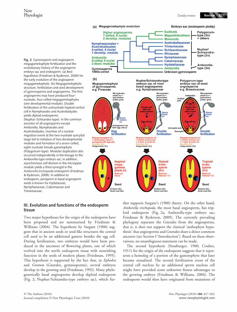
III. Evolution and functions of the endospermtissue
Two major hypotheses for the origin of the endosperm havebeen proposed and are summarized by Friedman &Williams (2004). The hypothesis by Sargant (1900) sug-gests that in ancient seeds or seed-like structures the centralcell used to be an additional gamete besides the egg cell.During fertilization, two embryos would have been pro-duced in the ancestors of flowering plants, one of whichevolved into the sterile endosperm tissue with nourishingfunction in the seeds of modern plants (Friedman, 1995).This hypothesis is supported by the fact that, in Ephedraand Gnetum (Gnetales, gymnosperms), several embryosdevelop in the growing seed (Friedman, 1992). Many phylo-genetically basal angiosperms develop diploid endosperm(Fig. 2; Nuphar ⁄ Schisandra-type embryo sac), which fur-
ther supports Sargant’s (1900) theory. On the other hand,Amborella trichopoda, the most basal angiosperm, has trip-loid endosperm (Fig. 2a; Amborella-type embryo sac;Friedman & Ryderson, 2009). The currently prevailingphylogeny separates the Gnetales from the angiosperms,that is, it does not support the classical ‘anthophyte hypo-thesis’ that angiosperms and Gnetales share a direct commonancestor (see Section I ‘Introduction’). Based on these obser-vations, no unambiguous statement can be made.
The second hypothesis (Strasburger, 1900; Coulter,1911) for the origin of the endosperm suggests that it repre-sents a homolog of a portion of the gametophyte that laterbecame sexualized. The second fertilization event of thecentral cell nucleus by an additional sperm nucleus cellmight have provided some unknown fitness advantages tothe growing embryo (Friedman & Williams, 2004). Theendosperm would then have originated from mutations of
(a)
(b)
Fig. 2 Gymnosperm and angiospermmegagametophyte fertilization and theevolutionary history of the angiospermembryo sac and endosperm. (a) Besthypothesis (Friedman & Ryderson, 2009) forthe early evolution of the angiospermmegagametophyte. (b) Megagametophytestructure, fertilization and seed developmentof gymnosperms and angiosperms. The firstangiosperms may have produced four-nucleate, four-celled megagametophytes(one developmental module). Doublefertilization of the uninucleate haploid centralcell in Nymphaeales and Austrobailyalesyields diploid endosperms(Nuphar ⁄ Schisandra-type). In the commonancestor of all angiosperms exceptAmborella, Nymphaeales andAustrobailyales, insertion of a nuclearmigration event at the two-nucleate syncytialstage led to initiation of two developmentalmodules and formation of a seven-celled,eight-nucleate female gametophyte(Polygonum-type). Modular duplication alsooccurred independently in the lineage to theAmborella-type embryo sac; in addition,asynchronous cell division in the micropylarmodule yields a third synergid in theAmborella trichopoda endosperm (Friedman& Ryderson, 2009). In addition toendosperm, perisperm in basal angiospermseeds is known for Hydataceae,Nymphaeaceae, Cabomaceae andTrimeniaceae.
NewPhytologist Tansley review Review 821
� The Authors (2010)
Journal compilation � New Phytologist Trust (2010)
New Phytologist (2010) 186: 817–831
www.newphytologist.com

the female gametophyte, which predestine these cells for asupporting nonreproductive role. This would explain theoccurrence of certain types of apomixes (Carman, 1997).According to Baroux et al. (2002), this theory is supportedby the fact that the addition of the paternal genome to thematernal central cell might create hybrid vigor. Both ofthese hypotheses have justifications, but a definite originof the endosperm tissue is still not clear (Berger, 2003;Friedman & Ryderson, 2009).
After fertilization in what is the most common type ofendosperm development, the nuclear type, the initial endo-sperm nucleus divides repeatedly without cell wall forma-tion, resulting in a characteristic coenocyte-stage endosperm(Baroux et al., 2002; Olsen, 2004; Friedman & Ryderson,2009). In many species, including Arabidopsis thaliana andcereals, this is subsequently followed by endosperm cellular-ization. Different developmental fate of chalazal and micro-pylar domains is a common pattern among the endospermsof all basal angiosperm taxa and suggests that this may be afeature of endosperm development in all angiosperms (Figs13–16 in Floyd & Friedman, 2000). Interactions and endo-sperm–embryo signaling is suggested from the fact that thebipolar endosperm development pattern of most angio-sperms is shared with the bipolar pattern of the embryos.For example, in all Nymphaeales (Fig. 2a; Nymphaeaceae,Cabomaceae, Hydatellaceae), the micropylar endospermundergoes division and becomes cellularized, whereas thechalazal domain remains undivided and acts as a hausto-rium, sometimes extending into the perisperm (Floyd &Friedman, 2000; Rudall et al., 2008, 2009). Other examplesof bipolar endosperm development are the BrassicaceaeA. thaliana and Lepidium virginicum, in which a multinucle-ated ‘chalazal’ region forms, and, at the same time, when therest of the endosperm including the micropylar domaincellularizes, this chalazal region remains multinucleated(Nguyen et al., 2000; Olsen, 2004). The radicle is embed-ded within the micropylar endosperm domain, whereas thetip of the cotyledons resides within the chazal endospermdomain of these Brassicaceae. The evolution of endospermdevelopmental patterns among basal flowering plants isreviewed in detail by Floyd & Friedman (2000), and endo-sperm development of other angiosperms is summarized byBaroux et al. (2002). The genera Arabidopsis and Lepidiumemerged as highly suited for cross-species work on seedsand fruits within the Brassicaceae family (Muller et al.,2006; Linkies et al., 2009; Mummenhoff et al., 2009).
Regardless of its origin, contemporary endosperm tissueserves not only as a nutrient source for the embryo duringseed development, but also as an integrator of seed growthand development which includes reciprocal signalingbetween seed compartments and parental effects caused byimprinting (Berger et al., 2006; Berger & Chaudhury,2009; Otho et al., 2009; Springer, 2009). The endospermis, depending on the species, partially or fully obliterated
upon seed maturity. However, most angiosperm specieshave retained an endosperm layer in their mature seeds(Fig. 19 in Floyd & Friedman, 2000; Fig. 3 in Forbis et al.,2002; Fig. 4 in Finch-Savage & Leubner-Metzger, 2006).In many cases, this endosperm in the mature seed is alsoinvolved in the control of germination by being a barrier forthe growing radicle. During germination, the micropylarendosperm weakens, allowing the radicle to protrude thesurrounding tissues. The hypothesis that weakening of theseed covering layers is achieved by enzymatic action wasfirst proposed by Ikuma & Thimann (1963). Endospermweakening was originally demonstrated for seeds of asteridspecies with either a thick endosperm layer (tomato,tobacco, coffee) or a thin endosperm layer (e.g. lettuce; Ni& Bradford, 1993; Bewley, 1997a,b; Toorop et al., 2000;Leubner-Metzger, 2002; Petruzzelli et al., 2003; Nonogaki,2006). More recent work demonstrated that endospermweakening also occurs in seeds of rosid species. The Brassic-aceae Lepidium sativum and A. thaliana have a thin endo-sperm layer and weakening of the micropylar endospermwas biomechanically quantified during the germination ofL. sativum (Muller et al., 2006; Bethke et al., 2007; Linkieset al., 2009). Comparison of the transcriptome of themicropylar and nonmicropylar endosperm of L. sativumsupports the bipolar character of this seed tissue (Linkieset al., 2009). It is the micropylar endosperm that canfunction as a barrier to radicle expansion and thereby cancontribute to the regulation of germination timing. Finch-Savage & Leubner-Metzger (2006) proposed that at leastsome of the molecular mechanisms of endosperm weaken-ing are widespread and are evolutionarily conserved traits.
IV. Evolution of dormancy
Seed dormancy is defined as an intrinsic block to the com-pletion of germination of a viable seed under favorable con-ditions for germination (e.g. temperature, humidity, light)of the corresponding nondormant seed. Seed dormancycontrols germination timing in response to the seasons andplays an important role in seed plant evolution and adapta-tion to climatic changes (Forbis et al., 2002; Baskin &Baskin, 2004; Evans & Dennehy, 2005; Leubner-Metzger,2007). Germination timing may strongly influence the rateat which species can expand their range, and may play animportant role in determining survival or extinction duringclimate change (Donohue, 2005).
Baskin & Baskin (1998, 2004) have proposed a compre-hensive ecophysiological classification system whichincludes five classes for ‘whole seed’ dormancy: physiologi-cal (PD), morphological (MD), morphophysiological(MPD), physical (PY) and combinational (PY+PD). Thesedifferent classes and their distribution among angiospermsare also summarized by Finch-Savage & Leubner-Metzger(2006).
822 Review Tansley reviewNewPhytologist
� The Authors (2010)
Journal compilation � New Phytologist Trust (2010)
New Phytologist (2010) 186: 817–831
www.newphytologist.com

Morphological dormancy is evident in seeds withembryos that are differentiated but very small comparedwith the size of the entire seed. The embryo to seed ratio(E : S ratio) describes the relative size of the embryo withinthe seed. A high E : S ratio (e.g. 0.9) means that the embryofills up most of the seed volume, whereas a low E : S ratio(e.g. 0.1) means that the embryo is tiny and the nutrientstorage tissue (endosperm, perisperm, megagametophyte)fills up most of the seed volume (Figs 3, 4 in Forbis et al.,2002; Figs 3, 4 in Finch-Savage & Leubner-Metzger,2006). Seeds with low E : S ratios often have long (a monthor more) germination times and the occurrence of abundantmegagametophyte (e.g. cycads, gymnosperms, Fig. 1c) orperisperm plus endosperm (e.g. Nuphar, basal angiosperms,Fig. 1d) tissue are typical for MD-class seeds. Forbis et al.(2002) used ancestral state reconstruction methods of con-tinuous characters using E : S family means for 179 familiescalculated from a large dataset of 1222 extant angiospermspecies. Their analysis showed that the E : S ratios haveincreased in derived angiosperms compared with ancestralangiosperms. They proposed, based on these results, that atiny embryo embedded in abundant endosperm ⁄ perisperm,and thereby classified as MD (and MPD), is the ancestraldormancy type of angiosperms. This hypothesis is inagreement with the results of Baskin & Baskin (2004). MDsimply delays germination timing by the time the embryoneeds to grow inside the seed before germination can takeplace. The dispersal of seeds with a small embryo that needstime to grow might have evolved as an ancient strategy todistribute germination times, since successful germination ishighly dependent on environmental conditions.
Among basal angiosperms (Fig. 2a), seeds with abundantperisperm that occupies a larger portion than the endo-sperm storage tissue is characteristic for extant and extinctNymphaeales (Nympheaceae, Cabomaceae, Hydatellaceae)(Floyd & Friedman, 2000; Friis et al., 2001; Yamadaet al., 2001; Chen et al., 2004; Baskin & Baskin, 2007;Friedman 2008; Rudall et al., 2009) and Austrobaileyales(Trimeniaceae) (Yamada & Marubashi, 2003; Yamadaet al., 2008). The presence of perisperm as the only nutritivetissue in the seed is rare. It is usually present together withendosperm in various proportions, locations and shapes.Abundant perisperm is not restricted to basal angiospermsand is also not necessarily associated with MD. Abundantperisperm is typical for most Caryophyllales seeds, such assugarbeet (nondormant, Amaranthaceae; Hermann et al.,2007) and cacti (PD, Cactaceae; Stuppy, 2002).
A trend to higher E : S ratios is also evident for gymno-sperms (Forbis et al., 2002). For example, extant basalgymnosperms, particularly cycads (Zamia, Cycas, Fig. 1c)and ginkgos, have smaller embryos than some of the morederived gymnosperm taxa, such as Callitropsis, Picea, Pinusand Juniperus (Baskin & Baskin, 1998; Fig. 4 in Forbiset al., 2002). Forbis et al. (2002) stated that available
gymnosperm embryo fossils are approximately in thesame shape and size ranges as extant gymnosperm embryosof related groups (see references cited in Forbis et al.,2002) and that there are no fossil gymnosperm seeds withextremely small or extremely large E : S ratios. They there-fore suggested that an unknown gymnosperm ancestor thatpredates these fossil specimens likely had a small embryoat dispersal. As several extant taxa have much largerembryos, they interpreted this as support for the hypothesisof increasing E : S ratios among gymnosperms.
Taken together, there seems to be a general trend ofincreasing relative embryo size during evolution (higherE : S ratio) for both angiosperms and gymnosperms (Forbiset al., 2002; Baskin & Baskin, 2004). Based on this, Forbiset al. (2002) proposed that morphological dormancy is theancestral dormancy type among gymnosperms and angio-sperms. This is consistent with the conclusion reached byBaskin & Baskin (1998, 2004). The evolution of largerembryo size likely resulted in occurrence of nondormantseeds; the embryo did not need to grow before germination.It is thought that increased relative embryo size is one of themain determinants (or requirements) for the evolution ofother classes of seed dormancy (Finch-Savage & Leubner-Metzger, 2006).
The core eudicots tend to have less endosperm than morebasal extant angiosperm species. At the same time, physio-logical dormancy developed, which is thought to be linkedto adaptation to seasonal weather changes as its releaserequires that the seeds perceived a specific environmentaltrigger(s). PD is the most abundant type of dormancy andis found in seeds of all major gymnosperm and angiospermclades (Fig. 1 in Baskin & Baskin, 2004; Fig. 4 in Finch-Savage & Leubner-Metzger, 2006). PD can be dividedinto different types; the most common form in both angio-sperm and gymnosperm is nondeep PD. Embryos excisedfrom seeds with nondeep PD will germinate normally andtreatment with gibberellins (GA) will break dormancy.Also, depending on species, dormancy can be broken byscarification (abrasion or cutting of the covering layers),after-ripening (a period of air-dry storage), and cold orwarm stratification. It has been shown that nondeep PDis determined by physiological factors in both theembryo and ⁄ or the covering layers (‘coats’ in a loose sense)(Bewley, 1997a,b; Koornneef et al., 2002; Kucera et al.,2005; Nonogaki, 2006; Bentsink & Koornneef, 2008;Holdsworth et al., 2008). Coat dormancy is mediated byany of the covering layers (the endosperm and ⁄ or the testa).Embryos excised from coat-dormant seeds develop andgrow readily. Abscisic acid (ABA) is an important positiveregulator of the coat-mediated nondeep PD in the seeds ofboth gymnosperms and angiosperms (Kucera et al., 2005).This suggests that gymnosperms and angiosperms sharecommon ABA-related molecular mechanisms regulatingdormancy and germination, and that ABA dependency is a
NewPhytologist Tansley review Review 823
� The Authors (2010)
Journal compilation � New Phytologist Trust (2010)
New Phytologist (2010) 186: 817–831
www.newphytologist.com

plesiomorphic trait for angiosperms and gymnosperms.The ABA-related transcription factor ABI3 ⁄ VP1 (ABAINSENSITIVE3 ⁄ VIVIPAROUS1) is widespread amonggreen plants and is involved in regulating dormancy ofangiosperm and gymnosperm seeds and buds (Holdsworthet al., 2008; Graeber et al., 2009). By contrast, DOG1(DELAY OF GERMINATION 1), a major quantitative traitgene more specifically involved in seed dormancy and ger-mination timing, is so far only known within the Brassica-ceae and its relation to ABA is the subject of ongoingresearch (Bentsink et al., 2006; Graeber et al., 2009).
Physiologically dormant and nondormant seeds are dis-tributed over the entire phylogenetic tree of gymnosperms,basal angiosperms, and eudicots (Fig. 4 in Finch-Savage &Leubner-Metzger, 2006). Therefore it has been proposedthat the gain and loss of PD quite likely occurred at severaltimes during evolution (Baskin & Baskin, 1998; Finch-Savage & Leubner-Metzger, 2006). The evolution of PDalso led to the appearance of MPD in seeds with a smallembryo, which upon gain in embryo size and concurrentloss of MD led to PD seeds (Fig. 5 in Finch-Savage &Leubner-Metzger, 2006). The most phylogeneticallyrestricted dormancy classes are PY and a combination ofboth PY and PD (Baskin & Baskin, 1998, 2004; Finch-Savage & Leubner-Metzger, 2006). PY is characterized by awater impermeability of the seed or fruit coat. It is believedto be an adaptation of the plant to specialized life habitats(Baskin & Baskin, 2004). PY is not found in gymnosperms,but only in angiosperm seeds, which indicates that it is amore derived form of dormancy.
V. Early seed-like structures and the transition toseeds
The origin and evolution of the seed habit is a fascinatingstory that started in late Devonian c. 370 million yr ago(Ma). Three major evolutionary trends were important forthe transition from the progymnosperms to the seed plants(Niklas, 1997; Doyle, 2006; Taylor & Taylor, 2009): theevolution from homospory to heterospory, meaning theproduction of specialized haploid female-like megasporesand male-like microspores; the evolution of the integu-ments; and the evolution of pollen-receiving structures.This includes the transition to water-independence of thepollination process.
The earliest seed plants emerged in the late Devonian froma paraphyletic group termed progymnosperms (Fig. 1a).Progymnosperm fossils show vegetative structures typicalfor seed plants combined with pteridophytic reproduction.Seed-like structures relating to the progymnosperm ⁄ seed–plant transition are often not preserved in fossil specimensand ⁄ or cannot be assigned unambiguously to the fossilspecimen. Archaeopteris, an extinct progymnosperm, wasthe first known modern tree (Judd et al., 2002; Crane et al.,
2004). Although it produced spores rather than seeds(Niklas, 1997; Judd et al., 2002), it exhibited an advancedsystem of spore production called heterospory. Hetero-spory, which has probably evolved independently in severallineages, is widely believed to be a precursor to seed repro-duction.
Fossils of paraphyletic seed ferns (Lyginopteridopsida,Fig. 1a) exhibit a variety of seed-like structures (Hemsley,1993; Taylor & Taylor, 1993, 2009; Doyle, 2006). Theoldest fossil pre-ovules are from the Middle Devonian(385 Ma, e.g. Runcaria; Gerrienne et al., 2004). Elksiniapolymorpha, the oldest known fossil seed plant, is a seed fernfrom the Late Devonian (Taylor & Taylor, 1993; Niklas,1997; DiMichele et al., 2006). This suggests that seedplants arose between 385 and 365 Ma, in the time intervalseparating Runcaria and the earliest known seed plants.The seed ferns Elksinia, as well as Archaeosperma andLagenostoma, produced pre-ovules or ovules on sterilestructures called cupules (Fig. 1a, inset). Cupules are cup-like structures that partially enclose the ovule. In these earlyovules, the nucellus was surrounded by integumentarytissue consisting of free lobes (Fig. 1a, inset). These lobescurved inwards at their tips, forming a ring around theapical end. So far, embryos have not been found inDevonian seed fern fossils.
The medullosan seed ferns are thought to be a mono-phyletic group of seed plants (Judd et al., 2002; Craneet al., 2004; DiMichele et al., 2006) and were abundanttrees in Carboniferous floodplains (> 290 Ma) andextended well into the Permian (> 250 Ma; Fig. 1a). Thisgroup includes Stephanospermum (Fig. 1b), Trigonocarpus,Pachytesta, Rhynchosperma, Medullosa, and Polypterospermum(Combourieu & Galtier, 1985; Drinnan et al., 1990; Taylor& Taylor, 1993; Dunn et al., 2002). Fossil seeds frommedullosan seed ferns are several mm to cm long. In somecases, even embryo structures have been preserved. In theseseed ferns, the cupule was replaced by a three-layered testa(Fig. 1b). There are indications that multiple origins ofcupules existed and that structures called cupules are not allhomologous among Paleozoic and Mesozoic seed ferns orgymnosperms (Hemsley, 1993; Doyle, 2006; Taylor &Taylor, 2009). The ovules usually have a round shape, withone end of the integument drawn out into a micropyle thatprobably helped guide pollen to the megagametophyte(Fig. 1b).
Schmeissneria has been proposed by Wang et al. (2007) asan Early Jurassic (> 160 Ma) missing link to angiospermsbecause it has angiospermous traits like closed carpels. Thisproposal is not widely accepted, however, and Schmeissneriais proposed by others to be a member of the Ginkgoales(Kirchner & Van Konijnenburg-Van Cittert, 1994; Zhou,2009). Phase-contrast X-ray microtomography links char-coalified fossil seeds from the Early Cretaceous (144 to100 Ma) with the gymnosperm Gnetales and Bennettitales
824 Review Tansley reviewNewPhytologist
� The Authors (2010)
Journal compilation � New Phytologist Trust (2010)
New Phytologist (2010) 186: 817–831
www.newphytologist.com

(Friis et al., 2007, 2009). These fossil seeds arec. 0.5–1.8 mm long and have two distinctly different layerssurrounding the nucellus: an inner, thin, membranousintegument, formed by thin-walled cells; and a robust,outer, sclerenchymatous seed envelope that completelyencloses the integument except for the micropylar opening.This outer seed envelope with distinctive anatomical struc-ture surrounds the nucellus and the integument. The integ-ument is extended apically into a long, narrow micropylartube. Only Gnetales (extant and extinct), Erdmanithecales(extinct) and Bennettitales (extinct) are known to have seedswith an additional seed envelope and the integumentextended into a long, narrow micropylar tube. The interpre-tation of the outer covering layer of Bennettitales seeds asan extra-integumentary outer envelope, as it is known fromGnetales seeds, is a matter of considerable debate (Friiset al., 2007, 2009; Rothwell et al., 2009).
Archaefructus, originally thought to be a stem-groupangiosperm of Jurassic age, is not; the fossil specimens havebeen redated as belonging to the Early Cretaceous(c. 125 Ma) flora of China (Sun et al., 2002; Friis et al.,2003; Frohlich & Chase, 2007). It seems to have been anaquatic plant and had fruits (c. 10 mm long and 2 mmwide) that contained two to 12 small seeds. It is possiblethat Archaefructus is on the stem lineage to angiosperms, butevidence for this is ambiguous (Friis et al., 2003; Pennisi,2009). Amborella trichopoda, an obscure shrub found onlyin New Caledonia, emerged as a crucial window to the past(Friedman & Ryderson, 2009; Pennisi, 2009; Williams,2009). Amborella sits at the base of the angiosperm familytree, the sister group of all other extant angiosperms (Fig.2a). The nuclear genome sequence of A. trichopoda will bean exceptional resource for comparative plant genomics(Soltis et al., 2008) and, based on its triploid endosperm(Fig. 2a), for re-examination of the evolutionary develop-mental history of the embryo sac (Williams, 2009).
VI. Seed size evolution
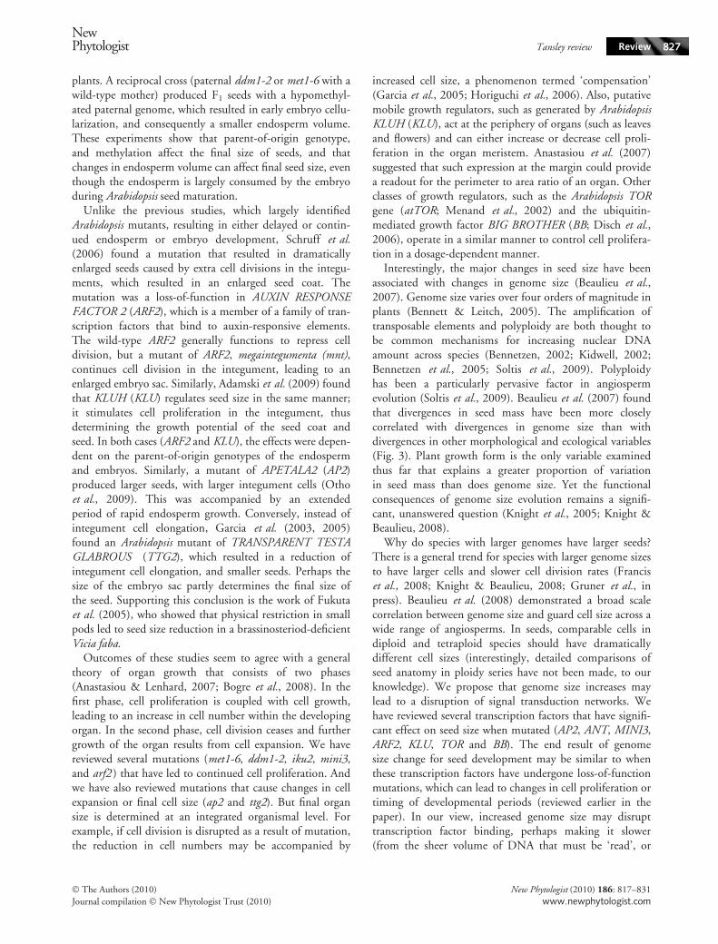
Seed size (mass) is central to many aspects of plant ecologyand evolution (Harper et al., 1970; Westoby et al., 1996;Leishman et al., 2000; Moles et al., 2005a,b). During aperiod of rapid angiosperm diversification (85–65 Ma),angiosperms moved out of the tropics and shifted frombeing predominantly small-seeded to having a much widerrange of seed sizes (Eriksson et al., 2000; Moles et al.,2005a,b). Extant angiosperms have seed masses spanning>11 orders of magnitude, from the lint-like seeds of orchidsup to the 20 kg seeds of the double coconut (Harper et al.,1970; Moles et al., 2005a,b). Gymnosperms have somewhatless variation in mass, but have larger seeds than the averageangiosperm (Fig. 3). Interestingly, individual orders andfamilies vary by up to eight orders of magnitude (Fig. 3).Moles et al. (2005a,b) found that plant size is the strongest
correlate with seed mass across a diverse assemblage of plantspecies (stronger than mode of dispersal or environmentalconditions). Several authors have suggested that species withlarge seeds have an advantage under low light conditions,when their greater protein and lipid reserves, or their moreadvanced development, can facilitate growth (Salisbury,1942; Mazer, 1989; Rees & Westoby, 1997; Geritz et al.,1999; Eriksson et al., 2000; Leishman et al., 2000; Moleset al., 2005a,b; Bruun & Ten Brink, 2008). However, largeseeds usually come at the cost of seed number per flower orfruit (Leishman, 2001). In addition, large seeds cannot bephysically borne on small plants because of the weight ofthe seed, which may partly explain the association betweenplant size and seed size (Grubb et al., 2005).
Another hypothesis to explain the plant size ⁄ seed size cor-relation is that there are common genetic components thatdetermine seed size, plant size, and the size of other plantorgans. There have been several recent reviews concerningthe genetic determinants of organ size (Sundaresan, 2005;Anastasiou & Lenhard, 2007; Bogre et al., 2008; Busovet al., 2008; Krizek, 2009). More cells or larger cells couldboth lead to larger organs, but in general it appears to be acombination of both (we will review several examples in thefollowing paragraphs). Cell cycle times, and thelength ⁄ duration of developmental periods, are thereforeimportant factors determining final organ size. In angio-sperm seeds, the size of each of the three major compart-ments (embryo, endosperm ⁄ perisperm, and seed coat)could increase individually. However, the growth of theseorgans is generally coordinated (Sundaresan, 2005; Othoet al., 2009), so selection for increased embryo size, maylead to a larger endosperm as well, and perhaps have conse-quences in other organs.
As with most developmental processes, the action of tran-scription factors has been shown to play a key role in deter-mining seed size. For example, in A. thaliana, large seedscan be generated by mutations in the APETALA2 (AP2)transcription factor (Jofuku et al., 2005; Ohto et al., 2005).Similarly, ectopic expression of the AINTEGUMENTA(ANT) transcription factor can also lead to larger seeds(Krizek, 1999; Mizukami & Fischer, 2000). Luo et al.(2005) found that mutations in either HAIKU2 (IKU2) orMINISEED3 (MINI3) led to reduced seed size, and thatthe mutant seed phenotypes depended on the parent-of-origin genotype of the endosperm and embryo. MINI3 is aWRKY family transcription factor, and IKU is a leucine-richrepeat (LRR) receptor kinase. IKU2 is expressed only inthe endosperm, while MINI3 is expressed in both theendosperm and the embryo. IKU2 expression was down-regulated in mini3 mutants, indicating that MINI3 actsupstream of IKU2. The reduced seed size of iku mutantsshowed reduced endosperm growth, premature cellulariza-tion of the endosperm, and a reduced proliferation of theembryo after the early torpedo stage (Garcia et al., 2003).
NewPhytologist Tansley review Review 825
� The Authors (2010)
Journal compilation � New Phytologist Trust (2010)
New Phytologist (2010) 186: 817–831
www.newphytologist.com

Zhou et al. (2009) recently discovered that SHORTHYPOCOTYL UNDER BLUE (SHB1) regulates seeddevelopment through changes in cell size and number. Kang& Ni (2006) found a mutant, shb1-D, that was a dominantgain-of-function allele that led to overexpression of SHB1.When Zhou et al. (2009) studied phenotypic effects in theseed, they found that shb1-D exhibited increased seed masslargely as a result of coordinated endosperm cellularizationand enlargement and continued embryo development. Theshb1-D mutants had more cells, larger cells, accumulatedmore proteins and fatty acids, and had delayed embryodevelopment (which was compensated for later in embryo-genesis). By utilizing chromatin immunoprecipitation
(ChIP), Zhou et al. (2009) were able to show that SHB1associates with MINI3 and IKU2 promoters, which indi-cates that these genes may all act in a coordinated fashion toaffect final seed mass through the proliferation or delay ofendosperm and embryo development.
Xiao et al. (2006) discovered changes in Arabidopsis seedmass that were associated with mutations in METHYL-TRANSFERASE 1 (MET1) and DECREASE IN DNAMETHYLATION1 (DDM1). Pistils of met1-6 or ddm1-2mutants pollinated with wild-type pollen produced F1
plants with hypomethylated maternal genomes. Seeds fromthese F1 plants showed delayed endosperm development,and larger endosperm volume compared with the wild-type
Fig. 3 Comparison of variation in seed mass of different plant species. Shown is a phylogeny of seed plants to the order level, and the corre-sponding seed masses are figured as whisker-box-plots. The gray boxes include 50% of all data points for a certain order, with the vertical lineshowing the median and the error bars indicating the range of seed masses. Numbers in round brackets indicate the number of species in thecorresponding order for which seed mass data were available. Arrows in the phylogenetic tree indicate major divergence points in genome size(G) and seed mass (S). Shown are the 20 largest contributions to present-day variation in 2C DNA content, ranked from 1 to 20 (superscriptnumbers following G) by their contribution score for genome size (Beaulieu et al., 2007). Major significant divergences for seed mass areshown (Moles et al., 2005a; Beaulieu et al., 2007). Note that often genome size and seed mass divergence points belong to the same node.Genome size and seed mass divergences that are within an order are shown in brackets directly behind the order’s name. All seed mass dataare from Liu et al. (2008). The phylogenetic tree was constructed using Phylomatic (http://www.phylodiversity.net/phylomatic).
826 Review Tansley reviewNewPhytologist
� The Authors (2010)
Journal compilation � New Phytologist Trust (2010)
New Phytologist (2010) 186: 817–831
www.newphytologist.com
plants. A reciprocal cross (paternal ddm1-2 or met1-6 with awild-type mother) produced F1 seeds with a hypomethyl-ated paternal genome, which resulted in early embryo cellu-larization, and consequently a smaller endosperm volume.These experiments show that parent-of-origin genotype,and methylation affect the final size of seeds, and thatchanges in endosperm volume can affect final seed size, eventhough the endosperm is largely consumed by the embryoduring Arabidopsis seed maturation.
Unlike the previous studies, which largely identifiedArabidopsis mutants, resulting in either delayed or contin-ued endosperm or embryo development, Schruff et al.(2006) found a mutation that resulted in dramaticallyenlarged seeds caused by extra cell divisions in the integu-ments, which resulted in an enlarged seed coat. Themutation was a loss-of-function in AUXIN RESPONSEFACTOR 2 (ARF2), which is a member of a family of tran-scription factors that bind to auxin-responsive elements.The wild-type ARF2 generally functions to repress celldivision, but a mutant of ARF2, megaintegumenta (mnt),continues cell division in the integument, leading to anenlarged embryo sac. Similarly, Adamski et al. (2009) foundthat KLUH (KLU) regulates seed size in the same manner;it stimulates cell proliferation in the integument, thusdetermining the growth potential of the seed coat andseed. In both cases (ARF2 and KLU), the effects were depen-dent on the parent-of-origin genotypes of the endospermand embryos. Similarly, a mutant of APETALA2 (AP2)produced larger seeds, with larger integument cells (Othoet al., 2009). This was accompanied by an extendedperiod of rapid endosperm growth. Conversely, instead ofintegument cell elongation, Garcia et al. (2003, 2005)found an Arabidopsis mutant of TRANSPARENT TESTAGLABROUS (TTG2), which resulted in a reduction ofintegument cell elongation, and smaller seeds. Perhaps thesize of the embryo sac partly determines the final size ofthe seed. Supporting this conclusion is the work of Fukutaet al. (2005), who showed that physical restriction in smallpods led to seed size reduction in a brassinosteriod-deficientVicia faba.
Outcomes of these studies seem to agree with a generaltheory of organ growth that consists of two phases(Anastasiou & Lenhard, 2007; Bogre et al., 2008). In thefirst phase, cell proliferation is coupled with cell growth,leading to an increase in cell number within the developingorgan. In the second phase, cell division ceases and furthergrowth of the organ results from cell expansion. We havereviewed several mutations (met1-6, ddm1-2, iku2, mini3,and arf2 ) that have led to continued cell proliferation. Andwe have also reviewed mutations that cause changes in cellexpansion or final cell size (ap2 and ttg2). But final organsize is determined at an integrated organismal level. Forexample, if cell division is disrupted as a result of mutation,the reduction in cell numbers may be accompanied by
increased cell size, a phenomenon termed ‘compensation’(Garcia et al., 2005; Horiguchi et al., 2006). Also, putativemobile growth regulators, such as generated by ArabidopsisKLUH (KLU), act at the periphery of organs (such as leavesand flowers) and can either increase or decrease cell proli-feration in the organ meristem. Anastasiou et al. (2007)suggested that such expression at the margin could providea readout for the perimeter to area ratio of an organ. Otherclasses of growth regulators, such as the Arabidopsis TORgene (atTOR; Menand et al., 2002) and the ubiquitin-mediated growth factor BIG BROTHER (BB; Disch et al.,2006), operate in a similar manner to control cell prolifera-tion in a dosage-dependent manner.
Interestingly, the major changes in seed size have beenassociated with changes in genome size (Beaulieu et al.,2007). Genome size varies over four orders of magnitude inplants (Bennett & Leitch, 2005). The amplification oftransposable elements and polyploidy are both thought tobe common mechanisms for increasing nuclear DNAamount across species (Bennetzen, 2002; Kidwell, 2002;Bennetzen et al., 2005; Soltis et al., 2009). Polyploidyhas been a particularly pervasive factor in angiospermevolution (Soltis et al., 2009). Beaulieu et al. (2007) foundthat divergences in seed mass have been more closelycorrelated with divergences in genome size than withdivergences in other morphological and ecological variables(Fig. 3). Plant growth form is the only variable examinedthus far that explains a greater proportion of variationin seed mass than does genome size. Yet the functionalconsequences of genome size evolution remains a signifi-cant, unanswered question (Knight et al., 2005; Knight &Beaulieu, 2008).
Why do species with larger genomes have larger seeds?There is a general trend for species with larger genome sizesto have larger cells and slower cell division rates (Franciset al., 2008; Knight & Beaulieu, 2008; Gruner et al., inpress). Beaulieu et al. (2008) demonstrated a broad scalecorrelation between genome size and guard cell size across awide range of angiosperms. In seeds, comparable cells indiploid and tetraploid species should have dramaticallydifferent cell sizes (interestingly, detailed comparisons ofseed anatomy in ploidy series have not been made, to ourknowledge). We propose that genome size increases maylead to a disruption of signal transduction networks. Wehave reviewed several transcription factors that have signifi-cant effect on seed size when mutated (AP2, ANT, MINI3,ARF2, KLU, TOR and BB). The end result of genomesize change for seed development may be similar to whenthese transcription factors have undergone loss-of-functionmutations, which can lead to changes in cell proliferation ortiming of developmental periods (reviewed earlier in thepaper). In our view, increased genome size may disrupttranscription factor binding, perhaps making it slower(from the sheer volume of DNA that must be ‘read’, or
NewPhytologist Tansley review Review 827
� The Authors (2010)
Journal compilation � New Phytologist Trust (2010)
New Phytologist (2010) 186: 817–831
www.newphytologist.com

because of a greater frequency of mismatches). Changes ingenome size may be analogous to changes in methylationobserved in the DDM1, MET1 mutants. Changes innuclear DNA content brought about by polyploidy lead tovariation in gene dosage and a doubling of orthologousgenes in the genome. Interestingly, orthologous genes havebeen observed to be down-regulated by methylation inpolyploids (Lee & Chen, 2001; Wang et al., 2004, 2006).
The studies reviewed here were largely done usingArabidopsis. These studies should become the basis of alarger cross-species comparison to see if the genetic factorsthat have been identified in mutant studies of Arabidopsisare the same ones that have led to the profound variationin seed mass across the angiosperms. Whether thesemechanisms are conserved in gymnosperms should also betested.
VII. Conclusion
The evolution of the seed represents a remarkable transitionfor photosynthetic organisms. Here, we have reviewed thedevelopment and dormancy of seeds, the rise and fall of theendosperm, and the genetic mechanisms and developmentalanatomy of large and small seeds. We would like to end bypresenting a series of unanswered questions that are ripe forfurther research in the field of seed biology. Whether gen-ome size increases lead to a disruption of signal transductionnetworks, causing continued cell proliferation and thereforeseed enlargement, is first on our list. We would also like toknow the biomechanical forces required to achieve embryogrowth, endosperm weakening, testa (seed coat) rupture,and the genes that are either up-regulated or down-regulatedduring these transitions. How does variation in cell size (andgenome size) affect germination, growth, and seed develop-ment? Why do some species have deep dormancy and othershave nondeep dormancy, and what genes are responsible forthese differences? Are the mechanisms that have led to theprofound variation in seed mass across the angiosperms thesame as the factors that have been identified in mutant stud-ies of Arabidopsis? There is still much to be learned from thecomparative anatomy and development of seeds. We suspectthat when looking at seeds of two species that vary consider-ably in size, the species with larger seeds will have more fullydeveloped embryos with a greater number of cells perembryo; larger cells in general; and will take longer todevelop to compensate for a slower cell division rate. Indeed,Leishman et al. (2000) found a close relationship betweenwhole-plant relative growth rates (RGRs) and seed mass,but whether whole-plant RGR is correlated with embryonicdevelopmental rates in seeds is unknown. Many of the ques-tions presented here are ideal for applying an integrative,phylogenic-based, cross-species, systems biology approach.And we predict great advances for our understanding of seedbiology and evolution in the coming decade.
Acknowledgements
Our work is supported by the Deutsche Forschungsgeme-inschaft (DFG-grants LE 720 ⁄ 6 and LE 720 ⁄ 7) and theERA-NET Plant Genomics grant vSEED to G.L.M., andby a sabbatical grant to C.K. by California Polytechnic StateUniversity.
References
Adamski NM, Anastasiou E, Eriksson S, O¢Neill CM, Lenhard M. 2009.
Local maternal control of seed size by KLUH ⁄ CYP78A5-dependent
growth signaling. Proceedings of the National Academy of Sciences, USA106: 20115–20120.
Anastasiou E, Kenz S, Gerstung M, MacLean D, Timmer J, Fleck C,
Lenhard M. 2007. Control of plant organ size by KLUH ⁄CYP78A5-dependent intercellular signaling. Developmental Cell 13:
843–856.
Anastasiou E, Lenhard M. 2007. Growing up to one’s standard. CurrentOpinion in Plant Biology 10: 63–69.
Baroux C, Spillane C, Grossniklaus U. 2002. Evolutionary origins
of the endosperm in flowering plants. Genome Biology 3: 1026.1–
1026.5.
Baskin CC, Baskin JM. 1998. Seeds – ecology, biogeography, and evolutionof dormancy and germination. London, UK: Academic Press.
Baskin CC, Baskin JM. 2007. A revision of Martin’s seed classification
system, with particular reference to his dwarf-seed type. Seed ScienceResearch 17: 11–20.
Baskin JM, Baskin CC. 2004. A classification system for seed dormancy.
Seed Science Research 14: 1–16.
Beaulieu JM, Leitch IJ, Patel S, Pendharkar A, Knight CA. 2008.
Genome size is a strong predictor of cell size and stomatal density in
angiosperms. New Phytologist 179: 975–986.
Beaulieu JM, Moles AT, Leitch IJ, Bennett MD, Dickie JB, Knight CA.
2007. Correlated evolution of genome size and seed mass. NewPhytologist 173: 422–437.
Bennett MD, Leitch IJ. 2005. Plant DNA C-values database, release 4.0,
October 2005. http://data.kew.org/cvalues/introduction.html.
Bennetzen JL. 2002. Mechanisms and rates of genome expansion and
contraction in flowering plants. Genetica 115: 29–36.
Bennetzen JL, Ma J, Devos KM. 2005. Mechanisms of recent genome size
variation in flowering plants. Annals of Botany 95: 127–132.
Bentsink L, Jowett J, Hanhart CJ, Koornneef M. 2006. Cloning of
DOG1, a quantitative trait locus controlling seed dormancy in
Arabidopsis. Proceedings of the National Academy of Sciences, USA 103:
17042–17047.
Bentsink L, Koornneef M. 2008. Seed dormancy and germination. In:
Somerville CR, Meyerowitz EM, eds. The Arabidopsis book. Rockville,
MD, USA: American Society of Plant Biologists, 1–18.
Berger F. 2003. Endosperm: the crossroad of seed development. CurrentOpinion in Plant Biology 6: 42–50.
Berger F, Chaudhury A. 2009. Parental memories shape seeds. Trends inPlant Science 14: 550–556.
Berger F, Grini PE, Schnittger A. 2006. Endosperm: an integrator of
seed growth and development. Current Opinion in Plant Biology 9:
664–670.
Berger F, Hamamura Y, Ingouff M, Higashiyama T. 2008. Double
fertilization – caught in the act. Trends in Plant Science 13: 437–443.
Bergfeld R, Schopfer P. 1986. Differentiation of a functional
aleurone layer within the seed coat of Sinapis alba L. Annals of Botany57: 25–33.
Bethke PC, Libourel IGL, Aoyama N, Chung Y-Y, Still DW, Jones RL.
2007. The Arabidopsis aleurone layer responds to nitric oxide,
828 Review Tansley reviewNewPhytologist
� The Authors (2010)
Journal compilation � New Phytologist Trust (2010)
New Phytologist (2010) 186: 817–831
www.newphytologist.com

gibberellin, and abscisic acid and is sufficient and necessary for seed
dormancy. Plant Physiology 143: 1173–1188.
Bewley JD. 1997a. Breaking down the walls – a role for endo-b-
mannanase in release from seed dormancy? Trends in Plant Science 2:
464–469.
Bewley JD. 1997b. Seed germination and dormancy. The Plant Cell 9:
1055–1066.
Black M. 2009. Darwin and seeds. Seed Science Research 19: 193–199.
Bogre L, Magyar Z, Lopez-Juez E. 2008. New clues to organ size control
in plants. Genome Biology 9: 226.1–226.7.
Bruun HH, Ten Brink D-J. 2008. Recruitment advantage of large seeds is
greater in shaded habitats. Ecoscience 15: 498–507.
Busov VB, Brunner AM, Strauss SH. 2008. Genes for control of plant
stature and form. New Phytologist 177: 589–607.
Cantino PD, Doyle JA, Graham SW, Judd WS, Olmstead RG, Soltis DE,
Soltis PS, Donoghue MJ. 2007. Towards a phylogenetic nomenclature
of Tracheophyta. Taxon 56: 822–846.
Carman JG. 1997. Asynchronous expression of duplicate genes in
angiosperms may cause apomixis, bispory, tetraspory, and
polyembryony. Biological Journal of the Linnean Society 61: 51–94.
Chen I, Manchester SR, Chen Z. 2004. Anatomically preserved seeds of
Nuphar (Nymphaeaceae) from Early Eocene of Wutu, Shandong
Province, China. American Journal of Botany 91: 1265–1272.
Combourieu N, Galtier J. 1985. Nouvelles observations sur
Polypterospermum, Polylophospermum, Colpospermum et Codonospermum,
ovules de Pteridospermales du Carbonifere superieur francais.
Palaeontographica Abt. B 196: 1–29.
Coulter JM. 1911. The endosperm of angiosperms. Botanical Gazette 52:
380–385.
Crane PR, Herendeen P, Friis EM. 2004. Fossils and plant phylogeny.
American Journal of Botany 91: 1683–1699.
Debeaujon I, Leon-Kloosterziel KM, Koornneef M. 2000. Influence of
the testa on seed dormancy, germination, and longevity in Arabidopsis.Plant Physiology 122: 403–413.
DiMichele WA, Phillips TL, Pfefferkorn HW. 2006. Paleoecology of Late
Paleozoic pteridosperms from tropical Euramerica. The Journal of theTorrey Botanical Society 133: 83–118.
Disch S, Anastasiou E, Sharma VK, Laux T, Fletcher JC, Lenhard M.
2006. The E3 ubiquitin ligase BIG BROTHER controls Arabidopsis
organ size in a dosage-dependent manner. Current Biology 16: 272–
279.
Donohue K. 2005. Niche construction through phenological plasticity:
life history dynamics and ecological consequences. New Phytologist 166:
83–92.
Doyle JA. 2006. Seed ferns and the origin of the angiosperms. The Journalof the Torrey Botanical Society 133: 169–209.
Drinnan AN, Schramke JM, Crane PR. 1990. Stephanospermumkonopeonus (Langford) comb. nov.: a medullosan ovule from the Middle
Pennsylvanian Mazon Creek Flora of northeastern Illinois, U.S.A.
Botanical Gazette 151: 385–401.
Dunn MT, Rothwell GW, Mapes G. 2002. Additional observations on
Rhynchosperma quinnii (Medullosaceae): a permineralized ovule from
the Chesterian (Upper Mississippian) Fayetteville Formation of
Arkansas. American Journal of Botany 89: 1799–1808.
Engler A, Prantl K. 1891. Die naturlichen Pflanzenfamilien. III. Teil, 2.Abteilung. Leipzig, Germany: Verlag von Wilhelm Engelmann.
Engler G. 1926. Die naturlichen Pflanzenfamilien, Volume 13,Gymnospermae. Leipzig, Germany: Wilhelm Engelmann.
Eriksson O, Friis EM, Pedersen KR, Crane PR. 2000. Seed size and
dispersal systems of Early Cretaceous angiosperms from Famalicao,
Portugal. International Journal of Plant Sciences 161: 319–329.
Evans MEK, Dennehy JJ. 2005. Germ banking: bet-hedging and variable
release from egg and seed dormancy. The Quarterly Review of Biology 80:
431–451.
Finch-Savage WE, Leubner-Metzger G. 2006. Seed dormancy and the
control of germination. New Phytologist 171: 501–523.
Floyd SK, Friedman WE. 2000. Evolution of endosperm developmental
patterns among basal flowering plants. International Journal of PlantSciences 161: S57–S81.
Forbis TA, Floyd SK, deQueiroz A. 2002. The evolution of embryo size in
angiosperms and other seed plants: implications for the evolution of seed
dormancy. Evolution 56: 2112–2125.
Francis D, Davies MS, Barlow PW. 2008. A strong nucleotypic effect on
the cell cycle regardless of ploidy level. Annals of Botany 101: 747–757.
Friedman WE. 1992. Evidence of a pre-angiosperm origin of endosperm:
implications for the evolution of flowering plants. Science 255: 336–339.
Friedman WE. 1995. Organismal duplication, inclusive fitness theory, and
altruism: understanding the evolution of endosperm and the angiosperm
reproductive syndrome. Proceedings of the National Academy of Sciences,USA 92: 3913–3917.
Friedman WE. 2008. Hydatellaceae are water lilies with gymnospermous
tendencies. Nature 453: 94–97.
Friedman WE, Madrid EN, Wiliams JH. 2008. Origin of the fittest and
survival of the fittest: relating female gametophyte development to
endosperm genetics. International Journal of Plant Sciences 169: 79–92.
Friedman WE, Ryderson KC. 2009. Reconstructing the ancestral female
gametophyte of angiosperms: insights from Amborella and other ancient
lines of flowering plants. American Journal of Botany 96: 129–143.
Friedman WE, Williams JH. 2004. Developmental evolution of the sexual
process in ancient flowering plant lineages. The Plant Cell 16(Suppl.):
S119–S132.
Friis EM, Crane PR, Pedersen KR, Bengtson S, Donoghue PC, Grimm
GW, Stampanoni M. 2007. Phase-contrast X-ray microtomography
links Cretaceous seeds with Gnetales and Bennettitales. Nature 450:
549–552.
Friis EM, Doyle JA, Endress PK, Leng Q. 2003. Archaefructus –
angiosperm precursor or specialized early angiosperm? Trends in PlantScience 8: 369–373.
Friis EM, Pedersen KR, Crane PR. 2001. Fossil evidence of water lilies
(Nymphaeales) in the Early Cretaceous. Nature 410: 357–360.
Friis EM, Pedersen KR, Crane PR. 2009. Early Cretaceous mesofossils
from Portugal and eastern north America related to the Bennettitales-
Erdtmanithecales-Gnetales group. American Journal of Botany 96: 252–
283.
Frohlich MW, Chase MW. 2007. After a dozen years of progress the
origin of angiosperms is still a great mystery. Nature 450: 1184–1189.
Fukuta N, Fukuzono K, Kawaide H, Abe H, Nakayama M. 2005.
Physical restriction of pods causes seed size reduction of a
brassinosteroid-deficient faba bean (Vicia faba). Annals of Botany 97:
65–69.
Garcia D, Fitz Gerald JN, Berger F. 2005. Maternal control of
integument cell elongation and zygotic control of endosperm growth are
coordinated to determine seed size in Arabidopsis. The Plant Cell 17:
52–60.
Garcia D, Saingery V, Chambrier P, Mayer U, Jurgens G, Berger F.
2003. Arabidopsis haiku mutants reveal new controls of seed size by
endosperm. Plant Physiology 131: 1661–1670.
Gasser CS, Broadhvest J, Hauser BA. 1998. Genetic analysis of ovule
development. Annual Review of Plant Physiology and Plant MolecularBiology 49: 1–24.
Geritz SAH, van der Meijden E, Metz JJ. 1999. Evolutionary dynamics of
seed size and seedling competitive ability. Theoretical Population Biology55: 324–343.
Gerrienne P, Meyer-Berthaud B, Fairon-Demaret M, Streel M, Steemans
P. 2004. Runcaria, a middle devonian seed plant precursor. Science 306:
856–858.
Graeber K, Linkies A, Muller K, Wunchova A, Rott A, Leubner-Metzger
G. 2009. Cross-species approaches to seed dormancy and germination:
NewPhytologist Tansley review Review 829
� The Authors (2010)
Journal compilation � New Phytologist Trust (2010)
New Phytologist (2010) 186: 817–831
www.newphytologist.com

conservation and biodiversity of ABA-regulated mechanisms and the
Brassicaceae DOG1 genes. Plant Molecular Biology Online First, doi:
10.1007/s11103-009-9583-x.
Grubb PJ, Coomes DA, Metcalfe DJ. 2005. Comment on ‘‘A brief history
of seed size.’’ Science 310: 783.
Gruner A, Hoverter N, Smith T, Knight CA. in press. Genome size is a
strong predictor of root meristem growth rate. Journal of Botany.Hajibabaei M, Xia J, Drouin G. 2006. Seed plant phylogeny: Gnetophytes
are derived conifers and a sister group to Pinaceae. MolecularPhylogenetics and Evolution 40: 208–217.
Harper JL, Lovell PH, Moore KG. 1970. The shapes and sizes of seeds.
Annual Review of Ecology and Systematics 1: 327–356.
Haughna G, Chaudhuryb A. 2005. Genetic analysis of seed coat
development in Arabidopsis. Trends in Plant Science 10: 472–477.
Hemsley AR. 1993. A review of Palaeozoic seed-megaspores.
Palaeontographica Abt. B 229: 135–166.
Hermann K, Meinhard J, Dobrev P, Linkies A, Pesek B, Heß B,
Machackova I, Fischer U, Leubner-Metzger G. 2007.
1-Aminocyclopropane-1-carboxylic acid and abscisic acid during the
germination of sugar beet (Beta vulgaris L.) – a comparative study of
fruits and seeds. Journal of Experimental Botany 58: 3047–3060.
Holdsworth MJ, Bentsink L, Soppe WJJ. 2008. Molecular networks
regulating Arabidopsis seed maturation, after-ripening, dormancy and
germination. New Phytologist 179: 33–54.
Horiguchi GA, Ferjani U, Fujikura H, Tsukaya J. 2006. Coordination of
cell proliferation and cell expansion in the control of leaf size in
Arabidopsis thaliana. Journal of Plant Research 119: 37–42.
Ikuma H, Thimann KV. 1963. The role of the seed-coats in
germination of photosensitive lettuce seeds. Plant and CellPhysiology 4: 169–185.
Jofuku KD, Omidyar PK, Gee Z, Okamuro JK. 2005. Control of seed
mass and seed yield by the floral homeotic gene APETALA2. Proceedingsof the National Academy of Sciences, USA 102: 3117–3122.
Judd WS, Campbell CS, Kellog EA, Stevens PF, Donoghue MJ. 2002.
Plant systematics: a phylogenetic approach. Sunderland, MA, USA:
Sinauer Associates.
Kang X, Ni M. 2006. Arabidopsis SHORT HYPOCOTYL UNDER
BLUE1 contains SPX and EXS domains and acts in cryptochrome
signaling. The Plant Cell 18: 921–934.
Kidwell MG. 2002. Transposable elements and the evolution of genome
size in eukaryotes. Genetica 115: 49–63.
Kirchner M, Van Konijnenburg-Van Cittert JHA. 1994. Schmeissneriamicrostachys (Presl, 1833) Kirchner et Van Konijnenburg-Van Cittert,
comb. nov. and Karkenia hauptmannii Kirchner et Van Konijnenburg-
Van Cittert, sp. nov., plants with ginkgoalean affinities from the Liassic
of Germany. Review of Palaeobotany and Palynology 83: 199–215.
Kirchner O, Loew E, Schroter C. 1938. Lebensgeschichte derBlutenpflanzen. Vol. II, 3. Dicotyledones, 41. Familie Nymphaeaceae.Stuttgart, Germany: Ulmer Verlag.
Knight CA, Beaulieu JM. 2008. Genome size scaling through phenotype
space. Annals of Botany 101: 759–766.
Knight CA, Molinari NA, Petrov DA. 2005. The large genome constraint
hypothesis: evolution, ecology, and phenotype. Annals of Botany 95:
177–190.
Koornneef M, Bentsink L, Hilhorst H. 2002. Seed dormancy and
germination. Current Opinion in Plant Biology 5: 33–36.
Krizek BA. 1999. Ectopic expression of AINTEGUMENTA in Arabidopsisplants results in increased growth of floral organs. DevelopmentalGenetics 25: 224–236.
Krizek BA. 2009. Making bigger plants: key regulators of final organ size.
Current Opinion in Plant Biology 12: 17–22.
Kucera B, Cohn MA, Leubner-Metzger G. 2005. Plant hormone
interactions during seed dormancy release and germination. Seed ScienceResearch 15: 281–307.
Lee HS, Chen ZJ. 2001. Protein-coding genes are epigenetically regulated
in Arabidopsis polyploids. Proceedings of the National Academy of Sciences,USA 98: 6753–6758.
Leishman M, Wright AJ, Moles AT, Westoby M. 2000. The
evolutionary ecology of seed size. In: Fenner M, ed. Seeds – the ecology ofregeneration in plant communities. Wallingford, UK: CAB International,
31–57.
Leishman MR. 2001. Does the seed size ⁄ number trade-off model
determine plant community structure? An assessment of the model
mechanisms and their generality Oikos 93: 294–302.
Leubner-Metzger G. 2002. Seed after-ripening and over-expression of
class I ß-1,3-glucanase confer maternal effects on tobacco testa rupture
and dormancy release. Planta 215: 659–698.
Leubner-Metzger G. 2007. Samendormanz und Keimungskontrolle:
Gene, Umweltfaktoren und Klimawandel. Vortrage fur Pflanzenzuchtung72: 87–104.
Li X, Yao Z. 1983. Fructifications of gigantopterids from South China.
Palaeontographica Abt. B 185: 11–26.
Linkies A, Muller K, Morris K, Tureckova V, Cadman CSC, Corbineau
F, Strnad M, Lynn JR, Finch-Savage WE, Leubner-Metzger G. 2009.
Ethylene interacts with abscisic acid to regulate endosperm rupture
during germination: a comparative approach using Lepidium sativumand Arabidopsis thaliana. The Plant Cell 21: 3803–3822.
Liu K, Eastwood RJ, Flynn S, Turner RM, Stuppy WH. 2008. SeedInformation Database, release 7.1, May 2008. http://www.kew.org/data/
sid.
Luo M, Dennis ES, Berger F, Peacock WJ, Chaudhury A. 2005.
MINISEED3 (MINI3), a WRKY family gene, and HAIKU2 (IKU2), a
leucine-rich repeat (LRR) KINASE gene, are regulators of seed size in
Arabidopsis. Proceedings of the National Academy of Sciences, USA 102:
17531–17536.
Mazer SJ. 1989. Ecological, taxonomic, and life history correlates of seed
mass among Indiana cune angiosperms. Ecological Monographs 59:
153–175.
Menand BT, Desnos T, Nussaume L, Berger F, Bouchez D, Meyer C,
Robaglia C. 2002. Expression and disruption of the Arabidopsis TOR(target of rapamycin) gene. Proceedings of the National Academy ofSciences, USA 99: 6422–6427.
Mizukami Y, Fischer RL. 2000. Plant organ size control:
AINTEGUMENTA regulates growth and cell numbers during
organogenesis. Proceedings of the National Academy of Sciences, USA 97:
942–947.
Moles AT, Ackerly DD, Webb CO, Tweddle JC, Dickie JB, Pitman AJ,
Westoby M. 2005a. Factors that shape seed mass evolution. Proceedingsof the National Academy of Sciences, USA 102: 10540–10544.
Moles AT, Ackerly DD, Webb CO, Tweddle JC, Dickie JB, Westoby M.
2005b. A brief history of seed size. Science 307: 576–580.
Muller K, Tintelnot S, Leubner-Metzger G. 2006. Endosperm-limited
Brassicaceae seed germination: abscisic acid inhibits embryo-induced
endosperm weakening of Lepidium sativum (cress) and endosperm
rupture of cress and Arabidopsis thaliana. Plant and Cell Physiology 47:
864–877.
Mummenhoff K, Polster A, Muhlhausen A, Theissen G. 2009. Lepidiumas a model system for studying the evolution of fruit development in
Brassicaceae. Journal of Experimental Botany 60: 1503–1513.
Nguyen H, Brown RC, Lemmon BE. 2000. The specialized chalazal
endosperm in Arabidopsis thaliana and Lepidium virginicum(Brassicaceae). Protoplasma 212: 99–110.
Ni BR, Bradford KJ. 1993. Germination and dormancy of abscisic acid-
deficient and gibberellin-deficient mutant tomato (Lycopersiconesculentum) seeds – sensitivity of germination to abscisic acid,
gibberellin, and water potential. Plant Physiology 101: 607–617.
Niklas KJ. 1997. The evolutionary biology of plants. Chicago, IL, USA: The
University of Chicago Press.
830 Review Tansley reviewNewPhytologist
� The Authors (2010)
Journal compilation � New Phytologist Trust (2010)
New Phytologist (2010) 186: 817–831
www.newphytologist.com

Nonogaki H. 2006. Seed germination – the biochemical and molecular
mechanisms. Breeding Science 56: 93–105.
Ohto M, Fischer RL, Goldberg RB, Nakamura K, Harada JJ. 2005.
Control of seed mass by APETALA2. Proceedings of the NationalAcademy of Sciences, USA 102: 3123–3128.
Olsen OA. 2004. Nuclear endosperm development in cereals and
Arabidopsis thaliana. The Plant Cell 16(Suppl.): S214–S227.
Otho M, Floyd SK, Fischer RL, Goldberg RR, Harada JJ. 2009. Effects of
APETALA2 on embryo, endosperm, and seed coat development
determine seed size in Arabidopsis. Sexual Plant Reproduction 22: 277–
289.
Pennisi E. 2009. Origins. On the origin of flowering plants. Science 324:
28–31.
Petruzzelli L, Muller K, Hermann K, Leubner-Metzger G. 2003. Distinct
expression patterns of ß-1,3-glucanases and chitinases during the
germination of Solanaceous seeds. Seed Science Research 13: 139–153.
Rees M, Westoby M. 1997. Game-theoretical evolution of seed mass in
multi-species ecological models. Oikos 78: 116–126.
Rothwell GW, Crepet WL, Stockey RA. 2009. Is the anthophyte
hypothesis alive and well? New evidence from the reproductive
structures of Bennettitales. American Journal of Botany 96: 296–322.
Rudall PJ, Eldridge T, Tratt J, Ramsay MM, Tuckett RE, Smith SY,
Collinson ME, Remizowa MV, Sokoloff DD. 2009. Seed fertilization,
development, and germination in Hydatellaceae (Nymphaeales):
implications for endosperm evolution in early angiosperms. AmericanJournal of Botany 96: 1581–1593.
Rudall PJ, Remizowa MV, Beer AS, Bradshaw E, Stevenson DW,
Macfarlane TD, Tuckett RE, Yadav SR, Sokoloff DD. 2008.
Comparative ovule and megagametophyte development in
Hydatellaceae and water lilies reveal a mosaic of features among the
earliest angiosperms. Annals of Botany 101: 941–956.
Salisbury EJ. 1942. The reproductive capacity of plants. London, UK: G.
Bell & Sons.
Sargant E. 1900. Recent work on the results of fertilization in
angiosperms. Annals of Botany 14: 689–712.
Schnarf W. 1937. Handbuch der Pflanzenanatomie, Vol. X ⁄ I, Anatomie derGymnospermensamen. Berlin, Germany: Gebruder Borntrager.
Schruff MC, Spielman M, Tiwari S, Adams S, Fenby N, Scott RJ. 2006.
The AUXIN RESPONSE FACTOR 2 gene of Arabidopsis links auxin
signalling, cell division, and the size of seeds and other organs.
Development 133: 251–261.
Scott DH. 1909. Studies in fossil botany, Vol.II, Spermatophyta. London,
UK: Adam and Charles Black.
Soltis D, Albert V, Leebens-Mack J, Palmer J, Wing R, dePamphilis C,
Ma H, Carlson J, Altman N, Kim S et al. 2008. The Amborellagenome: an evolutionary reference for plant biology. Genome Biology 9:
402.
Soltis DE, Albert VA, Leebens-Mack J, Bell CD, Paterson AH, Zheng C,
Sankoff D, dePamphilis CW, Wall PK, Soltis PS. 2009. Polyploidy
and angiosperm diversification. American Journal of Botany 96: 336–
348.
Springer NM. 2009. Small RNAs: how seeds remember to obey their
mother. Current Biology 19: R649–R651.
Strasburger E. 1900. Einige Bemerkungen zur Frage nach der ‘‘doppelten
Befruchtung’’ bei den Angiospermen. Botanische Zeitung 58: 294–315.
Stuppy W. 2002. Seed characters and generic classification of Opuntioideae.Surrey, UK: Richmond.
Sun G, Ji Q, Dilcher DL, Zheng S, Nixon KC, Wang X. 2002.
Archaefructaceae, a new basal angiosperm family. Science 296: 899–904.
Sundaresan V. 2005. Control of seed size in plants. Proceedings of theNational Academy of Sciences, USA 102: 17887–17888.
Taylor EL, Taylor TN. 2009. Seed ferns from the late Paleozoic and
Mesozoic: any angiosperm ancestors lurking there? American Journal ofBotany 96: 237–251.
Taylor TN, Taylor EL. 1993. The biology and evolution of fossil plants.New York, NY, USA: Prentice Hall.
Toorop PE, van Aelst AC, Hilhorst HWM. 2000. The second step of the
biphasic endosperm cap weakening that mediates tomato (Lycopersiconesculentum) seed germination is under control of ABA. Journal ofExperimental Botany 51: 1371–1379.
Wang J, Tian L, Lee H-S, Wei NE, Jiang H, Watson B, Madlung A,
Osborn TC, Doerge RW, Comai L et al. 2006. Genomewide
nonadditive gene regulation in Arabidopsis allotetraploids. Genetics 172:
507–517.
Wang J, Tian L, Madlung A, Lee H, Chen M, Lee JJ, Watson B,
Kagochi T, Comai L, Chen J. 2004. Stochastic and epigenetic
changes of gene expression in Arabidopsis polyploids. Genetics 167:
1961–1973.
Wang X, Duan S, Geng B, Cui J, Yang Y. 2007. Schmeissneria: a missing
link to angiosperms? BMC Evolutionary Biology 7: 14.
Westoby M, Leishman M, Lord J, Poorter H, Schoen DJ. 1996.
Comparative ecology of seed size and dispersal. PhilosophicalTransactions of the Royal Society of London. Series B, Biological Sciences351: 1309–1318.
Williams JH. 2009. Amborella trichopoda (Amborellaceae) and the
evolutionary developmental origins of the angiosperm progamic phase.
American Journal of Botany 96: 144–165.
Windsor JB, Symonds VV, Mendenhall J, Lloyd AM. 2000. Arabidopsis
seed coat development: morphological differentiation of the outer
integument. Plant Journal 22: 483–493.
Xiao W, Brown RC, Lemmon BE, Harada JJ, Goldberg RB, Fischer RL.
2006. Regulation of seed size by hypomethylation of maternal and
paternal genomes. Plant Physiology 142: 1160–1168.
Yamada T, Imaichi R, Kato M. 2001. Developmental morphology of
ovules and seeds of Nymphaeales. American Journal of Botany 88:
963–974.
Yamada T, Marubashi W. 2003. Overproduced ethylene causes
programmed cell death leading to temperature-sensitive lethality in
hybrid seedlings from the cross Nicotiana suaveolens · Nicotianatabacum. Planta 217: 690–698.
Yamada T, Nishida H, Umebayashi M, Uemura K, Kato M. 2008.
Oldest record of Trimeniaceae from the early Cretaceous of northern
Japan. BMC Evolutionary Biology 8: 135.1–135.7.
Zhou Y, Zhang XJ, Kang XJ, Zhao XY, Zhang XS, Ni M. 2009. SHORT
HYPOCOTYL UNDER BLUE1 associates with MINISEED3 and
HAIKU2 promoters in vivo to regulate Arabidopsis seed development.
The Plant Cell 21: 106–117.
Zhou Z-Y. 2009. An overview of fossil Ginkgoales. Palaeoworld 18: 1–22.
Zimmermann W. 1930. Die Phylogenie der Pflanzen. Jena, Germany:
Gustav Fischer Verlag.
NewPhytologist Tansley review Review 831
� The Authors (2010)
Journal compilation � New Phytologist Trust (2010)
New Phytologist (2010) 186: 817–831
www.newphytologist.com