table of contents - african veterinary information portal ... · web viewthe polymerase chain...
TRANSCRIPT

Livestock Health, Management and Production › High Impact Diseases › Contagious Diseases › Anthrax ›
AnthraxAuthor: Prof Valerius de Vos
Adapted from: DE VOS, V. & TURNBULL, P.C.B. 2004. Anthrax. In: Coetzer, J.A.W. & Tustin, R.C. Infectious Diseases of Livestock, Vol. 3, 3rd Ed. Oxford University Press Southern Africa. Pp 1788-1818.
Licensed under a Creative Commons Attribution license.
TABLE OF CONTENTSIntroduction....................................................................................................................2
Epidemiology..................................................................................................................3
Pathogenesis..................................................................................................................8
Diagnosis and differential diagnosis..........................................................................10
Clinical signs and pathology...................................................................................................10
Laboratory confirmation..........................................................................................................14
Differential diagnosis...............................................................................................................14
Control / Prevention.....................................................................................................15
Marketing and trade / Socio-economics....................................................................19
Important outbreaks....................................................................................................21
FAQs..............................................................................................................................22
References....................................................................................................................23
1 | P a g e
Livestock Health, Management and Production › High Impact Diseases › Contagious Diseases › Anthrax ›
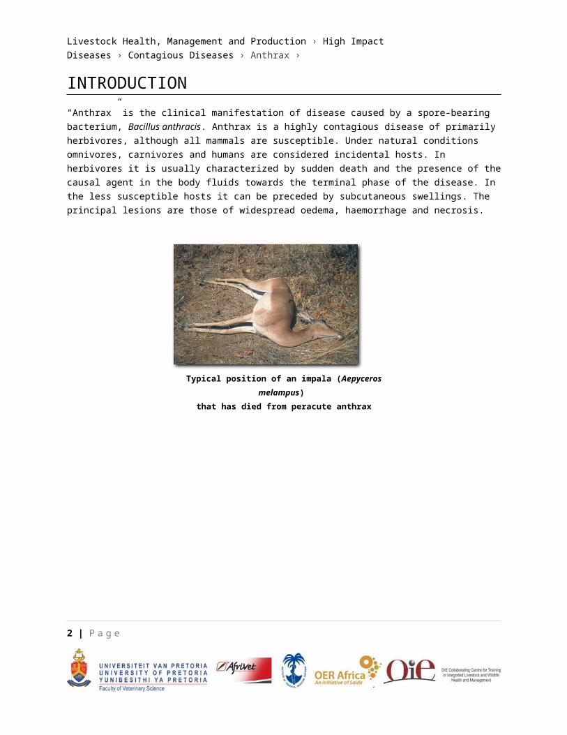
INTRODUCTION“Anthrax” is the clinical manifestation of disease caused by a spore-bearing bacterium, Bacillus anthracis. Anthrax is a highly contagious disease of primarily herbivores, although all mammals are susceptible. Under natural conditions omnivores, carnivores and humans are considered incidental hosts. In herbivores it is usually characterized by sudden death and the presence of the causal agent in the body fluids towards the terminal phase of the disease. In the less susceptible hosts it can be preceded by subcutaneous swellings. The principal lesions are those of widespread oedema, haemorrhage and necrosis.
Typical position of an impala (Aepyceros melampus) that has died from peracute anthrax
A Giemsa-stained blood smear from a recently-dead
2 | P a g e

Livestock Health, Management and Production › High Impact Diseases › Contagious Diseases › Anthrax ›
animal containing capsulated B. anthracis bacilli
When considering the eco-historical aspects of anthrax in southern Africa the following salient points emerge:
1. Early African settlers encountered and became familiar with anthrax (provided own name: “kwatsi”) in the Northern Cape Province where early European adventurers and travellers encountered it.
2. Isolation of anthrax from bone recovered from an archaeological digging at Pafuri, Kruger National Park (KNP), proves the existence of an anthrax outbreak at least 250±50 years ago.
3. Spread into the rest of the country equalled the expansion of commercial farming. The most severe outbreak documented occurred in 1923 with 60 000 livestock deaths recorded.
4. During more recent times, vaccination provided control of anthrax in commercial farming areas, but endemic anthrax persists in large wildlife areas such as the Kruger National Park (KNP) and Northern Cape Province. The rest of South Africa experiences sporadic point outbreaks. Livestock farming in some areas are however, being rapidly replaced by wildlife, exacerbating the anthrax problem, and making anthrax a re-emerging disease in southern Africa.
5. Genotyping of B. anthracis isolates suggests that the geographic origin of B. anthracis may be the sub-Saharan African continent, with South Africa featuring prominently. This means that before the appearance of man and his domestic animals, anthrax existed indigenously amongst free-living wild animals of the region. Remnants still exist in the KNP and Northern Cape in South Africa, where studies revealed an anthrax life cycle which is symbiotically integrated into the ecological processes of the region, and further corroborates the indigenous theory.
EPIDEMIOLOGYThe initiation of an outbreak or an epidemic of anthrax depends on interrelated factors that include specific properties of the bacterium, environmental factors, factors affecting dissemination of the organism, and certain human activities. The ability of the bacterium to survive outside its host, to enter and successfully infect its host, and to multiply in vivo is of particular importance. The anthrax cycle consists basically of: i) A biotic growth phase in a susceptible animal, where vegetative growth takes place, causing terminally a rapidly fatal septicaemia, and ii) An abiotic dormant phase, which starts with a transition into a very resistant spore form at the height of the growth phase under aerobic conditions and is carried passively and purely mechanically in carcass rests, soil, water and insects.
3 | P a g e
Livestock Health, Management and Production › High Impact Diseases › Contagious Diseases › Anthrax ›
The vegetative cells of B. anthracis are not particularly resistant to adverse environmental conditions, whilst spores are very resistant and are capable of surviving for very long periods until the opportunity to infect another host arises.
Within an infected host B. anthracis spores germinate to produce vegetative forms which multiply logarithmically, eventually killing the host. When conditions are not conducive to growth and multiplication, they tend to form spores but to do this need oxygen. Sporulation already starts within the live animal before oxygen levels drop below a level where growth does not occur, but occurs primarily after an infected fresh carcass has been opened by scavengers or man, and the tissues have been exposed to the air. Studies revealed that at the terminal stage there can already be about 1 spore for every 100 vegetative cells.
The clotting ability of the blood of an animal suffering from septicaemic anthrax is impaired. This can result in the formation of pools of blood and tissue fluid in and around the carcass of such an animal after it has been opened. In these pools, sufficiently aerobic conditions are present which, when favourable temperatures also prevail, allow B. anthracis to multiply and sporulate. Bacillus anthracis vegetative cells are “fragile” and require protein at a protective concentration around them to prevent them from lysis. It is therefore a race against time to become sporulated before the protein (blood or serum) becomes diluted, dispersed, or desiccated, and is the reason why vegetative cells do not sporulate on a thin blood smear.
The bacilli (vegetative form) cannot compete with putrefactive organisms and generally perish in the tissues of a carcass that has remained unopened for longer than three days at temperatures of 25 °C to 30 °C or higher. At temperatures of 5 °C to 10 °C, the rate of decomposition of a carcass is reduced, and B. anthracis can still be recovered from it for up to four weeks after death. The number of spores produced is therefore dependent on an early opening of an anthrax carcass, usually by predators, scavengers, carrion-eating birds or humans. Many of these spores remain at the site of the dead animal, but some are dispersed mechanically by run-off water and scavengers (mammals, birds and insects and humans).
It is believed that a variety of factors such as climate, topography, other microbial life, certain chemicals and certain plant material, may affect their survival. Thus, at most the duration of the survival period is probably limited to not more than three years, whilst in other circumstances, such as with burial, spores were found to remain dormant and viable in nature for some 200 + 50 years.
The “incubator area” or “soil capability” concept propagates the theory that B. anthracis can survive in soil in a dynamic state in which it undergoes cycles of germination, growth and sporulation, depending on fluctuating conditions in the micro-environment. Although it is accepted and proven by research that B. anthracis can grow in soil under special circumstances such as sterilized soil, existing evidence points to the fact that it is unable to multiply in competition with soil microbes, especially those containing phages and those that synthesize antibiotic substances. Not enough spores are produced to be a factor in the ecological processes of the anthrax cycle. B. anthracis therefore does not behave as a true saprophyte in
4 | P a g e
Livestock Health, Management and Production › High Impact Diseases › Contagious Diseases › Anthrax ›
nature. The “persistent spore” theory states that the vegetative growth cycle is host-dependent and that the external environment and soil act only as intermediary vehicles in which dormant spores are conveyed to new hosts. The survival of anthrax spores in nature is dependent on their initial numbers and the nature and composition of the environment where they eventually end-up. Spores may find themselves in a well-drained area and in a flowing river where they are eventually eliminated; or they end up in a low-lying poorly drained or stagnant area such as flood plains where spores accumulate, the so-called “anthrax concentrator areas”. Flood plains through centuries have become notorious for anthrax.
A variation of the persistent spore theory is the “concentrator area” concept in which host-independent growth is also discounted, but the role of calcium in spore preservation and subsequent germination requirements after dormancy are considered. It is proposed that, in calcium rich environments, exogenous calcium buffers leaching of calcium from the calcium rich core lattice of spores, thereby enhancing preservation of the spores until optimal nutrients and conditions for outgrowth are present. Calcium rich environments are therefore favourable to the survival of B. anthracis. However, strains vary in their calcium dependency. While some strains are found only in places with high calcium and pH soils, other strains may survive in a wider band of soils. It therefore means that B. anthracis is rather unique in that it has to kill its host in order to replicate and eventually survive. Warm climates favour the further growth and sporulation of B. anthracis in body fluids of opened infected carcasses with the potential for gross contamination of the surrounding soil and vegetation. In such climates, the occurrence of anthrax is closely integrated with a soil phase, leading to endemicity. In cold climates, the temperature is unfavourable for sporulation for much of the year. However summer temperatures are high enough to trigger the disease and allow significant sporulation, even at high latitudes where daylight is prolonged in summer.
Although the mortality rate is high and few animals recover spontaneously, evidence does exist that under exceptional circumstances, such as partially immunized animals, a carrier state or latent infection can develop in individual animals and some animal species. Although it is not clear what role such a state plays in the epidemiology of anthrax, it has been postulated that such carrier animals in an endemic anthrax area, when subjected to the occasional spore germination which macrophages normally clear successfully, could develop a mild form of anthrax which if unchecked when they are subjected to severe environmental stress, will convert into the peracute form of the disease.
Anthrax is primarily a disease of mammals, although birds (mostly ostriches) and reptiles (experimentally) have contracted the disease under special circumstances. In captive situations, such as in zoological gardens and on fur farms, outbreaks of anthrax are mostly limited to carnivorous species. In contrast, under free-ranging conditions, anthrax is most prevalent in herbivores, especially in ruminants; outbreaks in carnivores and omnivores are invariably incidental to outbreaks in herbivores.
Although anthrax deaths have been recorded in at least 59 mammal species, the susceptibility of different species to anthrax varies considerably. Discrepancies between susceptibility and prevalence of disease can also be explained in terms of differences in behaviour, such as feeding habits, and routes of infection;
5 | P a g e
Livestock Health, Management and Production › High Impact Diseases › Contagious Diseases › Anthrax ›
i.e. differences in vulnerability. In the KNP, the browsers have a higher vulnerability to anthrax than the grazers. In phosphorus-deficient areas in South Africa, osteophagia may increase the likelihood of cattle contracting anthrax; sheep do not usually manifest osteophagia under the same circumstances. In contrast to herbivores, pigs and carnivores are highly resistant to anthrax and the ingestion of large numbers of B. anthracis in infected meat is generally required to induce infection in them. The feeding behaviour of these animals, however, exposes them to much higher levels of infection than is the case with herbivores, and severe mortalities have been recorded. Age also affects the susceptibility of animals to anthrax; adults being generally more vulnerable than the young or subadults. Different behaviour patterns and feeding habits may predispose to this phenomenon, although there may also be a specific, but as yet, not understood, physiological basis. No sex differentiation could be found.
Anthrax outbreaks in carnivores in zoological gardens, and on fur farms most often occur when they are fed meat from carcasses which have not been adequately inspected.
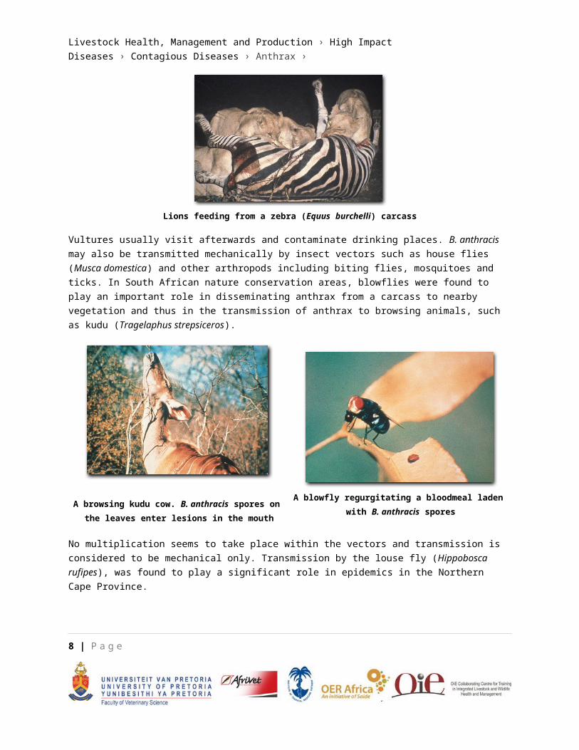
In the free-ranging state, dissemination and transmission is more complicated. Scavengers and predators play an important role, not only by opening up and dismembering infected carcasses, but also by dragging around portions and contaminating the environment.
Lions feeding from a zebra (Equus burchelli) carcass
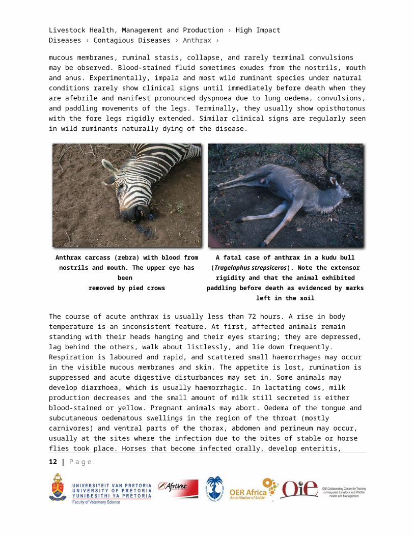
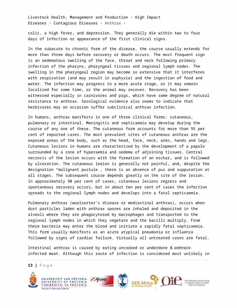
Vultures usually visit afterwards and contaminate drinking places. B. anthracis may also be transmitted mechanically by insect vectors such as house flies (Musca domestica) and other arthropods including biting flies, mosquitoes and ticks. In South African nature conservation areas, blowflies were found to play an important role in disseminating anthrax from a carcass to nearby vegetation and thus in the transmission of anthrax to browsing animals, such as kudu (Tragelaphus strepsiceros).
6 | P a g e

Livestock Health, Management and Production › High Impact Diseases › Contagious Diseases › Anthrax ›
A browsing kudu cow. B. anthracis spores on the leaves enter lesions in the mouth
A blowfly regurgitating a bloodmeal laden with B. anthracis spores
No multiplication seems to take place within the vectors and transmission is considered to be mechanical only. Transmission by the louse fly (Hippobosca rufipes), was found to play a significant role in epidemics in the Northern Cape Province.
Most tropical and subtropical countries have a peak of infection during dry summer seasons and a low point six months after the peak. This also applies to South Africa.
In the Etosha National Park, Namibia, and the Kgalagadi Transfrontier Park in South Africa/Botswana, anthrax outbreaks peak in summer after the major rains, following on the availability of water in man-made dams or gravel pits that attracts large numbers of game to certain areas. On the other hand, in the KNP, outbreaks of anthrax typically occur towards late winter/early summer before the first major rains when water is scarce and animals are concentrated around the remaining watering points. The onset of the rainy season terminates outbreaks.
In wildlife under free-ranging conditions, the general rule is an endemic anthrax situation interspersed with periodic epidemics. In the KNP these cyclic patterns of outbreaks occur approximately in ten year intervals, or multiples thereof, and are related to fluctuations in densities and concentrations of susceptible hosts.
Any epidemic is dependent on the availability of susceptible hosts. In the KNP it was found that all the major anthrax outbreaks had essentially normal epidemic curve patterns. This is an indication that a certain number or density of susceptible animals is necessary for an epidemic to start and progress into epidemic proportions. An anthrax epidemic in a natural setting is therefore density dependent and self-limiting. During these epidemics in the KNP anthrax killed less than 20% of its hosts, and being age linked, left in their wake a high percentage of young animals. Taking this into account it can be argued that in pristine nature anthrax can act as an ideal natural culling mechanism.
7 | P a g e
Livestock Health, Management and Production › High Impact Diseases › Contagious Diseases › Anthrax ›
PATHOGENESISNatural infection of animals is usually acquired by the ingestion of spores that germinate and gain entrance to the animal’s tissues, either in the pharynx or lower down the gastrointestinal tract. The bacterium is not invasive on its own and cannot penetrate the intact skin or mucosa but needs a portal of entry, such as an abrasion or insect bite; the exception being the lung alveoli. The spores soon germinate to form actively dividing encapsulated vegetative cells, which produce toxin. They are then transported from the primary site via lymphatics to regional lymph nodes and the mononuclear phagocytic system where further multiplication takes place and where vegetative bacilli continuously enter the bloodstream. During the final 10 to 14 hours of the host’s life, the bacteraemia increases progressively (at a doubling rate of 50 minutes in mice, guinea pigs and rhesus monkeys, 95 minutes in sheep, 125 minutes in rats and 150 minutes in chimpanzees) to reach a fairly constant terminal level (about 104 in Fischer rats, 106 in NIH black rats, 107 in mice, and 108 bacteria/ml in sheep). The host’s resistance to infection and intoxication determines the level. On this basis host species may be grouped into two categories: i) Species resistant to infection but highly susceptible to the effects of the toxin once infection is established (e.g., pigs, carnivores and primates). These species develop a relatively low terminal bacteraemia (± 104
bacilli/ml). ii) Species that are highly vulnerable to anthrax (e.g., ruminants). Most of these species show a high terminal bacteraemia (± 107-108 bacilli/ml).
Anthrax bacilli also possess two primary virulence factors, namely the poly-D-glutamate capsule and the tripartite protein toxin. The summative effect of the individual components of the toxin and the capsule is to inhibit phagocytosis, increase capillary endothelial permeability and delay blood clotting. Bacillus anthracis evades the immune system by producing an antiphagocytic capsule. In addition, it produces two related exotoxins namely, lethal toxin and oedema toxin. Lethal toxin comprises the protective antigen (PA) in combination with the lethal factor (LF), while the oedema toxin consists of PA in combination with the oedema factor (EF). All three proteins are well characterized and are produced simultaneously during the exponential phase of growth of the organism. Lethal toxin is responsible for the terminal shock in anthrax. Oedema factor induces altered water and ion movements resulting in the characteristic oedema of anthrax. The role of EF in the disease process may be to prevent mobilization and activation of polymorphonuclear leukocytes and thereby prevent phagocytosis of the bacteria. The genes for the virulence factors, the capsule and toxin complex, are associated with two large plasmids. The genes for PA, LF and EF are located on the plasmid pX01 and the genes encoding the enzymes mediating the polymerization of D-glutamic acid into the capsule on the plasmid pX02. Both plasmids have been sequenced. Plasmid pX02 is not present in the Sterne vaccine strain, thus rendering it attenuated.
Extreme hypoxia, hypoglycaemia and alkalosis may contribute to most of the clinical signs observed. Loss of body fluids into the tissues (oedema) and body cavities, such as the lungs, mediastinum, and peritoneal, pleural, and pericardial cavities, causes haemoconcentration in diseased animals. Respiratory distress and severe hypoxia in animals in extremis are caused by pulmonary oedema and utilization of available oxygen by the massive numbers of circulating bacilli and/or depression of the central nervous
8 | P a g e

Livestock Health, Management and Production › High Impact Diseases › Contagious Diseases › Anthrax ›
system. Hypoglycaemia, decreased levels of serum calcium, and increased levels of serum potassium and chlorine are recorded during the terminal stages of the disease. In some host species, leukocyte counts and haematocrit increase concurrently with the changes in calcium. The neuromuscular irritability, opisthotonus and possible convulsions exhibited by some animals in the terminal stages of anthrax are probably the consequence of decreased serum calcium and increased potassium. Extreme hypoxia and hypoglycaemia prevent the development of convulsions.
DIAGNOSIS AND DIFFERENTIAL DIAGNOSISClinical signs and pathology
The incubation period of anthrax under natural conditions is not known, but probably ranges from one to 14 days. Under experimental conditions, it varies considerably, and is influenced by the virulence of the organisms, the resistance of the animal, the route of infection and the number of infecting organisms. Some animals suffering from anthrax develop a fever but its severity is extremely variable. It appears to be characteristic for a species, and may depend on the number of infecting organisms. The fever usually declines before death.
Three different manifestations of anthrax are recognized: peracute or apoplectic, acute, and subacute to chronic forms. Cattle, sheep, goats and some wild ruminants, such as kudu, roan antelope and impala, mainly manifest the peracute and acute forms. Horses, donkeys and zebra suffer from the acute to subacute form. Pigs, carnivores and immunized animals (i.e. immunized animals challenged with high doses of infective material, or animals with low-grade immunity) usually contract the subacute to chronic form.
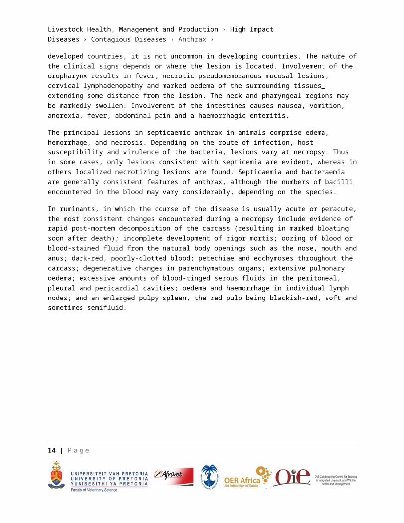
In peracute anthrax, the course of the clinical disease is usually less than two hours. The majority of animals are found dead without having shown signs of illness. In those in which clinical signs occur, pyrexia (depending on the species), restlessness or anxiety, muscle tremors, dyspnoea, congestion of mucous membranes, ruminal stasis, collapse, and rarely terminal convulsions may be observed. Blood-stained fluid sometimes exudes from the nostrils, mouth and anus. Experimentally, impala and most wild ruminant species under natural conditions rarely show clinical signs until immediately before death when they are afebrile and manifest pronounced dyspnoea due to lung oedema, convulsions, and paddling movements of the legs. Terminally, they usually show opisthotonus with the fore legs rigidly extended. Similar clinical signs are regularly seen in wild ruminants naturally dying of the disease.
9 | P a g e

Livestock Health, Management and Production › High Impact Diseases › Contagious Diseases › Anthrax ›
Anthrax carcass (zebra) with blood from nostrils and mouth. The upper eye has been
removed by pied crows
A fatal case of anthrax in a kudu bull (Tragelaphus strepsiceros). Note the extensor rigidity and that the animal exhibited paddling before death as evidenced
by marks left in the soil
The course of acute anthrax is usually less than 72 hours. A rise in body temperature is an inconsistent feature. At first, affected animals remain standing with their heads hanging and their eyes staring; they are depressed, lag behind the others, walk about listlessly, and lie down frequently. Respiration is laboured and rapid, and scattered small haemorrhages may occur in the visible mucous membranes and skin. The appetite is lost, rumination is suppressed and acute digestive disturbances may set in. Some animals may develop diarrhoea, which is usually haemorrhagic. In lactating cows, milk production decreases and the small amount of milk still secreted is either blood-stained or yellow. Pregnant animals may abort. Oedema of the tongue and subcutaneous oedematous swellings in the region of the throat (mostly carnivores) and ventral parts of the thorax, abdomen and perineum may occur, usually at the sites where the infection due to the bites of stable or horse flies took place. Horses that become infected orally, develop enteritis, colic, a high fever, and depression. They generally die within two to four days of infection or appearance of the first clinical signs.
In the subacute to chronic form of the disease, the course usually extends for more than three days before recovery or death occurs. The most frequent sign is an oedematous swelling of the face, throat and neck following primary infection of the pharynx, pharyngeal tissues and regional lymph nodes. The swelling in the pharyngeal region may become so extensive that it interferes with respiration (and may result in asphyxia) and the ingestion of food and water. The infection may progress to a more acute stage, or it may remain localized for some time, or the animal may recover. Recovery has been witnessed especially in carnivores and pigs, which have some degree of natural resistance to anthrax. Serological evidence also seems to indicate that herbivores may on occasion suffer subclinical anthrax infection.
In humans, anthrax manifests in one of three clinical forms: cutaneous, pulmonary or intestinal. Meningitis and septicaemia may develop during the course of any one of these. The cutaneous form accounts for
10 | P a g e

Livestock Health, Management and Production › High Impact Diseases › Contagious Diseases › Anthrax ›
more than 95 per cent of reported cases. The most prevalent sites of cutaneous anthrax are the exposed areas of the body, such as the head, face, neck, arms, hands and legs. Cutaneous lesions in humans are characterized by the development of a papule surrounded by a zone of hyperaemia and oedema of adjoining tissues. Central necrosis of the lesion occurs with the formation of an eschar, and is followed by ulceration. The cutaneous lesion is generally not painful, and, despite the designation “malignant pustule”, there is an absence of pus and suppuration at all stages. The subsequent course depends greatly on the site of the lesion. In approximately 90 per cent of cases, cutaneous lesions regress and spontaneous recovery occurs, but in about ten per cent of cases the infection spreads to the regional lymph nodes and develops into a fatal septicaemia.
Pulmonary anthrax (woolsorter's disease or mediastinal anthrax), occurs when dust particles laden with anthrax spores are inhaled and deposited in the alveoli where they are phagocytosed by macrophages and transported to the regional lymph nodes in which they vegetate and the bacilli multiply. From there bacteria may enter the blood and initiate a rapidly fatal septicaemia. This form usually manifests as an acute atypical pneumonia or influenza followed by signs of cardiac failure. Virtually all untreated cases are fatal.
Intestinal anthrax is caused by eating uncooked or underdone B. anthracis-infected meat. Although this route of infection is considered most unlikely in developed countries, it is not uncommon in developing countries. The nature of the clinical signs depends on where the lesion is located. Involvement of the oropharynx results in fever, necrotic pseudomembranous mucosal lesions, cervical lymphadenopathy and marked oedema of the surrounding tissues extending some distance from the lesion. The neck and pharyngeal regions may be markedly swollen. Involvement of the intestines causes nausea, vomition, anorexia, fever, abdominal pain and a haemorrhagic enteritis.
The principal lesions in septicaemic anthrax in animals comprise edema, hemorrhage, and necrosis. Depending on the route of infection, host susceptibility and virulence of the bacteria, lesions vary at necropsy. Thus in some cases, only lesions consistent with septicemia are evident, whereas in others localized necrotizing lesions are found. Septicaemia and bacteraemia are generally consistent features of anthrax, although the numbers of bacilli encountered in the blood may vary considerably, depending on the species.
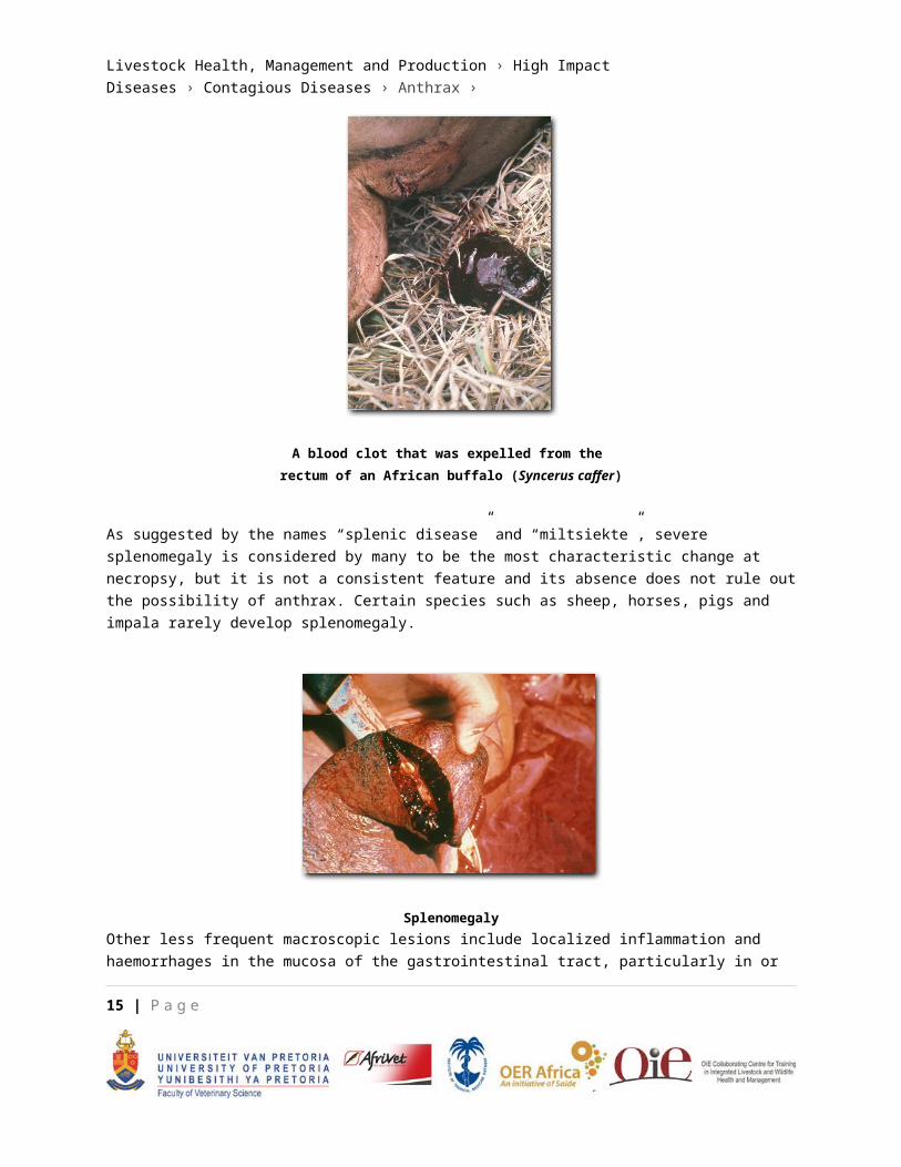
In ruminants, in which the course of the disease is usually acute or peracute, the most consistent changes encountered during a necropsy include evidence of rapid post-mortem decomposition of the carcass (resulting in marked bloating soon after death); incomplete development of rigor mortis; oozing of blood or blood-stained fluid from the natural body openings such as the nose, mouth and anus; dark-red, poorly-clotted blood; petechiae and ecchymoses throughout the carcass; degenerative changes in parenchymatous organs; extensive pulmonary oedema; excessive amounts of blood-tinged serous fluids in the peritoneal, pleural and pericardial cavities; oedema and haemorrhage in individual lymph nodes; and an enlarged pulpy spleen, the red pulp being blackish-red, soft and sometimes semifluid.
11 | P a g e
Livestock Health, Management and Production › High Impact Diseases › Contagious Diseases › Anthrax ›
A blood clot that was expelled from the rectum of an African buffalo (Syncerus caffer)
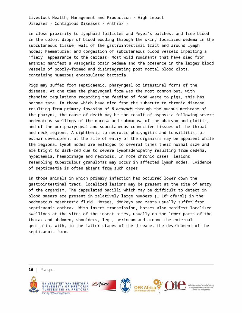
As suggested by the names “splenic disease” and “miltsiekte”, severe splenomegaly is considered by many to be the most characteristic change at necropsy, but it is not a consistent feature and its absence does not rule out the possibility of anthrax. Certain species such as sheep, horses, pigs and impala rarely develop splenomegaly.
SplenomegalyOther less frequent macroscopic lesions include localized inflammation and haemorrhages in the mucosa of the gastrointestinal tract, particularly in or in close proximity to lymphoid follicles and Peyer's patches,
12 | P a g e
Livestock Health, Management and Production › High Impact Diseases › Contagious Diseases › Anthrax ›
and free blood in the colon; drops of blood exuding through the skin; localized oedema in the subcutaneous tissue, wall of the gastrointestinal tract and around lymph nodes; haematuria; and congestion of subcutaneous blood vessels imparting a “fiery” appearance to the carcass. Most wild ruminants that have died from anthrax manifest a vasogenic brain oedema and the presence in the larger blood vessels of poorly-formed and disintegrating post mortal blood clots, containing numerous encapsulated bacteria.
Pigs may suffer from septicaemic, pharyngeal or intestinal forms of the disease. At one time the pharyngeal form was the most common but, with changing regulations regarding the feeding of food waste to pigs, this has become rare. In those which have died from the subacute to chronic disease resulting from primary invasion of B. anthracis through the mucous membrane of the pharynx, the cause of death may be the result of asphyxia following severe oedematous swellings of the mucosa and submucosa of the pharynx and glottis, and of the peripharyngeal and subcutaneous connective tissues of the throat and neck regions. A diphtheric to necrotic pharyngitis and tonsillitis, or eschar development at the site of entry of the organisms may be apparent while the regional lymph nodes are enlarged to several times their normal size and are bright to dark-red due to severe lymphadenopathy resulting from oedema, hyperaemia, haemorrhage and necrosis. In more chronic cases, lesions resembling tuberculous granulomas may occur in affected lymph nodes. Evidence of septicaemia is often absent from such cases.
In those animals in which primary infection has occurred lower down the gastrointestinal tract, localized lesions may be present at the site of entry of the organism. The capsulated bacilli which may be difficult to detect in blood smears are present in relatively large numbers (± 105 cfu/ml) in the oedematous mesenteric fluid. Horses, donkeys and zebra usually suffer from septicaemic anthrax. With insect transmission, horses also manifest localized swellings at the sites of the insect bites, usually on the lower parts of the thorax and abdomen, shoulders, legs, perineum and around the external genitalia, with, in the latter stages of the disease, the development of the septicaemic form.
13 | P a g e
Livestock Health, Management and Production › High Impact Diseases › Contagious Diseases › Anthrax ›
Ventral subcutaneous oedema in a horse
Subcutaneous oedema of the perineum of a horse
Severe inflammatory oedema of the soft tissues of the head, tongue and throat, stomach and intestines are characteristic features of anthrax in carnivores. In the African lion, localized lesions with no or late onset of septicaemia are often seen. These lesions are of variable severity, but are usually outspoken and localized in the tissues of the face, oral cavity and the regional lymph nodes. The changes are invariably characterized by localized necrotic glossitis or stomatitis to locally extensive necrotic cellulitis of the lips and the face accompanied by severe oedema causing severe swelling of the tissues of the head.
Facial oedema in a lion A fatal case of anthrax in a lioness. Note the swelling of the head
14 | P a g e
Livestock Health, Management and Production › High Impact Diseases › Contagious Diseases › Anthrax ›
Laboratory confirmation
The history—including clinical manifestations and necropsy findings (if by mistake the carcass has been opened)—is usually the first step in the diagnosis of anthrax and should lead to further confirmation procedures.
In stained smears of blood or tissue fluid obtained from infected animals, the organisms appear truncated, measure 1.0 to 1.5 by 3.0 to 10.0 mm, commonly occur singly or in short chains and are surrounded by a well-developed capsule. The capsule shows up well with polychrome methylene blue (M’Fadyean reaction stain) and reasonably well with Wright’s and Giemsa stains. The staining reaction is so typical that demonstration of the capsule by staining blood smears is accepted as confirmation of anthrax. The absence of bacilli does not necessarily exclude the possibility of anthrax.
If, after the examination of a blood smear, anthrax is still suspected, suitable samples (blood, soft tissue or bone, and even soil underneath) should be collected for bacteriological examination and submitted on ice to a laboratory.
Bacillus anthracis grows readily on artificial media. Best results are obtained on media that contains serum or blood. In contaminated specimens selective isolation techniques are necessary. The PLET medium of Knisely is generally recommended and is sensitive to concentrations as low as three spores per gram of soil. An isolate with the characteristic colonial morphology that is non-haemolytic or only weakly haemolytic, non-motile, sensitive to anthrax-specific gamma-phage and penicillin, and able to produce a capsule in vitro in defibrinated blood can be diagnosed with confidence as B. anthracis. Because of humane considerations, in vivo tests are not recommended.
The polymerase chain reaction (PCR) and effective serological enzyme immunoassay (EIA) are useful diagnostic, epidemiological, and research aids but presently are confined to a few specialist laboratories. A very promising development for future portable on-the-spot diagnosis is an immunochromatographic protective antigen assay. Although recent developments in molecular biology have made genotyping of B. anthracis isolates possible, it is still a specialist procedure.
Differential diagnosis
In general, all causes of sudden death, especially in herbivores, and causes of localised subcutaneous edematous swellings, especially in horse and pig families and carnivores, resemble anthrax.
CONTROL / PREVENTIONIn most countries anthrax is a controlled or notifiable disease by law, and control measures are prescribed and enforced by veterinary authorities. Anthrax control measures are aimed at breaking the cycle of
15 | P a g e
Livestock Health, Management and Production › High Impact Diseases › Contagious Diseases › Anthrax ›
infection and consist basically of a surveillance system, prophylactic procedures (immunization, treatment and disinfection), and disease regulatory actions (quarantine, immunization, treatment, proper disposal of carcasses and disinfection).
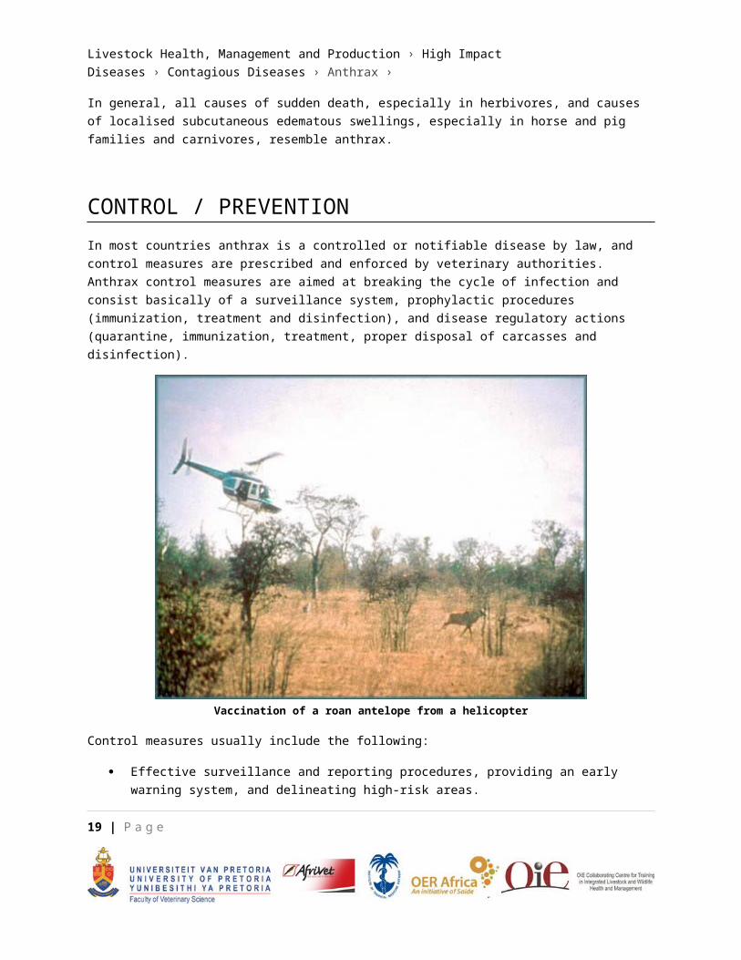
Vaccination of a roan antelope from a helicopter
Control measures usually include the following:
Effective surveillance and reporting procedures, providing an early warning system, and delineating high-risk areas.
Quarantine procedures, isolating an infected and contaminated area and animals until safe.
Preventing excess sporulation of the vegetative growth form of B. anthracis by not opening carcasses. For disposal of carcasses, burial should be discouraged in favour of incineration. Where burial is done, carcasses should be buried at a depth of two metres after being covered liberally with a mixture of chloride of lime, containing at least 25 per cent of active chlorine, and enclosed in plastic sheeting; the rationale being that chlorine gas develops and sterilizes the carcass.
The WHO has published guidelines for the disinfection of B. anthracis-infected material. For disinfection or decontamination of anthrax-infected/contaminated material and soil, formaldehyde (2%-10%) is generally preferred.
16 | P a g e

Livestock Health, Management and Production › High Impact Diseases › Contagious Diseases › Anthrax ›
In particularly valuable animals exposed to anthrax, treatment with bactericidal antibiotics may be used as a prophylactic measure. Simultaneous vaccination is contraindicated.
Vaccination with Sterne spore vaccine. Annual vaccination is recommended for all cattle in high-risk areas. During an outbreak vaccination of all cattle in the immediate surroundings and in-contact herbivores is recommended. This also applies to captive wild animals where possible. The Sterne vaccine is for all practical purposes non-pathogenic in most domestic and wild animal species. It however, appears to retain a degree of virulence for certain species such as goats and llamas. In such species, two inoculations one month apart, with the first being one quarter of the standard dose and the second being the full standard dose, are recommended. In horses, being slow to react, two standard doses one month apart and a single annual booster thereafter are recommended. In the other species a single inoculation provides effective immunity for about nine months to a year. Effective immunity generally develops within a week of vaccination, although in horses it may take a month or more. Outbreaks of anthrax in livestock are usually brought under control within a week to ten days of vaccination of affected herds.
While control of anthrax has become a distinct possibility in livestock areas, anthrax in free-ranging wildlife in several regions of the world retains a continued place in the ecology of African wildlife. In the southern African large wildlife parks, anthrax is now accepted as indigenous and one of a spectrum of natural culling mechanisms. However, at the interface with local populations and their livestock it can present risks and thus the need for control. While anthrax can be controlled and even eradicated in livestock, control in wildlife is largely impractical and almost impossible. Nevertheless, herd animals such as Burchell's zebra (Equus burchelli burchelli), blue wildebeest (Connochaetes taurinus), African buffalo (Syncerus caffer) and American bison (Bison bison) that can be corralled or captured, can be vaccinated. Long-stemmed, hand-held automatic vaccinating syringes and dart syringes are used from the sides of the boma or crush. An aerial method of immunizing free-ranging wildlife was also developed for especially small populations of endangered species.
In the major game reserves most of the control measures, as used for livestock, are difficult, if not impossible, to apply and/or enforce. In addition, anthrax being considered indigenous and a natural and integral part of the ecosystems of some of these areas, makes it debatable whether active control measures should actually be instituted. In the National Parks of South Africa, the current policy is to institute active control measures against anthrax only if it affects biodiversity negatively (e.g. by threatening the survival of low density or threatened species), and/or where the actions of humans (such as fencing or the provision of artificial watering points), are providing unnatural impetus to an outbreak. A general rule of thumb for deciding whether or not to implement control measures in wildlife areas is that the bigger and more natural and self-sufficient an area is, the less control measures should be implemented, and vice versa. Apart from immunization, anthrax control procedures which have been used in wildlife situations are
17 | P a g e
Livestock Health, Management and Production › High Impact Diseases › Contagious Diseases › Anthrax ›
inter alia: the fencing-off of known anthrax-contaminated water or burning of vegetation; the location and covering, incineration or burial (as specified above) of carcasses as soon as possible to prevent their dismemberment by scavengers (a helicopter or fixed-wing aircraft was found to be virtually essential in locating carcasses in an extensive area); and the replacement of natural waterholes by concrete drinking troughs in which the water (contaminated by scavengers) can be disinfected.
MARKETING AND TRADE / SOCIO-ECONOMICSEven though there has been a progressive and significant global reduction in livestock cases in response to effective control programmes, anthrax still occurs virtually worldwide. However, certain areas and conditions are recognized as being more conducive to anthrax outbreaks and should be kept in mind in any marketing and trade venture. These include:
Anthrax is considered uncommon in most of western Europe, Canada, USA and Australia and relatively common in southern and eastern Europe, several former USSR countries in Central Asia, southern and central America and Africa.
Anthrax is listed as a high impact disease by the World Organisation for Animal Health (OIE). All countries that are part of this international disease surveillance network are obliged to send records of all anthrax outbreaks to the OIE. This listing and records have an important impact on imports/exports of animals and products, especially from anthrax endemic areas. There are, however, reasons to believe that complacency has resulted in defective surveillance and reporting of the disease in many countries at all levels of development. This means that there is a higher prevalence of anthrax in virtually all countries of the world than the OIE and other records indicate.
There are still a large number of countries where the situation has remained unchanged and severe epidemics still occur. This is especially true for countries where poor socio-economic conditions occur. History has also shown that anthrax remains a potential danger in many regions of the world should anything happen to upset recognized practices of hygiene and control.
Because of practical difficulties encountered in vaccinating free-living wild animals, anthrax retains an endemic presence in large free-ranging wildlife areas in several regions of the world.
18 | P a g e
Livestock Health, Management and Production › High Impact Diseases › Contagious Diseases › Anthrax ›
IMPORTANT OUTBREAKSDuring 1923, the year during which its prevalence peaked, it was estimated that 30 000 to 60 000 animals died of anthrax. This is the biggest anthrax outbreak ever experienced in South Africa and led to concentrated and far-reaching efforts of research, amongst which the development of the Sterne spore vaccine, one of the most successful vaccines ever made.
During a massive outbreak of anthrax which occurred in Zimbabwe in the late 1970s, more than 10 000 cases in humans were reported and untold numbers of cattle succumbed. This outbreak was the result of the livestock vaccination programme which was interrupted during the war years of the late 1970s and serves as a vivid reminder (and is probably the best example) of what can happen if recognized practices of hygiene and control are interrupted.
Major epidemics have been, and are still periodically recorded in African wildlife conservation areas such as the Queen Elizabeth National Park in Uganda, the Selous Nature Reserve in Tanzania, the Luangwa Valley in Zambia, the Etosha National Park in Namibia, the Kruger National Park and the Northern Cape Province, South Africa. Being semi/pristine areas most of the knowledge on the natural ecology of the disease was accrued within these areas.
The Gruinard Island scenario (successful detonation of an anthrax bomb amongst sheep) during the last world war showed that biological warfare is a distinct possibility. This amongst others, led to the signing of the 1972 Biological Weapons Convention. However, since then an epidemic of anthrax in humans occurred near Sverdlovsk in Russia in April 1979, a facility suspected of being utilized for the production of materials for use in biological warfare. This caused international concern and appeared to violate the 1972 Biological Weapons Convention. This outbreak re-awakened interest and research in anthrax, especially as an agent for biological warfare in the rest of the world. Subsequent threats to use B. anthracis as an agent for biological warfare have been made as recently as 1990 during the Gulf war and afterwards as tensions between Iraq, neighbouring countries and the United Nations’ forces continued.
The potential use of anthrax for bioterrorism was vividly illustrated by the post office letter incident in the USA during the aftermath of the 09/11 tragedy. It was also calculated that the economic impact of such an act in the USA could amount to $26.2 billion per 100 000 persons exposed. This illustrates that the possibility of biological warfare/terrorism involving anthrax, will remain a nightmare for the international community in the foreseeable future.
19 | P a g e

Livestock Health, Management and Production › High Impact Diseases › Contagious Diseases › Anthrax ›
FAQs1. What is anthrax?
Anthrax is a highly contagious disease of primarily herbivores, although all mammals are susceptible, and caused by a spore-bearing bacterium, Bacillus anthracis. Under natural conditions humans, suids and carnivores are considered incidental hosts. In bio-warfare/terrorism man can however be targeted as the principal host. In herbivores it is usually characterized by sudden death and the presence of the causal agent in the body fluids towards the terminal phase of the disease. In the less susceptible hosts it can be preceded by subcutaneous swellings. The principal lesions are those of widespread oedema, haemorrhage and necrosis.
2. Is anthrax foreign or indigenous to southern Africa?
When considering the eco-historical aspects of anthrax in southern Africa, several salient points emerge. Evidence shows that anthrax existed in southern Africa (Northern Cape and northern Kruger National park) before early European adventurers and travellers entered these areas. Thereafter spread into rest of country equalled the development of commercial farming. South Africa experiences sporadic point outbreaks. This proves that anthrax is endemic in South Africa, and more specifically in areas of the Northern Cape and the northern regions of the KNP. Studies on genotype grouping of anthrax isolates also suggest that the geographic origin of B. anthracis may be the sub-Saharan African continent, including South Africa. This means that before the entrance of man and his domestic animals, anthrax existed indigenously amongst free-living wild animals of the region. “Indigenous” implies co-evolution of disease with host/s and environment leading to symbiosis and possible inter/dependency as a natural and interacting element of the natural ecology of the area, typically reaching a dynamic balance.
3. What are the implications of the indigenous nature of anthrax?
The implications of anthrax being considered indigenous and a natural and integral part of the ecosystems of some of the more pristine and larger wildlife areas, makes it debatable whether active control measures should actually be instituted. In the National Parks of South Africa, the current policy is to institute active control measures against anthrax only if it affects biodiversity negatively (e.g. by threatening the survival of low density or threatened species), and/or where the actions of humans (such as fencing or the provision of artificial watering points), are providing unnatural impetus to an outbreak. A general rule of thumb for deciding whether or not to implement control measures in wildlife areas is that the bigger and more natural and self-sufficient an area is, the less control measures should be implemented, and vice versa. Anthrax, being endemic and indigenous in certain wildlife areas, can also have implications for export of animals and by-products, such as hunting trophies.
20 | P a g e
Livestock Health, Management and Production › High Impact Diseases › Contagious Diseases › Anthrax ›
4. Which animals are susceptible to anthrax and does susceptibility vary?
Anthrax is a disease primarily of herbivores, although all mammals are susceptible. Anthrax deaths have been recorded in at least 59 mammal species, but the susceptibility of different species to anthrax varies considerably. Discrepancies between susceptibility and prevalence of disease can also be explained in terms of differences in behaviour, such as feeding habits, and routes of infection; i.e. differences in vulnerability. In the Kruger National Park, the browsers have a higher vulnerability to anthrax than the grazers. In phosphorus-deficient areas in South Africa, osteophagia may increase the likelihood of cattle contracting anthrax; sheep do not usually manifest osteophagia under the same circumstances and are less vulnerable. In contrast to herbivores, pigs and carnivores are highly resistant to anthrax and the ingestion of large numbers of B. anthracis in infected meat is generally required to induce infection in them. The feeding behaviour of these animals, however, exposes them to much higher levels of infection than is the case with herbivores, and severe mortalities have been recorded. In captive situations, such as in zoological gardens and on fur farms, outbreaks of anthrax are mostly limited to carnivorous species fed infected meat. Age also affects the susceptibility of animals to anthrax; adults being generally more vulnerable than the young or subadults. Different behaviour patterns and feeding habits may predispose to this phenomenon, although there may also be a specific, but as yet, not understood, physiological basis. No sex differentiation could be found.
Humans are regarded as fairly resistant to infection. Normally humans are infected incidentally, almost always acquiring the disease directly or indirectly from infected animals, when handling, or eating meat from, the carcasses of animals that have died of anthrax, or through occupational exposure to animal products contaminated with anthrax spores.
5. What is the recommended vaccination procedure for various species?
Vaccination with the Sterne spore vaccine is done annually for all cattle in high-risk areas. This also applies to captive wild animals where possible. The Sterne vaccine is, for all practical purposes, non-pathogenic in most domestic and wild animal species. It however, appears to retain a degree of virulence for certain species such as goats and llamas. In such species, two inoculations one month apart, with the first being one quarter of the standard dose and the second being the full standard dose, are recommended. In horses two standard doses one month apart and a single annual booster thereafter are recommended. In the other species a single inoculation provides effective immunity for about nine months to a year. Effective immunity generally develops within a week of vaccination, although in horses it may take a month or more. Outbreaks of anthrax in livestock are usually brought under control within a week to ten days of vaccination of affected herds.
6. What constitutes a well-designed control action plan in domestic species once an outbreak has taken place?
21 | P a g e

Livestock Health, Management and Production › High Impact Diseases › Contagious Diseases › Anthrax ›
Anthrax control measures are aimed at breaking the cycle of infection and consist basically of a surveillance system, prophylactic procedures (immunization, treatment and disinfection), and disease regulatory actions (quarantine, immunization, treatment, proper disposal of carcasses and disinfection). Control measures usually include the following:
Effective surveillance and reporting procedures, providing an early warning system, and delineating high-risk areas.
Quarantine procedures, isolating an infected and contaminated area and animals until safe.
To prevent excess sporulation of the vegetative growth form of B. anthracis, carcasses should not be opened. For disposal of carcasses, where possible, burial should be discouraged in favour of incineration. Where burial is the only option carcasses should be buried at a depth of two metres after being covered liberally with a mixture of chloride of lime, containing at least 25 per cent of active chlorine, and enclosed in plastic sheeting; the rationale being that chlorine gas develops and sterilizes the carcass.
The WHO has published guidelines for the disinfection of B. anthracis-infected material. For the disinfection or decontamination of anthrax-infected/contaminated material and soil, formaldehyde (2%-10%) is generally preferred.
In particularly valuable animals exposed to anthrax, treatment with bactericidal antibiotics may be used as a prophylactic measure. Simultaneous vaccination is contraindicated.
7. Taking into account the very effective Sterne spore vaccine, is anthrax still a major problem?
Yes, anthrax bacilli can convert into a very resistant spore form. Spores can survive for hundreds of years in carcass rests (especially bone) and calcareous soils. Anthrax spores can also survive in opened and unopened (spore formation already start shortly before death in vivo) carcasses buried for many years (80 years have been documented). During the 1923 anthrax epidemic in the RSA it was estimated that 30 000 to 60 000 animals died. Most of them were buried. The locality of very few was documented. Inadvertently opening these burial pits have caused anthrax outbreaks in the past and will so in future. Because of practical difficulties encountered in vaccinating free-living wild animals, anthrax retains an endemic presence in large free-ranging wildlife areas in several regions of the world. In southern Africa in large wildlife areas such as Etosha, Kruger Park and Northern Cape, anthrax is endemic with cyclic epidemics. Livestock farming are also rapidly being replaced by wildlife in areas of the Eastern Cape, Northern Cape and the northern bushveld of South Africa, exacerbating the anthrax problem. It therefore seems
22 | P a g e

Livestock Health, Management and Production › High Impact Diseases › Contagious Diseases › Anthrax ›
that anthrax is in southern Africa to stay, at least for the foreseeable future. It is expected that sporadic point outbreaks will occur in livestock and sporadic to cyclic epidemics in wildlife areas.
8. How would naturally occurring anthrax differ from a terrorist or warfare event?
It is important to clearly distinguish natural and bio-aggression situations. The massive and overwhelming exposure doses that can be created in man-made, deliberate release scenarios cannot be remotely copied by nature. The natural disease is readily controllable. The potentially much more severe epidemic that could result from a biowarfare or bioterrorist act would be a much greater challenge to bring under control. This also explains why there is no conflict between the statements that humans are moderately resistant to anthrax infection and, on the other hand, the choice of anthrax spores by an aggressor.
REFERENCES1. CLEGG, S.B. et al. 2006. Preparedness for anthrax epizootics in wildlife areas. Emerging
Infectious Diseases. http://w.w.w.cdc.gov/ncidod/EID/vol12no07/06-0458.htm
2. DE VOS, V. & TURNBULL, P.C.B. 2004. Anthrax. In: Coetzer, J.A.W. & Tustin, R.C. Infectious Diseases of Livestock, Vol. 3, 3rd Ed. Oxford University Press Southern Africa. Pp 1788-1818.
3. HUGH-JONES, M.E. & DE VOS, V. 2002. Anthrax and wildlife. Revue Scientifique et Technique de l’Office International des Epizooties, 21, 359-383.
4. LINDEQUE, P.M. & TURNBULL, P.C.B. 1994. Ecology and epidemiology of anthrax in the Etosha National Park, Namibia. Onderstepoort Journal of Veterinary Research, 61, 71–83.
5. PARRY, J.M., TURNBULL, P.C.B. & GIBSON, R.J. 1983. A Colour Atlas of Bacillus Species. London: Wolfe Medical Publications Ltd.
6. Proceedings of the Conference on Progress in the understanding of anthrax. Federation Proceedings. Federation of American Societies for Experimental Biology. Bethesda, Maryland, USA. 1967. 1485-1571.
7. Proceedings of the ARC-Onderstepoort OIE International Congress with WHO-Co-sponsorship on Anthrax, Brucellosis, CBPP, Clostridial and Mycobacterial Diseases. Berg-en Dal, Kruger National Park. 1988. Sigma Press, Pretoria, South Africa.
8. Proceedings of an International Workshop on Anthrax. Winchester, England. Salisbury Medical Bulletin. No 68. Special Supplement. 1990.
23 | P a g e

Livestock Health, Management and Production › High Impact Diseases › Contagious Diseases › Anthrax ›
9. Proceedings of an International Workshop on Anthrax. Winchester, England. Salisbury Medical Bulletin. No 87. Special Supplement. 1996.
10. TURNBULL, P.C.B. (Ed). 2008. Anthrax in humans and animals (Fourth Edition). World Health Organization NLM classification WC 305.
11. TURNBULL, P.C.B., BÖHM, R., COSIVI, O., DOGANAY, M., HUGH-JONES, M., JOSHU, D.D., LALITHA, M.K. & DE VOS, V. 1998. Guidelines for the Surveillance and Control of Anthrax in Humans and Animals. 3rd edn. World Health Organization, Department of Communicable Disease Surveillance and Response. Who/EMC/ZDI/98.6.
12. WHO anthrax guidelines. 1980: http://www.who.int/csr/resources/publications/anthrax/WHO EMC ZDI 98/en/
24 | P a g e