synthesis of new paramagnetic retinal analogues
TRANSCRIPT
ORIGINAL PAPER
Synthesis of new paramagnetic retinal analogues
Tamas Kalai • Noemi Lazsanyi • Gergely Gulyas-Fekete •
Kalman Hideg
Received: 6 September 2013 / Accepted: 18 December 2013
� Springer-Verlag Wien 2014
Abstract We synthesized new paramagnetic retinal ana-
logues using Horner-Wadsworth-Emmons and Wittig
reactions. In these new analogues, the pyrroline nitroxide
moiety is situated in the place of the b-ionone ring or at the
end of the polyene chain.
Keywords Nitroxides � Radicals � Terpenoids �Wittig reaction
Introduction
The use of retinal and its synthetic analogues to probe the
binding site and the photochemistry of both the visual
pigment rhodopsin and bacteriorhodopsin have been well
established. Paramagnetic modifications of retinal have
also been published earlier [1, 2]. These efforts are part of
an effort to determine the accessibility and penetration of
small molecules to specific sites of proteins. The mecha-
nism by which small molecules reach various domains in
proteins is of fundamental interest in the study of protein
dynamics and enzyme mechanisms [3]. Retinal and its
metabolites (retinoids) are essential to the proper function
of a number of biological processes. The visual cycle is
perhaps the most thoroughly described field, but repro-
duction, cell growth and differentiation, embryonic
development, immune response, and intermediacy metab-
olism are also regulated by all-E retinoic acid and 9-Z-
retinoic acid [4]. The role of retinal and retinoids in anti-
oxidant defense is still controversial: they are used in the
treatment of diseases associated with oxidative stress, but
several studies report that they may increase oxidative
stress by impairing mitochondrial function [5].
In our laboratory, we have had a long-standing interest
to synthesize the paramagnetic analogues of amino acids
[6], carbohydrates [7], drugs [8, 9], and antioxidants [10] in
order to study the receptor binding via EPR spectroscopy
[7–9] and to study their antioxidant properties. Most of
these paramagnetically modified molecules exhibited better
antioxidant activity than the original biomolecules did [9,
10]. As part of our ongoing interest in the synthesis of spin-
labeled biomolecules, we have lately focused on the syn-
thesis of paramagnetic analogues of diterpenes such as
retinal and paramagnetic retinoic acid. Although para-
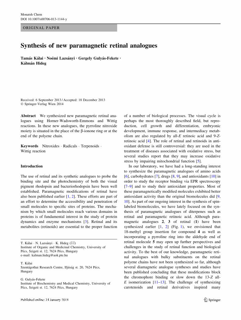
magnetic analogues 2, 3 of retinal (1) have been
synthesized earlier [1, 2] (Fig. 1), we envisioned that
18-methyl group insertion for compound 4 as well as
incorporating a pyrroline ring into the aldehyde end of
retinal molecule 5 may open up further perspectives and
challenges in the study of retinal function and biological
activity. To the best of our knowledge, paramagnetic reti-
nal analogues with bulky substituents on the retinal
polyene chains have not been synthesized so far, although
several diamagnetic analogue syntheses and studies have
been published concluding that these modifications block
the chromophore binding or slow down the 13-Z all-
E isomerization [11–13]. The challenge of synthesizing
carotenoids and retinal derivatives inspired many
T. Kalai � N. Lazsanyi � K. Hideg (&)
Institute of Organic and Medicinal Chemistry, University of
Pecs, Szigeti st. 12, 7624 Pecs, Hungary
e-mail: [email protected]
T. Kalai
Szentagothai Research Centre, Ifjusag st. 20, 7624 Pecs,
Hungary
G. Gulyas-Fekete
Institute of Biochemistry and Medical Chemistry, University of
Pecs, Szigeti st. 12, 7624 Pecs, Hungary
123
Monatsh Chem
DOI 10.1007/s00706-013-1144-y
distinguished organic chemists both in laboratories and in
the industry [14], however, reports on spin-labeled retinal
derivatives are still limited [1, 2], probably because of the
difficulty of utilizing organometallic reagents in the pre-
sence of nitroxides and the difficulties of NMR
investigation of the paramagnetic species formed. In this
paper, we report the extension of the Horner-Wadsworth-
Emmons olefination and Wittig reaction-based approach
for paramagnetic retinal and retinoic acid synthesis for
further biological studies, including antioxidant and
receptor-binding investigations.
Results and discussion
We began our synthesis with 1-oxyl-2,2,4,5,5-pentamethyl-
2,5-dihydro-1H-pyrrol-3-carbaldehyde (6), available via a
Suzuki reaction [15]. For chain elongation, compound 6
was treated with the anion of ethyl 4-diethoxyphosphinyl-
3-methyl-2-butenoate [16] in THF at -78 �C, giving
compound 7. Transformation of the ester group into alde-
hyde by the previously reported protocols [1, 2] did not
give satisfactory results in our hands. Therefore, the
resulting ester 7 was hydrolyzed to carboxylic acid 8
cautiously in aqueous sodium hydroxide-methanol solu-
tion. Compound 8 was converted to mixed anhydride ester
with ethyl chloroformate in the presence of Et3N, and this
was reduced with 1.1 equivalent sodium borohydride in
ethanol to an alcohol [17]. Oxidation of this alcohol with
activated MnO2 provided aldehyde 9.
The configuration of two double bonds in the chain of 9
was proven by HMQC, HMBC, COSY, and NOESY
measurements, and they were found to be E,E-isomers.
Further elongation of the chain from aldehyde 9 with the
lithium salt of ethyl 4-diethoxyphosphinyl-3-methyl-2-
butenoate in THF at -78 �C gave ester 10. The 2D mea-
surements, 1H NMR, and 13C NMR studies of compound
10 suggested the presence of both Z and E isomers. This
was also confirmed by HPLC studies [18], revealing that
the product contains 33 % of the 11-Z-isomer and 64 % of
the all-E-isomer, as well as two additional minor isomers in
amounts of 1 and 2 %. Compound 10 was hydrolyzed with
aqueous sodium hydroxide in methanol to the paramag-
netic analogue of retinoic acid 11. This acid was converted
to mixed anhydride ester, which was reduced with 1.1
equivalent NaBH4 in ethanol at 0 �C and the alcohol
achieved was not isolated, but oxidized immediately to
paramagnetic retinal 4 with activated MnO2 in CH2Cl2 at
room temperature (Fig. 2). The 2D measurements, 1H
NMR, and 13C NMR studies of compound 4 suggested the
presence of both Z and E isomers also. This was confirmed
by HPLC studies as well, confirming that the paramagnetic
retinal analogue contains 24 % of the 11-Z-isomer and
73 % of the all-E-isomer, along with 0.5 and 2 % of
another two minor isomers.
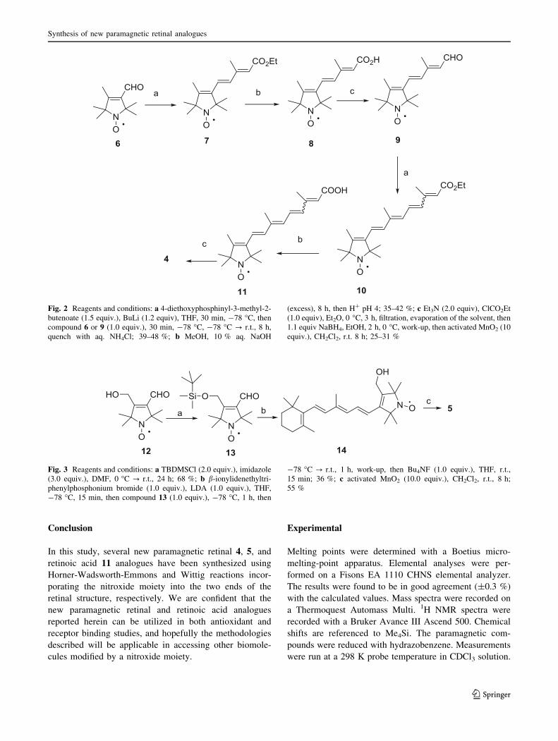
To study further the steric constraints in the retinal
binding pocket by EPR spectroscopy, we envisioned that
the paramagnetic 13-Z-locked retinal analogue might be a
useful substrate. To incorporate the pyrroline ring into the
C(13)-C(14) positions of the retinal molecule, we used
1-oxyl-4-(hydroxymethyl)-2,2,5,5-tetramethyl-2,5-dihydro-
1H-pyrrol-3-carbaldehyde (12) [19] as a starting material.
It was silylated on the hydroxyl group [20], and the
treatment of aldehyde 13 with ylide, generated from
b-ionylidenethyltriphenylphosphonium bromide [21] with
LDA at -78 �C in THF, gave a mixture of silylated and
desilylated products. After removing the silyl group from
the crude product with Bu4NF in THF during the work-up,
we achieved alcohol 14. Oxidation of compound 14 with
activated MnO2 in CH2Cl2 at room temperature provided
aldehyde 5, a paramagnetic retinal derivative with a C(13)-
C(14) Z double bond. Otherwise, the structure of com-
pound 5 was confirmed using HMQC, HMBC, COSY, and
NOESY, and revealed the E configuration for all three
double bonds in question in Fig. 3.
Fig. 1 Retinal (1), previously reported (2, 3) and herein reported
paramagnetic retinal derivatives (4, 5)
T. Kalai et al.
123
Conclusion
In this study, several new paramagnetic retinal 4, 5, and
retinoic acid 11 analogues have been synthesized using
Horner-Wadsworth-Emmons and Wittig reactions incor-
porating the nitroxide moiety into the two ends of the
retinal structure, respectively. We are confident that the
new paramagnetic retinal and retinoic acid analogues
reported herein can be utilized in both antioxidant and
receptor binding studies, and hopefully the methodologies
described will be applicable in accessing other biomole-
cules modified by a nitroxide moiety.
Experimental
Melting points were determined with a Boetius micro-
melting-point apparatus. Elemental analyses were per-
formed on a Fisons EA 1110 CHNS elemental analyzer.
The results were found to be in good agreement (±0.3 %)
with the calculated values. Mass spectra were recorded on
a Thermoquest Automass Multi. 1H NMR spectra were
recorded with a Bruker Avance III Ascend 500. Chemical
shifts are referenced to Me4Si. The paramagnetic com-
pounds were reduced with hydrazobenzene. Measurements
were run at a 298 K probe temperature in CDCl3 solution.
Fig. 2 Reagents and conditions: a 4-diethoxyphosphinyl-3-methyl-2-
butenoate (1.5 equiv.), BuLi (1.2 equiv), THF, 30 min, -78 �C, then
compound 6 or 9 (1.0 equiv.), 30 min, -78 �C, -78 �C ? r.t., 8 h,
quench with aq. NH4Cl; 39–48 %; b MeOH, 10 % aq. NaOH
(excess), 8 h, then H? pH 4; 35–42 %; c Et3N (2.0 equiv), ClCO2Et
(1.0 equiv), Et2O, 0 �C, 3 h, filtration, evaporation of the solvent, then
1.1 equiv NaBH4, EtOH, 2 h, 0 �C, work-up, then activated MnO2 (10
equiv.), CH2Cl2, r.t. 8 h; 25–31 %
Fig. 3 Reagents and conditions: a TBDMSCl (2.0 equiv.), imidazole
(3.0 equiv.), DMF, 0 �C ? r.t., 24 h; 68 %; b b-ionylidenethyltri-
phenylphosphonium bromide (1.0 equiv.), LDA (1.0 equiv.), THF,
-78 �C, 15 min, then compound 13 (1.0 equiv.), -78 �C, 1 h, then
-78 �C ? r.t., 1 h, work-up, then Bu4NF (1.0 equiv.), THF, r.t.,
15 min; 36 %; c activated MnO2 (10.0 equiv.), CH2Cl2, r.t., 8 h;
55 %
Synthesis of new paramagnetic retinal analogues
123
ESR spectra were taken on a Miniscope MS 200 in 10-4 M
CHCl3 solution and all monoradicals gave triplet line
aN = 14.4 G. The IR spectra were taken with a Bruker
Alpha FT-IR instrument with ATR support on a diamond
plate. UV spectra were taken with a Specord 40 instrument
(Analytic Jena). The HPLC system was interfaced to a
gradient pump Dionex P680 and Dionex PDA-100 detec-
tor; the acquisitions were performed with k = 450 nm
detection at 22 �C. Data acquisitions were performed by
Chromeleon 6.70 software. The HPLC separations were
carried out on an end-capped column (250 9 4.6 mm i.d.;
YMC C30, 3 lm). The eluents consisted of: A: 81 %
MeOH, 15 % TBME, 4 % H2O, and B: 6 % MeOH, 90 %
TBME, 4 % H2O. The linear gradient used was: 00 100 %
A–150 85 % A, 15 % B eluent, and the flow rate was
1.00 cm3/min. Flash column chromatography was per-
formed on a Merck Kieselgel 60 (0.040–0.063 mm).
Qualitative TLC was carried out on commercially available
plates (20 9 20 9 0.02 cm) coated with Merck Kieselgel
GF254. Compounds 6 [15], 12 [19], ethyl 4-diethoxypho-
sphinyl-3-methyl-2-butenoate [16], and b-ionylidene-
thyltriphenylphosphonium bromide [21] were prepared
according to published procedures and other reagents were
purchased from Aldrich.
General procedure for Horner-Wadsworth-Emmons
reaction
A solution of 2.4 cm3 BuLi (6.0 mmol, 2.5 M in hexanes)
was added dropwise at -78 �C to a stirred solution of
1.98 g 4-diethoxyphosphinyl-3-methyl-2-butenoate (7.5
mmol) in 20 cm3 anhydrous THF. The mixture was stirred
under N2 at this temperature for 30 min, then 910 mg
aldehyde 6 (5.0 mmol) or 1.24 g aldehyde 9 (5.0 mmol)
was added dropwise in 10 cm3 THF at -78 �C. The mix-
ture was stirred at this temperature for 30 min, and then it
was allowed to warm to room temperature and was stirred
overnight. Following the addition of 10 cm3 sat. aq. NH4Cl
solution, 20 cm3 EtOAc was added and the organic phase
was separated. The aqueous phase was extracted with
10 cm3 EtOAc, the combined organic phase was dried
(MgSO4), filtered, and evaporated. The residue was puri-
fied by flash column chromatography with gradient elution
(hexane/ether: 90/10 % to 60/40 %) to furnish compounds
7 as a yellow and 10 as deep yellow solids.
(2E,4E)-Ethyl 5-(1-oxyl-2,2,4,5,5-pentamethyl-2,5-
dihydro-1H-pyrrol-3-yl)-3-methylpenta-2,4-dienoate
radical (7, C17H26NO3)
Yield: 605 mg (48 %); m.p.: 53 �C; Rf = 0.40 (hexane/
Et2O 2:1); IR (neat): m = 1,701, 1,645, 1,606 cm-1; UV–
Vis (ethanol, c = 2.36 9 10-5 mol dm-3): kmax (e) = 303
(26,400) nm (mol-1 dm3 cm-1); MS (70 eV): m/z = 292
(M?, 100), 277 (32), 262 (23), 91 (83).
Ethyl 9-(1-oxyl-2,2,4,5,5-pentamethyl-2,5-dihydro-1H-
pyrrol-3-yl)-3,7-dimethylnona-2,4,6,8-tetraenoate
radical (10, C22H32NO3)
Yield: 698 mg (39 %); m.p.: 98 �C; Rf = 0.35 (hexane/
Et2O 2:1); 1H NMR (500 MHz, CDCl3): d = 1.30 (s, 6H,
CH3), 1.28 (t, 3H, CH3), 1.45 (s, 6H, CH3), 1.86 (s, 3H,
CH3), 2.09 (s, 3H, CH3), 2.45 (s, 3H, CH3), 4.27 (m, 2H,
CH2), 6.22 (d, 1H, CH), 6.31 (d, 1H, CH), 6.40 (dd, 1H,
CH), 6.57 (d, 1H, CH), 6.67 (d, 1H, CH), 7.06 (m, 1H, CH)
ppm; 13C NMR (125 MHz, CDCl3): d = 10.80 (CH3),
13.72 (CH3), 14.25 (CH3), 23.87 (CH3), 24.99 (CH3), 25.60
(CH3), 59.56 (CH2), 68.76 (C), 69.50 (C), 121.67 (CH),
130.44 (CH), 130.57 (CH), 131.99 (CH), 133.70 (CH),
135.46 (C), 135.68 (CH), 138.68 (C), 139.26 (C), 142.66
(C), 167.02 (C) ppm; IR (neat): m = 1,694, 1,601,
1,569 cm-1; UV–Vis (ethanol, c = 2.24 9 10-5
mol dm-3): kmax (e) = 361 (52,100), 262 (4,500) nm
(mol-1 dm3 cm-1); MS (70 eV): m/z = 358 (M?, 2), 344
(28), 328 (11), 192 (79).
General procedure for ester hydrolysis
Aq. NaOH (10 %, 10 cm3) was added to a solution of
1.17 g ester 7 (4.0 mmol) or 1.43 g 10 (4.0 mmol) in
20 cm3 MeOH. The mixture was allowed to stand over-
night at ambient temperature in the dark. The MeOH was
evaporated in vacuo (\40 �C), and the pH was adjusted to
4 by cautious addition of 5 % H2SO4 at 0 �C. Then the
aqueous phase was immediately extracted with CHCl3(2 9 15 cm3). The organic phase was dried (MgSO4), fil-
tered, and evaporated. The carboxylic acids 8 and 11 were
isolated as yellow solids after flash column chromatogra-
phy by gradient elution (hexane/EtOAc 66/33 % for
5 9 30 cm3 fraction and then CHCl3/Et2O from 10/90 %
to 50/50 %).
(2E,4E)-5-(1-Oxyl-2,2,4,5,5-pentamethyl-2,5-dihydro-1H-
pyrrol-3-yl)-3-methylpenta-2,4-dienoic acid radical
(8, C15H22NO3)
Yield: 443 mg (42 %); m.p.: 152 �C; Rf = 0.33 (CHCl3/
Et2O 2:1); IR (neat): m = 3,034, 1,674, 1,602, 1,584 cm-1;
UV–Vis (ethanol, c = 2.74 9 10-5 mol dm-3): kmax
(e) = 299 (25,300) nm (mol-1 dm3 cm-1); MS (70 eV):
m/z = 264 (M?, 13), 249 (14), 234 (8), 43 (100).
9-(1-Oxyl-2,2,4,5,5-pentamethyl-2,5-dihydro-1H-pyrrol-3-
yl)-3,7-dimethylnona-2,4,6,8-tetraenoic acid radical
(11, C20H28NO3)
Yield: 462 mg (35 %); m.p.: 208 �C; Rf = 0.27 (CHCl3/
Et2O 2:1); IR (neat): m = 3,046, 1,672, 1,596, 1,564 cm-1;
T. Kalai et al.
123
UV–Vis (ethanol, c = 1.68 9 10-5 mol dm-3): kmax
(e) = 257 (4,000), 357 (51,700) nm (mol-1 dm3 cm-1);
MS (70 eV): m/z = 330 (M?, 9), 316 (8), 300 (5), 282
(15), 91 (68) 44 (100).
General procedure for conversion of acids to aldehydes
To a stirred solution of carboxylic acids 8 (2.0 mmol) or 11
(2.0 mmol) and 404 mg Et3N (4.0 mmol) in 20 cm3 an-
hydr. Et2O 217 mg ethyl chloroformate (2.0 mmol) in
5 cm3 Et2O was added dropwise at 0 �C. The mixture was
stirred at this temperature for 3 h, then the triethylamine
hydrochloride was filtered off in a glass sintered funnel,
washed with 10 cm3 Et2O, and the ether was evaporated in
vacuo (\40 �C). The residue was immediately dissolved in
15 cm3 dry EtOH and 84 mg NaBH4 (2.2 mmol) was
added in three portions during 30 min at 0 �C. After the
consumption of the starting mixed anhydride ester, moni-
tored by TLC (*90 min), the EtOH was evaporated
(\40 �C), the residue was dissolved in 20 cm3 CHCl3,
washed with 10 cm3 brine, and the organic phase was dried
(MgSO4), filtered, and evaporated. The residue was
immediately dissolved in 20 cm3 dry CH2Cl2, and 1.72 g
activated MnO2 (20.0 mmol) was added in one portion and
stirred overnight at room temperature in the dark. Then the
reaction mixture was filtered through Celite, washed with
10 cm3 CH2Cl2, the solvent was evaporated, and the resi-
due was purified by flash column chromatography
(gradient: hexane/Et2O 90/10 % to 75/25 % 10 9 30 cm3
then hexane/EtOAc 60/40 %) to give aldehydes 9 and 4 as
yellow solids.
(2E,4E)-5-(1-Oxyl-2,2,4,5,5-pentamethyl-2,5-dihydro-1H-
pyrrol-3-yl)-3-methylpenta-2,4-dienal radical
(9, C15H22NO2)
Yield: 153 mg (35 %); m.p.: 104 �C; Rf = 0.17 (hexane/
Et2O 2:1); 1H NMR (500 MHz, CDCl3): d = 1.31 (s, 6H,
CH3), 1.44 (s, 6H, CH3), 1.87 (s, 3H, CH3), 2.37 (s, 3H,
CH3), 6.09 (d, 1H, CH), 6.55 (d, 1H, CH), 6.82 (d, 1H,
CH), 10.20 (s, 1H, CHO) ppm; 13C NMR (125 MHz,
CDCl3): d = 11.14 (CH3), 12.79 (CH3), 23.97 (CH3),
25.11 (CH3), 68.70 (C), 69.78 (C), 128.12 (CH), 129.38
(CH), 131.83 (CH), 135.32 (C), 144.84 (C), 154.82 (C),
191.09 (CHO) ppm; IR (neat): m = 1,649, 1,616,
1,600 cm-1; UV–Vis (ethanol, c = 2.71 9 10-5
mol dm-3): kmax (e) = 323 (32,600) nm (mol-1 dm3
cm-1); MS (70 eV): m/z = 248 (M?, 78), 218 (11), 91
(82), 42 (100).
9-(1-Oxyl-2,2,4,5,5-pentamethyl-2,5-dihydro-1H-pyrrol-3-
yl)-3,7-dimethylnona-2,4,6,8-tetraenal radical
(4, C20H28NO2)
Yield: 157 mg (25 %); m.p.: 140 �C; Rf = 0.47 (hexane/
EtOAc 2:1); 1H NMR (500 MHz, CDCl3): d = 1.30 (s, 6H,
CH3), 1.44 (s, 6H, CH3), 1.86 (s, 3H, CH3), 2.10 (s, 3H,
CH3), 2.38 (s, 3H, CH3), 6.24 (d, 1H, CH), 6.33 (d, 1H,
CH), 6.46 (d, 1H, CH), 6.56 (d, 1H, CH), 6.66 (d, 1H, CH),
7.20 (m, 1H, CH), 10.17 (s, 1H, CH) ppm; 13C NMR
(125 MHz, CDCl3): d = 10.87 (CH3), 13.01 (CH3), 23.80
(CH3), 24.94 (CH3), 25.54 (CH3), 68.88 (C), 69.67 (C),
122.56 (CH), 129.16 (CH), 130.73 (CH), 131.97 (CH),
133.53 (CH), 135.06 (CH), 135.46 (C), 140.59 (C), 140.93
(C), 142.62 (C), 190.97 (CH) ppm; IR (neat): m = 1,652,
1,597, 1,567 cm-1; UV–Vis (ethanol, c = 1.98 9 10-5
mol dm-3): kmax (e) = 377 (33,600), 270 (10,800) nm
(mol-1 dm3 cm-1); MS (70 eV): m/z = 314 (M?, 41), 300
(15), 288 (28), 91 (63), 44 (100).
1-Oxyl-4-(t-butyldimethylsilyloxymethyl)-2,2,5,5-tetra-
methyl-2,5-dihydro-1H-pyrrol-3-carbaldehyde
radical (13, C15H30NO3Si)
To a stirred solution of 990 mg alcohol 12 (5.0 mmol) and
1.02 g imidazole (15.0 mmol) in 7 cm3 dry DMF 1.50 g t-
butyldimethylchlorosilane was added in 3–4 portions at
0 �C, then the solution was stirred for 24 h at ambient
temperature. The solution was poured onto a mixture of ice
and 50 cm3 sat. aq. NaHCO3 solution, extracted with Et2O
(3 9 20 cm3), the organic phase was dried (MgSO4),
filtered, evaporated, and the residue was purified by flash
column chromatography (gradient: hexane/Et2O 90/10 %
to 70/30 %) to give the title compound (1.06 g, 68 %) as a
yellow solid. M.p.: 74 �C; Rf = 0,44 (hexane/Et2O 2:1); 1H
NMR (500 MHz, CDCl3): d = 0.12 (s, 3H, SiCH3), 0.13
(s, 3H, SiCH3), 0.94 (s, 9H, C(CH3)3), 1.34 (s, 6H, CH3),
1.39 (s, 6H, CH3), 4.59 (s, 2H, CH2), 10.27 (s, 1H, CHO)
ppm; 13C NMR (125 MHz, CDCl3): d = -5.64 (SiCH3),
18.08 (SiC), 24.01 (CH3), 24.22 (CH3), 25.67 (CH3), 58.28
(CH2), 67.95 (C), 69.60 (C), 139.38 (C), 159.96 (C),
189.24 (CHO) ppm; IR (neat): m = 1,658, 1,625 cm-1; MS
(70 eV): m/z = 312 (M?, 2), 240 (12), 183 (27), 75 (100).
1-Oxyl-3-hydroxymethyl-2,2,5,5-tetramethyl-4-
[(1E,3E,5E)-4-methyl-6-(2,6,6-trimethylcyclohex-1-en-1-
yl)hexa-1,3,5-trien-1-yl]-2,5-dihydro-1H-pyrrole
radical (14, C25H38NO2)
To a stirred solution of 2.72 g b-ionylidenethyltriphenyl-
phosphonium bromide (5.0 mmol) in 40 cm3 anhydr. THF,
2.8 cm3 LDA solution (5.0 mmol in THF/heptane/ethyl-
benzene) was added dropwise at -78 �C. After stirring the
solution for 15 min, 1.56 g of compound 13 (5.0 mmol)
dissolved in 10 cm3 THF was added dropwise at -78 �C to
the dark red solution and the stirring was continued for 1 h
at -78 �C, then the reaction mixture was allowed to warm
to room temperature and was stirred at this temperature
overnight. The solution was diluted with 30 cm3 Et2O and
10 cm3 sat. aq. NH4Cl solution was added. The organic
phase was separated, dried (MgSO4), filtered, and evapo-
rated. The residue was dissolved in 20 cm3 THF, 1.30 g
Synthesis of new paramagnetic retinal analogues
123
Bu4NF 9 H2O (5.0 mmol) was added in one portion, and
the reaction mixture was stirred for 15 min at room
temperature. Then, 20 cm3 Et2O was added, the reaction
mixture was washed with 20 cm3 water, the organic phase
was separated, dried (MgSO4), filtered, and evaporated.
The chromatographic purification of the crude product
(gradient: hexane/EtOAc 90/10 % to 70/30 %) offered
compound 14 as a pale yellow solid (691 mg, 36 %). M.p.:
106 �C; Rf = 0.47 (hexane/EtOAc 2:1); IR (neat):
m = 3,481, 1,591 cm-1; UV–Vis (ethanol, c = 1.85 9
10-5 mol dm-3): kmax (e) = 334 (37900), nm (mol-1
dm3 cm-1); MS (70 eV): m/z = 348 (M?, 70), 369 (10),
354 (41), 42 (100).
1-Oxyl-2,2,5,5-tetramethyl-4-[(1E,3E,5E)-4-methyl-6-
(2,6,6-trimethylcyclohex-1-en-1-yl)hexa-1,3,5-trien-1-yl]-
2,5-dihydro-1H-pyrrol-3-carbaldehyde radical
(5, C25H36NO2)
To a stirred solution of 384 mg alcohol 14 (1.0 mmol) in
10 cm3 CH2Cl2, 860 mg activated MnO2 (10.0 mmol) was
added and the mixture was stirred overnight at ambient
temperature in the dark. Then, the reaction mixture was
filtered through Celite, washed with 5 cm3 CH2Cl2, the
solvent was evaporated, and the residue was purified by
flash column chromatography with gradient elution (hex-
ane/Et2O 90/10 % to 60/40 %) to give aldehyde 5
(210 mg, 55 %) as a yellow solid. M.p.: 100 �C;
Rf = 0.57 (hexane/Et2O 2:1); 1H NMR (500 MHz,
CDCl3): d = 1.10 (s, 6H, CH3), 1.46 (s, 12H, CH3), 1.55
(s, 2H, CH2), 1.69 (s, 2H, CH2), 1.79 (s, 3H, CH3), 2.06 (s,
3H, CH3), 2.09 (m, 2H, CH2), 6.22 (m, 2H, CH), 6.41 (d,
1H, CH), 6.53 (d, 1H, CH), 7.09 (dd, 1H, CH), 10.02 (s,
1H, CHO) ppm; 13C NMR (125 MHz, CDCl3): d = 12.92
(CH3), 19.06 (CH2), 21.59 (CH3), 24.31 (CH3), 24.80
(CH3), 28.82 (CH3), 32.98 (CH2), 34.12 (C), 39.47 (CH2),
67.80 (C), 69.58 (C), 120.36 (CH), 129.17 (CH), 129.61
(CH), 130.23 (C), 135.52 (CH), 136.74 (CH), 137.50 (C),
138.52 (C), 140.79 (C), 159.48 (C), 187.62 (CHO) ppm; IR
(neat): m = 1,646, 1,565, 1,540 cm-1; UV–Vis (ethanol,
c = 1.73 9 10-5 mol dm-3): kmax (e) = 384 (21,300),
258 (8,300) nm (mol-1 dm3 cm-1); MS (70 eV): m/
z = 382 (M?, 16), 352 (29), 377 (17), 43 (100).
Acknowledgments We are grateful to Prof. Jozsef Deli (Depart-
ment of Pharmacognosy, University of Pecs) for HPLC measurement
and helpful discussions, Viola Csokona for elemental analyses, and to
the Hungarian National Research Fund (OTKA K81123, K104956)
for the financial support.
References
1. Renk GE, Or SY, Crouch RK (1987) J Am Chem Soc 109:6163
2. Groesbeek M, Lugtenburg J (1995) Rec Travaux Chim Pays-Bas
114:403
3. Steinhoff HJ, Savitsky A, Wegener C, Pfeiffer M, Plato M,
Mobius K (2000) Biochim Biophys Acta Bioenerg 1457:253
4. Wada A, Fukunaga K, Ito M, Mizuguchi Y, Nakagawa K, Okano
T (2004) Bioorg Med Chem 12:3931
5. Siems W, Sommerburg O, Schild L, Augustin W, Langhans CD,
Wiswedel I (2002) FASEB J 16:1289
6. Kalai T, Schindler J, Balog M, Fogassy E, Hideg K (2008) Tet-
rahedron 64:1094
7. Zhao M, Kalai T, Hideg K, Altenbach C, Hubbell WL, Kaback
HR (2000) Biochemistry 39:11381
8. Miyazaki J, Hideg K, Marsh D (1992) Biochim Biophys Acta
1103:62
9. Petrlova J, Kalai T, Maezawa I, Altman R, Harishchandra G,
Hong HS, Bricarello DA, Parikh AN, Lorigan GA, Jin LW, Hideg
K, Voss JC (2012) PLoS ONE 7:e35443
10. Kalai T, Borza E, Antus C, Radnai B, Gulyas-Fekete G, Feher A,
Sumegi B, Hideg K (2011) Bioorg Med Chem 19:7311
11. Lewis JW, Pinkas I, Sheves M, Ottolenghi M, Kliger DS (1995) J
Am Chem Soc 117:918
12. Danshina SV, Drachev AL, Drachev LA, Eremin SV, Kaulen
AD, Khytrina LV, Mitsner BI (1990) Arch Biochem Biophys
279:225
13. Yan B, Xie A, Nienhaus UG, Katsuta Y, Spudich JL (1993)
Biochemistry 32:10224
14. Britton G, Liaaen-Jensen S, Pfander H (1996) Carotenoids, vol 2.
Birkhauser, Basel
15. Kalai T, Balog M, Jek}o J, Hubbell WL, Hideg K (2002) Synthesis
34:2365
16. Magoulas GE, Bariamis E, Athanassopoulos MC, Haskopoulos
A, Dedes PG, Krokidis MG, Karamanos NK, Kletsas D, Papa-
ioannou D, Maroulis G (2011) Eur J Med Chem 46:721
17. Hideg K, Hankovszky HO, Lex L, Kulcsar GY (1980) Synthesis
12:911
18. Gulyas-Fekete G, Murillo E, Kurtan T, Papp T, Illyes ZT, Drahos
L, Visy J, Agocs A, Turcsi E, Deli J (2013) J Nat Prod 76:607
19. Kalai T, Jek}o J, Hideg K (2000) Synthesis 32:831
20. Greene TW, Wuts PG (1999) Protective groups in organic syn-
thesis. Wiley, Hoboken
21. Tietze LF, Eicher TH (1989) Reactions and syntheses in the
organic chemistry laboratory. University Science Books, Mill
Valley
T. Kalai et al.
123