surgical treatment of the neglected achilles tendon rupture
TRANSCRIPT

3,250+OPEN ACCESS BOOKS
106,000+INTERNATIONAL
AUTHORS AND EDITORS112+ MILLION
DOWNLOADS
BOOKSDELIVERED TO
151 COUNTRIES
AUTHORS AMONG
TOP 1%MOST CITED SCIENTIST
12.2%AUTHORS AND EDITORS
FROM TOP 500 UNIVERSITIES
Selection of our books indexed in theBook Citation Index in Web of Science™
Core Collection (BKCI)
Chapter from the book Achilles TendonDownloaded from: http://www.intechopen.com/books/achilles-tendon
PUBLISHED BY
World's largest Science,Technology & Medicine
Open Access book publisher
Interested in publishing with InTechOpen?Contact us at [email protected]

7
Surgical Treatment of the Neglected Achilles Tendon Rupture
Jake Lee and John M. Schuberth Kaiser Foundation Hospital, San Francisco, CA
USA
1. Introduction
The true frequency of acute Achilles tendon rupture is unknown but historically it was
regarded as a rare injury comprising less than 0.2% of the general population (Cetti et al.,
1993; Nillius et al., 1976). However, in the past decade the incidence of Achilles tendon
rupture has increased (Maffuli et al., 1999). At the present time, Achilles tendon ruptures are
the most common tendon rupture of the lower extremity and may account for up to 40% of
all operated tendon ruptures (Habusta, 1995; Jozsa et al., 1989). The increase in frequency is
thought to be due to an increased interest and participation in recreational sports by middle-
aged and older patients and also to better reporting (Coughlin et al, 2007).
In many patients the initial symptoms after an Achilles tendon rupture diminish quickly. In a study of 57 patients with acute Achilles rupture, 19 of them reported to be painless (Christensen, 1953). Patients with Achilles tendon ruptures frequently are unable to stand on the toes of the involved side, however, active plantarflexion maybe intact due to partial ruptures, recruitment of plantar flexors, and an intact plantaris muscle. The lack of pain and no obvious loss of plantarflexion can be misleading and up to 20-25% of cases the diagnosis is missed initially (Maffuli, 1996; Arner & Lindholm, 1959; Nillius et al., 1976). The failure to establish the diagnosis at the initial presentation is the most common reason for delayed treatment.
There are many terms used to describe this condition and treatment, including neglected or chronic rupture, late or old repair, and delayed reconstruction (Abraham & Pankovich, 1975; Ozaki et al., 1989; Porter et al., 1997). There is no consensus regarding the specific time in which an acute becomes a neglected rupture although 4 weeks may be the most widely accepted interval (Leppilahti & Orava, 1998; Porter et al., 1997). Contraction of the triceps surae complex has been observed 3 to 4 days post-injury (Bosworth, 1956). Regardless of the lack of a chronological definition, neglected ruptures are characterized by the difficulty of achieving an end-to-end apposition of the tendon ends with plantarflexion of the foot during surgical reconstruction.
Neglected ruptures can heal without surgery as abundant scar tissue has been shown to form in the rupture interval (Barnes & Hardy, 1986). However, due to the contracture of the triceps surae complex, the resulting functional length of the muscle-tendon unit may be too long even with re-establishment of the continuity of the muscle tendon complex through
www.intechopen.com

Achilles Tendon 116
scar tissue formation. This leads to comprised plantarflexion power, reducing ankle stability and an impaired gait pattern.
1.1 Clinical evaluation
A palpable gap is rarely felt on physical examinations at the previous rupture site due to scar tissue formation. However with careful digital palpation or direct visual inspection, the site of the neglected rupture can often be determined due to a change in the consistency of the tissue and a change in contour of the posterior leg [Figure 1]. The additional findings on clinical examination will depend on the functional length of the healed tendon. Patients will display increased dorsiflexion of the ankle joint and decreased plantarflexion power compared to the contralateral limb. Patients often report that they are easily fatigued with sports. It is highly unlikely that they are able to perform a single limb heel rise. During gait there is delayed heel-off and a shortened stride. Magnetic Resonance Imaging (MRI) is a useful tool in confirming the clinical diagnosis but more so for assessing the amount of functional defect within the Achilles tendon for preoperative planning [Figure 2].
Fig. 1. Delayed presentation with clinically evident defect in the Achilles tendon
www.intechopen.com

Surgical Treatment of the Neglected Achilles Tendon Rupture 117
Fig. 2. Magnetic Resonance Imaging demonstrating a large defect in a patient with a neglected Achilles tendon rupture
2. Conservative treatment
The best functional outcomes are achieved through surgical reconstruction but non-surgical treatment may be preferable for patients with poor skin condition, history of smoking, soft tissue complications from previous surgery, and poorly controlled long-standing diabetes mellitus. Conservative treatment could be as simple as lace up ankle brace or custom made leather ankle brace (i.e. – Arizona brace). In patients with severe Achilles dysfunction, an Ankle-Foot-Orthosis (AFO) can be considered. Any bracing method can be coupled with physical therapy to strengthen the gastrocnemius and recruitment of the entire deep posterior compartment muscles.
The use of immobilization for treatment of neglected ruptures is suspect, but may be more useful prior to the maturation of the interposed scar in the post-injury period. If conservative treatment is chosen, it is important to realize that the immobilization period
www.intechopen.com

Achilles Tendon 118
will be much longer. Serial casting with reduction of the equinus position of the foot at each visit may allow for consolidation and re-establishment of functional continuity. However each respective casting stage will be extended compared to non-operative treatment of an acute rupture. Ultrasound can offer some assistance in assessing the extent of fibrous tissue in the gap. It can serve as a prognostic indicator as well as a tool in guiding how much equinus is needed for tendon apposition. Initial immobilization in a long leg cast with the knee at 25 degrees and the appropriate level of equinus of the ankle has been proposed. This initial cast is kept for 4 weeks. Subsequent serial casting is done every 3 weeks with successive reduced equinus over the span of 7-10 weeks or once the tendon continuity is ensured clinically. This is followed by conversion to a short leg cast with gradual serial reduction of any residual equinus. (Schuberth, 1996)
3. Surgical treatment
Many surgical techniques have been described for the management of neglected Achilles
ruptures. The primary goal of any surgical treatment is to restore the function and
strength of the gastrocnemius-soleus complex by recreating the optimal length-tension
relationship. End-to-end repair is ideal if the gap between tendon ends allow direct
apposition after resection of the interposed scar tissue. This will allow for maximum
isokinetic strength of Achilles because re-establishment of the pre-injury tendon length
can only be achieved. It is generally accepted that approximately 1-2 cm gap will allow
end-to-end repair (Myerson, 2010) [Figure 3]. However, primary repair is still an
uncommon form of treatment for most chronic ruptures because of the potential for
shortening and contracture of the gastrocnemius-soleus muscle-tendon unit. (Bosworth,
1956). Excision of scar tissue from neglected rupture often results in a sizable gap
requiring other modalities to bridge the defect. If the gap exceeds 1-2 cm and primary
repair is still deemed feasible, proximal lengthening of the gastro-soleal complex may be
utilized to achieve mobilization of the proximal tendon end to facilitate primary repair.
These techniques were developed primarily because of the dissatisfaction with the fascial
turn down techniques (Abraham & Pankovich, 1975). Porter et al. reported on end-to-end
primary repair without augmentation of chronic ruptures (greater than or equal to 4
weeks and less than equal to 12 weeks from injury) in 11 patients. Proximal gastro-soleal
complex release was performed with imbrication of the fibrous scar tissue without
excision of local tissue. Primary repair of the tendon ends was then performed. In an
average follow up of 3.5 years no re-ruptures were observed and patients were able to
return to pre-injury level of activities in an average of 5.8 months. Total ankle range of
motion (ROM) was comparable to the uninjured side. The loss of plantarflexion power
and pain scale ratings were similar to the patients surgically treated after an acute rupture
repair performed by the same surgeon (Porter et al., 1997).
Gastrocnemius slide lengthening techniques have also been utilized to achieve end-to-end anastomosis (Barnes and Hardy, 1987) (Abraham & Pankovich, 1975). In this technique, an inverted V incision is made into the aponeurosis then with traction on the distal tendon it is repaired in a Y fashion. The arms of the V incision should at least one and a half times the length of the defect to allow suturing in a Y shape. The size of defects after excision of scar tissue ranged from 5cm to 6cm with the ankle in plantarflexion in their series and in 3 out 4 patients in their study full plantarflexion strength was restored (Abraham & Pankovich,
www.intechopen.com

Surgical Treatment of the Neglected Achilles Tendon Rupture 119
Fig. 3. Intraoperative photo showing large gap that exceeds the capability of end to end repair
1975). An alternative method of advancement is a tongue-in-groove configuration [Figures
4-7]. However, more recently the argument is made against greater than 5cm of advancement as this can lead to detachment from the underlying muscle and cause weakness and decreased peak torque in plantarflexion when compared to the uninjured side (Kissel et al., 1994; Us et al., 1997).
www.intechopen.com

Achilles Tendon 120
Fig. 4. Intraoperative photo showing interposed scar tissue in neglected rupture
www.intechopen.com

Surgical Treatment of the Neglected Achilles Tendon Rupture 121
Fig. 5. More proximally a tongue-in-groove lengthening is performed to mobilize distally (right) in order to bridge the gap.
Fig. 6. The mobilized proximal portion of the gastrosoleal complex has been sutured to the distal stump of the Achilles.
www.intechopen.com

Achilles Tendon 122
Fig. 7. At 6 months postoperative, the patient is able to do a single heel rise.
www.intechopen.com

Surgical Treatment of the Neglected Achilles Tendon Rupture 123
On many occasions direct primary repair is not feasible due to contracture of the ruptured
tendon ends over time and a more extensive reconstructive effort is needed. In general, the
longer the interval between injury and repair, the more likely primary repair will not be
possible, even with mobilization of the proximal segment. When delayed primary repair is not
possible, some surgeons advocate bridging of the gap with other augmentation methods at the
site of the defect. The materials available for augmentation can be categorized into autologous,
synthetic, or allograft augmentation techniques (Dalton, 1996). Several techniques with distant
or local autologous tendon transfers have been described in order to reinforce or reconstruct
neglected Achilles tendon rupture. Synthetic materials have also been used for augmentation.
The advantage of using synthetic materials is that they avoid sacrificing other active tendons.
In turn, the morbidity associated with larger incisions and dissections involved in autologous
techniques can be bypassed. However, the use of synthetic materials in the area well-known
for tenuous wound healing is a major disadvantage. More recently, Achilles tendon allografts
have been used for reconstruction of neglected Achilles tendon rupture. The allograft
technique can used to reconstruct large defects without sacrificing other autologous lower
extremity tendons with relative technical ease.
Instead of advancement of the proximal gastrosoleal complex to negotiate the resultant gap, various gastrocnemius-soleus fascia turn-down techniques have been described. A longitudinal strip of the gastrocnemius fascia can be turned down with the distal end still attached. The 1.5 cm wide strip is then weaved in-out of the proximal and distal ruptured ends to bridge the gap (Bosworth, 1956). Other modifications of the turn down fascial flap have included the use of two flaps measuring 1 x 8 cm that are raised from the proximal gastrocnemius fascia. The distal portions of the flaps are left attached distally 3 cm proximal to the tendon end and turned 180 degrees on themselves. The flaps are sutured into the distal stump as well as to each other (Arner & Lindholm, 1959; Lindholm, 1959). Alternatively, a centrally based turn-down flap can be developed from the proximal segment which is then turned 180 degrees on itself and approximated to the distal stump (Coughlin et al, 2007). In this technique the proximal flap is passed deep to the proximal portion to decrease the bulk. Although these methods are useful in bridging the gap in continuity, strength deficits of up to 23% have been reported (Takao et al., 2003).
3.1 Free fascia-tendon graft
Several authors have reported on the use of free distant fascial or tendon grafts for the reconstruction of neglected ruptures (Maffulli et al., 2005) (Bugg & Boyd, 1968). Free tendinous autograft, utilizing a tongue-in-groove gastrocnemius recession has been described as well (Schuberth et al, 1984). Either the fascia lata or gracilis tendon can be utilized. The usual posterior approach is made and the scar tissue is excised. An ipsilateral incision is made in the thigh to harvest a section of fascia lata 7.5 by 15 cm in dimension. Three 1 cm wide sections are fashioned and laid across the defect between the tendon defects obliquely [Figure 8]. The remaining fascia lata is then wrapped around the repair with the serosal side facing outward (Bugg & Boyd, 1968). Maffulli et al used a free gracilis tendon graft in 21 patients with neglected ruptures. In a minimum follow up of 2 years, no re-ruptures were reported all patients were able to stand on tip-toes with no visible limp during gait. However, the calf circumference remained significantly reduced and the operative limb was significant weaker than the uninjured side at final review (Maffulli et al., 2005).
www.intechopen.com

Achilles Tendon 124
Fig. 8. Intraoperative photo showing strip of fascia lata rolled up and placed around repair site for augmentation.
3.2 Local tendon transfers
The increased technical difficulty of utilizing free tendon grafts as well as need for a separate incision has made local tendon transfers more popular. The use of a local tendon utilizes a viable structure with an intact vascular supply and can augment the plantar flexion strength. Although the biomechanical characteristics and caliber of the transferred tendon is dissimilar to that of the recipient, it can provide additional blood supply to the deficit and avoids the host rejection possible with the use of allografts. The current rule of thumb is to utilize tendon augmentation for defects greater than 2-3 cm (Den Hartog, 2008). The most commonly used are flexor hallucis longus (FHL) and flexor digitorum longus (FDL) tendons. Peroneus brevis (PB) tendon transfer has been utilized in both neglected and recurrent ruptures. (White & Kraynick, 1959; Schuberth, 1984). Plantaris and posterior tibial (PT) tendon transfers have been described as well (Schedl & Faso, 1979; Platt, 1931).
www.intechopen.com

Surgical Treatment of the Neglected Achilles Tendon Rupture 125
3.3 FHL tendon transfer
The use of FHL tendon has become popular in the repair of the neglected Achilles tendon rupture. In part it is due to the mechanical advantage compared to the other autologous transfers, as it has been shown to be stronger than the PB and almost twice as strong as the FDL tendon. Further, it is active during the same phase as the triceps surae complex and helps maintain normal ankle function (Pintore et al., 2001; Leppilahti & Orava, 1998; Den Hartog, 2008).The close proximity to the Achilles tendon affords a readily accessible harvest site. The abundant vascular supply to the muscle belly of the FHL extends to the distal avascular region of the Achilles tendon, improving the blood supply to the injured area (Wilcox et al., 2000; Wapner et al., 1993; Wapner et al, 1995; Carr & Norris, 1989; Martin et al., 2005; Monroe et al., 2000). The FHL tendon can be sewn to the Achilles in a side-to-side fashion, or transferred directly to the calcaneus. Good to excellent functional results have been reported even though there was a reduction in plantar flexion strength (Wapner et al., 1993). Similar good to excellent results were obtained in later studies utilizing the more proximal harvest (at the distal tip of the medial malleolus) in which the FHL tendon transfer was used after extensive debridement for chronic Achilles tendinosis (Den Hartog, 2003; Wilcox et al., 2000; Coull et al., 2003). The argument for using a separate medial incision to harvest the FHL tendon is to obtain the longest working length of the tendon possible. The average tendon length from the posterior incision was 5.16 cm compared to 8.09 cm that can be obtained from a separate medial incision (Tashjian et al., 2003). Although a much longer tendon can be harvested from a separate incision, the more proximal harvest was found be sufficient for transfer and solid fixation into the calcaneus. The loss of FHL function and alteration of the forefoot loading pattern have been shown to be minimal after FHL tendon transfers (Wapner et al., 1995; Coull et al., 2003). After FHL harvest, there is little pressure change to the plantar first or second metatarsophalangeal joint with no clinical functional deficit of the first ray (Coull et al., 2003).
The surgical exposure is approached through a posteromedial incision with the patient in prone position. Once the Achilles tendon is exposed and interposing scar tissue has been excised the deep fascia anterior to the Achilles tendon is incised to expose the FHL tendon and muscle belly. The tendon is then coursed through the fibro-osseous tunnel alongside the calcaneal tuberosity. The neurovascular bundle is in close proximity to the FHL tendon distally and should be protected. The great toe and the ankle are plantarflexed and the FHL tendon is cut from medial to lateral orientation as far distal as possible (Den Hartog, 2008; Hansen, 1991). The FHL tendon is then mobilized and assessed for sufficient length for transfer and the end of the tendon is secured with a Krackow stitch (Den Hartog, 2008; Grove & Hardy, 2008). Some advocate the resection of the superior aspect of the calcaneal tuberosity to create sufficient space for the FHL tendon (Den Hartog, 2008). The FHL tendon is placed under tension with the foot in 20 degrees of plantarflexion (Den Hartog, 2008). If sufficient length of the tendon is available an interference screw is used through drill in the calcaneus. Suture anchors can also be used if the length of the harvested tendon is too short for the interference technique (Den Hartog, 2008).
3.4 PB tendon transfer
The routine use of peroneus brevis is not widely practiced because of the loss of eversion and presumed frontal plane stability. Although subjectively, there seems to be a loss of
eversion power, the loss of ankle stability does not seem to develop as a consequence
www.intechopen.com

Achilles Tendon 126
(Gallant et al. 1995) (Miskulin, 2005). In addition, the availability of other autologous options has made the use of this tendon almost obsolete. Historically PB tendon transfer has been
described in repairing acute Achilles tendon ruptures (Teuffer, 1974). Later, there were reports of good to excellent results using this technique in cases with large defects in tendon
continuity or poor quality tissue (Hepp & Blauth, 1978) (Schuberth, 1984). No obvious functional deficits secondary to the loss of the function of the PB tendon were noted.
The published reports regarding the use of PB tendon in the treatment of neglected rupture of the Achilles tendon have generally noted good functional results. (White & Kraynick, 1959) (Miskulin, 2005). Miskulin et al performed PB tendon transfer in conjunction with plantaris tendon augmentation in 5 patients with neglected rupture with an average of 19.8 weeks (range 5-40 weeks) of delay in injury to presentation. They found that all 5 patients were able to return to pre-injury activity level. There were no reported wound complications, postoperative pain, or function limitations. Using an isokinetic dynamometer, they found that all 5 patients had increase in peak plantarflexion torque approximately 1 year after surgery and all were able to perform single toe rise on the involved side after the reconstruction.
Surgical exposure is obtained through a posterolateral incision. The sural nerve is identified and protected throughout the procedure. The deep fascia is then incised and the PB muscle belly with the tendon is visualized. The interposing scar tissue is resected from the ruptured ends of the Achilles tendon. In order the harvest the PB tendon distally a separate incision is made (1 cm to 1.5 cm) directly over the base of the fifth metatarsal. The tendon is transected at this point and pulled through the original posterolateral incision. The foot is then plantarflexed 20 degrees and end-to-end anastomosis is attempted when feasible. Some use the plantaris tendon to augment the primary anastomosis. If the distal stump of the Achilles tendon appears to be in good condition the PB tendon is pulled through the distal stump (lateral to medial) and sutured to the proximal and the distal tendon stumps. It can also be secured to the calcaneus with an interference screw or suture anchors (Miskulin, 2005).
3.5 FDL tendon transfer
The use of FDL tendon has been advocated as it mimics the course of the Achilles tendon without comprising the lesser digit function postoperatively. There use of this tendon also avoids the loss of eversion and ankle balance seen with transfer of the PB. Mann et al first described the technique in 7 patients with duration of symptoms ranging from 3 to 36 months with an average follow up of 39 months. They achieved excellent result in 4 patients, good in 2, and fair in 1. The 6 patients who achieved good to excellent result were all able to return to pre-injury activities without pain. Two patients with good result had wound complications requiring a secondary procedure. No re-ruptures were reported in their series and active plantarflexion of the digits were preserved and no hammer-toe deformities were seen postoperatively (Mann et al., 1991). The exposure involves a hockey stick shaped incision beginning medial to the Achilles tendon and continues distally to the insertion. The incision is curved laterally to expose the entire Achilles tendon unit. A second linear incision is made just distal and inferior to the navicular tuberosity but superior to the abductor hallucis muscle. The abductor hallucis muscle is retracted plantarly and the FHL, FDL, and FHB (flexor hallucis brevis) tendons are identified. The master knot of Henry can be released in order to improve the visualization of the tendons. The FDL tendon is identified proximal to the division to
www.intechopen.com

Surgical Treatment of the Neglected Achilles Tendon Rupture 127
digital branches and resected. The proximal aspect of the distal FDL tendon segment is then sutured to the FHL tendon while the digits are held with the interphalangeal joints in neutral position. If the patient had pre-existing hammer-toe deformities, the distal portion can be left free. The FDL tendon is then freed and pulled through the original posterior incision. The tendon sheath is incised and the transferred tendon is placed next to the Achilles tendon. A drill hole is made in the posterior aspect of the calcaneus and the tendon is passed through from medial to lateral and sutured onto itself while the foot is held in 10 to 15 degrees of plantarflexion. If fortification of the tendon interface is deemed necessary, a central slip from the proximal Achilles stump is mobilized and flipped distally and cross-sutured to the FDL tendon and the distal stump (Mann et al., 1991).
3.6 Fascia advancement in conjunction with local tendon transfer
Recently FHL tendon transfer in conjunction with fascia advancement has been advocated
for neglected ruptures with defects greater than 5 cm (Elias, 2007; Den Hartog, 2008). The
argument for the combined procedures is that fascia advancement, whether in forms of turn-down flap, V-Y plasty, etc., provides continuity of the Achilles tendon while the
transferred tendon can provide plantarflexory power. Elias et al. reported on 15 patients with a neglected rupture that underwent a FHL tendon transfer in conjunction with a V-Y
plasty with an average follow-up of 106 weeks. Subjectively all patients were satisfied with the outcome. No re-ruptures were reported in their series. The authors concluded that their
result is at least comparable to previous studies in which fascial advancement or a FHL tendon transfer was performed alone.
3.7 Allograft
3.7.1 Achilles tendon allograft
Tendon allograft has become popular especially for the reconstructive knee and shoulder surgery. Achilles tendon allografts have shown to be effective in anterior cruciate ligament
(ACL) reconstruction with similar functional outcomes compared to autografts. (Poehling et al., 2005; Indelli et al., 2004). The use of Achilles tendon allograft for reconstruction of the
neglected Achilles ruptures have been reported but mostly limited to case reports (Nellas et al., 1996; Yuen & Nicholas, 2000; Lepow & Green, 2006). All authors reported favorable
outcome after the operation. The use of allograft has been recommended when significant segmental defect is encountered, such as, greater than 10 cm when fascia advancement or
tendon transfer is not able to provide sufficient bridging between the tendon ends (Den Hartog, 2008; Lepow, 2006). The use of an allograft allows bridging of a large tendon defect
with an adequate graft, avoidance of donor site morbidity, and relative ease of surgical
technique. However, any allograft carries the small risk of transmission of disease and graft rejection by the host. The risk of viral disease transmission has been shown to be low,
however, with the most recent report by the American Association of Tissue Banks showing no incidence of viral disease transmission in more than 2 million musculoskeletal allografts
distributed within 5 years at the time of the report (Mahirogullari et al., 2007). In addition, functional outcomes over a long follow-up period have not been established.
In an animal study the mechanical strength of an allograft tendon is similar to that of an autograft (Mahirogullari et al, 2007). However, the maturation process (remodeling) is
www.intechopen.com

Achilles Tendon 128
longer for the allograft and it is this phase that the tendon is most vulnerable to injury (Lepow & Green, 2006). This process has been shown to vary from 26 weeks to 18 months in animal studies (Shino et al., 1998; Arnoczky et al., 1986., Jackson et al., 1987). The allograft serves as a scaffold for remodeling and once the maturation process is complete, histological studies have shown similar cellular composition to a native tendon (Drez et al., 1991). However, the correlation of this process with the return to normal function has yet to be established.
The surgical approach is made through the standard posteromedial incision with the patient in the prone position. A surgical plane is created between the subcutaneous tissue and the paratenon which is then incised. All fibrotic tissue interposed between the ruptured ends is resected until normal appearing tendon is visualized on both ends of the native tendon [Figure 9]. The Achilles allograft is thawed and rehydrated in sterile normal saline solution for 30 minutes prior to insertion. The graft is cut to the appropriate size to fill defect with the
Fig. 9. All fibrotic tissue has been debrided in this neglected rupture, creating a large defect.
www.intechopen.com

Surgical Treatment of the Neglected Achilles Tendon Rupture 129
foot in approximately 20 degrees of plantarflexion [Figures 10-11]. The suturing technique can vary depending on the surgeon’s preference. Common tendon suture such as a running Krackow, Kessler or modified Bunnel stitch are used at either end to secure the allograft. Proximally the allograft gastrocnemius aponeurosis is placed over the native aponeurosis prior to suturing. Distally, the allograft comes with attached portion of a calcaneus and in cases where the distal tendon end is insufficient for repair, the calcaneal portion can be fixed to the recipient calcaneus with either some drill holes from dorsal to plantar at the insertion site [Figure 12] or with an inset technique using the allograft bone portion and internal fixation at the insertion site [Figures 13-14].
Fig. 10. The Achilles tendon allograft with attached bone is being prepared for insertion
www.intechopen.com

Achilles Tendon 130
Fig. 11. The allograft Achilles tendon is sewn into place with the appropriate amount of tension.
www.intechopen.com

Surgical Treatment of the Neglected Achilles Tendon Rupture 131
Fig. 12. The dorsal aspect of the calcaneus has been prepared with drill holes to accept sutures for anchoring of the allograft to the bone.
www.intechopen.com

Achilles Tendon 132
Fig. 13. MRI with insufficient Achilles tendon at the calcaneus.
www.intechopen.com

Surgical Treatment of the Neglected Achilles Tendon Rupture 133
Fig. 14. Technique showing utilization of bone block with the Achilles tendon allograft. (Courtesy of David Deng, DPM)
www.intechopen.com

Achilles Tendon 134
3.7.2 Synthetic grafts
Several synthetic materials have been used with success in some early studies. These materials include vascular grafts, carbon fiber composites, polyglycol threads, and polyester mesh (Lieberman et al, 1988; Parsons et al, 1989; Shedl & Fasol, 1979; Ozaki et al., 1989). The use of synthetic materials avoids the sacrifice of functional tendon structures and extensive incision and dissection. Foreign body reaction has been observed with the use of carbon or polyester fiber (Amis et al., 1984). However, the introduction of a foreign material into an area notorious for tenuous healing makes the use of a synthetic graft unfavorable. Unlike synthetic grafts, an acellular dermal matrix graft has been shown in animal studies to be able to b incorporate into the native tissue and resemble autologous tendon histologically (Mandelbaum et al., 1995). Lee in 2007 reported on 9 patients who underwent primary repair of a neglected Achilles rupture with augmentation by an acellular dermal matrix graft. The follow-up ranged from 20 to 30 months with no incidence of re-rupture. All patients were able to perform single heel raise on the reconstructed side (Lee, 2007).
4. Complications of surgical treatment
Although most operations result in reasonably successful and functional outcomes, significant complications can occur. One of the variables during surgical reconstruction is determining the optimal tension of the repaired tendon. If the tendon complex is too tight, then the patient will have some difficulty in attaining a plantigrade foot. Extensive physical therapy in the postoperative period may diminish some of the equinus position but generally the resultant deformity is not easily treated because the resultant scar tissue that forms in the gap has a more limited capacity to stretch than native tissue. The collagen structure is more loosely organized with irregular cross-linking, resulting in a less resilient tendon.
On the other hand, the reconstructed tendon may have healed in a lengthened position which causes some functional weakness. The ultimate determinant of a good result is the capability to do a single limb heel-rise. In most cases, this is attainable around 6 months postoperatively but will not be possible if the tendon reconstruction has too much laxity and not enough tension. The patient may complain of weakness particularly on hills, but on physical exam there is a distinct asymmetry with the affected leg assuming a more dorsiflexed posture in the prone position [Figure 15].
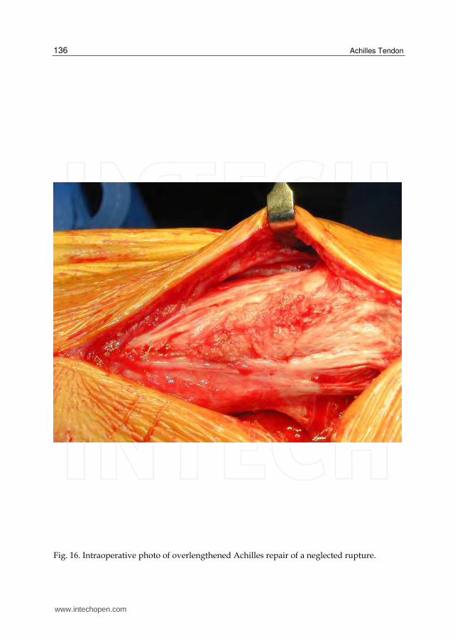
One way to avoid this complication during the repair is to match the position of the unaffected leg intraoperatively. Often this requires that the unaffected leg be draped free as well for comparison. Modulation of the position is actually easier in the neglected rupture because the tendon is not frayed and more accurate purchase of the tissues by the suture allows for better control of the length. If however, the patient has unacceptable weakness, a shortening of the reconstructed tendon can be performed [Figures 16-17].
Although the incidence of rerupture is far less after repair of a neglected rupture compared to repair of the acute rupture, the incidence is not zero. Given the high tensile strength sutures available today, the failure point is almost always at the suture tissue junction rather than a failure of the suture material. From a speculative standpoint, the quality of tissue after a negelected rupture is not as robust and may handle the tension from sutures poorly. Other possibilities of failure include the unraveling of the suture material due to the slippery nature of the knot. [Figure 18].
www.intechopen.com

Surgical Treatment of the Neglected Achilles Tendon Rupture 135
Fig. 15. Postoperative result after repair of delayed rupture. Note the accentuated on the right side.
www.intechopen.com
Achilles Tendon 136
Fig. 16. Intraoperative photo of overlengthened Achilles repair of a neglected rupture.
www.intechopen.com

Surgical Treatment of the Neglected Achilles Tendon Rupture 137
Fig. 17. Cylinder of resected tissue to correct for overlenghtening.
www.intechopen.com

Achilles Tendon 138
Fig. 18. Rerutpure after failure of the suture knot.
Wound problems are not uncommon after the repair of a neglected rupture. The local blood supply to the posterior aspect of the leg is often precarious and may be further disrupted by surgical intervention, introduction of foreign material such as allograft and the denser scar tissue that diminishes the healing capability of the skin [Figures 19-20]. Generally surgical exposures are more extensive compared to repair of the acute rupture, which also increases the chance of wound problems.
www.intechopen.com

Surgical Treatment of the Neglected Achilles Tendon Rupture 139
Fig. 19. Full thickness skin loss 4 weeks after routine repair of neglected rupture using a local gastrocnemius recession for tendon apposition.
www.intechopen.com

Achilles Tendon 140
Fig. 20. Markedly hypertrophic scar after delayed operative repair of ruptured Achilles tendon
www.intechopen.com

Surgical Treatment of the Neglected Achilles Tendon Rupture 141
5. Conclusion
Many surgical treatments are available for reconstruction of a neglected Achilles tendon rupture. There is no concrete data to support one technique over another; hence, there is no “gold standard”. Most agree, however, in order to achieve the optimal functional outcome surgical reconstruction is required. Regardless of the chosen technique, the ultimate goal of surgical treatment is to restore the length tension relationship such that sufficient plantar flexion power is attained. The ability of the patient to achieve a single limb heel rise on the affected side, most often indicates a successful outcome, although some patients are satisfied without attaining this goal.
Patients who have sufficient risk factors and/or low functional capacity may be better treated with bracing as local and more global surgical complications from reconstruction can be devastating (Boyden et al., 1995). The physician needs to decide on the proper treatment course appropriate for the individual patient. The length of the delay, risk factors, desired outcome, and functional requirement of each patient should be taken into account in implementing the appropriate treatment.
6. References
Abraham, E. & Pankovich, A. (1975). Neglected rupture of the Achilles tendon. Treatment by V-Y tendinous flap. J Bone Joint Surg, 57A, pp. 253– 255
Amis, A.; Campbell, J.; Kempson, S. & Miller, J. (1984). Comparison of the structure of neotendons induced by implantation of carbon or polyester fiber. J Bone Joint Surg, 66B, pp. 131-139
Anzel, S.; Covey, K.; Weiner. A. & Lipscomb, P (1959). Disruption of muscles and tendons; an analysis of 1,1014 cases. Surgery, 45, 3, pp. 406-414
Arner, O. & Lindholm, A. (1959). Subcutaneous rupture of the Achilles tendon; a study of 92 cases. Acta Chir Scand, 116, supp 239, pp. 1-51
Arnoczky, S.; Warren, R. & Ashlock, M. (1986). Replacement of the anterior cruciate ligament using a patellar tendon allograft: an experimental study. J Bone Joint Surg Am, 68, pp. 376 –385
Barnes, M. & Hardy, A. (1986). Delayed reconstruction of the calcaneal tendon. J Bone Joint Surg Br, 68, 1, pp. 121-124
Bosworth, D. (1956). Repair of defects in the tendo achillis. J Bone Joint Surg Am, 38-A, 1, pp. 111-114
Boyden, E.; Kitaoka, H.; Cahalan, T. & An, K. (1995). Late versus early repair of Achilles tendon rupture. Clinical and biomechanical evaluation. Clin Orthop Relat Res, 317, pp. 150-158
Bugg, E. Jr. & Boyd, B. (1968). Repair of neglected rupture or laceration of the Achilles tendon.Clin Orthop Relat Res, 56, pp. 73-75
Carden, D.; Noble, J.; Chalmers, J.; Lunn, P. & Ellis, J. (1987). Rupture of the calcaneal tendon. The early and late management. J Bone Joint Surg Br, 69, 3, pp. 416-20
Carr, A. & Norris, S. (1989). The bood supply of the calcaneal tendon. J Bone Joint Surg, 71B, pp. 100-101
www.intechopen.com

Achilles Tendon 142
Cetti, R.; Christensen, S. & Ejsted, R. et al (1993). Operative versus non-operative treatment of Achilles tendon rupture. A prospective randomized study and review of the literature. Am J Sports Med, 21, 6, pp. 791-799
Christensen, I. (1953). Rupture of the Achilles tendon: analysis of 57 cases. Acta Chir. Scand, 106, pp. 50-60
Coughlin, M.; Mann, R. & Saltzman, C. (2007): Disorders of tendons, In: Surgery of the foot and ankle, Eighth Edition, pp. 1249-1261, Mosby, Inc., Philadelphia
Coull, R.; Flavin, R. & Stephens, M. (2003). Flexor hallucis longus tendon transfer: Evaluation of postoperative morbidity. Foot Ankle Int, 24, 12, pp. 931-934
Dalton, G. (1996). Achilles tendon rupture. Foot Ankle Clin, 1, pp. 225-236 Den Hartog, B. (2003). Flexor Hallucis Longus Tendon Transfer for Chronic Achilles
Tendinosis. Foot Ankle Int, 24, 3, pp. 233 – 237 Drez, D. Jr.; DeLee, J.; Holden, J.; Arnoczky, S.; Noyes, F. & Roberts, T. (1991). Anterior
cruciate ligament reconstruction using bone-patellar tendonbone allografts: a biological and biomechanical evaluation in goats. Am J Sports Med, 19, pp. 256 –263
Elias, I.; Besser, M.; Nazarian, L. & Rankin, S. (2007). Reconstruction for missed or neglected Achilles tendon rupture with V-Y lengthening and flexor hallucis longus tendon transfer through one incision. Foot Ankle Int, ,28, 12, pp. 1238-1248
Gallant, G.; Massie, C. & Turco, V. (1995). Assessment of eversion and plantarflexion strength after repair of the Achilles tendon rupture using peroneus brevis tendon transfer. Am J Orthop, 24, pp. 257–261
Grove, J. & Hardy, M. (2008). Autograft, allograft, and xenograft options in the treatment of neglected Achilles tendon ruptures: a historical review with illustration of surgical repair, In: The Foot and Ankle Online Journal, Available from: http://faoj.files.wordpress.com/2008/04/autograft-allograft-and-xenograft-options-in-treatment-of-neglected-achilles-tendon-ruptures.pdf
Habusta, S. (1995). Bilateral simultaneous rupture of the Achilles tendon. A rare traumatic injury. Clin Orthop Relat Res, 320, pp. 231-234
Hansen, S. (1991): Trauma to the Heel Cord, In: Disorders of the Foot and Ankle, Second Edition, Jahss, M., pp. 2357, W.B. Saunders, Philadelphia
Hepp, W. & Blauth, W. (1978). Zur Behandlung von Achilessehnendefekten mitder “peronaeus-brevis-plastik“. Arch Orth Traum Surg, 9, pp. 195–200
Indelli, P.; Dillingham, M.; Fanton, G. & Schurman, D. (2004). Anterior cruciate ligament reconstruction using cryopreserved allografts. Clin Orthop, 420, pp. 268 –275
Jackson, D.; Grood, E.; Arnoczky, S.; Butler, D. & Simon, T. (1987). Freeze-dried anterior cruciate ligament with freeze dried fascia lata allografts. Preliminary studies in a goat model. Am J Sports Med, 15, pp. 295–303
Jozsa, L.; Kvist, M. & Balint, B. et al. (1989). The role of recreational sport activity in Achilles tendon rupture: a clinical, pathoanatomical, and sociological study of 292 cases. Am J Sports Med, 17, pp. 338-343
Kissel, C.; Blacklidge, D. & Crowley, D. (1994). Repair of neglected Achilles tendon ruptures--procedure and functional results. J Foot Ankle Surg, 33, 1, pp. 46-52
Lee, D. (2007). Achilles tendon repair with acellular tissue graft augmentation in neglected ruptures. J Foot Ankle Surg, 46, 6, pp. 451-455
Lepow, G. & Green, J. (2006). Reconstruction of a neglected Achilles tendon rupture with an Achilles tendon allograft: a case report, J Foot Ankle Surg, 45, 5, pp. 351-355
www.intechopen.com

Surgical Treatment of the Neglected Achilles Tendon Rupture 143
Leppilahti, J. & Orava, S. (1998). Total Achilles tendon rupture. A review. Sports Med, 25, 2, pp. 79-100
Lieberman, J.; Lozman, J.; Czajka, J. & Dougherty, J. (1988). Repair of Achilles tendon rupture with Dacron vascular graft. Clin Orthop, 243, pp. 204-208
Lindholm, A. (1959). A new method of operation in subcutaenous ruptureof the Achilles tendon. Acta Chir Scand, 117, pp. 261-270
longus tendon transfer/augmentation. Foot Ankle Int, 21, 12, pp. 1004-1010 Maffulli, N. & Leadbetter, W. (2005). Free gracilis tendon graft in neglected tears of the
Achilles tendon. Clin J Sport Med, 15, 2, pp. 56-61 Maffulli, N. (1996). Clinical tests in sports medicine: more on Achilles tendon. Br J Sports
Med, 30, pp. 250 Maffulli, N.; Waterston, S. & Squair, J. et al. (1999). Changing incidence of Achilles tendon
rupture in Scotland: a 15-year study. Clin J Sport Med, 9, pp. 157-160 Mahirogullari, M.; Ferguson, C.; Whitlock, P.; Stabile, K. & Poehling, G. (2007). Freeze-dried
allografts for anterior cruciate ligament reconstruction. 26, pp. 625-637 Mandelbaum, B.; Myerson, M. & Forster, R. (1995). Achilles tendon ruptures. A new method
of repair, early range of motion, and functional rehabilitation. Am J Sports Med, 2, pp. 392–395
Mann, R.; Holmes, G. & Seale, K. (1991). Chronic rupture of the Achilles tendon: a new technique of repair. J Bone Joint Surg, 73A, pp. 214–219
Martin, R.; Manning, C.; Carcia, C. & Conti, S. (2005). An outcome study of chronic Achilles tendinosis after excision of the Achilles tendon and flexor hallucis longus tendon transfer. Foot Ankle Int, 26, 9, pp. 691-697
Mendicino, S. & Reed, T. (1996). Repair of neglected Achilles tendon ruptures with a triceps surae muscle tendon advancement. J Foot Ankle Surg, 35, 1, pp. 13-18
Miskulin, M.; Miskulin, A.; Klobucar, H. & Kuvalja, S. (2005). Neglected rupture of the Achilles tendon treated with peroneus brevis transfer: a functional assessment of 5 cases. J Foot Ankle Surg, 44, 1, pp. 49-56
Monroe, M.; Dixon, D. & Beals, T. et al. (2000). Plantarfelxion torque following reconstruction of Achilles tendinosis or rupture with flexor hallucis longus augmentation. Foot Ankle Int, 21, pp. 324-329
Myerson, M. (2010): Disorders of the Achilles tendon, In: Reconstruction foot and ankle surgery: management of complications, Second Edition, pp. 341, Saunders, Philadelphia
Nellas, Z.; Lorder, B. & Wertheimer, S. (1996). Reconstruction of an Achilles tendon defect utilizing an Achilles tendon allograft. J Foot Ankle Surg, 35, pp. 144-148
Nillius, S.; Nilsson, B. & Westlin, N (1976). The incidence of Achilles tendon rupture. Acta Orthop Scand, 47, 1, pp. 118-121
of chronic Achilles tendinopathy. Foot Ankle Int, 24, 9, pp. 673-676 Ozaki, J.; Fujiki, J. & Sugimoto, K. et al. (1989). Reconstruction of the neglected Achilles
tendon rupture with Marlex mesh. Clin Orthop Relat Res, 238, pp. 204-208 Parsons, J.; Weiss, A. & Schenk, RS. et al. (1989). Long-term follow-up of Achilles tendon
repair with an absorbable polymer carbon fiber composite. Foot Ankle, 9, 4, pp. 179-184
Pintore, E.; Barra, V.; Pintore, R. & Maffulli, N. (2001). Peroneus brevis tendon transfer in neglected tears of the Achilles tendon. J Trauma, 50, 1, pp. 71-78
Platt, H. (1931). Observations on Some Tendon Ruptures. British Med J, 1, pp. 611-615
www.intechopen.com

Achilles Tendon 144
Poehling, G.; Curl, W.; Lee, C.; Ginn, T.; Rushing, J.; Naughton, M.; Holden, M.; Martin, D. & Smith, B. (2005). Analysis of outcomes of anterior cruciate ligament repair with 5 year follow-up: allograft versus autograft. Arthroscopy, 21, 7, pp. 774-785
Porter, D.; Mannarino, F.; Snead, D.; Gabel, S. & Ostrowski, M. (1997). Primary repair without augmentation for early neglected Achilles tendon ruptures in the recreational athlete. Foot Ankle Int, 18, 9, pp. 557-564
Schedl, R. & Fasol, P. (1979). Achilles tendon repair with the plantaris tendon compared with repair using polyglycol threads. J Trauma, 19, pp. 189-194
Schuberth, J.; Dockery, G. & McBride R. (1984). Recurrent rupture of the tendo Achillis: repair by free tendinous autograft. J Am Podiatr Med Assoc, 74, pp.157-162
Schuberth, JM, B. (1996):Achilles tendon trauma, In: Foot and ankle trauma, Scurran, B. pp. 225, Churchill Livingston, Inc., New York
Shino, K.; Inoue, M.; Horibe, S.; Nagano, J. & Ono, K. (1998). Maturation of allograft tendons transplanted into the knee: an arthroscopic and histological study. J Bone Joint Surg Br, 70, pp. 556 –560
Tashjian, R.; Hur, J.; Sullivan, R.; Campbell, J. & DiGiovanni, C. (2003). Flexor hallucis longus transfer for repair
Teuffer, A. (1974). Traumatic rupture of the Achilles tendon: reconstruction by transplant and graft using lateral peroneus brevis. Orthop Clin North Am, 5, pp. 89 –93
Turco, V. & Spinella, A. (1987). Achilles tendon ruptures: peroneus brevis transfer. Foot Ankle, 7, pp. 253–259
Wapner, K.; Hecht, P. & Mills, R. Jr. (1995). Reconstruction of neglected Achilles tendon injury. Orthop Clin North Am, 26, 2, pp. 249-263
Wapner, K.; Pavlock, G. & Hecht, P. et al. (1993). Repair of chronic Achilles tendon rupture with flexor hallucis longus tendon transfer. Foot Ankle, 14, 8, pp. 443-449
White, R. & Kraynick, B. (1959). Surgical uses of the peroneus brevis tendon. Surg Gynecol Obstet, 108, 1, pp. 117-121
Wilcox, D.; Bohay, D. & Anderson, J. (2000). Treatment of chronic Achilles tendon disorders with flexor hallucis
Yuen, J. & Nicholas, R. (2000). Reconstruction of a total Achilles tendon and soft-tissue defect utilizing an Achilles allograft combined with a rectus muscle free flap. J Plast Reconstr Surg, 107, pp. 1807-1811
www.intechopen.com

Achilles TendonEdited by Prof. Andrej Cretnik
ISBN 978-953-51-0264-9Hard cover, 144 pagesPublisher InTechPublished online 08, January, 2012Published in print edition January, 2012
InTech EuropeUniversity Campus STeP Ri Slavka Krautzeka 83/A 51000 Rijeka, Croatia Phone: +385 (51) 770 447 Fax: +385 (51) 686 166www.intechopen.com
InTech ChinaUnit 405, Office Block, Hotel Equatorial Shanghai No.65, Yan An Road (West), Shanghai, 200040, China
Phone: +86-21-62489820 Fax: +86-21-62489821
Achilles tendon has always attracted a great attention. Its disorders include various problems from pain andswelling with bumps to functional impairment or even ruptures. Debates concerning aetiology and optimaltreatment are still going on. A lot of efforts and research have already been put on to find the answers tounsolved problems and this book is an attempt to share (some of) these findings to the readers. If only one ofthe papers helps the therapists or patients in understanding and solving their problems, we will consider thatthe mission of the book was accomplished.
How to referenceIn order to correctly reference this scholarly work, feel free to copy and paste the following:
Jake Lee and John M. Schuberth (2012). Surgical Treatment of the Neglected Achilles Tendon Rupture,Achilles Tendon, Prof. Andrej Cretnik (Ed.), ISBN: 978-953-51-0264-9, InTech, Available from:http://www.intechopen.com/books/achilles-tendon/surgical-treatment-of-neglected-achilles-tendon-rupture