supplementary materials a subpopulation of high il-21 … · poidinger, juan j. lafaille, maria a....
TRANSCRIPT

1
SUPPLEMENTARY MATERIALS
A subpopulation of high IL-21-producing CD4+ T cells in Peyer’s Patches is
induced by the microbiota and regulates germinal centers
Leigh Jones, Wen Qi Ho, Sze Ying, Lakshmi Ramakrishna, Kandhadayar G. Srinivasan,
Marina Yurieva, Wan Pei Ng, Sharrada Subramaniam, Nur H. Hamadee, Sabrina
Joseph, Jayashree Dolpady, Koji Atarashi, Kenya Honda, Francesca Zolezzi, Michael
Poidinger, Juan J. Lafaille, Maria A. Curotto de Lafaille
SUPPLEMENTARY FIGURES
SUPPLEMENTARY METHODS

2
SUPPLEMENTARY FIGURES
Supplementary Figure S1
Generation and characterization of IL-21eGFP mice.
Supplementary Figure S2
GFP+CXCR5+PD1+ CD4+ T cells express a polarized Tfh phenotype.
Supplementary Figure S3
GFP+CXCR5+PD-1+CD4+ cells are highly differentiated Tfh cells.
Supplementary Figure S4
GFP+Tfh and GFP-Tfh cells from PP have a diverse polyclonal TCRβ repertoire.
Supplementary Figure S5
The TCRβ CDR3 repertoire of PP CD4+ T cell subsets
Supplementary Figure S6
Induction of IL-21 expression in CD4+ T cells activated in the presence of TGFβ, IL-6
and RA.
Supplementary Figure S7
Splenic GFP-CD4+ T cells IL-21eGFP mice traffic to PP where they differentiate into
GFP+Tfh cells and drive B cell activation.
Supplementary Figure S8
DT depletion of GFP+ cells in IL-21eGFP mice results in alterations of local B cell
activation and antibody production in the PP.
Supplementary Figure S9
Overall composition of gut microbiome was similar between WT and IL21eGFP mice.

3
Supplementary Figure S1
Supplementary Figure S1 Generation and characterization of IL-21eGFP mice.
(a) Schematic representation of the IL-21eGFP BAC transgenic construct. The DTR-
eGFP-SV40pA gene cassette was inserted downstream of the IL-21 promoter (IL-21P),

4
leaving intact splicing signals and intron 1 sequences. (b) Representative flow cytometry
analysis of GFP expression within the CD4+ cell population from Peyer’s Patches,
mesenteric lymph node, inguinal lymph node, spleen and thymus of IL-21eGFP
transgenic mice (upper row) and non-transgenic littermates (lower row). (c) Gating
strategy for analyzing GFP expression in CD4+ Tfh cells. Complements Figure 1 of the
manuscript.

5
Supplementary Figure S2
Supplementary Figure S2 GFP+CXCR5+PD1+ CD4+ T cells express a polarized Tfh
phenotype. (a) QPCR analysis of IL21 mRNA expression in sorted PP CD4+ cell
populations from IL-21eGFP mice (data from three independent sorting experiments).
(b) Representative flow cytometry histograms (left) and quantification of ICOS levels by
MFI (right) in gated GFP+CXCR5+PD1+ (GFP+Tfh), GFP-CXCR5+PD1+ (GFP-Tfh), and
GFP-CXCR5-PD1- (Non-Tfh) CD4+ populations from PP (n=5). (c) MFI levels of TIGIT
(left), CD226 (center) and IL-7rα (right) determined by flow cytometry in gated GFP+Tfh,
GFP-Tfh, and GFP- Non-Tfh CD4+ populations from PP (n=3). The data in (c) is
representative of two experiments. Statistical significance was determined by unpaired,
two-tailed, Mann-Whitney U-test. Complements Figure 1 of the manuscript.

6
Supplementary Figure S3

7
Supplementary Figure S3 GFP+CXCR5+PD-1+CD4+ cells are highly differentiated Tfh
cells. (a-c) Heatmap representation of differentially expressed genes (DEG) in Tfh cells,
identified by comparison between all CXCR5+PD-1+ CD4+ Tfh cell samples (GFP+ and
GFP-), and GFP-CXCR5-PD-1- CD4+ non-Tfh cells from PP cells of IL-21eGFP mice.
Only DEG with log2 fold change (logFC) value ≥ 0.58 and p≤ 0.05 were included. (a) Tfh
DEGs with similar expression levels in GFP+CXCR5+PD-1+ and GFP-CXCR5+PD-1+
CD4+ cells. (b) Tfh DEG subset further downregulated in GFP+Tfh than in to GFP-Tfh
cells. (c) Tfh DEG subset with higher upregulation in GFP+Tfh than in GFP-Tfh cells.
Genes in the heatmaps are shown in Supplementary Tables 1-3. Complements Figure 1
of the manuscript.

8
Supplementary Figure S4

9
Supplementary Figure S4 GFP+Tfh and GFP-Tfh cells from PP have a diverse
polyclonal TCRβ repertoire. Percentage of Vβ gene usage in sorted PP CD4+ T cell
populations from a representative IL-21eGFP mouse. Complements Figure 2 of the
manuscript.

10
Supplementary Figure S5
Supplementary Figure S5 The TCRβ CDR3 repertoire of PP CD4+ T cell subsets.
(a) Overlap of the repertoire of unique TCR Vβ CDR3 sequences from GFP-Tfh cells,
with the repertoire of GFP+Tfh and non-Tfh CD4+ cells in the same mouse. Each pie
graphic represents all GFP-Tfh cells’ unique CDR3 sequences from one mouse (n=3).
(b) Repertoire relatedness between paired CD4+ populations of individual mice as

11
determined by computational β diversity analysis. Complements Figure 2 of the
manuscript.

12
Supplementary Figure S6
Supplementary Figure S6 Induction of IL-21 expression in CD4+ T cells activated in
the presence of TGFβ, IL-6 and RA. Splenocytes from naïve TBmc mice were
stimulated with anti-CD3 and anti-CD28 with addition of indicated cytokines, blocking
antibodies or RA. Number of (a) IL-17+ and (b) Foxp3+ CD4+ cells as determined by
intracellular staining. (c) IL-17 and (d) IL-21 levels in culture supernatants collected
daily and measured by ELISA. Complements Figure 3 of the manuscript.

13
Supplementary Figure S7

14
Supplementary Figure S7 Splenic GFP-CD4+ T cells IL-21eGFP mice traffic to PP
where they differentiate into GFP+Tfh cells and drive B cell activation. Splenic polyclonal
GFP-CD4+ T cells from IL-21eGFP BALB/c mice were transferred alone (T) or with
polyclonal B cells from CD45.1+ BALB/c mice (T+B) into TBmc mice. Groups of TBmc
mice also received CD45.1+ B cells without T cells (B). 4 weeks post transfer, PP cells T
and B cell populations in the recipient mice were analysed by flow cytometry. (a, b)
Donor T cells, identified as CD4+KJ126-, expanded similarly in PP of recipient mice
infused or not with polyclonal B cells. (a) Representative flow cytometry plots of gated
CD4+ T cells showing endogenous KJ1-26+ CD4 T cell gate and donor KJ1-26- CD4 T
cell gate. (b) Percentage of donor CD4+ cells in total PP CD4+ cells. (c) Percentage of
donor T cells in the CD4+ T cell population of PP, mesenteric LN (MLN) and spleen
(Spl) of recipient mice. (d) Representative flow cytometry plots of gated endogenous
KJ1-26+CD4+ T cells in PP of recipient mice demonstrating absence of Tfh cells. (e-f)
Larger expansion of donor CD45.1+ B cells in PP of recipient mice co-transferred with
polyclonal T cells. (e) Representative plots of gated B220+ B cells showing endogenous
CD45.2+ B cell gate and donor CD45.2- B cell gate. (f) Percentage of donor CD45.2-
B220+ cells in total B220+ cells from PP. (g-i) Differentiation of endogenous CD45.2+ B
cells into (g) CD95+GL7+ GC cells, (h) IgA+ cells and (i) IgG1+ cells. All bar graphs show
mean + s.e.m of 6-8 mice per group. Statistical significance was determined by
unpaired, two-tailed Mann Whitney U-test (f) or Kruskal-Wallis followed by Dunn’s
multiple comparisons test (g-i). P values are only shown for statistically significant
differences (p<0.05). Complements Figure 4 of the manuscript.

15
Supplementary Figure S8
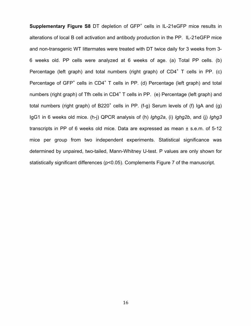
16
Supplementary Figure S8 DT depletion of GFP+ cells in IL-21eGFP mice results in
alterations of local B cell activation and antibody production in the PP. IL-21eGFP mice
and non-transgenic WT littermates were treated with DT twice daily for 3 weeks from 3-
6 weeks old. PP cells were analyzed at 6 weeks of age. (a) Total PP cells. (b)
Percentage (left graph) and total numbers (right graph) of CD4+ T cells in PP. (c)
Percentage of GFP+ cells in CD4+ T cells in PP. (d) Percentage (left graph) and total
numbers (right graph) of Tfh cells in CD4+ T cells in PP. (e) Percentage (left graph) and
total numbers (right graph) of B220+ cells in PP. (f-g) Serum levels of (f) IgA and (g)
IgG1 in 6 weeks old mice. (h-j) QPCR analysis of (h) Ighg2a, (i) Ighg2b, and (j) Ighg3
transcripts in PP of 6 weeks old mice. Data are expressed as mean ± s.e.m. of 5-12
mice per group from two independent experiments. Statistical significance was
determined by unpaired, two-tailed, Mann-Whitney U-test. P values are only shown for
statistically significant differences (p<0.05). Complements Figure 7 of the manuscript.

17
Supplementary Figure S9
Supplementary Figure S9 Overall composition of gut microbiome was similar between
WT and IL21eGFP mice. IL-21eGFP mice and non-transgenic WT littermates were
treated with DT twice daily for 3 weeks from 3-6 weeks old. Genomic DNA was
extracted from fecal samples and the 16S variable regions were amplified and
sequenced. (a) α diversity analysis of the gut microbiome of WT and IL21eGFP mice
treated with PBS or DT. No significant differences were observed. (b) Distribution of gut

18
microbe families within fecal samples from WT and IL21eGFP mice treated with PBS or
DT. Complements Figure 7 of the manuscript.

19
SUPPLEMENTARY METHODS
• Generation of IL-21eGFP mice
• Isolation of PP lymphocytes
• Antibiotic treatment
• Diphtheria toxin (DT) treatment
• 16S RNA sequencing of fecal microbiome
• Flow cytometry and cell sorting
• RNA expression analysis by QPCR and microarray
• High throughput TCR sequencing
• ELISA
• Immunohistology
• List of antibodies used
• Primer sequences
• References

20
Generation of IL-21eGFP mice. IL-21eGFP transgenic mice were generated using
BAC technology. First, Il21 upstream and downstream homologous sequences were
ligated to the 5’ and 3’ ends respectively of a cassette encoding a DTR-eGFP fusion
protein and the SV40 polyadenylation site in pGEM T vector 1. The cassette was then
recombined with the wild type Il21 gene in BAC RP23-137F6 (Invitrogen-Life
Technologies). The BAC insert was gel purified and injected into C57Bl/B6 blastocysts
at the Transgenic Facility of the New York University School of Medicine (NYUSM). IL-
21eGFP mice were genotyped by PCR using the primers
TTATCCTCCAAGCCACAAGC and AAGTCGTGCTGCTTCATGTG. Of the 7 founders,
one expressing GFP in CD4+ T cells of Peyer’s Patches (PP) was backcrossed to the
BALB/c strain (The Jackson Laboratories) and to TBmc mice 2 for use in the study.
Isolation of Peyer’s Patches lymphocytes. PP lymphocytes were prepared as
previously described 3 with some modifications: PP were excised and epithelial cells
removed by incubation in PBS with 1mM EDTA for two rounds of 10 minutes at 37°C
and with 200 rpm rotation. Following washing with PBS, the remaining tissue was
incubated with 50µg/ml Liberase™ (Roche) and 333µg/ml DNAse (Sigma) at 37°C
during centrifugation at 100 rpm for 30 minutes, before a second 15 minute incubation
with DNAse. A single cell suspension was generated by pressing the tissue through a
cell strainer. Cells were then washed in RPMI-1640 supplemented with 10% fetal bovine
serum (Invitrogen).
Antibiotic treatment. For broad ablation of the intestinal microbiota, a cocktail of
antibiotics – ampicillin (0.5mg/ml; Sigma), metronidazole (0.5mg/ml; Acros Organic),

21
neomycin sulfate (1mg/ml; Fisher) and vancomycin hydrochloride (0.25mg/ml; Fisher)
was supplied to mice in the drinking water. For ablation of gram negative bacteria a
cocktail of metronidazole (0.5mg/ml), neomycin sulfate (1mg/ml) and polymyxin B
(0.2mg/ml) was used. For ablation of gram positive bacteria, mice were treated with
vancomycin (0.25mg/ml). The artificial sweetener Splenda was added to the water at
4mg/ml to encourage drinking. Treatments commenced at 2 weeks of age when mice
were still in the breeding cages and continued after weaning. Control cages were kept
on water containing Splenda alone. After weaning, experimental cages contained same
sex transgenic and non-transgenic BALB/c littermates undergoing the same treatment.
Both males and females were included in these experiments.
Diphtheria toxin (DT) treatment. To deplete GFP+ cells, IL-21eGFP mice were
injected i.p. with DT (5ng/g body weight; Sigma) diluted in 100µl PBS twice daily for
three weeks. Control mice received 100µl PBS under the same treatment regimen. Non-
transgenic WT BALB/c littermates underwent identical treatment.
Flow cytometry and cell sorting. Single cell suspensions were incubated with 10µg/ml
anti-CD16/32 for 10 minutes at 4°C prior to surface labeling with antibody cocktails in
labeling buffer (2% FBS and 0.1% NaN3 in PBS) for 40 minutes at 4°C. Labeling for
CXCR5 was carried out at 37°C for 30 minutes prior to any other surface labeling.
Where indicated, intra-nuclear detection of FoxP3 or Bcl6 was conducted using the
FoxP3/Transcription Factor staining buffer kit (eBioscience). Intracellular staining of
cytokines was performed using BD Biosciences Cytofix/CytopermTM kit. Cells were
analyzed using a FACsCanto or LSR II 5-laser flow cytometer (BD Biosciences) or

22
sorted using an Influx or FACsAria II 4-laser cell sorter (BD Biosciences), with FlowJo
software (Tree Star) for data analysis. Purified cells were used for QPCR or adoptive
transfer experiments. Details of all antibodies used are in a table below.
RNA expression analysis by QPCR and microarray. For quantitative PCR analysis
(QPCR) of gene expression, RNA was obtained from cells using TRIzol (Invitrogen) and
cDNA synthesized with SuperScript II reverse transcriptase as per manufacturer’s
instructions. QPCR for selected gene transcripts was carried out using primers (as listed
below) on a BioRad cFX96 RT-PCR instrument. Gene expression was normalized to β-
Actin.
Microarray expression analysis of triplicate samples of sorted GFP+CXCR5+PD1+ Tfh,
GFP-CXCR5+PD1+ Tfh, and GFP- CXCR5-PD1- non-Tfh CD4+ T cells from PP was
performed as previously described using Affymetrix MoGene 1.0 ST arrays 4. A list of
Tfh differentially expressed genes (DEG) was generated using log2 fold change (logFC)
value ≥ 0.58 and p≤ 0.05 as selection criteria. Microarray datasets can be found in NCBI
GEO GSE77899.
High throughput TCRβ sequencing. CD4+ T cells from PP of IL-21eGFP mice were
sorted into CXCR5+PD-1+GFP+Tfh, CXCR5+PD-1+GFP-Tfh and CXCR5-PD-1- naive cell
populations using a LSRII 4-laser cell sorter (BD Biosciences). Total RNA was isolated
using ARCTURUS® PicoPure® RNA Isolation Kit (Life Technologies) and immune
repertoire amplicon libraries were prepared using 2ng of RNA as described 5. Briefly,
reverse transcription and amplification were performed with Mouse TCR beta, Illumina
MiSeq, V-C gene Primers (iRepertoire Inc., Huntsville, AL, USA) and Qiagen One-Step

23
RT-PCR kit (Qiagen). In the primary PCR reaction barcode sequences for identifying
and demultiplexing individual samples were added to the template while the Illumina
adapters were added in the secondary PCR reaction using Qiagen Multiplex PCR Kit.
Equimolar concentrations of secondary PCR products were pooled, electrophoresed in
2% agarose gel, and amplicons in the 400-450 bp range were gel purified. qPCR was
performed (Kapa Biosystems) on the amplicon library to ascertain the loading
concentration. The library was sequenced using Illumina MiSeq to generate 250bp
paired end reads at a sequencing depth of 1 million reads per sample. Raw sequencing
reads were submitted to iRepertoire® for analysis. In brief, raw reads were
demultiplexed and processed using a SMART (Sequencing, Mosaic, Amplification,
Reference, Frequency Threshold) filter to remove sequencing errors, chimeric
sequences, PCR artifacts, mismatches with reference sequences and low frequency
CDR3s. Identical reads were collapsed and assigned to V,D,J segments using Smith-
Waterman algorithm 6 for local sequence alignment with germline reference from IMGT
(www.imgt.org). Raw and processed reads were provided by iRepertoire® for further
analysis.
α and β diversity indices were generated using the vegan package in R
v2.15.2/Bioconductor 6,7. The α diversity index was determined using Shannon’s Index.
The β diversity index was determined using the “z” method of the betadiver function
from vegan, specifically (log(2)-log(2*a+b+c)+log(a+b+c)/log(2), where “a” represents
the number of shared sequences, and b and c represent the unique sequences in the
two samples. The datasets of the TCRβ sequence analysis were deposited in NCBI
Bioproject, accession PRJNA311496.

24
16S RNA sequencing of the fecal microbiome. Genomic DNA (gDNA) was isolated
from stool samples of mice using the QIAmp DNA stool kit (QIagen, Valencia, CA)
following manufacturers instructions. For amplification of the 16S variable regions (V4
to V5), PCR was performed using 10ng of stool gDNA with Long Amp Taq polymerase
(New England Biolabs, Ipswich, MA) according to manufacturer’s instructions. The
reaction mix for the first round of amplification contains a specific forward primer and
reverse primer binding to V4 & V5 regions, respectively. The forward primer (U519F
Mod –CAGCMGCCGCGGTAAYWC) was modified from Baker et al. 8, while the reverse
primer MetGen R-BSCCCGYCAATTYMTKTRAGT was modified from Teske and
Sorenson 9. Secondary PCR was carried out to further enrich for variable region
sequences using communal primers. During the secondary PCR reaction, barcodes for
identifying and de-multiplexing individual samples, and illumina adapter sequences,
were added to the template. PCR cycling parameters consisted of initial denaturation for
30s at 94°C, followed by 15 cycles of 15 s at 94°C, 30 s at 45°C, and 30 s at 65°C with
a final extension for 10 min at 65°C for the primary PCR reaction. For secondary PCR,
the cycling parameters were the same except that amplification was carried out for 25
cycles. Equimolar concentrations of secondary PCR products were pooled and
electrophoresed using 2% agarose gel. Amplicons in the size range of 550 bp were gel
purified using the Qiaquick Gel Extraction Kit (Qiagen). Concentrations of gel purified
libraries were estimated using the DNA 1000 kit (Agilent Technologies, Santa Clara,
CA). QPCR was performed using quantified amplicon libraries (Kapa Biosystems,
Wilmington, MA) to ascertain the loading concentration. The library was sequenced
using Illumina MiSeq to generate 250bp Single end reads at a sequencing depth of
about half a million reads per sample. These reads were trimmed for primers in Accelrys

25
Pipeline Pilot, USA (version 9.2.0, www.accelrys.com) and quality (cutoff=20) in BBDuk
[http://sourceforge.net/projects/bbmap/]. Trimmed reads were mapped to Silva 119 OTU
database 10 with QIIME version 1.8.0 11 OTU picking script
pick_closed_reference_otus.py using uclust 12. β diversities were calculated using
Unifrac 13 distances with beta_diversity_through_plots.py script, and alpha_diversity.py
scripts in QIIME and alpha-diversity simpson index was calculated and compared for all
the samples using compare_alpha_diversity.py script in QIIME. The association
between metadata and microbial abundance was determined in MaAsLin
(http://huttenhower.org/galaxy) by pairwise comparisons of DT-treated and PBS-treated
control mice within WT and IL-21eGFP groups. QIIME and TIBCO Spotfire, USA
(version 5.5.0, http://spotfire.tibco.com/) were used to make visualisations. Datasets for
the microbiome analysis are in NCBI BioProject, accession number PRJNA293925.
ELISA. Feces were collected and weighed before disruption in protease inhibitor
(Complete Mini, EDTA free 14) at 100mg feces/ml. Fecal supernatants were prepared by
centrifuging at 10,000 x g for 10 minutes at 4°C. ELISA was carried out on fecal
supernatants for determination of the concentration of IgA, and on serum samples for
IgA and IgG1 as described2. Cell culture supernatants were tested for levels of IL-17
and IL-21. Typically, pairs of unlabeled and biotinylated antibodies were used for plate
coating and detection of the analyte, respectively. Plates were incubated with
streptavidin conjugated to Horse Radish Peroxidase (HRP) and a colorimetric substrate
to measure optical density. Purified cytokines and antibody isotypes were used to obtain
a standard curves. All antibodies used are listed below.

26
Immunohistology. Segments of intestine containing PP were frozen in OCT (Tissue-
Tek) and sectioned at 8µM. Sections were fixed with 1:1 acetone/methanol, washed in
PBS and then incubated for 1 hour at room temperature in blocking buffer (PBS with 1%
BSA and 20µg/ml anti-mouse CD16/32). Sections were then incubated overnight at 4°C
with primary antibodies, washed with PBS and counterstained with Hoescht for 15
minutes at room temperature. Fluorescent images were captured with an Olympus
FV1000 confocal microscope and analyzed with Olympus FluoView software.
List of antibodies used
Antigen Clone Conjugate Source Bcl6 K112-91 Alexa Fluor 647 BD Biosciences
CD3e 145-2C11 Unlabelled BD Biosciences CD4 GK1.5 eFluor 450 eBioscience CD4 GK1.5 eFluor 605NC eBioscience CD4 L3T4 PerCP BD Biosciences CD4 RM4-5 APC eBioscience CD8 53-6.7 PE eBioscience
CD11b M1/70 PE eBioscience CD19 eBio1D3 (1D3) eFluor 450 eBioscience CD19 eBio1D3 (1D3) PeCy7 eBioscience CD25 PC61.5 APC eBioscience CD25 PC61.5 PE BD Biosciences CD28 37.51 Unlabelled BD Biosciences
CD45R (B220) RA3-6B2 PerCP BD Biosciences CD45R (B220) RA3-6B2 PE-Cy7 eBioscience CD45R (B220) RA3-6B2 APC-eFluor780 eBioscience
CD45.2 104 APC eBioscience CD49b DX5 PE eBioscience CD95 Jo2 PE-Cy7 BD Biosciences
CD138 281-2 Brilliant Violet 421
BioLegend
CD138 281-2 APC BD Biosciences CD278 (ICOS) 7E.17G9 PE eBioscience CD279 (PD1) J43 APC BD Biosciences CD279 (PD1) RMP1-30 eFluor450 eBioscience CD279 (PD1) J43 PE BD Biosciences
CD326 (EPCAM) G8.8 APC eBioscience

27
Antigen Clone Conjugate Source CD326 (EPCAM) G8.8 PE eBioscience
CXCR5 2GB PE-Cy7 BD Biosciences FoxP3 FJK-16s Alexa Fluor 647 eBioscience GFP Polyclonal IgG Alexa Fluor 488 Invitrogen IFNγ XMG1.2 Unlabelled BioLegend IgA Polyclonal IgG Unlabelled Southern Biotech IgA Polyclonal IgG PE Southern Biotech IgA Polyclonal IgG HRP Southern Biotech IgD 11-26 PE eBioscience
IgG1 F(ab’)2 Polyclonal Unlabelled Southern Biotech IgG1 F(ab’)2 Polyclonal Biotin Southern Biotech
IgG1 RMG1-1 APC BioLegend IgG1 M1-14D12 PE-Cy7 eBioscience IgG1 M1-14D12 PerCP-eFluor
710 eBioscience
IL-4 11B11 Unlabelled BioLegend IL-17A TC11-18H10.1 PE BioLegend IL-17 50101 Unlabelled R&D Systems IL-17 Polyclonal IgG Biotin R&D Systems IL-21 Polyclonal IgG Unlabelled R&D Systems IL-21 Polyclonal IgG Biotin R&D Systems Ly76 TER-110 PE eBioscience
MHCII M5/114.15.2 APC eBioscience MHCII M5/114.15.2 PE eBioscience
T and B cell activation antigen
GL7 Alexa Fluor 647 BD Biosciences
T and B cell activation antigen
GL7 FITC BD Biosciences
TCR DO11.10 KJ126 PE BioLegend TCR Vβ2 B20.6 PE BD Biosciences TCR Vβ3 KJ25 PE BD Biosciences TCR Vβ4 KT4 PE BD Biosciences
TCR Vβ5.1/5.2 MR9-4 PE BD Biosciences TCR Vβ6 RR4-7 APC eBioscience TCR Vβ7 TR310 PE BioLegend TCR Vβ8 F23.1 PE BD Biosciences
TCR Vβ8.1/8.2 MR5-2 PE BioLegend Biosciences
TCR Vβ8.3 8C1 PE BioLegend TCR Vβ10 B21.5 PE eBioscience TCR Vβ11 RR3-15 PE BD Biosciences TCR Vβ12 MR11-1 PE BioLegend

28
Primer sequences
Gene Forward primer Reverse primer Β-Actin TGACAGGATGCAGAAGGAGA GTACTTGCGCTCAGGAGGA IL-21 CGCCTCCTGATTAGACTTC GCCCCTTTACATCTTGTGGA IgA AGTGGCGCATCATTCAAGT CTGGGGTGGGAAGGTGTT
IgG1 AGTCTGACCTCTACACTCTG CTTACAACCACAATCCCTG IgG2a TGACCTCTACACCCTCAGCA AGGACAGGGCTTGATTGTGG IgG2b CACCACGGTGGACAAAAAAC ACAGGGGTTGATTGTTGAA IgG3 GCCAGCAAGACTGAGTTGAT GGGTACTGGGCTTGGGTATT

29
References
1 Lahl, K. et al. Selective depletion of Foxp3+ regulatory T cells induces a scurfy-
like disease. J Exp Med 204, 57-63 (2007).
2 Curotto de Lafaille, M. A. et al. Hyper immunoglobulin E response in mice with
monoclonal populations of B and T lymphocytes. J Exp Med 194, 1349-1359 (2001).
3 Sheridan, B. S. & Lefrancois, L. Isolation of mouse lymphocytes from small
intestine tissues. Curr Protoc Immunol Chapter 3, Unit3 19 (2012).
4 He, J. S. et al. The distinctive germinal center phase of IgE+ B lymphocytes limits
their contribution to the classical memory response. J Exp Med 210, 2755-2771(2013).
5 Wang, C. et al. High throughput sequencing reveals a complex pattern of
dynamic interrelationships among human T cell subsets. Proc Natl Acad Sci U S A 107,
1518-1523 (2010).
6 Black, P. E. Smith-Waterman algorithm, in Dictionary of Algorithms and Data
Structures [online]. Vreda Pieterse and Paul E. Black, eds. 2006.
7 Jari Oksanen, F. G. B., Roeland Kindt, Pierre Legendre,, Peter R. Minchin, R. B.
O. H., Gavin L. Simpson, Peter Solymos, M. & Wagner, H. H. S. a. H. vegan:
Community Ecology Package. R package version 2.0-10. http://CRAN.R-
project.org/package=vegan (2013).
8 Baker, G. C., Smith, J. J. & Cowan, D. A. Review and re-analysis of domain-
specific 16S primers. J Microbiol Methods 55, 541-555 (2003).
9 Teske, A. & Sorensen, K. B. Uncultured archaea in deep marine subsurface
sediments: have we caught them all? ISME J 2, 3-18 (2008).
10 Quast, C. et al. The SILVA ribosomal RNA gene database project: improved data
processing and web-based tools. Nucleic Acids Res 41, D590-596 (2013).

30
11 Caporaso, J. G. et al. QIIME allows analysis of high-throughput community
sequencing data. Nat Methods 7, 335-336 (2010).
12 Edgar, R. C. Search and clustering orders of magnitude faster than BLAST.
Bioinformatics 26, 2460-2461 (2010).
13 Lozupone, C. & Knight, R. UniFrac: a new phylogenetic method for comparing
microbial communities. Appl Environ Microbiol 71, 8228-8235 (2005).
14 Tian, J. et al. Toll-like receptor 9-dependent activation by DNA-containing
immune complexes is mediated by HMGB1 and RAGE. Nat Immunol 8, 487-496 (2007).