sumoylation differentially regulates sp1 to control cell ... · sumoylation differentially...
TRANSCRIPT

Sumoylation differentially regulates Sp1 to controlcell differentiationLili Gonga,b,1, Wei-Ke Jia,1, Xiao-Hui Hua,c,1, Wen-Feng Hua,c,1, Xiang-Cheng Tanga,b,1, Zhao-Xia Huangc, Ling Lic,Mugen Liud, Shi-Hua Xiange, Erxi Wuf, Zachary Woodwarda, Yi-Zhi Liub,2, Quan Dong Nguyena,2,and David Wan-Cheng Lia,b,c,2
aDepartment of Ophthalmology and Visual Sciences, Truhlsen Eye Institute, University of Nebraska Medical Center, Omaha, NE 68198-5540; bState KeyLaboratory of Ophthalmology, Zhongshan Ophthalmic Center, Sun Yat-Sen University, Guangzhou, Guangdong 510060, China; cKey Laboratory of ProteinChemistry and Developmental Biology of Ministry of Education, College of Life Sciences, Hunan Normal University, Changsha, Hunan 410081, China; dKeyLaboratory of Molecular Biophysics of Ministry of Education, College of Life Science and Technology, Center for Human Genome Research, HuazhongUniversity of Science and Technology, Wuhan, Hubei 430074, China; eNebraska Center for Virology, School of Veterinary Medicine and BiomedicalSciences, University of Nebraska–Lincoln, Lincoln, NE 68583-0900; and fDepartment of Pharmaceutical Sciences, College of Pharmacy, North Dakota StateUniversity, Fargo, ND 58105-6050
Edited* by Michael Karin, University of California, San Diego School of Medicine, La Jolla, CA, and approved March 3, 2014 (received for reviewAugust 8, 2013)
The mammalian small ubiquitin-like modifiers (SUMOs) are activelyinvolved in regulating differentiation of different cell types. However,the functional differences between SUMO isoforms and their mech-anisms of action remain largely unknown. Using the ocular lens as amodel system, we demonstrate that different SUMOs display distinctfunctions in regulating differentiation of epithelial cells into fibercells. During lens differentiation, SUMO1 and SUMO2/3 displayeddifferent expression, localization, and targets, suggesting differen-tial functions. Indeed, overexpression of SUMO2/3, but not SUMO1,inhibited basic (b) FGF-induced cell differentiation. In contrast,knockdown of SUMO1, but not SUMO2/3, also inhibited bFGF action.Mechanistically, specificity protein 1 (Sp1), a major transcription factorthat controls expression of lens-specific genes such as β-crystallins,was positively regulated by SUMO1 but negatively regulated bySUMO2. SUMO2 was found to inhibit Sp1 functions through severalmechanisms: sumoylating it at K683 to attenuate DNA binding, and atK16 to increase its turnover. SUMO2 also interfered with the interac-tion between Sp1 and the coactivator, p300, and recruited a repressor,Sp3 to β-crystallin gene promoters, to negatively regulate their ex-pression. Thus, stable SUMO1, but diminishing SUMO2/3, during lensdevelopment is necessary for normal lens differentiation. In supportof this conclusion, SUMO1 and Sp1 formed complexes during earlyand later stages of lens development. In contrast, an interaction be-tween SUMO2/3 and Sp1 was detected only during the initial lensvesicle stage. Together, our results establish distinct roles of differentSUMO isoforms and demonstrate for the first time, to our knowledge,that Sp1 acts as a major transcription factor target for SUMO controlof cell differentiation.
transcription regulation | eye development | crystallin gene expression
The conjugation of small ubiquitin-like modifiers (SUMOs) toprotein substrates (named sumoylation) is a critical post-
translational modification with diverse cellular functions (1).Three major SUMO isoforms (SUMO1, -2, and -3) were iden-tified in vertebrates. Although the mature SUMO2 and SUMO3share a very high level of sequence identity (97%) and cannot beimmunologically discriminated (thus referred to as SUMO2/3),they significantly differ from SUMO1, with only 45% identity(2, 3). Recent studies using proteomics revealed that SUMO1and SUMO2/3 can be targeted to both distinct and overlappingsets of substrates (4). However, whether SUMO1 and SUMO2/3have redundant or different functions in vivo is not clear becauseinconsistent results have been reported in SUMO1 knockoutmice (5, 6).SUMO conjugation is executed by three enzymes. The acti-
vating enzyme E1, a heterodimer of SAE1 and SAE2, transfersSUMO to the single E2-conjugating enzyme Ubc9, which eithersumoylates the substrate alone, or cofunctions with different E3
ligases. Sumoylation is highly dynamic and can be rapidly revertedby sentrin-specific proteases (SENPs) (7). Functionally, it regu-lates many cellular processes, including cell differentiation (8–11).In ocular tissues, sumoylation helps to determine the differentia-tion of cone versus rod photoreceptors (11). Our recent studyrevealed that SUMO1-mediated sumoylation is an indispensablestep toward activation of p32 Pax-6, a master regulator of eye andbrain development (12). Although the effects of sumoylation onindividual targets in regulating cell differentiation and other bi-ological processes are being unraveled, it remains largely unknownhow SUMO isoforms regulate cell differentiation and whetherSUMO1 and SUMO2/3 display distinct functions.The vertebrate lens is an attractive model for studying cell
differentiation (13). In this tissue, cell proliferation and differ-entiation occur throughout life. Differentiation of the vertebratelens starts from the lens vesicle (LV) stage. Cells in the anteriorof the LV retain epithelial morphology and proliferative capacitywhereas the posterior cells elongate and differentiate into pri-mary lens fiber cells (LFCs). The lens constantly grows throughoutlife via continued proliferation and differentiation of lens epi-thelial cells (LECs) in the germinal zone into secondary LFCs.Formation of both primary and secondary LFCs is characterizedby elongated cell shape, accumulation of differentiation-specificproteins such as β-crystallins, and loss of subcellular organelles.
Significance
The mammalian small ubiquitin-like modifiers (SUMOs) areactively involved in regulating differentiation of different celltypes. However, the exact functions of SUMO1 and SUMO2/3have not been defined. Here, we demonstrate for the firsttime, to our knowledge, that SUMO1 promotes cell differenti-ation whereas SUMO2/3 inhibits cell differentiation. Mecha-nistically, we demonstrate that specificity protein 1 is the majortarget activated by SUMO1 conjugation but is repressed bySUMO2/3-mediated sumoylation via our newly identified K683residue, as well as the known K16 site.
Author contributions: L.G., W.-K.J., X.-H.H., W.-F.H., X.-C.T., Y.-Z.L., Q.D.N., and D.W.-C.L.designed research; L.G., W.-K.J., X.-H.H., W.-F.H., X.-C.T., Z.-X.H., and L.L. performedresearch; M.L., S.-H.X., E.W., and Y.-Z.L. contributed new reagents/analytic tools; L.G.,W.-K.J., X.-H.H., W.-F.H., X.-C.T., Z.-X.H., M.L., S.-H.X., E.W., Y.-Z.L., Q.D.N., and D.W.-C.L.analyzed data; and L.G., Z.W., Q.D.N., and D.W.-C.L. wrote the paper.
The authors declare no conflict of interest.
*This Direct Submission article had a prearranged editor.1L.G., W.-K.J., X.-H.H., W.-F.H., and X.-C.T. contributed equally to this work.2To whom correspondence may be addressed. E-mail: [email protected], [email protected], or [email protected].
This article contains supporting information online at www.pnas.org/lookup/suppl/doi:10.1073/pnas.1315034111/-/DCSupplemental.
5574–5579 | PNAS | April 15, 2014 | vol. 111 | no. 15 www.pnas.org/cgi/doi/10.1073/pnas.1315034111

In vitro and in vivo, lens differentiation can be triggered byextracellular growth factors, such as basic fibroblast growth factor(bFGF) (14). When cultured in high concentration of bFGF(50∼100 ng/mL), LECs elongate, migrate, and become multi-layered to form LFCs (13, 14). Furthermore, accumulation ofdifferentiation-specific β-crystallin is induced upon prolongedculture (14). However, loss of nuclei was not detected in thiscondition (14), implying that the in vitro model mimics early stagesof in vivo fiber differentiation.Using this system, we explored the functions of SUMO1 and
SUMO2/3 in cell differentiation. Our results reveal that SUMO1and SUMO2/3 differ significantly in abundance, localization, andsubstrate targets. Although SUMO1 promotes cell differentia-tion, SUMO2/3 inhibits this process. Such distinct functionaldifferences are in part mediated through transcription factorSp1. Although SUMO1 directly sumoylates Sp1 to positivelyregulate β-crystallin genes, SUMO2 inhibits Sp1 function by at-tenuating its DNA binding activity, decreasing its protein stability,and suppressing its interactions with the coactivator p300. To-gether, our results demonstrate distinct functional roles andmechanisms of action for SUMO1 and SUMO2/3 in regulatinglens cell differentiation.
ResultsSUMO1 and SUMO2/3 Display Significant Differences in Expressionand Cellular Localization During Mouse-Lens Development. To ex-plore the possible roles of SUMO1 and SUMO2/3 in lens-celldifferentiation, we examined their expression and localizationpatterns from embryonic day 9.5 (ED9.5) to day 3 lens usingimmunohistochemistry (IHC). As shown in Fig. S1, strong signalsof SUMO1 and SUMO2/3 were ubiquitously detected in all LVcells (Fig. S1) (ED11.5), and a similar pattern of SUMO1 andSUMO2/3 expression was detected at ED13.5 (Fig. S1). AtED16.5, when active fiber differentiation takes place, homoge-nous SUMO1 distribution was detected in nuclei of LECs at theanterior portion of the lens (Fig. 1 A, b and b2). At the transitionzone, SUMO1 labeling started to move toward the nuclear pe-riphery (NP) (Fig. 1 A, b and b1). In elongating LFCs moving outof the transition zone, SUMO1 labeling was primarily detectedat the NP (Fig. 1 A, b and b1). For LFCs localized at deeperlayers undergoing terminal differentiation, the NP localization ofSUMO1 was more prominent (Fig. 1 A, b and b2). By contrast,SUMO2/3 staining was prominently represented by intensitychange during lens differentiation. Strong SUMO2/3 labelingwas detected in LEC nuclei, with a decreased signal in elongatingprimary LFCs (Fig. 1 A, d and d1), and eventually the signaldisappeared in terminally differentiated LFCs (Fig. 1 A, d andd2). Similar patterns were observed in the day 3 lens (Fig. S1). Inthe adult lens, SUMO1 labeling was mainly seen in LECs, witha detectable level in the differentiating LFCs of the transitionzone (Fig. S1). In contrast, the SUMO2/3 signal was much lowerin the LECs and became hardly detectable in LFCs of thetransition zone (Fig. S1).
SUMO1 and SUMO2/3 Are Conjugated to Different Targets DuringMouse-Lens Differentiation. The different distributions of SUMO1and SUMO2/3 in differentiating LFCs suggest that they maybe conjugated to different targets. Indeed, Western-blot analysis(WB) suggested that SUMO1 and SUMO2/3 modified distincttargets (Fig. 1B). Neither SUMO1 nor SUMO2/3 was detectedin their free forms (in Fig. 1B, no bands with 11 kDa wereobserved), indicating that the observed IHC signals (Fig. 1A andFig. S1) represent sumoylated proteins. Interestingly, an in-creased SUMO1 modification of several lower molecular weightproteins was observed in adult lens (Fig.1B, red arrow heads).The older lens has a higher content of secondary LFCs thanembryonic or younger lenses, and WB conducted on differentlens compartments showed that the newly SUMO1-modified
species indeed came from the secondary LFCs (the Co-LF laneof Fig. 1C, Left). A comparison of the WB result (Fig. 1B, Left)with the Coomassie blue staining (Fig. S2A) or silver staining(Fig. S2B) of the same samples (indicated by black or whitearrow heads, respectively) suggested that the increased SUMO1signals appeared to come from enhanced sumoylation instead ofaltered expression of SUMO1 substrates. By contrast, SUMO2/3modifications were limited to the same substrates but showeda significant reduction in density as lens ages (Fig. 1B, Right).Furthermore, SUMO2/3 conjugates were detected only in LECsof adult mice (Fig. 1C, Right and Fig. S1). Reduced SUMO2/3conjugation is likely derived from decreased SUMO2/3 expres-sion (Fig. S2D) rather than enhanced desumoylation because nofree SUMO2/3 were detected (Fig. 1C, Left). In addition, dotblot-based WB (Fig. S2 C and D) showed that SUMO1 is moreabundant than SUMO2 and is thus the major SUMO isoformin the lens. Decreased SUMO2/3 expression is necessary fornormal lens differentiation to occur (see the section below).
SUMO1 Promotes, Whereas SUMO2/3 Inhibits, Lens-Fiber Differentiation.The differential expression profiles, cellular localization, and con-jugation patterns of SUMO isoforms imply that SUMO1 andSUMO2/3 may play distinct roles in lens differentiation. To ex-plore this possibility, mouse lens epithelial cells (MLECs) weresubjected to bFGF treatment to induce fiber differentiation (Fig.S3). Although bFGF enhanced SUMO1 conjugation, it modestlydecreased SUMO2 modification (FGF lanes of Fig. 1C). Toanalyze the functions of SUMO isoforms in bFGF-induced LFC
Fig. 1. SUMO1 and SUMO2/3 are differentially expressed and conjugatedduring lens differentiation. (A) Cryosections of mouse eye at embryonic day(ED) 16.5 were processed for IHC and observed under a confocal microscope.Lens epithelial (LE) cells and lens fiber (LF) cells are indicated by arrow andarrow head, respectively. (a and c) Overlapping of SUMO1 or SUMO2 (green)image with nuclei (DAPI staining, blue). (b and d) IHC staining of SUMO1 orSUMO2. (Magnification: a–d, 100×.) (b1, b2, d1, and d2) Magnifications ofboxed areas in b and d, respectively. (B) WB showing conjugation of SUMO1(Left) and SUMO2/3 (Right) in mouse lenses of indicated ages. NB, newborn;7D, 7 d; 2M, 2 mo. The newly sumoylated protein species in the adult lens areindicated by red arrow head. (C) WB showing SUMO1 (Left) and SUMO2(Right) in adult mouse lenses and bFGF-induced αTN4-1 cells. Protein specieswith enhanced SUMO1 modification or decreased SUMO2 modification un-der bFGF treatment were indicated by red asterisk or blue asterisk, re-spectively. Those also modified by SUMO1 or SUMO2 in vivo were labeledwith red or blue arrow head, respectively.
Gong et al. PNAS | April 15, 2014 | vol. 111 | no. 15 | 5575
CELL
BIOLO
GY

differentiation, we stably expressed SUMO1, SUMO2, or SUMO3in MLECs (αTN4-1 line); stable clones were established andexpression of SUMO1/2/3 was confirmed (Fig. S4B). The stableclones were treated with or without 100 ng/mL bFGF to inducefiber differentiation. After bFGF treatment, both vector GFP- andSUMO1-transfected αTN4-1 cells demonstrated multilayered LFCmorphology with lentoid bodies (Fig. 2 A, a′–c′ and d′–f′). Incontrast, SUMO2- and SUMO3-transfected αTN4-1 cells stillremained as a monolayer after 15-d bFGF treatment (Fig. 2 A, g′–i′and j′–l′). Consistent with these morphological differences, bFGF-induced β-crystallin was suppressed in both SUMO2- and SUMO3-transfected αTN4-1 cells (Fig. 2B). To confirm that SUMO1 isnecessary for lens differentiation, SUMO1 was stably silenced inαTN4-1 cells (Fig. S4C). As a result, bFGF-induced fiber differ-entiation was significantly inhibited by SUMO1 knockdown (Fig. 2C and D) but not by SUMO2 or SUMO2/3 knockdown (Fig. 2 Cand D). The observed suppression of differentiation correlatedwith enhanced cell proliferation of SUMO1-silenced cells (Fig. 2E).Together, our results reveal that SUMO1 promotes, but SUMO2and -3 inhibit bFGF-induced LFC differentiation.
SUMO1 and SUMO2/3 Differentially Regulate Sp1 Activity. To explorethe possible molecular mechanisms mediating the regulation oflens differentiation by SUMO1 and SUMO2/3, we explored theirtargets. Knowing that, during lens differentiation, β-crystallinsare greatly induced, we examined their core promoters andfound that the most common consensus cis-element is the site
bound by the Sp1/Sp3 transcription factors (Fig. S5 A–C) (15).Electrophoretic mobility-shift assay (EMSA) confirmed that Sp1and Sp3 directly bound to the core promoter regions of humanβB1-, βB2-, and βB3-crystallins (Fig. S5 D–F). Furthermore, Sp1significantly transactivated the β-crystallin gene promoter-drivenluciferase reporter constructs (Fig. S5 G, a–c) and endogenousβ-crystallin expression (Fig. S5 G, d–f). This Sp1-mediated acti-vation disappeared when the Sp1 binding sites were mutated inthe reporter-gene constructs (Fig. S5 G, a–c).Next, we explored whether SUMO1 and SUMO2/3 can
modulate Sp1 activity. Coexpression of Sp1 with SUMO1 en-hanced luciferase reporter gene activity (Fig S5 G, a–c) (P < 0.05in all cases) and endogenous β-crystallin expression (Fig. S5 G,d–f). In contrast, coexpression of Sp1 with SUMO2 significantlydown-regulated reporter-gene activity and endogenous β-crys-tallin expression (Fig. S5G). As control, expression of SUMO1or SUMO2 alone did not yield obvious changes in luciferaseactivities and endogenous β-crystallin expression (Fig. S5G).Thus, SUMO1 and SUMO2 differentially regulate Sp1 tran-scriptional activity in the lens system.
Conjugation of Sp1 by SUMO1 and SUMO2 at K683 Has DifferentialEffects on DNA Binding Activity. To understand how SUMO1 andSUMO2 differentially affect Sp1 activity, we examined theireffects on Sp1 DNA binding. Previous studies have revealed thatSp1 has a conserved SUMO acceptor site at K16 (16). A carefulexamination of the C-terminal portion of the Sp1 primary struc-ture identified another putative sumoylation site at K683 (Fig.S6 A, a). In vitro-translated Sp1 DNA binding domain (DBD)containing this site was subjected to sumoylation by SUMO1 orSUMO2. Sp1 K683 was preferentially sumoylated by SUMO2(Fig. S6 A, b), and SUMO2-conjugated Sp1 DBD displayedweaker binding to the βB1-crystallin gene promoter than SUMO1-conjugated Sp1 DBD did (compare lane 6 with lane 8 in Fig. S6B, b). When K683 was mutated to arginine (R), the differencein the DNA binding patterns by Sp1 DBD after SUMO1 orSUMO2 conjugation disappeared (compared lane 12 with lane 14in Fig. S6 B, b). Thus, differential conjugation of Sp1 by SUMO1and SUMO2 at K683 led to differential effects on Sp1 DNAbinding activity.
SUMO1 and SUMO2 Exert Opposing Effects on Sp1 Stability. We nextexamined Sp1 stability in response to conjugation with SUMO1or SUMO2. Sp1 protein expression was examined in nuclear(NE) and cytoplasmic fraction (Cyto) extracted from mockor bFGF-induced cells transfected with vector, SUMO1, orSUMO2 (Fig. 3A). bFGF treatment increased nuclear Sp1 levelin both GFP- and SUMO1-transfected αTN4-1 cells, but Sp1expression was reduced in SUMO2-transfected αTN4-1 cellseither before or after bFGF treatment, compared with thevector-transfected cells under the same conditions (Fig. 3 A, a).Consistently, cotransfection with SUMO2 into human lens epi-thelial (HLE) cells also decreased Sp1 expression (Fig. 3 A, b).This SUMO2-mediated Sp1 decrease is not derived from de-creased mRNA level (Fig. 3 A, c), but from the decreased pro-tein stability as revealed with cyclohexamide treatment (to blockprotein synthesis, Fig. 3 A, d). In contrast, cotransfection withSUMO1 significantly increased the Sp1 protein expression (Fig.3 A, d). This result is interesting because a previous study withcancer cells showed that modification by SUMO1 at K16 de-creased Sp1 stability (16). To confirm the above results, wemutated K16 into R in Sp1 and cotransfected Sp1-K16R withSUMO1 or SUMO2. Mutation of K16 did not affect SUMO1-induced Sp1 increase but suppressed SUMO2-dependent Sp1degradation (Fig. 3 A, d). Thus, SUMO1-enhanced Sp1 expressionmay be derived from enhanced mRNA translation (17) but is notdue to direct modification of Sp1. Taken together, SUMO1 andSUMO2 have differential effects on Sp1 expression.
Fig. 2. Contrasting effects of SUMO1 and SUMO2/3 on bFGF-induced fiberdifferentiation. (A) αTN4-1 cells stably expressing GFP, GFP-SUMO1, GFP-SUMO2, or GFP-SUMO3 were left untreated (a–l) or treated with bFGF for 15 d(a′–l′). Cell morphology was observed under a phase contrast microscope(PH) or under a fluorescence microscope to detect GFP or nuclei (DAPI).(Magnification: 50×.) (B) WB to show β-crystallin in four stable cell lineswithout (Mock; 15 d) or with (FGF; 15 d) bFGF induction. (C ) Establishedmock (Mock), SUMO1 knockdown (SUMO1 sh), SUMO2 knockdown(SUMO2 sh), or SUMO2/3 knockdown (SUMO2/3 sh) αTN4-1 cells weretreated with bFGF to induce fiber differentiation for 8 d. (Magnification:50×.) (D). WB to show β-crystallin expression in Mock, SUMO1 sh, SUMO2sh, and SUMO2/3 sh cells during bFGF-induced fiber differentiation. (E )MTT assay to evaluate the cell proliferation in different cells indicated.Experiments were done in triplicate and represent mean values ± SD.
5576 | www.pnas.org/cgi/doi/10.1073/pnas.1315034111 Gong et al.

SUMO1 Promotes, but SUMO2/3 Suppresses, the Interactions BetweenSp1 and p300. Besides the effects on DNA binding and stability,SUMO2 may suppress the transcriptional activity of Sp1 byrecruiting a repressor or preventing coactivator binding toβ-crystallin promoters (18). Sp3 is known to act as a repressor ofthe Sp family (19), and our results showed that it could directlybind to the β-crystallin promoters (Fig. S5 D–F) and represstheir activation by Sp1 (Fig. S6 C, a–c). Histone acetyltransferase
p300 is an important coactivator for Sp1 during cell differ-entiation (18). Quantitative chromatin immunoprecipitation(qChIP) showed that, although the Sp1 enrichment was notaltered in each cell line, overexpression of SUMO2 led to a sig-nificantly higher Sp3 occupancy at β-crystallin promoters, andp300 enrichment was greatly suppressed (Fig. 3B). In contrast,higher p300 occupancy was found in SUMO1-αTN4 cells. Wehypothesized that SUMO2 expression may interfere with theinteraction between Sp1 and p300; thus, a coimmunoprecipi-tation (Co-IP)-linked WB was conducted. Indeed, cotransfec-tion with SUMO2 led to decreased interaction between Sp1 andp300 (Fig. 3 C, a). Additionally, the endogenous Sp1 and p300interaction was further investigated under bFGF treatment.Without the differentiation signal, interaction between en-dogenous Sp1 and p300 was detectable only in SUMO1-transfected cells. In the presence of bFGF, such interactionwas induced in GFP-transfected cells and dramatically increasedin SUMO1-transfected cells but was undetectable in the SUMO2-transfected αTN4-1 cells (Fig. 3 C, b). To further confirm theinterference of SUMO2 conjugation with the interaction betweenp300 and Sp1, an in vitro direct binding assay was conducted. Asshown in Fig. S6D, sumoylation of Sp1 with SUMO2 inhibitedits interaction with p300, but sumoylation with SUMO1 en-hanced the interaction.Taken together, SUMO2 negatively regulated Sp1 activity by
attenuating DNA binding, decreasing Sp1 protein stability, and in-terfering with the interaction between Sp1 and its coactivator p300.To confirm the functional importance of sumoylation at K16
and K683 of Sp1 by SUMO1 and SUMO2/3, we conductedrescue study under Sp1 knockdown background with a shRNAplasmid targeting at the 3′-non translation region. Transfectionof K16R mutant Sp1 into the Sp1 knockdown cells could notrestore bFGF-induced differentiation (Fig. S7). In contrast, ex-pression of the K683R mutant Sp1 rescued bFGF-induced dif-ferentiation (Fig. S7). Together, these results further confirm theimportance of Sp1 sumoylation by SUMO1 and SUMO2 inregulating lens differentiation.
SUMO1 and SUMO2/3 Differentially Sumoylate Sp1 at DifferentDevelopmental Stages of Mouse Lens. To demonstrate that Sp1 isactually subjected to SUMO modulation in vivo, we first ex-amined the possible colocalization of Sp1 and SUMO1, or Sp1and SUMO2/3, in mouse embryonic lens. As shown in Fig. 4A, Sp1and SUMO1 or SUMO2/3 can be colocalized in the lens vesicle asearly as ED11.5. Notably, the colocalization of Sp1 and SUMO1is relatively homogenous in the anterior LECs and posteriordifferentiating cells of the lens vesicle (Fig. 4A, Upper, arrow). Incontrast, colocalization of Sp1 and SUMO2/3 is mainly seen inthe anterior epithelium (arrow) but greatly reduced in the pos-terior cells undergoing differentiation into primary LFCs (Fig.4A, Lower, arrow head). To confirm differential interactionsbetween Sp1 and different SUMOs, Co-IP experiments wereconducted. As shown in Fig. 4B, although Sp1 and SUMO1formed a complex at both ED11.5 and newborn (NB) stages,a complex between Sp1 and SUMO2/3 was detected only atED11.5 but not in the NB stage (Fig. 4 B, a), which mainlycomprise differentiating lens fiber (LF) cells. Similar resultswere obtained when SUMO antibodies were used for the im-munoprecipitation and when Sp1 antibody was used for im-munoblotting (Fig. 4 B, b). To further confirm the differentialregulation of the β-crystallin gene promoters by SUMO1 andSUMO2/3, we conducted qChIP assays. As shown in Fig. 4C,a sequential qChIP using anti-SUMO1 first and then anti-Sp1antibodies revealed that SUMO1 and Sp1 were found bound tothe β-crystallin gene promoters in the regions where the Sp1binding sites were present. In contrast, binding by SUMO2/3 orSUMO2/3-conjugated Sp1 to these promoters in NB lens was atbackground levels because the sequential qChIP assays with
Fig. 3. SUMO1 and SUMO2 differentially regulate Sp1 stability and inter-actions with other factors. (A) SUMO2 overexpression decreased Sp1 stabil-ity. (a) WB to show Sp1 level in nucleus and cytoplasm of the indicated celllines without or with bFGF treatment. (b) Myc-tagged Sp1 (Myc-Tag-Sp1)was cotransfected with equal amount of HA-tagged SUMO1 or -SUMO2 intoHLE cells. (c) mRNA level of Sp1 was examined by qPCR at the indicatedtransfections. (d) Cycloheximide (100 μg/mL) treatment to assess the effectof SUMO1 or SUMO2 on the steady level of Sp1 wild type (WT) or K16Rmutant (K16R) in HLE cells. The numbers below the β-Actin lanes are therelative levels of Sp1 expression under different conditions. (B). qChIPshowing that SUMO2 overexpression enhances Sp3 but suppresses p300binding into βB1, βB2, and βB3 promoters. (C) SUMO2 overexpressioninhibits interactions between Sp1 and p300. (a) Myc-Sp1 was cotransfectedwith SUMO1 or SUOM2 and then precipitated by Myc-tagged magnetic beadconjugate. Interactions with p300 under indicated transfections weredetected by WB using anti-p300 and anti-IgG antibodies (an equal amountof normal IgG was also added into input samples for loading comparison).(b) SUMO2 overexpression inhibits interactions between Sp1 and p300 dur-ing bFGF induction. GFP, SUMO1, and SUMO2-αTN4-1 cells were untreated(Mock) or treated with 100 ng/mL bFGF to induce fiber differentiation. Atotal of 5 mg of proteins were used in each IP. The resulting precipitateswere separated by 6% SDS/PAGE gel and analyzed by WB.
Gong et al. PNAS | April 15, 2014 | vol. 111 | no. 15 | 5577
CELL
BIOLO
GY
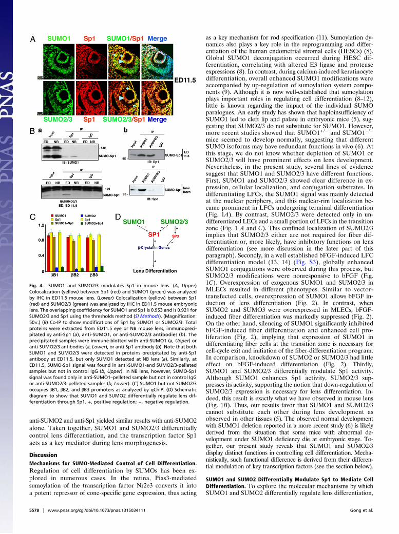
anti-SUMO2 and anti-Sp1 yielded similar results with anti-SUMO2alone. Taken together, SUMO1 and SUMO2/3 differentiallycontrol lens differentiation, and the transcription factor Sp1acts as a key mediator during lens morphogenesis.
DiscussionMechanisms for SUMO-Mediated Control of Cell Differentiation.Regulation of cell differentiation by SUMOs has been ex-plored in numerous cases. In the retina, Pias3-mediatedsumoylation of the transcription factor Nr2e3 converts it intoa potent repressor of cone-specific gene expression, thus acting
as a key mechanism for rod specification (11). Sumoylation dy-namics also plays a key role in the reprogramming and differ-entiation of the human endometrial stromal cells (HESCs) (8).Global SUMO1 deconjugation occurred during HESC dif-ferentiation, correlating with altered E3 ligase and proteaseexpressions (8). In contrast, during calcium-induced keratinocytedifferentiation, overall enhanced SUMO1 modifications wereaccompanied by up-regulation of sumoylation system compo-nents (9). Although it is now well-established that sumoylationplays important roles in regulating cell differentiation (8–12),little is known regarding the impact of the individual SUMOparalogues. An early study has shown that haploinsufficiency ofSUMO1 led to cleft lip and palate in embryonic mice (5), sug-gesting that SUMO2/3 do not substitute for SUMO1. However,more recent studies showed that SUMO1+/− and SUMO1−/−
mice seemed to develop normally, suggesting that differentSUMO isoforms may have redundant functions in vivo (6). Atthis stage, we do not know whether depletion of SUMO1 orSUMO2/3 will have prominent effects on lens development.Nevertheless, in the present study, several lines of evidencesuggest that SUMO1 and SUMO2/3 have different functions.First, SUMO1 and SUMO2/3 showed clear difference in ex-pression, cellular localization, and conjugation substrates. Indifferentiating LFCs, the SUMO1 signal was mainly detectedat the nuclear periphery, and this nuclear-rim localization be-came prominent in LFCs undergoing terminal differentiation(Fig. 1A). By contrast, SUMO2/3 were detected only in un-differentiated LECs and a small portion of LFCs in the transitionzone (Fig. 1 A and C). This confined localization of SUMO2/3implies that SUMO2/3 either are not required for fiber dif-ferentiation or, more likely, have inhibitory functions on lensdifferentiation (see more discussion in the later part of thisparagraph). Secondly, in a well established bFGF-induced LFCdifferentiation model (13, 14) (Fig. S3), globally enhancedSUMO1 conjugations were observed during this process, butSUMO2/3 modifications were nonresponsive to bFGF (Fig.1C). Overexpression of exogenous SUMO1 and SUMO2/3 inMLECs resulted in different phenotypes. Similar to vector-transfected cells, overexpression of SUMO1 allows bFGF in-duction of lens differentiation (Fig. 2). In contrast, whenSUMO2 and SUMO3 were overexpressed in MLECs, bFGF-induced fiber differentiation was markedly suppressed (Fig. 2).On the other hand, silencing of SUMO1 significantly inhibitedbFGF-induced fiber differentiation and enhanced cell pro-liferation (Fig. 2), implying that expression of SUMO1 indifferentiating fiber cells at the transition zone is necessary forcell-cycle exit and initiation of the fiber-differentiation program.In comparison, knockdown of SUMO2 or SUMO2/3 had littleeffect on bFGF-induced differentiation (Fig. 2). Thirdly,SUMO1 and SUMO2/3 differentially modulate Sp1 activity.Although SUMO1 enhances Sp1 activity, SUMO2/3 sup-presses its activity, supporting the notion that down-regulation ofSUMO2/3 expression is necessary for lens differentiation. In-deed, this result is exactly what we have observed in mouse lens(Fig. 1B). Thus, our results favor that SUMO1 and SUMO2/3cannot substitute each other during lens development asobserved in other tissues (5). The observed normal developmentwith SUMO1 deletion reported in a more recent study (6) is likelyderived from the situation that some mice with abnormal de-velopment under SUMO1 deficiency die at embryonic stage. To-gether, our present study reveals that SUMO1 and SUMO2/3display distinct functions in controlling cell differentiation. Mecha-nistically, such functional difference is derived from their differen-tial modulation of key transcription factors (see the section below).
SUMO1 and SUMO2 Differentially Modulate Sp1 to Mediate CellDifferentiation. To explore the molecular mechanisms by whichSUMO1 and SUMO2 differentially regulate lens differentiation,
Fig. 4. SUMO1 and SUMO2/3 modulates Sp1 in mouse lens. (A, Upper)Colocalization (yellow) between Sp1 (red) and SUMO1 (green) was analyzedby IHC in ED11.5 mouse lens. (Lower) Colocalization (yellow) between Sp1(red) and SUMO2/3 (green) was analyzed by IHC in ED11.5 mouse embryoniclens. The overlapping coefficiency for SUMO1 and Sp1 is 0.953 and is 0.921 forSUMO2/3 and Sp1 using the thresholds method (SI Methods). (Magnification:50×.) (B) Co-IP to show modifications of Sp1 by SUMO1 or SUMO2/3. Totalproteins were extracted from ED11.5 eye or NB mouse lens, immunopreci-pitated by anti-Sp1 (a), anti-SUMO1, or anti-SUMO2/3 antibodies (b). Theprecipitated samples were immune-blotted with anti-SUMO1 (a, Upper) oranti-SUMO2/3 antibodies (a, Lower), or anti-Sp1 antibody (b). Note that bothSUMO1 and SUMO2/3 were detected in proteins precipitated by anti-Sp1antibody at ED11.5, but only SUMO1 detected at NB lens (a). Similarly, atED11.5, SUMO-Sp1 signal was found in anti-SUMO1-and SUMO2/3-pelletedsamples but not in control IgG (b, Upper). In NB lens, however, SUMO-Sp1signal was found only in anti-SUMO1–pelleted sample but not in control IgGor anti-SUMO2/3–pelleted samples (b, Lower). (C) SUMO1 but not SUMO2/3occupies βB1, βB2, and βB3 promoters as analyzed by qChIP. (D) Schematicdiagram to show that SUMO1 and SUMO2 differentially regulate lens dif-ferentiation through Sp1. +, positive regulation; −, negative regulation.
5578 | www.pnas.org/cgi/doi/10.1073/pnas.1315034111 Gong et al.

we explored their conjugated targets. We found here that themajor lens differentiation marker genes coding for β-crystallinswere expressed upon bFGF-induced differentiation in vitro or invivo (Fig. 2). Moreover, we demonstrated that all of the β-crys-tallin gene promoters have well-conserved Sp1/Sp3 binding sites(Fig. S5 A–C). Sp1 is a universal transcription factor that hasbeen shown to control expression of many genes from viral tocellular (15, 20). In the present study, the results of EMSAexperiments demonstrated that Sp1 strongly binds to β-crystallingene promoters (Fig. S5 D–F) and positively regulates their ex-pression (Fig. S5G). Moreover, coexpression of the exogenousSUMO1 and SUMO2 with Sp1 differentially modulates thesegenes. Although SUMO1 enhanced β-crystallin gene expression,SUMO2 significantly inhibited these promoters (Fig. S5G).Furthermore, the Sp1 knockdown cells can only be rescued bya K683R (a SUMO2-favored sumoylation site) mutant but not byK16R mutant (Fig. S7).At the molecular level, Sp1 conjugation by SUMO1 and
SUMO2 has different outcomes. We found that Sp1 has a novelsumoylation site at K683 in the DNA binding domain, whichshows preferred conjugation by SUMO2 (Fig. S6 A, b). SUMO2conjugation at this site attenuates Sp1 DNA binding activity (Fig.S6B). Although both SUMO1 and SUMO2 can be conjugated toSp1 at K16, conjugation of SUMO2 to this site significantlydecreases Sp1 stability (Fig. 3A). In contrast, SUMO1 conjuga-tion to Sp1 has no direct effect on protein stability (Fig. 3A). Thisresult differs from a previous study with cancer cells whereSUMO1 modification led to Sp1 degradation (16). Such an in-consistence may be due to cell-specific effects. Finally, SUMO1and SUMO2 differentially direct Sp1 interactions with repressorsand coactivators. Previous studies showed that the interactionwith p300 enhanced Sp1 DNA binding activity (18). We foundthat coexpression of Sp1 with SUMO1 prevents its interactionswith the sibling repressor, Sp3, but enhanced its interaction withthe coactivator, p300 (Fig. 3C and Fig. S6D). In contrast, coex-pression of Sp1 with SUMO2 led to the opposite effects:SUMO2 recruits Sp3 to the β-crystallin gene promoters (Fig. 3B)but prevents the association between Sp1 with p300 (Fig. 3C andFig. S6D).Because the outcomes of Sp1 conjugation by SUMO1 and
SUMO2 are so different, progression of lens differentiationwould require one of two conditions: either the lens must havea fine mechanism to ensure specific Sp1 sumoylation by SUMO1vs. SUMO2/3 at specific developmental stages if they areexpressed at similar levels, or, more simply, they display differ-ential expression patterns such that SUMO1 is present when it isneeded and SUMO2 disappears when it is not required. Our
results demonstrate that the lens apparently adopts the latterstrategy (Fig. 1 and Fig. S1). Although SUMO1 is stronglyexpressed and conjugated during the embryonic stage and ismaintained at a certain level in the postnatal stages (Fig. 1 andFig. S1), expression of SUMO2/3 is gradually decreased and ispresent only in the epithelial cells of the adult lens (Fig. 1C andFig. S1). In this way, the presence of SUMO1 allows positiveregulation of lens differentiation (Fig. 2 A and B). In contrast,the absence of SUMO2/3 in lens-fiber cells would pose no in-hibition on lens differentiation. Moreover, the confinement ofSUMO2/3 in LECs also ensured that LECs remain in the epi-thelial status. These observations would be consistent withresults from both in vitro and in vivo studies. In the in vitrobFGF induction of lens differentiation, although SUMO1-con-jugated proteins are gradually increased from day 0 to day 15(Fig. 1B), the SUMO2/3-conjugated substrates are graduallydecreasing during the same period. During normal lens de-velopment, Sp1 conjugation by SUMO1 was detected by ho-mogenous localization in lens vesicle (ED11.5) (Fig. 4A) and byCo-IP in both ED11.5 and NB lenses (Fig. 4B). In contrast, Sp1conjugation by SUMO2/3 was detected only by colocalization inthe anterior epithelial cells of the lens vesicle (ED11.5) and wasfurther confirmed by Co-IP (Fig. 4 A and B). At the NB lens, Co-IPconfirmed that Sp1 was not modified by SUMO2/3 (Fig. 4B). Thus,our study here provides an important conclusion: down-regulationof SUMO2/3 as lens differentiation proceeds is necessary fornormal lens to complete differentiation.In summary, our present study has clearly demonstrated that
SUMO1 and SUMO2/3 have distinct functions in regulatinglens-cell differentiation, which is derived from their differentialeffects on Sp1 and possibly other transcription factors (Fig. 4D).
MethodsMice used in this study were handled in compliance with the Guide for theCare and Use of Laboratory Animals. Embryonic and adult mice wereobtained from the University of Nebraska Medical Center breeding facility.
Other analytical methods used in this study are detailed in SI Methods.Oligo primers used in the generation of various plasmids, qRT-PCR, andqChIP are listed in Tables S1 and S2.
ACKNOWLEDGMENTS. This work was supported in part by NationalInstitutes of Health Grant 1R01EY018380, National Natural Science Founda-tion of China Grant 81272228, the Lotus Scholar Program (D.W.-C.L.),a collaborative grant from Zhongshan Ophthalmic Center (Grant 8282012)(to X.-C.T., Y.-Z.L., and D.W.-C.L.), a University of Nebraska Medical CenterGraduate Fellowship (to L.G.), and the Chinese Scholarship Council (W.-K.J.,X.-H.H., and W.-F.H.).
1. Geiss-Friedlander R, Melchior F (2007) Concepts in sumoylation: A decade on. Nat RevMol Cell Biol 8(12):947–956.
2. Lapenta V, et al. (1997) SMT3A, a human homologue of the S. cerevisiae SMT3 gene,maps to chromosome 21qter and defines a novel gene family. Genomics 40(2):362–366.
3. Kamitani T, Kito K, Nguyen HP, Fukuda-Kamitani T, Yeh ET (1998) Characterization ofa second member of the sentrin family of ubiquitin-like proteins. J Biol Chem 273(18):11349–11353.
4. Vertegaal AC, et al. (2006) Distinct and overlapping sets of SUMO-1 and SUMO-2target proteins revealed by quantitative proteomics. Mol Cell Proteomics 5(12):2298–2310.
5. Alkuraya FS, et al. (2006) SUMO1 haploinsufficiency leads to cleft lip and palate.Science 313(5794):1751.
6. Zhang FP, et al. (2008) Sumo-1 function is dispensable in normal mouse development.Mol Cell Biol 28(17):5381–5390.
7. Kim KI, Baek SH, Chung CH (2002) Versatile protein tag, SUMO: Its enzymology andbiological function. J Cell Physiol 191(3):257–268.
8. Jones MC, et al. (2006) Regulation of the SUMO pathway sensitizes differentiatinghuman endometrial stromal cells to progesterone. Proc Natl Acad Sci USA 103(44):16272–16277.
9. Deyrieux AF, Rosas-Acosta G, Ozbun MA, Wilson VG (2007) Sumoylation dynamicsduring keratinocyte differentiation. J Cell Sci 120(Pt 1):125–136.
10. La Salle S, Sun F, Zhang XD, Matunis MJ, Handel MA (2008) Developmental control ofsumoylation pathway proteins in mouse male germ cells. Dev Biol 321(1):227–237.
11. Onishi A, et al. (2009) Pias3-dependent SUMOylation directs rod photoreceptor
development. Neuron 61(2):234–246.12. Yan Q, et al. (2010) Sumoylation activates the transcriptional activity of Pax-6, an
important transcription factor for eye and brain development. Proc Natl Acad Sci
USA 107(49):21034–21039.13. Chamberlain CG, McAvoy JW (1989) Induction of lens fibre differentiation by acidic
and basic fibroblast growth factor (FGF). Growth Factors 1(2):125–134.14. McAvoy JW, Chamberlain CG (1989) Fibroblast growth factor (FGF) induces different
responses in lens epithelial cells depending on its concentration. Development 107(2):
221–228.15. Suske G (1999) The Sp-family of transcription factors. Gene 238(2):291–300.16. Wang YT, et al. (2008) Sumoylation of specificity protein 1 augments its degradation
by changing the localization and increasing the specificity protein 1 proteolytic
process. J Mol Biol 380(5):869–885.17. Xu X, Vatsyayan J, Gao C, Bakkenist CJ, Hu J (2010) Sumoylation of eIF4E activates
mRNA translation. EMBO Rep 11(4):299–304.18. Billon N, et al. (1999) Cooperation of Sp1 and p300 in the induction of the CDK in-
hibitor p21WAF1/CIP1 during NGF-mediated neuronal differentiation. Oncogene
18(18):2872–2882.19. Dennig J, Beato M, Suske G (1996) An inhibitor domain in Sp3 regulates its glutamine-
rich activation domains. EMBO J 15(20):5659–5667.20. Briggs MR, Kadonaga JT, Bell SP, Tjian R (1986) Purification and biochemical charac-
terization of the promoter-specific transcription factor, Sp1. Science 234(4772):47–52.
Gong et al. PNAS | April 15, 2014 | vol. 111 | no. 15 | 5579
CELL
BIOLO
GY