sulfate assimilation mediates tellurite reduction and ...ec.asm.org/content/9/10/1635.full.pdf ·...
TRANSCRIPT

EUKARYOTIC CELL, Oct. 2010, p. 1635–1647 Vol. 9, No. 101535-9778/10/$12.00 doi:10.1128/EC.00078-10Copyright © 2010, American Society for Microbiology. All Rights Reserved.
Sulfate Assimilation Mediates Tellurite Reduction and Toxicity inSaccharomyces cerevisiae�†
Lars-Goran Ottosson,1 Katarina Logg,1,2 Sebastian Ibstedt,1 Per Sunnerhagen,1 Mikael Kall,2Anders Blomberg,1 and Jonas Warringer1,3*
Department of Cell and Molecular Biology, University of Gothenburg, Gothenburg, Sweden1; Department of Applied Physics,Chalmers University of Technology, 41296 Gothenburg, Sweden2; and Centre for Integrative Genetics,
Norwegian University of Life Sciences, Ås, Norway3
Received 29 March 2010/Accepted 22 July 2010
Despite a century of research and increasing environmental and human health concerns, the mechanisticbasis of the toxicity of derivatives of the metalloid tellurium, Te, in particular the oxyanion tellurite, Te(IV),remains unsolved. Here, we provide an unbiased view of the mechanisms of tellurium metabolism in the yeastSaccharomyces cerevisiae by measuring deviations in Te-related traits of a complete collection of gene knockoutmutants. Reduction of Te(IV) and intracellular accumulation as metallic tellurium strongly correlated withloss of cellular fitness, suggesting that Te(IV) reduction and toxicity are causally linked. The sulfate assimi-lation pathway upstream of Met17, in particular, the sulfite reductase and its cofactor siroheme, was shown tobe central to tellurite toxicity and its reduction to elemental tellurium. Gene knockout mutants with alteredTe(IV) tolerance also showed a similar deviation in tolerance to both selenite and, interestingly, selenomethi-onine, suggesting that the toxicity of these agents stems from a common mechanism. We also show that Te(IV)reduction and toxicity in yeast is partially mediated via a mitochondrial respiratory mechanism that does notencompass the generation of substantial oxidative stress. The results reported here represent a robust basefrom which to attack the mechanistic details of Te(IV) toxicity and reduction in a eukaryotic organism.
The metalloid tellurium (Te) is one of the rarest elements inthe Earth’s crust, and its derivatives, particularly the oxyaniontellurite, TeO3
2� or Te(IV), are also highly toxic to livingorganisms. Despite more than 100 years of physiological andmolecular research, the mechanism by which tellurium speciesexert their toxicity still constitutes a scientific conundrum (7).This toxicity is of concern because the expanding use of tellu-rium in electronic appliances, optics, and batteries, togetherwith its natural occurrence in sulfide-rich ores, gives rise tohigh local concentrations in connection with waste dumps andmetallurgical plants, with detrimental effects on the environ-ment and human health. In fact, although tellurium is currentlynot known to be an essential element, it is nevertheless one ofthe most abundant trace elements in human bone, supersededonly by iron, zinc, and rubidium (45). An intriguing twist is thatTe(IV) is reduced by living cells to volatile methylated formssuch as dimethyltellurium or to elemental tellurium, Te(0) (21,24, 52). In bacteria, the latter is typically deposited as blackaggregates with strain-specific nanostructures resulting in adistinct darkening of cells and tissues (30, 55), a phenomenonthat is exploitable in environmental viability screens (31).
It has long been known that different bacterial species varyin their tolerance to Te(IV), which has prompted the inclusionof TeO3
2� in a variety of selective bacteriological tests (48).However, the mechanistic basis of this variance remains elu-
sive. In general, Te(IV) resistance in bacteria has been thoughtto be associated with accumulation of tellurium (53), althoughTe(IV) resistance without tellurium accumulation has beenobserved in phototrophic bacteria (72). For some bacterialspecies, the capacity to grow at high Te(IV) concentrations hasbeen shown to depend on the presence of Te(IV) resistance-encoding genetic determinants carried on IncHI, IncHII, andIncP plasmids (65). In addition, chromosomal genes importantfor growth in the presence of Te(IV) have been identified in afew species, but the physiological basis of their involvement hasnot been clearly determined (28, 54, 58). A favorite culprit ofmetal toxicity is oxidative damage of proteins, DNA, and lipids.It has been proposed that this toxicity results from an ability ofTe(IV) to act as a strong oxidizing agent (33), as Te(IV)directly interacts with and oxidizes cellular thiol groups (63,64). Te(IV) has also been implicated in redox reactions involv-ing the respiratory chain (59). Recent reports favor an oxida-tive mode of action; Te(IV) increases the production of reac-tive oxygen species (ROS) in vitro as well as in vivo, increasesprotein carbonylation, a common sign of oxidative damage,and inactivates oxidative stress-sensitive Fe-S enzymes (41).Furthermore, the transcription, as well as the activity, of thesuperoxide dismutases, critical components of the oxidativestress defense, is elevated in Te(IV)-stressed bacteria (4, 41).Nevertheless, the absence of genome-wide data on telluriumexposure leaves open the question of whether oxidative dam-age constitutes the dominant source of Te(IV) toxicity. Alter-native toxicity mechanisms for Te(IV) and the chemophysicallyrelated selenium derivative Se(IV) have been suggested, suchas the inactivation of proteins by the replacement of sulfhydrylgroups or the direct quenching of respiratory chain compo-nents (30, 59, 61, 62).
* Corresponding author. Mailing address: Department of Cell andMolecular Biology, University of Gothenburg, Medicinaregatan 9c,Gothenburg 41390, Sweden. Phone and fax: 46-317732587. E-mail:[email protected].
† Supplemental material for this article may be found at http://ec.asm.org/.
� Published ahead of print on 30 July 2010.
1635
on May 28, 2018 by guest
http://ec.asm.org/
Dow
nloaded from
To provide an unbiased and system-wide view of the mech-anisms of Te(IV) toxicity and reduction in eukaryotes, we herequantified the tolerance of the Saccharomyces cerevisiae geneknockout collection to Te(IV), as well as measured Te(0) ac-cumulation. Our results demonstrated that Te(IV) toxicity andreduction to tellurium are inversely correlated; low Te(0) ac-cumulation coincided with high Te(IV) tolerance and viceversa, suggesting a causal relationship between accumulationand toxicity. In addition, we show that two cellular routesmediate the bulk of Te(IV) reduction and toxicity in yeast, thesulfate assimilation pathway and a mitochondrial mechanismthat encompasses respiration but not the generation of oxida-tive damage. We also found that Te(IV) toxicity and reductionstrongly overlapped with Se(IV) toxicity and reduction andwith selenomethionine toxicity, suggesting a common mode ofaction.
MATERIALS AND METHODS
Strains and media. Throughout this study, the diploid S. cerevisiae typestrain S288c and diploid (BY4743 [MATa/MAT� his3�1/his3�1 leu2�0/leu2�0met17�0/MET17 LYS2/lys2�0 ura3�0/ura3�0]) or haploid (BY4741 [MATahis3�1 leu2�0 met17�0 ura3�0] and BY4742 [MAT� his3�1 leu2�0 lys2�0ura3�0]) derivatives thereof were used (5). A genome-wide deletion strainscreen was carried out using the haploid BY4741 deletion collection (as de-scribed above, ORF::kanMX4) and strains with significant deviations were con-firmed by rescreening using the diploid homozygote BY4743 collection (as de-scribed above, ORF::kanMX4/ORF::kanMX4). For Schizosaccharomyces pombe,the standard wild-type (WT) strain 972h� was used. S. cerevisiae strains werecultivated in synthetic complete medium (0.14% yeast nitrogen base, 0.5% am-monium sulfur, 1% succinic acid, 2% [wt/vol] glucose, and 0.077% completesupplement mixture supplemented with 20 mg/liter cysteine, pH 5.8) with andwithout K2TeO3, Na2SeO3, and selenomethionine (all chemicals were fromSigma Aldrich). For the study of respiratory growth, glucose was replaced with amixture of 2% (wt/vol) ethanol and 3% (wt/vol) glycerol. Where indicated,methionine-free medium was also used. S. pombe strains were cultivated in YESmedium (0.5% yeast extract, 3% [wt/vol] glucose, 225 mg/liter histidine, leucine,lysine, arginine, adenine, and uracil) where indicated. Glutathione suppressionexperiments were performed with synthetic complete medium supplementedwith 5 mM reduced glutathione (GSH). Agar at 20 g/liter was added to solidmedium for growth experiments. All strains were stored in 20% (wt/vol) glycerolsolution at �80°C and cultivated at 30°C.
Liquid medium microcultivation. Liquid medium growth curves of yeaststrains growing for 72 h in the presence and or absence of the indicatedconcentrations of K2TeO3 were obtained using a high-resolution microculti-vation approach as previously described (66, 67). Strains were tested induplicate (n � 2).
Optical microscopy of Te(IV)-stressed cells. Te(0) plaque formation in indi-vidual exponentially growing cells was monitored by time-lapse microscopy. S.cerevisiae and S. pombe cells were precultivated overnight in liquid medium andresuspended to an optical density at 610 nm (OD610) of �0.1. When an OD610
of �0.2 was reached, 0.5 mM K2TeO3 was added and cells were placed in anenclosed 160-�l chamber, heated to 30°C, and mounted in an inverted micro-scope (Nikon TE2000 PFS). Except where otherwise stated, bright-field imageswere acquired every 5 min for 20 h using a 60�/numerical aperture (NA) 1.4 ora 100�/NA 1.45 oil immersion objective (Nikon) and a charge-coupled devicecamera (iXon; Andor). Te(0) plaque formation in S. cerevisiae cells grown onagar plates containing 0.5 or 2 mM K2TeO3 at 30°C was followed by imaging ofthe cells every 24 h for 5 days. Prior to imaging, cells were resuspended in liquidmedium, placed between two cover glasses, and imaged in bright-field mode (asdescribed above). To follow Te(0) plaque localization with regard to the vacuole,time-lapse measurements were performed with S. cerevisiae and S. pombe cellsfluorescently stained with the vacuolar membrane styryl dye FM 4-64 (T-3166;Invitrogen). Cells were precultivated in medium (as described above), resus-pended to an OD610 of �0.3 in 0.5 mM K2TeO3 medium, and cultivated for 4 h(S. pombe) or �18 h (S. cerevisiae). Cultivated cells were concentrated �10� bycentrifugation (10,000 rpm, 2 min) and resuspension in 100 to 200 �l medium,and 1 to 4 �l of dye was added. Following 20 to 30 min of preincubation at 30°Cto facilitate uptake of the dye, cells were pelleted (10,000 rpm, 2 min), resus-
pended in 1 ml of 0.5 mM K2TeO3 medium, cultivated for �2 h at 30°C, andconcentrated �5� (as described above) for imaging. A 160-�l volume of culturewas placed between cover glasses precoated with immobilizing concanavalin Aand heated to 30°C. Cells were imaged every 30 s (as described above). Fluo-rescence images were acquired using excitation filter D540/25x, dichroic mirror565DCLP, and emission filter D605/55m (Chroma Corporation).
Raman measurements. Raman spectra were collected using a Raman spec-trometer (Renishaw 2000) in combination with an inverted microscope(TE2000E; Nikon). The microscope was equipped with a 60�/NA 0.7 objective(Nikon) and an argon laser (Spectra Physics 2060) tuned to 514.5 nm for exci-tation. The integration time was 30 s with a power of �3.5 mW at the sample. Anangle-tuned holographic notch filter was used to block the Rayleigh scatteredlight. Raman spectra were collected from WT S. cerevisiae cells cultivated with-out K2TeO3 or with 2 mM K2TeO3, oxidized tellurium TeO2, or elementaltellurium Te(0). Cells were grown on synthetic complete medium plates (20g/liter agar) for 11 days and resuspended in phosphate-buffered saline, and 5 �lwas withdrawn and put between two cover glasses, of which one was precoatedwith concanavalin A to immobilize the cells. Te(0)and TeO2 spectra were re-corded from the interior and the oxidized surface of freshly cleaved Te(0) grains(Sigma Aldrich), respectively.
Segregation analysis of tellurium traits in the cross BY4741 � BY4742. Tofollow the potential cosegregation of tellurium traits and the Met17�0 and lys2�0markers in a cross between BY4741 and BY4742, strains were cospotted onto afresh YPD plate and allowed to mate for 1 day at 30°C. Following suspension inwater, cells were streaked onto sporulation plates (1% potassium acetate), sporu-lated for 7 days at 22°C, and suspended in Lyticase solution (5,000 U/ml; SigmaAldrich) for 30 min at 37°C. Lysed asci were streaked onto YPD plates, and 10tetrads were dissected using a micromanipulator (Singer Instruments, UnitedKingdom). Following incubation for 2 days at 30°C, tetrads were replica platedonto synthetic complete medium plates lacking methionine or lysine but con-taining 0.1 mM K2TeO3. Plates were incubated for 1 day at 30°C and imaged (asdescribed above), and the potential cosegregation of Met17�0 and lys2�0 (datanot shown) with tellurium traits was evaluated visually.
A genome-wide screen for Te(IV) toxicity and tellurium accumulation. Agenome-wide deletion strain screen for Te(IV) toxicity and tellurium accumula-tion was carried out using the haploid BY4741 deletion collection (as describedabove, ORF::kanMX4). Strains were pinned in 1536 format on media containing0, 0.1, 0.2, 0.3, 0.4, and 0.5 mM K2TeO3 using a benchtop RoToR HDA robot(Singer Instruments, United Kingdom) with default settings. After 1, 2, 3, and 6days, colonies were imaged using a 10-megapixel digital camera (PowershotA640; Canon) and stored as JPEG images. Images were evaluated by scoring ofcolony size and coloration using a manual evaluation system with 10 grades,colony size reflecting Te(IV) tolerance and colony darkening reflecting telluriumaccumulation (n � 8, replicate colonies evaluated in aggregate). Absolute valuesof colony size and color were normalized to the strain average for that concen-tration and day, resulting in relative measurements of tellurium accumulationand Te(IV) tolerance. Compensating for general growth defects, colony sizein the presence of Te(IV) was further normalized to colony size in theabsence of Te(IV). Gene knockout mutants showing very slow growth in theabsence of stress were excluded from evaluation in order to avoid scoringartifacts; a total of 4,311 gene knockout mutants were retained. Gene knock-out mutants with similar Te(IV)-specific aberrations in colony size or color inat least three out of six independent evaluations in both screens were re-screened using the diploid homozygote BY4743 deletion collection (as de-scribed above, ORF::kanMX4/ORF::kanMX4). Rescreening was performedin 384 pinning format using 0, 0.1, and 0.4 mM K2TeO3; colony size and colorwere evaluated (as described above) after 1, 2, 3, and 6 days of growth (n �4). BY4743 gene knockout mutants with confirmed K2TeO3 phenotypes werefurther tested for Na2SeO3 and selenomethionine phenotypes by pinningonto agar plates containing 0, 0.2, and 0.8 mM Na2SeO3 or 0, 0.03, and 0.06mM selenomethionine.
RESULTS
Tellurium accumulation in S. cerevisiae and S. pombe. Cul-tivation of commonly used laboratory strains of the modelyeasts S. cerevisiae and S. pombe in the presence of TeO3
2�
resulted in the distinct cell darkening that is a hallmark oftellurium accumulation in bacteria. For batch-cultivated S.pombe cells, this Te(IV)-induced cell darkening was reflectedin an OD/turbidity increase in stationary phase of about 8-fold
1636 OTTOSSON ET AL. EUKARYOT. CELL
on May 28, 2018 by guest
http://ec.asm.org/
Dow
nloaded from

compared to that of cells cultivated in the absence of Te(IV)(Fig. 1A). Only marginal increases were observed during ex-ponential fermentative growth, suggesting that Te(0) accumu-lation is coupled primarily to respiration. In the presence ofhigh concentrations of Te(IV), yeast also failed to utilize therespiratory carbon sources ethanol and glycerol, confirming atight link between Te(IV) toxicity and respiration (Fig. 1B). Totime the appearance of intracellular darkening followingTe(IV) exposure, individual Te(IV)-stressed cells were moni-tored by time-lapse microscopy. In the average cell, darkeningoccurred as localized plaque formation with a very distinctonset at 8 to 8.5 h of Te(IV) exposure in S. pombe and 14 to18 h of Te(IV) exposure in S. cerevisiae; however, followinglong exposure times, plaque formation spread throughout thecell until it was filled completely (Fig. 1C and D). By stainingTe(IV)-exposed S. cerevisiae and S. pombe cells with a vacuolarmembrane dye, we found that the intracellular plaques wereabsent from the vacuolar lumen but were formed in closeproximity to the vacuolar membrane (Fig. 1E; see videos S1 toS3 in the supplemental material). After prolonged Te(IV) ex-posure, the integrity of the vacuole was compromised and thevacuole subsequently fractionated and disintegrated. This wasfollowed by rapid cell shrinkage to about 75% of the initial size(Fig. 1F). In Te(IV)-exposed bacteria, cellular darkening cor-responds to the accumulation of elemental tellurium aggre-gates with little complex binding to any organic matter (60). Toconfirm that cellular darkening is due to elemental telluriumaccumulation also in yeast, we performed Raman spectroscopyof Te(IV)-stressed cells. Raman spectroscopy is a powerfulmethod to obtain vibration “fingerprint” spectra of moleculesand solids via inelastic scattering of laser light. No distinctRaman signatures were observed in the sample withoutTe(IV), but a distinct Raman active vibration peak was ob-served at around 270 cm�1 in Te(IV)-stressed cells. The peakposition is in excellent agreement with what is expected forelemental tellurium (peak position, 268 cm�1, Fig. 1G). Itshould be noted that elemental tellurium can be oxidized toTeO2 in the presence of oxygen and that TeO2 thus is analternative reaction product following cellular metabolizationof Te(IV). However, the 390- to 395-cm�1 band characterizingTeO2 is absent from Raman spectra measured inside Te(IV)-stressed cells. We therefore conclude that Te(IV)-induced cel-lular darkening in yeast indeed results from intracellular accu-mulation of elemental tellurium.
Investigating Te(IV) toxicity and tellurium accumulation intwo essentially isogenic derivatives of the reference strain,BY4741 (met17�0) and BY4742 (lys2�0), we noted very strongdifferences (Fig. 1H). These strains differ only in their matingtypes and auxotrophic markers, providing a clear indication asto the genetic basis of their trait differences. In fact, controlledmating of the two strains, followed by tetrad dissection andphenotyping and genotyping of the F1 haploid progeny,showed a complete cosegregation of the tellurium accumula-tion trait and the met17�0 marker; absence of Met17 gave riseto high tellurium accumulation and correspondingly low tol-erance to Te(IV) (Fig. 1I). MET17 encodes a cysteine andmethionine synthase at the root of the divergence of themethionine and cysteine biosynthesis pathways, implicatingsulfur assimilation as one determinant of tellurium accumu-lation in yeast.
Sulfate assimilation mediates tellurium accumulation inyeast. To provide a genome-wide view of the genetic determi-nants that control the intracellular fate of Te(IV), we screeneda collection of nonessential haploid S. cerevisiae gene knockoutmutants in the BY4741 background for aberrations in Te(IV)tolerance and Te(0) accumulation. Changes in Te(IV) toler-ance were quantified as Te(IV)-specific deviations in colonysize on agar, whereas Te(0) accumulation variations werescored as deviations in colony darkening. Evaluating 4,310gene knockout mutants, we found 3.1% to have deviations inTe(0) accumulation and 6.3% to feature variations in Te(IV)tolerance. We observed very strong links between toleranceand accumulation variations; gene knockout mutants with hightellurium accumulation also tended to show low tolerance toTe(IV) (7.3-fold enrichment [P � 5 � 10�9; Fisher’s exacttest]) whereas gene knockout mutants with reduced telluriumaccumulation tended to have high tolerance to Te(IV) (9.4-fold enrichment [P � 2 � 10�11; Fisher’s exact test]). Thissuggests that reduction of Te(IV) to bioaccumulated telluriumis an important cause of Te(IV) toxicity, in complete contrastto observations from bacterial studies indicating that accumu-lation of tellurium is frequently associated with Te(IV) resis-tance (53). To identify cellular functions linked to variations intellurium accumulation and Te(IV) tolerance, gene ontologyfunctions overrepresented among gene knockout mutants withvariations in tellurium phenotypes were identified. Essentiallyall of the biological processes that were enriched amongTe(IV)-tolerant gene knockout mutants were also enrichedamong gene knockout mutants with low tellurium accumula-tion, confirming the inverse functional link between toleranceand accumulation (Fig. 2A). To exclude the possibility of re-cessive secondary site mutations penetrating in the haploidbackground and also control for the influence of the met17�0marker, gene knockout mutants with tellurium phenotypeswere rescreened using homozygous diploid knockout mutants(BY4743 [met17�0/MET17 lys2�0/LYS2]). In the diploidknockout mutants, 75% of the knockout phenotypes could bestringently verified while the remaining 25% were discarded.
The most striking functional characteristic among geneknockout mutants with low tellurium accumulation was thestrong overrepresentation of genes involved in sulfate assimi-lation (Fig. 2A). Knockout mutants lacking all of the genes inthe sulfur assimilation pathway upstream of MET17, i.e.,MET3, MET10, MET14, MET16, MET22, and ECM17, consis-tently showed low tellurium accumulation and correspondinglyhigh Te(IV) tolerance (Fig. 2A and 3A). These effects wereobserved in both the haploid BY4741 and the diploid BY4743and were thus independent of met17�0. A knockout mutantlacking the key transcriptional regulator of sulfur assimilationgenes, MET28, featured phenotypes similar to those of theabove-mentioned MET genes, as did met12� and met13� in theintersecting branch from folate biosynthesis (Fig. 3A). Knock-out mutants lacking SAM1, SAM2, and MET6 downstream ofMet17 showed normal or strongly increased tellurium accumu-lation, depending on the absence or presence of MET17, andsam1� and sam2� also caused low Te(IV) tolerance (Fig. 2B).Interestingly, met1� and met8� knockout mutants lacking en-zymes that act downstream of Sam1, Sam2, and Met6 showedvery low tellurium accumulation and high Te(IV) tolerance(Fig. 2B and 3A). This is very likely explained by the fact that
VOL. 9, 2010 TELLURIUM IN YEAST 1637
on May 28, 2018 by guest
http://ec.asm.org/
Dow
nloaded from

FIG. 1. Tellurium is accumulated in yeast following Te(IV) exposure. (A) Growth curves were recorded for S. cerevisiae and S. pombe cellsmicrocultivated with and without 0.1 mM K2TeO3 for 70 h by automated measurements of medium OD/turbidity. (B) S. cerevisiae cells (BY4741)were spotted onto fermentative (glucose [Glu]) and respiratory media with and without 0.5 mM K2TeO3 and cultivated for 6 days. EtOH, ethanol;Gly, glycerol. (C) Cellular darkening and plaque formation in a single S. pombe cell (red arrow) through 20 h of 0.5 mM K2TeO3 stress. (D) Cellulardarkening and plaque formation in S. pombe (3 days, liquid cultivation) and S. cerevisiae (11 days, solid medium cultivation) after exposure to 0.5mM K2TeO3. (E) Tellurium plaque formation with respect to the vacuole was followed by time-lapse microscopy of S. cerevisiae S288c cells stainedwith a fluorescent vacuolar membrane dye. The time sequence shows a typical cell. Time zero is �19 h after the addition of 0.5 mM K2TeO3. Seealso videos S1 to S3 in the supplemental material. (F) The relative cell size decreases to about 75% of the initial cell size in Te(IV)-exposed cellsfollowing collapse of the vacuole. The relative size of a typical S. cerevisiae cell is shown. Time zero is �21 h after the addition of 0.5 mM K2TeO3.(G) Raman spectra of S. cerevisiae cells grown for 11 days in the absence or presence of 2 mM K2TeO3, as well as Raman spectra of oxidized (TeO2)and nonoxidized elemental tellurium, Te. (H) Tellurium accumulation and Te(IV) tolerance of WT S. cerevisiae strains BY4741 (MATa met17�0)and BY4742 (MAT� lys2�0), which are isogenic except for their auxotrophic markers and mating types. (I) High tellurium accumulation andTe(IV) sensitivity cosegregate with the met17�0 marker in the F1 haploid progeny of the BY4741 � BY4742 cross. Shown is colony growth 2 daysafter replica plating from rich medium onto a medium supplemented with 0.1 mM K2TeO3. Met, methionine.
1638 OTTOSSON ET AL. EUKARYOT. CELL
on May 28, 2018 by guest
http://ec.asm.org/
Dow
nloaded from

FIG. 2. A genome-wide screen for gene knockout-caused variations in tellurium accumulation and tolerance in S. cerevisiae. A genome-widescreen for gene knockout-induced variations in tellurium accumulation and tolerance in S. cerevisiae was carried out. About 4,200 gene knockoutmutants of haploid reference strain BY4741 were robotically pinned onto K2TeO3 (0.1 or 0.4 mM)-containing solid medium, and colony size andcolor were recorded after 1, 2, 3, and 6 days of growth. (A) Cellular functions enriched (Fisher’s exact test, P � 0.001) among gene knockoutmutants with significant aberrations in Te(IV) tolerance and/or tellurium accumulation (colony color) using biological process annotations fromMIPS (http://mips.gsf.de/genre/proj/yeast). Heat map colors reflect degrees of enrichment. (B) Gene knockout mutants influencing Te(IV)tolerance and/or tellurium accumulation were confirmed using homozygote diploid knockout mutants in the BY4743 background. Confirmed geneknockout mutants were hierarchically clustered (Pearson correlation coefficients, average-linkage clustering) according to degree of deviation inTe(IV) tolerance and tellurium accumulation in relation to the BY4743 reference strain.
VOL. 9, 2010 TELLURIUM IN YEAST 1639
on May 28, 2018 by guest
http://ec.asm.org/
Dow
nloaded from

they are required for the production of siroheme, an indispens-able cofactor of the sulfite reductase encoded by MET10 andECM17, in the upper parts of the pathway (Fig. 4A). The lowtellurium accumulation in met1� and met10� mutant cells wasverified by time-resolved microscopic monitoring of telluriumplaque formation in single cells; the decreased rate of plaqueformation was clear in both met1� and met10� mutant cells,and only after 5 days of Te(IV) exposure did substantial Te(0)accumulation occur (Fig. 3B). Taken together, these observa-tions are consistent with the idea that the activity of the sulfateassimilation pathway upstream of Met17 is a major determi-nant of Te(IV) toxicity and tellurium accumulation in yeast.
Our screens were performed in the presence of 1 mMexternal methionine, repressing the activity of the endogenoussulfur assimilation system to less than 10% of its maximalcapacity (9). Not surprisingly, loss of MUP1, encoding thehigh-affinity methionine permease in yeast (19), and the ac-companying activation of the sulfate assimilation activity re-sulted in strongly reduced tolerance of Te(IV) (Fig. 2B and3A) and, in the met17�0 background, high tellurium accumu-lation. Methionine removal also strongly increased Te(IV) tox-icity: in the absence of methionine, WT cells failed to grow inhigh (1 mM) concentrations of Te(IV) (data not shown).Hence, both the rate of Te(0) accumulation and the toxicity ofTe(IV) were linked to the activity of the sulfur assimilationpathway. MET gene transcription is largely controlled by theMet28-Met31-Met32 and Met28-Cbf1-Met4 complexes. MET4is essential, Met 31/Met32 is functionally redundant, and Cbf1
is not known to lead to defects in sulfate assimilation; hence, itis not surprising that met28� is alone among the correspondingknockout mutants in showing strong tellurium phenotypes.However, many MET gene promoters also contain inositol/choline-responsive elements which are typically bound bythe Ino2p/Ino4p transcription activation complex (1). Conse-quently, the dependence of MET gene expression on Ino2/4promoter activation may explain the very low tellurium accu-mulation observed in ino2� and ino4� mutants (Fig. 2B; seeTable S1 in the supplemental material). Loss of THR1 orTHR4, converting homoserine to threonine, lowered telluriumaccumulation, whereas loss of HOM6, which catalyzes the pro-duction of homoserine, conferred increased tellurium accumu-lation and a corresponding lower tolerance (Fig. 2B and 3A).External cysteine was supplied to allow screening of cysteine-auxotrophic gene knockout mutants which all showed normaltellurium phenotypes. Furthermore, loss of GSH2, which en-codes the glutathione synthase acting downstream of cysteinebiosynthesis, conferred only a mild increase in Te(IV) toxicityand somewhat lower Te(0) accumulation. Surprisingly, exter-nal addition of an excess of GSH caused an immediate anddrastic darkening of the growth medium, suggesting glutathi-one-mediated extracellular conversion of Te(IV) to Te(0) (Fig.3C). This external reduction of Te(IV) suppressed the growthdefects almost completely, in agreement with the lower toxicityof elemental Te(0). Taken together, these observations suggestthat GSH is capable of reducing Te(IV) to Te(0) extracellu-larly in the yeast growth medium but that the GSH generated
FIG. 3. Tellurium accumulation is mediated via the sulfate assimilation pathway. (A) Significant deviations in tellurium accumulation (colonycolor) of homozygote diploid gene knockout mutants in the sulfur assimilation superpathway after 1, 2, 3, and 6 days of exposure to 0.1 or 0.4 mMK2TeO3. THF � tetrahydrofolate. The phenotype caused by mup1� was only observed in the met17� mutant background. (B) Time-resolvedmicroscopy of tellurium accumulation in WT and met1� and met10� mutant cells exposed to 0.5 mM Te(IV) stress. Representative cells aredisplayed. (C) Growth of S. cerevisiae (S288c) and S. pombe (972h�) cells after 2 to 14 days of growth in the presence or absence of 0.5 mM K2TeO3and in the presence or absence of 5 mM GSH. Observe that the medium in the presence of both K2TeO3 and GSH immediately turns black becauseof extracellular conversion of Te(IV) to Te(0).
1640 OTTOSSON ET AL. EUKARYOT. CELL
on May 28, 2018 by guest
http://ec.asm.org/
Dow
nloaded from
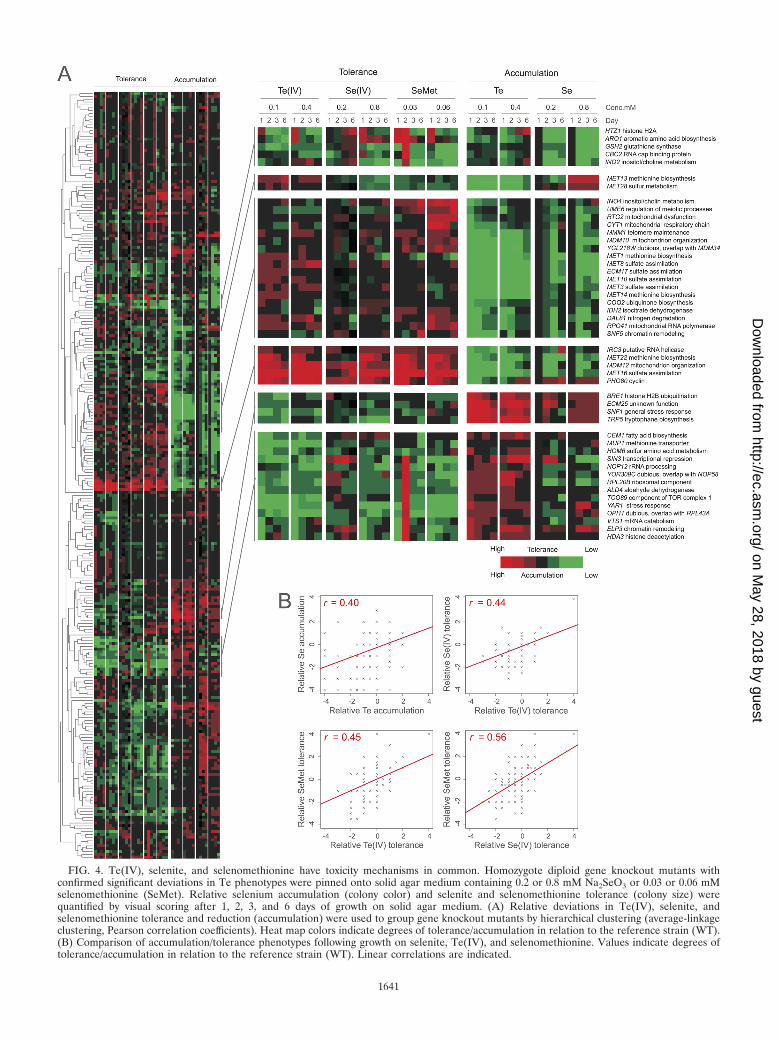
FIG. 4. Te(IV), selenite, and selenomethionine have toxicity mechanisms in common. Homozygote diploid gene knockout mutants withconfirmed significant deviations in Te phenotypes were pinned onto solid agar medium containing 0.2 or 0.8 mM Na2SeO3 or 0.03 or 0.06 mMselenomethionine (SeMet). Relative selenium accumulation (colony color) and selenite and selenomethionine tolerance (colony size) werequantified by visual scoring after 1, 2, 3, and 6 days of growth on solid agar medium. (A) Relative deviations in Te(IV), selenite, andselenomethionine tolerance and reduction (accumulation) were used to group gene knockout mutants by hierarchical clustering (average-linkageclustering, Pearson correlation coefficients). Heat map colors indicate degrees of tolerance/accumulation in relation to the reference strain (WT).(B) Comparison of accumulation/tolerance phenotypes following growth on selenite, Te(IV), and selenomethionine. Values indicate degrees oftolerance/accumulation in relation to the reference strain (WT). Linear correlations are indicated.
1641
on May 28, 2018 by guest
http://ec.asm.org/
Dow
nloaded from

intracellularly only has a limited effect on Te(IV) toleranceand reduction. Major regulators of the amino acid starvationresponse, Ure2 mediating nitrogen repression and Npr2-Npr3mediating nitrogen activation, also featured some of the stron-gest tellurium phenotypes in the screen, with the npr2� andnpr3� mutants showing high Te(0) accumulation and the ure2�mutant featuring low Te(0) accumulation (Fig. 2B; see TableS1 in the supplemental material). Although nitrate reductasespossess the ability to reduce Te(IV) to Te(0) in Escherichia coli(2), S. cerevisiae lacks nitrate reductases. Hence, mechanismsother than nitrate reduction must form the basis of the linkbetween nitrogen metabolism and tellurium accumulation inyeast.
Imbalances in intracellular oxidation status and mitochon-drial respiration affect tellurium accumulation. Sulfate assim-ilation consumes vast amounts of reductive power; the sulfitereductase alone requires three NADPH molecules in the re-duction of each sulfite (IV) molecule to sulfide (69). Hence,the enrichment of genes involved in NAD/NADP binding, andconsequently in redox status, among gene knockout mutantswith low Te(0) accumulation is not surprising (Fig. 2A).Among knockout mutants with low tellurium accumulation, wealso see an enrichment of proteins with a known localization inthe mitochondria (enrichment � 1.6-fold [P � 0.01; Fisher’sexact test]). Mitochondrial respiration is the dominant intra-cellular source of ROS and has profound effects on cellularredox status. Knockout mutants lacking the key respiratorygenes yield petite colonies that are too small to allow stringentevaluation of Te phenotypes; however, loss of nonpetite com-ponents such as CYT1, encoding cytochrome c1, Coq1 andCoq2, catalyzing the first steps in ubiquinone biosynthesis, andthe cytochrome c reductase subunit Qcr9 conferred low Te(0)accumulation (Fig. 2B). Similarly, a large group of genes in-volved in mitochondrial import featured very low Te(0) accu-mulation. Knockout mutants lacking the components of theMdm10-Mdm12-Mmm1-Mdm34 complex, mediating assemblyof mitochondrial import channels (25), or of their genetic in-teraction partners MDM31 and MDM35 (12) all showed highTe(IV) tolerance and low Te(0) accumulation (Fig. 2B; seeTable S1 in the supplemental material). These mitochondrialimport mutants all have respiratory deficiencies due to prob-lems in assembling fully functional respiratory chain complexes(11). Respiratory defects may also cause the very low Te(0)accumulation observed following loss of the functionally inter-dependent SNF5 and SNF6 genes (26) (Fig. 2B); these SWI/SNF chromatin remodeling complex components are abso-lutely required for respiratory growth because of their role inthe transcriptional regulation of genes in the respiratory chain.
A potential explanation for the strong tellurium phenotypesof mitochondrial deletion mutants is the effect on Fe-S proteinassembly due to Te replacement of, or strong binding to, S.Fe-S centers catalyze key reactions in the respiratory chain andare assembled in the mitochondrial matrix in a series of reac-tions known to involve 19 proteins. Ten of these proteins areessential, precluding the screening of Te phenotypes. How-ever, of the nine viable Fe-S assembly deletion mutants, onlythose with the above-mentioned qcr9� mutation, which indi-rectly interferes with Fe-S assembly by destabilizing the RieskeFe-S complex, showed altered Te phenotypes. Furthermore,GAL1-driven individual overexpression (49) of the 19 Fe-S
assembly components produced normal Te(IV) tolerance and,at most, marginal deviations in tellurium accumulation (datanot shown). Hence, Fe-S assembly does not seem to be directlyconnected to tellurium toxicity in yeast.
Tellurite, selenite, and selenomethionine share a toxicitymechanism. A key question is whether Te(IV) exerts its tox-icity by mechanisms exclusive to Te(IV) or through mecha-nisms shared with other chemically related elements. To re-solve this issue, we first investigated a potential overlap withthe selenite SeO3
2�, where the chalcogen selenium occurs asSe(IV). Se(IV) is chemophysically related to Te(IV) and isconverted inside the cell to Se(0), producing a reddish colonypigmentation, via a set of reactions that is poorly understood inyeast but that in bacteria involves glutathione and the releaseof ROS (46, 62). Screening gene knockout mutants with sig-nificant Te(IV) phenotypes on comparable concentrations ofSe(IV), we found substantial overlap between Te(IV) andSe(IV) phenotypes (linear correlation coefficient [r], 0.4)(Fig. 4A); gene knockout mutants with low tellurium accumu-lation tended to show low accumulation of Se(0) (Fig. 4 and 5).Genes required for both tellurium and selenium accumulationincluded the components of the Mmm1-Mdm10-Mdm12-Mdm34 and Ino2-Ino4 transcription complexes, the respira-tory chain components CYT1 and COQ2, and almost all of theMET genes. Interestingly, gsh2�, which only produced some-what reduced Te(0) accumulation, produced much lower Se(0)accumulation, suggesting a more direct involvement of gluta-thione in selenite conversion to selenium (Fig. 4A and 5).Gene knockout mutants with high tellurium accumulation, e.g.,snf1� and snf4� mutants lacking the glucose repression switch,produced a similar high accumulation of selenium pigmenta-tion (Fig. 4A). Hence, despite the fact that Te is bulkier thanSe and less prone to form stable bonds with carbon and hy-drogen, intracellular conversion of Te(IV) to Te(0) appears toproceed largely along the same metabolic routes as conversionof Se(IV) to Se(0). One potential cause for the toxicity ofSe(IV) and Te(IV) is the intracellular conversion into sel-enomethionine and telluromethionine, respectively, which, fol-lowing replacement of methionine during protein biosynthesis,may cause protein inactivation. To investigate Se-methionineand Te-methionine formation as potential toxicity mechanismsfor Se(IV) and Te(IV), we screened gene knockout mutantswith aberrations in Te phenotypes following selenomethionineexposure. Telluromethionine is unstable and rapidly decom-poses, impeding phenotypic screening. No quantifiable Se(0)accumulation was observed in selenomethionine-exposed cells,suggesting that the conversion of Se(IV) to selenomethionineis unidirectional (Fig. 5). However, comparing gene knockouttolerance traits, we found a strong overlap of Se(IV), Te(IV),and selenomethionine, showing that toxicity and detoxifica-tion mechanisms are partially shared by these compounds(Fig. 4). Specifically, loss of MET genes conferred toleranceto Se(IV), Te(IV), and selenomethionine whereas loss ofHOM6 or MUP1 resulted in sensitivity, suggesting a similarrole for the sulfate assimilation system in Se(IV), Te(IV),and selenomethionine toxicity.
To address whether more general metal/metalloid tolerancesystems are involved in Te(IV) detoxification, we also com-pared genes revealed in this study as important for Te(IV)tolerance to genes previously reported to be important for
1642 OTTOSSON ET AL. EUKARYOT. CELL
on May 28, 2018 by guest
http://ec.asm.org/
Dow
nloaded from

optimal cadmium (Cd) (20, 43, 47, 57) or arsenite [As(III)] (18,20, 57) tolerance. Circumventing the issue of a generally smalloverlap between screens, core sets, as defined by Thorsen et al.(57), corresponding to genes identified as required for resis-tance in at least two As(III) or two Cd screens were used. Wefound significant (P � 10�5; Fisher’s exact test) enrichments ofboth Cd-sensitive (enrichment � 3.7-fold) and As(III)-sensi-tive (enrichment � 4.2-fold) gene knockout mutants amongthe Te(IV)-sensitive mutants identified here. Hence, the tox-icity of Te(IV) partially follows routes that overlap not onlywith Se(IV) toxicity but apparently also with chemophysicallymore dissimilar metals. These routes involved actin interaction(RVS161, SLA1, and BEM4), metal homeostasis regulators(MAC1), redox metabolism (GSH2 and HAC1), and nitrogenmetabolism (URE2, UME6, and NPR2), as well as MET22 andHOM6, which are involved in the metabolism of sulfur-containingamino acids (Fig. 6). The overlap with cadmium specificallyincluded vacuolar/endosomal transport (VPS1, VMA21, andVPS24) and chromatin remodeling via SNF5 and SNF6 (Fig.6). However, it should be emphasized that the similaritybetween the Te(IV) and Cd/As mutant sensitivity profiles,although significant, was less pronounced than the strongoverlap of the Te, Se, and selenomethionine phenotypes.Specifically, none of the most prominent Te, Se, and selenome-thionine toxicity effects, such as the hypertolerance of sulfateassimilation knockout mutants, were observed for Cd or As.
DISCUSSION
The mechanistic causes of the toxicity and intracellular ac-cumulation of Te species are classical enigmas in molecularmicrobiology. Here we shed light on the Te conundrum byshowing that conversion of Te(IV) to Te(0) and the biotox-
icity of Te(IV) are mediated largely by the same cellularroutes in yeast. A genome-wide screen for tellurium-relatedtraits among yeast gene knockout mutants implicated twomain cellular routes, sulfur assimilation and redox-relatedmitochondrial respiration. From a metal biology perspec-tive, a link between sulfur assimilation and tellurium pheno-types is not unexpected; sulfur metabolism affects the biotox-icity of essentially all well-studied heavy metals and metalloidsin yeast, either directly or indirectly via its role in glutathionebiosynthesis (68). However, the involvement of sulfur metab-olism is typically the reverse of what is observed here; that is,unperturbed sulfur metabolism is a key factor in maintainingoptimal heavy metal/metalloid tolerance. Mutants resistant toselenate, Se(VI), which are known to be defective in compo-nents of the sulfate assimilation pathway, constitute exceptions(6, 8). Se(VI) is rapidly converted inside the cell to Se(IV),which is chemophysically similar to Te(IV). Hence, the con-nection between Se- and Te-related traits and sulfate assimi-lation reported here is not surprising. Sulfate assimilation in S.cerevisiae represents a sequence of redox reactions that arecostly in terms of ATP and NADPH (56). The key reductionreaction enabling utilization of sulfur compounds with a highoxidation state is the six-electron reduction of sulfite to sulfideby the two-component sulfite reductase at the expense of threeNADPH molecules (69, 70). We identified the sulfate/sulfurassimilation pathway as the most important cellular route me-diating Te(IV) toxicity and intracellular accumulation of tellu-rium in yeast. This directly points to a role for the sulfitereductase in Te(IV) reduction, which is somewhat surprising asthere is no evidence from bacterial studies for a sulfite reduc-tase-mediated reduction of either Te(IV) or Se(IV) to H2Te orH2Se, respectively; in addition, reductase component muta-
FIG. 5. Te(IV), selenite, and selenomethionine toxicity and bioaccumulation. Homozygous diploid gene knockout mutants with confirmedsignificant deviations in Te(IV) phenotypes were pinned onto solid agar medium containing 0.2 or 0.8 mM Na2SeO3 or 0.03 or 0.06 mMselenomethionine (SeMet) and evaluated manually after 1, 2, 3, and 6 days of growth on solid agar medium.
VOL. 9, 2010 TELLURIUM IN YEAST 1643
on May 28, 2018 by guest
http://ec.asm.org/
Dow
nloaded from

tions do not produce substantial Se phenotypes in bacteria (14,62). Instead, bacterial Te metabolism is believed to mimic Semetabolism, which, due to the reactivity of selenium com-pounds in low oxidation states with sulfur, is largely centeredon thiol-mediated reactions (62). In bacteria, glutathione andcysteine are believed to reduce Te(IV) and Se(IV) via thePainter reaction to Te and Se trisulfides which, although stableunder acidic conditions, will rapidly decompose to yield Te(0)and Se(0) under neutral conditions (17, 22, 40). This mayexplain why, in E. coli, Te(IV) tolerance correlates with therelative expression of cysteine biosynthetic genes (16, 64). Inyeast, glutathione metabolism and the closely connected cys-teine metabolism had a surprisingly limited effect on the intra-cellular fate of Te species; mutants with cysteine metabolismgene knockout mutants featured normal Te phenotypes, andloss of the glutathione synthase Gsh2 conferred only minorreductions in Te(IV) tolerance and a slight reduction in theconversion of Te(IV) to Te(0). However, external addition ofan excess of GSH to Te(IV)-containing growth medium causedan immediate and drastic darkening and suppressed the Te-induced growth defects. This suggests that GSH is capable ofreducing Te(IV) to Te(0) extracellularly. It has recently beenshown that yeast cells export GSH during As(III) stress inorder to complex bind and detoxify the harmful metalloidoutside the cell (68); a similar extracellular detoxificationmechanism may have a role in Te detoxification.
The inverse correlation observed here between Te(0) accu-mulation and Te(IV) tolerance suggests that the conversion ofTe(IV) to Te(0) is a potent cause of Te(IV) toxicity. Keyquestions are whether Te(IV), similarly to Se(IV), is bioassimi-lated; whether the resulting tellurocysteine, telluromethionine,or derivatives thereof are incorporated into peptides; and
whether this bioassimilation contributes to Te(IV) toxicity. Incontrast to selenium, which is required for the activity of manyproteins in higher organisms, no biological functions have yetbeen discovered for tellurium. The probable reason is the moremetallic properties of tellurium, which forms weaker covalentbonds with hydrogen and carbon (34). In bacteria, selenome-thionine formation from selenite is believed to pass via reduc-tion of glutathione-bound GS-SeH into H2Se and then furtherinto selenomethionine via the methyl cycle (62). Selenomethi-onine is then randomly incorporated into proteins due to theediting tolerance of methionyl-tRNA synthetase (10, 35). Al-though the intracellular fate of telluromethionine is less wellknown, the existence of telluromethionine, methyltellu-romethionine, and, to a lesser extent, tellurocysteine hasbeen reported from the cultivation of yeast and bacteria inthe presence of high concentrations of Te(IV) (42, 71).Telluromethionine has also been shown to be able to replacemethionine residues in about 40% of the copies of the proteindihydrofolate reductase in E. coli (3). Telluromethionine rap-idly decomposes in aqueous solutions (38) and does not permitstraightforward tests of toxicity mechanisms. However, wefound a remarkable overlap between Te(IV) toxicity and sel-enomethionine toxicity, suggesting that they stem from sharedmodes of action. This raises the prospect that the detrimentaleffect of Te(IV) on yeast growth partially stems from tellu-romethionine formation. Although it is unclear whether tellu-romethionine incorporation into peptides would significantlyalter protein properties, it has been shown that introduction oftellurocysteine into the active site of a glutathione transferasealtered the protein’s functionality to a glutathione peroxidase(29). An alternative toxicity mechanism is the incorporation oftellurocysteine into glutathione with concomitant depletion of
FIG. 6. Te(IV) toxicity is partially mediated via general metal toxicity routes. Hierarchical clustering (average-linkage clustering based onPearson correlation coefficients) of gene knockout mutants with reduced Te(IV) tolerance (reduced colony size) and reduced tolerance tocadmium and/or arsenite. Colors indicate degrees of tolerance in relation to that of the WT; for cadmium and arsenite, an arbitrary intermediatevalue was used, as the data were qualitative.
1644 OTTOSSON ET AL. EUKARYOT. CELL
on May 28, 2018 by guest
http://ec.asm.org/
Dow
nloaded from
reducing power. However, the absence of strong Te pheno-types in cysteine and glutathione biosynthesis gene knockoutmutants suggests that this is an unlikely toxicity mechanism inyeast. The absence of such phenotypes also fails to support amodel where inhibition of glutathione by direct binding of Teto the SH group of glutathione would form the mechanisticbasis of Te(IV) toxicity. Such a mechanism has been con-templated for various metals and metalloids (e.g., see ref-erence 51).
Oxidative stress is considered a dominant cause of Te(IV)toxicity in bacterial systems, particularly during respiratorygrowth (4, 41, 59). In particular, generation of the toxic super-oxide radical during Te(IV) reduction is a major contributor toTe(IV) toxicity, in analogy to what has been suggested forselenium oxyanions (46, 50). Surprisingly, we found no supportfor oxidative damage being an important mechanism of Te(IV)toxicity in yeast. Knockout mutants lacking genes involved inthioredoxin and glutaredoxin reactions, which have beenlinked to Te(IV) tolerance in bacteria (7), failed to show ab-errant Te phenotypes, as did knockout mutants lacking per-oxidases, catalases, and the major transcriptional regulators ofthese systems. The absence of links to oxidative stress protec-tion suggests that ROS generation is not a major source ofTe(IV) toxicity in yeast and that routes for Te(IV) toxicity anddetoxification may differ substantially between yeast and bac-teria. However, genes involved in respiratory functions, as wellas transport into the mitochondria, showed low tellurium ac-cumulation and high Te(IV) tolerance. Furthermore, initiationof accumulation of Te(0) coincided with the switch from fer-mentative to respiratory metabolism in S. cerevisiae and respir-ing yeast cells were hypersensitive to Te(IV). These observa-tions agree well with observations on Pseudomonas aeruginosaand P. pseudoalcaligenes which show that the rate of Te(IV)reduction is strongly correlated with the rate of respiration,that inhibition of respiration partially inhibits Te(IV) reduc-tion, and that Te(IV) exposure induces changes in multiplecomponents of the respiratory chain (13, 59). Hence, our re-sults identified two cellular routes as key to the intracellularfate of Te species in yeast: sulfate assimilation and a mitochon-drial mechanism that encompasses respiration. Interestingly,both sulfur metabolism and mitochondrial genes, including allof the major mitochondrial contributors to Te phenotypes re-ported here, were recently reported as having low or no rustcoloration during bismuth sulfite exposure (23). Bismuth sul-fite is converted into bismuth sulfide in yeast in a processdependent on the sulfate assimilation pathway, resulting in areddish-blackish rust coloration (15, 36). Consequently, bis-muth sulfite reduction and Te(IV) reduction proceed along thesame cellular routes, both with a strong emphasis on sulfateassimilation and mitochondrial respiration.
Data regarding the toxicity and fate of tellurium compoundsin higher eukaryotes, including humans, is scarce. Te speciesare absorbed in the body, but telluroamino acids have not beenfound in humans. The present view holds that Te(IV) is me-tabolized in a manner mimicking that of selenite, implyingrapid reduction to telluride followed by methylation to mono-,di-, or trimethylated Te species (37). However, in contrast toselenium, tellurium does not appear to be glycosylated inhigher eukaryotes, and trimethyltelluronium, rather than tel-lurosugars, is the dominating Te species (39). We found no
indications that methyltransferases are involved in Te metab-olism in yeast, suggesting that Te methylation is of little im-portance in this organism. In higher eukaryotes, tellurium-induced cell death has been suggested to occur by an oxidativemechanism that at least partially involves oxidation-inducedDNA damage (44). Se(IV) exposure is, in fact, well known tolead to DNA double-strand breaks (27, 32). In yeast, however,viable knockout mutants lacking genes involved in DNA repairand replication and in cell cycle checkpoints showed normal Tephenotypes, suggesting that DNA damage contributes little toTe toxicity. Similar to arsenic, which is accumulated as di-methyl arsenical, but in contrast to selenium, tellurium reacheshigh concentrations in red blood cells as dimethylated Te (37).Hence, arsenic and tellurium may share mechanistic featuresin mammals. We found a significant overlap between geneknockout sensitivities to Te(IV) and As(III), suggesting thatmechanistic similarities exist also in yeast. A common detoxi-fication mechanism of many metals/metalloids, notably, Cd,Sb, As, Hg, and Pb, is sequestration in the vacuole as GSHconjugates (68), a process that is mainly mediated by the ABCtransporter Ycf1. However, a ycf1� mutant showed normal Tephenotypes, as did knockout mutants lacking the Ycf1 paralogsBpt1 and Vmr1. Te plaques were also clearly absent from thevacuolar lumen. Intriguingly, however, Te plaques initiallyemerged in close proximity to the vacuolar membrane, and atlater stages of Te stress, the integrity of the vacuole was com-promised, followed by vacuole disintegration and rapid cellshrinkage. Several vacuolar mutants also showed substantialTe sensitivity phenotypes. Taken together, these findings sug-gest that the vacuole may play a role in Te toxicity and detox-ification in yeast but that this role does not encompass seques-tration.
ACKNOWLEDGMENTS
We express our gratitude to Lianming Tong, Department of AppliedPhysics, Chalmers University of Technology, for his assistance with theRaman measurements and to Markus Tamas, University of Gothen-burg, for thoughtful comments on the manuscript.
J.W. was supported by the Carl Trygger Research Foundation andthe Royal Swedish Academy of Sciences.
REFERENCES
1. Ambroziak, J., and S. A. Henry. 1994. INO2 and INO4 gene products,positive regulators of phospholipid biosynthesis in Saccharomyces cerevisiae,form a complex that binds to the INO1 promoter. J. Biol. Chem. 269:15344–15349.
2. Avazeri, C., R. J. Turner, J. Pommier, J. H. Weiner, G. Giordano, and A.Vermeglio. 1997. Tellurite reductase activity of nitrate reductase is respon-sible for the basal resistance of Escherichia coli to tellurite. Microbiology143(Pt. 4):1181–1189.
3. Boles, J. O., K. Lewinski, M. Kunkle, J. D. Odom, B. Dunlap, L. Lebioda,and M. Hatada. 1994. Bio-incorporation of telluromethionine into buriedresidues of dihydrofolate reductase. Nat. Struct. Biol. 1:283–284.
4. Borsetti, F., V. Tremaroli, F. Michelacci, R. Borghese, C. Winterstein, F.Daldal, and D. Zannoni. 2005. Tellurite effects on Rhodobacter capsulatuscell viability and superoxide dismutase activity under oxidative stress condi-tions. Res. Microbiol. 156:807–813.
5. Brachmann, C. B., A. Davies, G. J. Cost, E. Caputo, J. Li, P. Hieter, and J. D.Boeke. 1998. Designer deletion strains derived from Saccharomyces cerevi-siae S288c: a useful set of strains and plasmids for PCR-mediated genedisruption and other applications. Yeast 14:115–132.
6. Breton, A., and Y. Surdin-Kerjan. 1977. Sulfate uptake in Saccharomycescerevisiae: biochemical and genetic study. J. Bacteriol. 132:224–232.
7. Chasteen, T. G., D. E. Fuentes, J. C. Tantalean, and C. C. Vasquez. 2009.Tellurite: history, oxidative stress, and molecular mechanisms of resistance.FEMS Microbiol. Rev. 33:820–832.
8. Cherest, H., J. C. Davidian, D. Thomas, V. Benes, W. Ansorge, and Y.
VOL. 9, 2010 TELLURIUM IN YEAST 1645
on May 28, 2018 by guest
http://ec.asm.org/
Dow
nloaded from

Surdin-Kerjan. 1997. Molecular characterization of two high affinity sulfatetransporters in Saccharomyces cerevisiae. Genetics 145:627–635.
9. Cherest, H., F. Eichler, and H. Robichon-Szulmajster. 1969. Genetic andregulatory aspects of methionine biosynthesis in Saccharomyces cerevisiae. J.Bacteriol. 97:328–336.
10. Cowie, D. B., and G. N. Cohen. 1957. Biosynthesis by Escherichia coli ofactive altered proteins containing selenium instead of sulfur. Biochim. Bio-phys. Acta 26:252–261.
11. Dimmer, K. S., S. Fritz, F. Fuchs, M. Messerschmitt, N. Weinbach, W.Neupert, and B. Westermann. 2002. Genetic basis of mitochondrial functionand morphology in Saccharomyces cerevisiae. Mol. Biol. Cell 13:847–853.
12. Dimmer, K. S., S. Jakobs, F. Vogel, K. Altmann, and B. Westermann. 2005.Mdm31 and Mdm32 are inner membrane proteins required for maintenanceof mitochondrial shape and stability of mitochondrial DNA nucleoids inyeast. J. Cell Biol. 168:103–115.
13. Di Tomaso, G., S. Fedi, M. Carnevali, M. Manegatti, C. Taddei, and D.Zannoni. 2002. The membrane-bound respiratory chain of Pseudomonaspseudoalcaligenes KF707 cells grown in the presence or absence of potas-sium tellurite. Microbiology 148:1699–1708.
14. Fimmel, A. L., and R. E. Loughlin. 1977. Isolation and characterization ofcysK mutants of Escherichia coli K12. J. Gen. Microbiol. 103:37–43.
15. Forbes, B. A., D. F. Sahm, and A. S. Weissfeld. 1998. Bailey & Scott’sdiagnostic microbiology, 10th ed. C.V. Mosby Company, St. Louis, MO.
16. Fuentes, D. E., E. L. Fuentes, M. E. Castro, J. M. Perez, M. A. Araya, T. G.Chasteen, S. E. Pichuantes, and C. C. Vasquez. 2007. Cysteine metabolism-related genes and bacterial resistance to potassium tellurite. J. Bacteriol.189:8953–8960.
17. Ganther, H. E. 1999. Selenium metabolism, selenoproteins and mechanismsof cancer prevention: complexities with thioredoxin reductase. Carcinogen-esis 20:1657–1666.
18. Haugen, A. C., R. Kelley, J. B. Collins, C. J. Tucker, C. Deng, C. A. Afshari,J. M. Brown, T. Ideker, and B. Van Houten. 2004. Integrating phenotypicand expression profiles to map arsenic-response networks. Genome Biol.5:R95.
19. Isnard, A. D., D. Thomas, and Y. Surdin-Kerjan. 1996. The study of methi-onine uptake in Saccharomyces cerevisiae reveals a new family of amino acidpermeases. J. Mol. Biol. 262:473–484.
20. Jin, Y. H., P. E. Dunlap, S. J. McBride, H. Al-Refai, P. R. Bushel, and J. H.Freedman. 2008. Global transcriptome and deletome profiles of yeast ex-posed to transition metals. PLoS Genet. 4:e1000053.
21. Karlson, U., and W. Frankenberger. 1993. Biological alkylation of seleniumand tellurium, p. 185–227. In H. Sigel and A. Sigel (ed.), Metal ions inbiological systems. Marcel Dekker Inc., New York, NY.
22. Kice, J. L., T. W. S. Lee, and S.-T. Pan. 1980. Mechanism of the reaction ofthiols with selenite. J. Am. Chem. Soc. 102:4448–4455.
23. Kim, H. S., J. Huh, and J. C. Fay. 2009. Dissecting the pleiotropic conse-quences of a quantitative trait nucleotide. FEMS Yeast Res. 9:713–722.
24. Konetzka, W. A. 1977. Microbiology of metal transformations, p. 317–342. InE. D. Weinberg (ed.), Microorganisms and minerals. Marcel Dekker Inc.,New York, NY.
25. Kornmann, B., E. Currie, S. R. Collins, M. Schuldiner, J. Nunnari, J. S.Weissman, and P. Walter. 2009. An ER-mitochondria tethering complexrevealed by a synthetic biology screen. Science 325:477–481.
26. Laurent, B. C., M. A. Treitel, and M. Carlson. 1991. Functional interdepen-dence of the yeast SNF2, SNF5, and SNF6 proteins in transcriptional acti-vation. Proc. Natl. Acad. Sci. U. S. A. 88:2687–2691.
27. Letavayova, L., D. Vlasakova, J. E. Spallholz, J. Brozmanova, and M. Cho-vanec. 2008. Toxicity and mutagenicity of selenium compounds in Saccha-romyces cerevisiae. Mutat. Res. 638:1–10.
28. Liu, M., and D. E. Taylor. 1999. Characterization of gram-positive telluriteresistance encoded by the Streptococcus pneumoniae tehB gene. FEMSMicrobiol. Lett. 174:385–392.
29. Liu, X., L. A. Silks, C. Liu, M. Ollivault-Shiflett, X. Huang, J. Li, G. Luo,Y. M. Hou, J. Liu, and J. Shen. 2009. Incorporation of tellurocysteine intoglutathione transferase generates high glutathione peroxidase efficiency. An-gew. Chem. Int. Ed. Engl. 48:2020–2023.
30. Lloyd-Jones, G., A. M. Osborn, D. A. Ritchie, P. Strike, J. L. Hobman, N. L.Brown, and D. A. Rouch. 1994. Accumulation and intracellular fate of tel-lurite in tellurite-resistant Escherichia coli: a model for the mechanism ofresistance. FEMS Microbiol. Lett. 118:113–119.
31. Lloyd-Jones, G., W. M. Williamson, and T. Slootweg. 2006. The Te-Assay: ablack and white method for environmental sample pre-screening exploitingtellurite reduction. J. Microbiol. Methods 67:549–556.
32. Manikova, D., D. Vlasakova, J. Loduhova, L. Letavayova, D. Vigasova, E.Krascsenitsova, V. Vlckova, J. Brozmanova, and M. Chovanec. 2010. Inves-tigations on the role of base excision repair and non-homologous end-joiningpathways in sodium selenite-induced toxicity and mutagenicity in Saccharo-myces cerevisiae. Mutagenesis 25:155–162.
33. Moore, M. D., and S. Kaplan. 1992. Identification of intrinsic high-levelresistance to rare-earth oxides and oxyanions in members of the class Pro-teobacteria: characterization of tellurite, selenite, and rhodium sesquioxidereduction in Rhodobacter sphaeroides. J. Bacteriol. 174:1505–1514.
34. Moroder, L. 2005. Isosteric replacement of sulfur with other chalcogens inpeptides and proteins. J. Pept. Sci. 11:187–214.
35. Munier, R., and G. N. Cohen. 1959. Incorporation of structural analogues ofamino acids into bacterial proteins during their synthesis in vivo. Biochim.Biophys. Acta 31:378–390.
36. Nickerson, W. J. 1953. Reduction of inorganic substances by yeasts. I. Ex-tracellular reduction of sulfite by species of Candida. J. Infect. Dis. 93:43–56.
37. Ogra, Y. 2009. Toxicometallomics for research on the toxicology of exoticmetalloids based on speciation studies. Anal. Sci. 25:1189–1195.
38. Ogra, Y., T. Kitaguchi, N. Suzuki, and K. T. Suzuki. 2008. In vitro translationwith [34S]-labeled methionine, selenomethionine, and telluromethionine.Anal. Bioanal. Chem. 390:45–51.
39. Ogra, Y., R. Kobayashi, K. Ishiwata, and K. T. Suzuki. 2008. Comparison ofdistribution and metabolism between tellurium and selenium in rats. J. In-org. Biochem. 102:1507–1513.
40. Painter, E. P. 1941. The chemistry and toxicity of selenium compounds whichspecial reference to the selenium problem. Chem. Rev. 28:179–213.
41. Perez, J. M., I. L. Calderon, F. A. Arenas, D. E. Fuentes, G. A. Pradenas,E. L. Fuentes, J. M. Sandoval, M. E. Castro, A. O. Elias, and C. C. Vasquez.2007. Bacterial toxicity of potassium tellurite: unveiling an ancient enigma.PLoS One 2:e211.
42. Ramadan, S. E., A. A. Razak, A. M. Ragab, and M. el-Meleigy. 1989. Incor-poration of tellurium into amino acids and proteins in a tellurium-tolerantfungi. Biol. Trace Elem. Res. 20:225–232.
43. Ruotolo, R., G. Marchini, and S. Ottonello. 2008. Membrane transportersand protein traffic networks differentially affecting metal tolerance: agenomic phenotyping study in yeast. Genome Biol. 9:R67.
44. Sandoval, J. M., P. Leveque, B. Gallez, C. C. Vasquez, and P. Buc Calderon.2010. Tellurite-induced oxidative stress leads to cell death of murine hepa-tocarcinoma cells. Biometals 23:623–632.
45. Schroeder, H. A., J. Buckman, and J. J. Balassa. 1967. Abnormal traceelements in man: tellurium. J. Chronic Dis. 20:147–161.
46. Seko, Y., Y. Saito, J. Kitahara, and N. Imura. 1998. Active oxygen generationby the reaction of selenite with reduced glutathione in vitro, p. 70–73. In A.Wendel (ed.), Selenium in biology and medicine. Springer-Verlag, Berlin,Germany.
47. Serero, A., J. Lopes, A. Nicolas, and S. Boiteux. 2008. Yeast genes involvedin cadmium tolerance: identification of DNA replication as a target ofcadmium toxicity. DNA Repair (Amst.) 7:1262–1275.
48. Smith, D. G. 1974. Tellurite reduction in Schizosaccharomyces pombe.J. Gen. Microbiol. 83:389–392.
49. Sopko, R., D. Huang, N. Preston, G. Chua, B. Papp, K. Kafadar, M. Snyder,S. G. Oliver, M. Cyert, T. R. Hughes, C. Boone, and B. Andrews. 2006.Mapping pathways and phenotypes by systematic gene overexpression. Mol.Cell 21:319–330.
50. Spallholz, J. E. 1994. On the nature of selenium toxicity and carcinostaticactivity. Free Radic. Biol. Med. 17:45–64.
51. Stohs, S. J., and D. Bagchi. 1995. Oxidative mechanisms in the toxicity ofmetal ions. Free Radic. Biol. Med. 18:321–336.
52. Summers, A. O., and S. Silver. 1978. Microbial transformations of metals.Annu. Rev. Microbiol. 32:637–672.
53. Taylor, D. E. 1999. Bacterial tellurite resistance. Trends Microbiol. 7:111–115.
54. Taylor, D. E., Y. Hou, R. J. Turner, and J. H. Weiner. 1994. Location of apotassium tellurite resistance operon (tehA tehB) within the terminus ofEscherichia coli K-12. J. Bacteriol. 176:2740–2742.
55. Taylor, D. E., E. G. Walter, R. Sherburne, and D. P. Bazett-Jones. 1988.Structure and location of tellurium deposited in Escherichia coli cellsharbouring tellurite resistance plasmids. J. Ultrastruct. Mol. Struct. Res.99:18–26.
56. Thomas, D., and Y. Surdin-Kerjan. 1997. Metabolism of sulfur amino acidsin Saccharomyces cerevisiae. Microbiol. Mol. Biol. Rev. 61:503–532.
57. Thorsen, M., G. Perrone, E. Kristiansson, M. Traini, T. Ye, I. W. Dawes, O.Nerman, and M. Tamas. 2009. Genetic basis of arsenite and cadmiumtolerance in Saccharomyces cerevisiae. BMC Genomics 10:105.
58. Toptchieva, A., G. Sisson, L. J. Bryden, D. E. Taylor, and P. S. Hoffman.2003. An inducible tellurite-resistance operon in Proteus mirabilis. Microbi-ology 149:1285–1295.
59. Trutko, S. M., V. K. Akimenko, N. E. Suzina, L. A. Anisimova, M. G.Shlyapnikov, B. P. Baskunov, V. I. Duda, and A. M. Boronin. 2000. Involve-ment of the respiratory chain of gram-negative bacteria in the reduction oftellurite. Arch. Microbiol. 173:178–186.
60. Tucker, F. L., J. F. Walper, M. D. Appleman, and J. Donohue. 1962. Com-plete reduction of tellurite to pure tellurium metal by microorganisms. J.Bacteriol. 83:1313–1314.
61. Turner, R. J., Y. Aharonowitz, J. H. Weiner, and D. E. Taylor. 2001. Gluta-thione is a target in bacterial tellurite toxicity and is protected by telluriteresistance determinants in Escherichia coli. Can. J. Microbiol. 47:33–40.
62. Turner, R. J., J. H. Weiner, and D. E. Taylor. 1998. Selenium metabolism inEscherichia coli. Biometals 11:223–227.
63. Turner, R. J., J. H. Weiner, and D. E. Taylor. 1999. Tellurite-mediated thioloxidation in Escherichia coli. Microbiology 145(Pt. 9):2549–2557.
1646 OTTOSSON ET AL. EUKARYOT. CELL
on May 28, 2018 by guest
http://ec.asm.org/
Dow
nloaded from

64. Vasquez, C. C., C. P. Saavedra, C. A. Loyola, M. A. Araya, and S. Pichuantes.2001. The product of the cysK gene of Bacillus stearothermophilus V me-diates potassium tellurite resistance in Escherichia coli. Curr. Microbiol.43:418–423.
65. Walter, E. G., and D. E. Taylor. 1992. Plasmid-mediated resistance to tellu-rite: expressed and cryptic. Plasmid 27:52–64.
66. Warringer, J., D. Anevski, B. Liu, and A. Blomberg. 2008. Chemogeneticfingerprinting by analysis of cellular growth dynamics. BMC Chem. Biol. 8:3.
67. Warringer, J., and A. Blomberg. 2003. Automated screening in environmen-tal arrays allows analysis of quantitative phenotypic profiles in Saccharomy-ces cerevisiae. Yeast 20:53–67.
68. Wysocki, R., and M. J. Tamas. 2010. How Saccharomyces cerevisiae copes
with toxic metals and metalloids. FEMS Microbiol. Rev. [Epub ahead ofprint.] doi:10.1111/j.1574–6976.2010.00217.x.
69. Yoshimoto, A., and R. Sato. 1968. Studies on yeast sulfite reductase. I.Purification and characterization. Biochim. Biophys. Acta 153:555–575.
70. Yoshimoto, A., and R. Sato. 1968. Studies on yeast sulfite reductase. II.Partial purification and properties of genetically incomplete sulfite reducta-ses. Biochim. Biophys. Acta 153:576–588.
71. Yu, L., K. He, D. Chai, C. Yang, and O. Zheng. 1993. Evidence for tellu-roamino acid in biological materials and some rules of assimilation of inor-ganic tellurium by yeast. Anal. Biochem. 209:318–322.
72. Yurkov, V. V., and J. T. Beatty. 1998. Aerobic anoxygenic phototrophicbacteria. Microbiol. Mol. Biol. Rev. 62:695–724.
VOL. 9, 2010 TELLURIUM IN YEAST 1647
on May 28, 2018 by guest
http://ec.asm.org/
Dow
nloaded from