genomics of a phototrophic nitrite oxidizer: insights into...
TRANSCRIPT
ORIGINAL ARTICLE
Genomics of a phototrophic nitrite oxidizer: insightsinto the evolution of photosynthesis and nitrification
James Hemp1,6, Sebastian Lücker2,3,6, Joachim Schott4, Laura A Pace5, Jena E Johnson1,Bernhard Schink4, Holger Daims3 and Woodward W Fischer11Department of Geological and Planetary Sciences, California Institute of Technology, Pasadena, CA, USA;2Department of Microbiology, Radboud University Nijmegen, Nijmegen, The Netherlands; 3Division of MicrobialEcology, Department of Microbiology and Ecosystem Research, University of Vienna, Vienna, Austria; 4Divisionof Microbial Ecology, Department of Biology, University of Konstanz, Konstanz, Germany and 5Division ofGastroenterology, Department of Medicine, University of California, San Diego, La Jolla, CA, USA
Oxygenic photosynthesis evolved from anoxygenic ancestors before the rise of oxygen ~2.32 billionyears ago; however, little is known about this transition. A high redox potential reaction center is aprerequisite for the evolution of the water-oxidizing complex of photosystem II. Therefore, it is likelythat high-potential phototrophy originally evolved to oxidize alternative electron donors that utilizedsimpler redox chemistry, such as nitrite or Mn. To determine whether nitrite could have had a role inthe transition to high-potential phototrophy, we sequenced and analyzed the genome of ThiocapsaKS1, a Gammaproteobacteria capable of anoxygenic phototrophic nitrite oxidation. The genomerevealed a high metabolic flexibility, which likely allows Thiocapsa KS1 to colonize a great variety ofhabitats and to persist under fluctuating environmental conditions. We demonstrate that ThiocapsaKS1 does not utilize a high-potential reaction center for phototrophic nitrite oxidation, whichsuggests that this type of phototrophic nitrite oxidation did not drive the evolution of high-potentialphototrophy. In addition, phylogenetic and biochemical analyses of the nitrite oxidoreductase (NXR)from Thiocapsa KS1 illuminate a complex evolutionary history of nitrite oxidation. Our resultsindicate that the NXR in Thiocapsa originates from a different nitrate reductase clade than the NXRs inchemolithotrophic nitrite oxidizers, suggesting that multiple evolutionary trajectories led to modernnitrite-oxidizing bacteria.The ISME Journal advance online publication, 19 April 2016; doi:10.1038/ismej.2016.56
Introduction
The evolution of oxygenic photosynthesis in theancestors of Cyanobacteria was the most importantmetabolic innovation in Earth’s history; however, theevolutionary trajectory from ancestral anoxygenicphototrophy to oxygenic photosynthesis remainsuncertain. Comparative biology of extant phototrophicmicrobes places important constraints on this process.There are currently seven bacterial phyla that containmembers capable of chlorophyll-based phototrophy:Cyanobacteria, Alpha-, Beta- and Gammaproteobac-teria, Chloroflexi, Chlorobi, Firmicutes, Acidobacteriaand Gemmatimonadetes (Bryant et al., 2007;Overmann and Garcia-Pichel, 2013; Shih et al.,2013; Zeng et al., 2014). A fundamental divisionbetween phototrophs is based on the types of reaction
centers that they use. In type I reaction centers (RCIand PSI) the final electron acceptor is a ferredoxinprotein, whereas in type II reaction centers (RCII andPSII) the electrons are donated to quinones(Blankenship, 2014). Anoxygenic phototrophs onlyutilize a single reaction center, whereas oxygenicCyanobacteria couple type I and type II reactioncenters in series. Many substrates can be used aselectron donors for phototrophy; however, there areimportant energetic limits (Supplementary Table S1).Extant anoxygenic reaction centers have redox poten-tials that range from +300 to +500mV, and primarilyoxidize lower potential (o+100mV) electron donors(Supplementary Figure S1). In contrast, PSII used inoxygenic photosynthesis produces a very strongoxidant (~+1250mV; Rappaport and Diner, 2008)that is capable of oxidizing water to molecular oxygen(+810mV). The water-oxidizing complex in PSIIcould have only evolved after the origin of high-potential phototrophy (Fischer et al., 2016), suggestingthat some type of high-potential anoxygenic photo-trophy bridged the evolutionary gap betweenlow-potential anoxygenic and high-potential oxygenicphototrophy (Olson, 1970; Rutherford and Faller,
Correspondence: J Hemp, Department of Geological and PlanetarySciences, California Institute of Technology, 1200 E. CaliforniaBoulevard, Pasadena, CA 91125, USA.E-mail: [email protected] authors contributed equally to this work.Received 18 June 2015; revised 24 February 2016; accepted4 March 2016
The ISME Journal (2016), 1–10© 2016 International Society for Microbial Ecology All rights reserved 1751-7362/16www.nature.com/ismej
2003). The highest redox potential substrate oxidizedby known extant anoxygenic phototrophs is nitrite(+430mV); therefore, phototrophic nitrite oxidizersmight provide insight into the evolution of high-potential phototrophy (Griffin et al., 2007; Schottet al., 2010).
Phototrophic nitrite oxidizers are also important forunderstanding the evolution of chemolithotrophicnitrite-oxidizing bacteria (NOB). NOB catalyzes thesecond step of nitrification, which is a key process ofthe biogeochemical nitrogen cycle in oxic ecosystems.The highly structured intracytoplasmic membranesystems (ICMs) of the NOB Nitrobacter (Alphaproteo-bacteria) and Nitrococcus (Gammaproteobacteria)closely resemble the ICM of phototrophic purplebacteria. This ultrastructural similarity, and therelatively close phylogenetic affiliation of Nitrobacterand Nitrococcus with purple bacteria, led to thehypothesis that these chemolithotrophic NOB evolvedfrom phototrophic ancestors (Teske et al., 1994). Thisscenario seemed to gain support from the surprisingdiscovery of anoxygenic phototrophic nitrite oxidizersin the genera Rhodopseudomonas and Thiocapsawithin the Alpha- and Gammaproteobacteria,respectively (Griffin et al., 2007; Schott et al., 2010).Recent studies revealed a second evolutionaryorigin of chemolithotrophic nitrite oxidation, wherethis metabolism evolved independently in thenon-proteobacterial NOB genera Nitrospira andNitrospina, probably through horizontal gene transferof the nitrite-oxidizing enzyme and other functionallyimportant proteins with anaerobic ammonium oxidi-zers (Lücker et al., 2010, 2013). Until now, it hasremained unclear whether the extant anoxygenicphototrophic nitrite oxidizers developed along eitherof these two lines of NOB evolution or representa third independent lineage.
To reveal the mechanism of phototrophic growthon nitrite, constrain the energetics of high-potentialanoxygenic phototrophy, shed light on possibleevolutionary links between photolithotrophic andchemolithotrophic nitrite oxidizers and elicudate themetabolic capabilities and ecophysiology of a photo-trophic nitrite oxidizer, we sequenced and analyzedthe genome of the nitrite-oxidizing phototrophThiocapsa sp. strain KS1. In addition, we carriedout biochemical experiments with fractionatedcell-free extracts of a Thiocapsa KS1 pure cultureto characterize the enzymatic nitrite-oxidizing andnitrate-reducing activities.
Materials and methods
Genome sequencing and analysisThiocapsa KS1 (JCM 15485) was grown as describedin Supplementary Information. High-molecular-weight genomic DNA was isolated following thehexadecyltrimethylammonium bromide protocol asdescribed elsewhere (Lücker et al., 2013). Sequencingand assembly were performed at LGC Genomics
(Berlin, Germany) using GS FLX Titanium-sequencingtechnology. The Thiocapsa KS1 draft genome wasannotated using the MicroScope annotation platform(Vallenet et al., 2013). Data from this sequencingproject have been deposited at the European Nucleo-tide Archive under study ID PRJEB9229.
RCII and NXR sequences from genomes andmetagenomes were retrieved from National Centerfor Biotechnology Information and DOE JointGenome Institute. Sequence alignments were calcu-lated using Muscle 3.8 (Edgar, 2004) or ARB(Ludwig, 2004). Phylogenetic analyses were per-formed on the CIPRES (Miller et al., 2010) clusteror on a desktop PC using RAxML (Stamatakis, 2014)and MrBayes (Ronquist and Huelsenbeck, 2003).
Cell-free extracts and protein analysesCell-free extracts from Thiocapsa KS1 were preparedand fractionated as described in SupplementaryInformation. The enzymatic activities of NXR weremeasured continuously with spectrophotometric assays(Supplementary Information). SDS-PAGE was per-formed according to Muller et al. (2009). Protein bandswere sent to TopLab (Martinsried, Germany) for trypticdigestion and peptide mass fingerprinting withoutdestaining. The fingerprints were matched (Mascotsearch engine) against the NCBI protein database.
Results
Genome analysisThiocapsa KS1 is a gammaproteobacterium isolatedfrom a sewage treatment facility in Konstanz,Germany, by selecting for photoautotrophic growthon nitrite (Griffin et al., 2007; Schott et al., 2010). Thisversatile phototroph can utilize a range of simpleorganic carbon molecules, sulfur compounds, H2
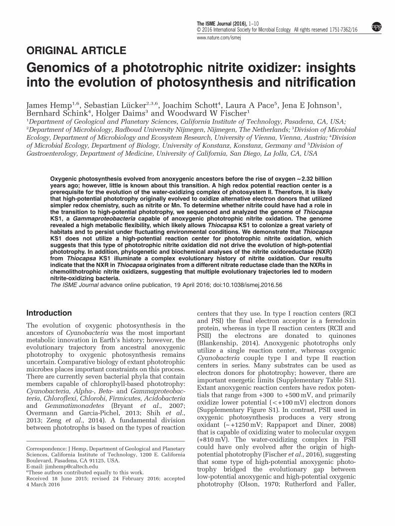
and nitrite as electron donors. We briefly describethe genes associated with respiration, phototrophy,nitrogen metabolism and other functions that conferThiocapsa KS1 with exceptionally high ecophysiolo-gical flexibility, which makes it the most metaboli-cally versatile nitrite-oxidizing microorganism known(Figure 1 and Supplementary Table S2).
General respirationThe genome of Thiocapsa KS1 contained one copy ofa 14-subunit NADH dehydrogenase (complex I) thatallows organotrophic respiration. Under photolithoau-totrophic growth conditions, complex I will operatein reverse to produce NADH required for carbonfixation (Elbehti et al., 2000; Supplementary FigureS2). Biosynthetic pathways for both menaquinone andubiquinone were present, consistent with the detec-tion of both quinone types in the close relativeT. roseopersicina (Imhoff, 1984). Thiocapsa KS1had one cytochrome (cyt.) bc1 complex (complex III)that conserves energy during phototrophic growth,aerobic respiration and denitrification. Three high-
Genomics of a phototrophic nitrite oxidizerJ Hemp et al
2
The ISME Journal
affinity oxygen reductases were present; two C-familyheme–copper oxidoreductases (cyt. cbb3 oxidase,complex IV) and a quinol-oxidizing cyt. bd oxidase.Low-affinity A-family oxygen reductases were absent.Details on ATP production and reverse electrontransport are provided in Supplementary Information.
PhototrophyThiocapsa KS1 encoded one set of RCII genes: PufL,PufM, PufC and PuhA. These had very high sequenceidentities (98% for PufL and PufM) to the closestsequenced strain, T. marina DSM 5653, which wasconfirmed using PCR. Thiocapsa KS1 and T. marinacontained a second copy of the RCII cyt. c subunitPufC, which appeared to be fused to an outermembrane protein. Multiple copies of genes for the
light-harvesting complexes LH1 and LH2 were alsopresent. Thiocapsa KS1 utilizes bacteriochlorophylla for phototrophic growth, with genes responsible forits biosynthesis distributed throughout the genome.The pathway required for the biosynthesis ofspirilloxanthin, the major pigment in T. roseopersi-cina (Kovács et al., 2003), was complete. Several cyt.c4-like di-heme proteins, along with two copies of high-potential iron–sulfur proteins (HiPIPs), were available toact as diffusible periplasmic electron carriers.
Sulfur metabolismThiocapsa KS1 encoded a wide diversity of enzymesinvolved in oxidative sulfur metabolism (Gregersenet al., 2011; Figure 1, Supplementary Information).Systems for the oxidation of sulfur, thiosulfate and
Figure 1 Metabolic diversity of Thiocapsa KS1. For details see main and supplemental text. APR, adenylylsulphate reductase complex;bd, cytochrome bd quinol oxidase; BFR, bacterioferritin; CA, carbonic anhydrase; CYN, cyanate hydratase; Cys, assimilatory sulfatereduction complexes; DSR, reverse dissimilatory sulfite reductase; FCC, sulfide dehydrogenase; FDH, formate dehydrogenase; HOX, HUP,HYD, HYN, hydrogenases (with enzyme classification indicated in brackets); NAP, periplasmic nitrate reductase; NIF, nitrogenase;NOR, nitric oxide reductase; NOS, nitrous oxide reductase; NXR, nitrite oxidoreductase; OTR, octaheme tetrathionate reductase;PHA, polyhydroxyalkanoate; PS, photosystem (type II reaction center); PTS, phosphotransferase system; RNF, H+/Na+-translocatingNAD-ferredoxin reductase; SAT, sulfate adenylyltransferase; SIR, sulfite reductase; SOD, superoxide dismutase; SOE, sulfite-oxidizingenzyme; SOX, sulfur/thiosulfate oxidation protein complex; SQR, sulfide-quinone reductase; TCA cycle, tricarboxylic acid cycle; URE,urease. Enzyme complexes of the electron transport chain are labeled by Roman numerals: I, NADH dehydrogenase; II, succinatedehydrogenase/fumarate reductase; III, cytochrome bc1 complex; IV, cbb3-type cytochrome c oxidase; Orange, red, green and bluediamonds represent quinones, cytochrome c proteins, HiPIPs and ferredoxins, respectively.
Genomics of a phototrophic nitrite oxidizerJ Hemp et al
3
The ISME Journal
sulfide were identified, which allow for the utilizationof these sulfur compounds as electron donors foranoxygenic photosynthesis. During phototrophicgrowth with sulfide or thiosulfate, elemental sulfur(S0) is stored in extracytoplasmic sulfur globuleswithin chromatophores (Pattaragulwanit et al., 1998),a property shared with other purple sulfur bacteria.These stores can later be used as electron donorswhen environmental sulfide and thiosulfate concen-trations decrease. The complete assimilatory pathwayfor sulfate reduction via adenosinephosphosulfate and3'-phosphoadenosinephosphosulfate was also presentin the genome.
HydrogenasesThiocapsa KS1 encoded at least five Ni–Fe hydro-genases that can oxidize H2 as an electron source forphotosynthesis, recycle H2 formed during diazo-trophic growth or produce H2 during phototrophicgrowth on reduced sulfur or carbon compounds(Maróti et al., 2010; Supplementary Information).
Carbon metabolismThiocapsa KS1 assimilates CO2 via the Benson–Bassham cycle, which was complete in the genome.Genes for the ribulose-bisphosphate carboxylase (type IRuBisCO) large and small subunits were duplicated anda type IV RuBisCO, which is not involved in carbonfixation (Tabita et al., 2007), was also present. Carboxy-some shell proteins and carbonic anhydrases indicatedthe presence of carboxysomes for concentrating CO2
(Yeates et al., 2008). Phosphoglycolate formed by theoxygenase activity of RuBisCO may be fed into thetricarboxylic acid cycle via the glycolate salvage path-way and glyoxylate shunt. The complete C4-dicar-boxylic acid cycle allows additional CO2 fixationby phosphoenolpyruvate carboxylation. ThiocapsaKS1 also possessed a full gene inventory for photo-or chemoorganoheterotrophic growth (SupplementaryInformation).
Nitrogen metabolismAs a diazotroph, Thiocapsa KS1 encoded a completeset of nif genes for nitrogen fixation includingmolybdenum-iron nitrogenase (NifDK) and nitrogen-ase reductase (NifH). Thiocapsa KS1 can assimilateammonium and also utilize nitrite and nitrate asnitrogen sources in culture (Schott et al., 2010),although the organism did not possess genes forcanonical assimilatory nitrate or nitrite reductases.Interestingly, the genome encoded several alternativemechanisms (Supplementary Information). In addi-tion, the presence of genes encoding nickel-dependenturease, cyanate hydratase, thiocyanate hydrolase andethanolamine ammonia-lyase indicated a broad spec-trum of reduced nitrogen sources for Thiocapsa KS1.
Nitrite oxidation in Thiocapsa KS1 was mediated bya Mo-bis-MGD-binding nitrite oxidoreductase (NXR), an
enzyme that can catalyze nitrite oxidation and nitratereduction (NO2
−+H2O ↔ NO3−+2e− + 2H+ ; Tanaka
et al., 1983; Sundermeyer-Klinger et al., 1984). The NXRof Thiocapsa KS1 was similar to the NXR forms foundin the chemolithotrophic NOB Nitrobacter, NitrococcusandNitrolancea (Starkenburg et al., 2008; Sorokin et al.,2012) and to the dissimilatory membrane-bound nitratereductase (NAR) system found in many nitrate-reducingorganisms. The NXR complex consisted of the α subunit(NxrA), which contains the catalytic site, the electron-channeling β subunit (NxrB) with four cysteine-richbinding motifs for [Fe-S] clusters and the γ subunit(NxrC), a membrane protein that putatively binds twoheme b groups. Electrons derived from nitrite flow fromNxrA through NxrB to NxrC, which anchors NXR in themembrane and transfers the electrons to the down-stream electron carriers. Like in Nitrobacter (Kirsteinand Bock, 1993; Spieck et al., 1996) the NxrA and NxrBsubunits were oriented toward the cytoplasm andresembled the NarGH subunits of bacterial NARs. ATorD-like chaperone, which probably inserts the Mo-bis-MGD cofactor into NxrA (Blasco et al., 1998; Ilbertet al., 2003), was encoded between the nxrB andnxrC genes.
Although Thiocapsa KS1 has candidate genes fora complete denitrification pathway (Figure 1)and can use a range of organic and inorganic low-potential electron donors for energy conservation, nogrowth was observed under anoxic conditions in thepresence of these electron donors and nitrate aselectron acceptor (Griffin et al., 2007; Schottet al., 2010). Nitrate reduction to nitrite could beperformed by a periplasmic NAR (NapDAGHB). Thiscomplex is missing NapC, similar to the periplasmicNARs found in many Epsilonproteobacteria (Simonet al., 2003), suggesting that an alternative electrontransfer pathway to NapA is utilized. In addition,NXR could function as a membrane-bound NAR(see below). Iron- (NirS) or copper- (NirK) dependentnitrite reductases are missing. Instead, the ThiocapsaKS1 genome contained two copies of hydroxylaminedehydrogenase-related proteins, which have beenimplied in nitrite reduction to NO in Methylococcuscapsulatus strain Bath (Campbell et al., 2011).In addition, strain KS1 had both a nitric oxidereductase and a nitrous oxide reductase that wouldenable the sequential reduction of NO to N2.
Enzymatic activities of NXRCell-free extracts were prepared from Thiocapsa KS1cultures to test the nitrite-oxidizing and nitrate-reducing enzyme activities. Although cultures grownphotolithoautotrophically with nitrite as the soleelectron donor were analyzed, no nitrite-oxidizingenzyme activity was detected by the two assaysapplied (Meincke et al., 1992). In contrast,a pronounced nitrate-reducing activity was measur-able (Supplementary Table S3). The highest specificNAR activity (up to 1700mU·(mg protein)−1) was foundin the membrane fraction, whereas the cytosolic
Genomics of a phototrophic nitrite oxidizerJ Hemp et al
4
The ISME Journal
fraction showed less than half of this activity(Supplementary Table S3). The protein contents of themembrane and cytosolic fractions were roughly equal.No activity was detected with NAD(P)H as an alter-native electron donor and dithiothreitol as reducingagent. Cells grown with the alternative electron donorH2 had no NAR activity when grown with ammoniumas nitrogen source (Supplementary Table S3), and grewonly poorly with nitrate as nitrogen source so that nocell extracts could be prepared from these cultures.Extracts from cells grown on fructose and nitrate hada very low NAR activity that was found exclusively inthe cytosolic fraction (Supplementary Table S3), indi-cating the involvement of a distinct enzyme system forassimilatory nitrate reduction.
Cell fractions were also analyzed using one-dimensional SDS-PAGE (Supplementary Figure S3).The protein patterns from nitrite-grown cells containedtwo strong bands that were absent from H2-grown cellsand thus assumed to be involved in nitrite oxidation(Supplementary Figure S3). These bands had estimatedsizes of 130–150 and 55–60 kDa, respectively, whichresemble the sizes of NxrA and NxrB from Nitrobacter(Meincke et al., 1992). Consistently, mass spectrometricfingerprint analysis and in silico comparison of theobtained peaks to publicly available protein sequencesconfirmed that the large bands (130–150 kDa) inthe cytosolic and membrane fractions represented theNXR α subunit, whereas the smaller bands (55–60 kDa)were identified as the NXR β subunit (SupplementaryFigure S3).
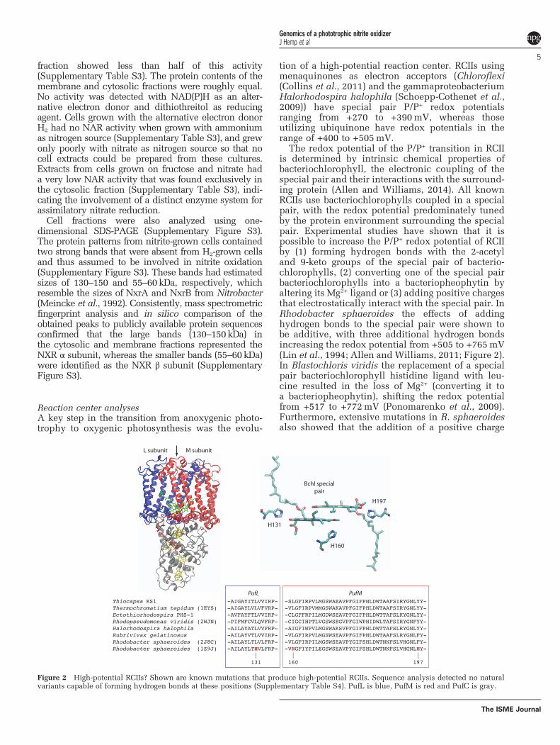
Reaction center analysesA key step in the transition from anoxygenic photo-trophy to oxygenic photosynthesis was the evolu-
tion of a high-potential reaction center. RCIIs usingmenaquinones as electron acceptors (Chloroflexi(Collins et al., 2011) and the gammaproteobacteriumHalorhodospira halophila (Schoepp-Cothenet et al.,2009)) have special pair P/P+ redox potentialsranging from +270 to +390mV, whereas thoseutilizing ubiquinone have redox potentials in therange of +400 to +505mV.
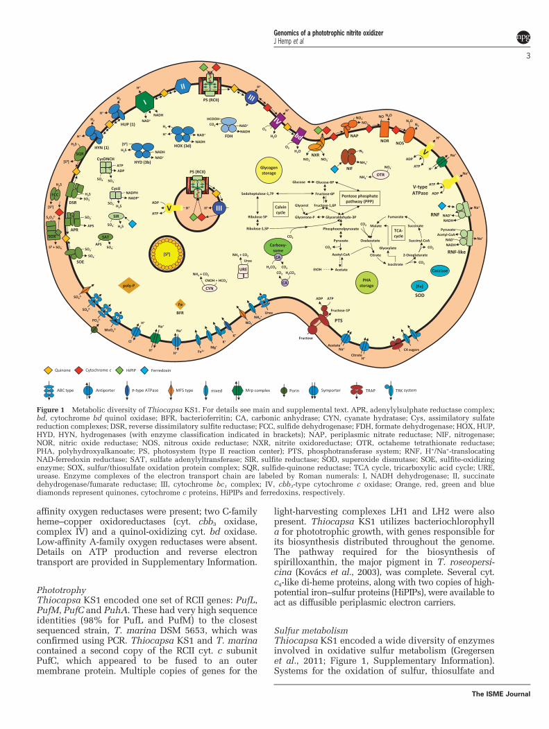
The redox potential of the P/P+ transition in RCIIis determined by intrinsic chemical properties ofbacteriochlorophyll, the electronic coupling of thespecial pair and their interactions with the surround-ing protein (Allen and Williams, 2014). All knownRCIIs use bacteriochlorophylls coupled in a specialpair, with the redox potential predominately tunedby the protein environment surrounding the specialpair. Experimental studies have shown that it ispossible to increase the P/P+ redox potential of RCIIby (1) forming hydrogen bonds with the 2-acetyland 9-keto groups of the special pair of bacterio-chlorophylls, (2) converting one of the special pairbacteriochlorophylls into a bacteriopheophytin byaltering its Mg2+ ligand or (3) adding positive chargesthat electrostatically interact with the special pair. InRhodobacter sphaeroides the effects of addinghydrogen bonds to the special pair were shown tobe additive, with three additional hydrogen bondsincreasing the redox potential from +505 to +765mV(Lin et al., 1994; Allen and Williams, 2011; Figure 2).In Blastochloris viridis the replacement of a specialpair bacteriochlorophyll histidine ligand with leu-cine resulted in the loss of Mg2+ (converting it toa bacteriopheophytin), shifting the redox potentialfrom +517 to +772mV (Ponomarenko et al., 2009).Furthermore, extensive mutations in R. sphaeroidesalso showed that the addition of a positive charge
Figure 2 High-potential RCIIs? Shown are known mutations that produce high-potential RCIIs. Sequence analysis detected no naturalvariants capable of forming hydrogen bonds at these positions (Supplementary Table S4). PufL is blue, PufM is red and PufC is gray.
Genomics of a phototrophic nitrite oxidizerJ Hemp et al
5
The ISME Journal
within 10 Å of the special pair increased the redoxpotential by 50mV. Protein modifications such asthese can be easily identified in multiple sequencealignments.
The RCII from Thiocapsa KS1 has none of themodifications described above that could raise its redoxpotential, and has the exact same residues interactingwith the special pair as Thermochromatium tepidum(Ivancich et al., 1996), implying a redox potential of~+500mV (Figure 2, Supplementary Table S4).
To determine whether RCIIs found in natureexhibited any modifications that would enable high-potential phototrophy, we analyzed the RCII genesfrom all sequenced genomes and publically availablemetagenomes. Sequence alignments of 43000 RCIIproteins (Supplementary Table S5) identified novariants in positions that could form additionalhydrogen bonds with either the 2-acetyl and 9-ketogroups of the special pair (Supplementary Table S4).In addition, no sequences were found that hadmodified bacteriochlorophyll ligands, and no variantswere identified that would modify electrostatic inter-actions with the special pair (Krammer et al., 2009).Together, this implies that extant RCIIs are unable toachieve redox potentials greater than ~+500mV.
Discussion
Ecophysiology of Thiocapsa KS1Although chemolithoautotrophic NOB have beenstudied for decades, we are just beginning to under-stand the physiology of phototrophic nitrite oxidizers.Traditionally, the chemolithoautotrophic NOBswere assumed to be highly specialized and metaboli-cally restricted organisms. This picture changedwith discoveries such as the chemoorganohetero-trophic Nitrobacter (Bock, 1976) and the aerobicallyH2-oxidizing, or anaerobically formate-consuming andnitrate-reducing, Nitrospira (Koch et al., 2014, 2015).However, the Thiocapsa KS1 genome has revealed anexceptionally high degree of metabolic versatility notfound in other NOB. This flexibility likely allowsThiocapsa KS1 to colonize a wide variety of ecologi-cal niches and to persist under changing environ-mental conditions, using photolithotrophic nitriteoxidation, as only one of several alternative lifestyles.Further studies on the ecophysiology of phototrophicNOB will be crucial for assessing the relativecontribution of photolithotrophic nitrite oxidation tooverall nitrification in the environment. The whole-genome analysis of Thiocapsa KS1 presented hereprovides a number of hypotheses on the ecophysiol-ogy of phototrophic NOB that can be tested in targetedexperiments in either pure cultures of Thiocapsa KS1or in microbial communities.
Evolution of nitrite oxidation in Thiocapsa KS1 andchemolithotrophic nitrite oxidizersThiocapsa KS1 is the first identified (Griffin et al.,2007) and genomically characterized (this study)
nitrite-oxidizing phototroph. As several genomes ofchemolithotrophic NOB had been sequenced earlier,we were able to compare the nitrite-oxidizingsystems of these organisms to elucidate whetherand how the evolutionary pathways of photo- andchemolithotrophic nitrite oxidation are related.
To enable photolithotrophic growth on nitrite,Thiocapsa KS1 utilizes NXR. Notably, in the knownaerobic chemolithotrophic NOB two different formsof NXR have been identified. They differ in thelocalization of their α and β subunits on eitherthe cytoplasmic or periplasmic side of the mem-brane, and thus in the bioenergetics of nitriteoxidation. The NXR of Nitrospira (Lücker et al.,2010) and Nitrospina (Lücker et al., 2013) is locatedon the periplasmic side of the membrane, indicatingthat nitrite oxidation occurs in the periplasm of theseorganisms. This is energetically ideal, given thatprotons generated by the reaction directly contributeto the pmf. In contrast, NOB from the Proteobacteria,such as Nitrobacter (Starkenburg et al., 2006), andfrom the Chloroflexi (Sorokin et al., 2012), oxidizesnitrite on the cytoplasmic side of the membrane.In this case protons from nitrite oxidation are releasedinto the cytoplasm, with no contribution to pmf,and electrons are transferred across the membrane toa soluble cyt. c550 on the periplasmic side (Figure 1 andSupplementary Figure S4). The redox potential of thiscyt. c550 appears to be low (~+280mV in Nitrobacter;Ketchum et al., 1969), and this step of the electrontransport chain might require energy from theelectrochemical membrane potential (Cobley, 1976;Ferguson, 1982). Assuming a membrane potential of~150mV, the electrons from the nitrite/nitratecouple may reach a potential of ~ +280mV whentransferred along the electrochemical gradient to thepositive side of the membrane.
The NXR of Thiocapsa KS1 faces similar con-straints as the Nitrobacter system because itsα subunit with the active site is also located on thecytoplasmic side of the membrane. In phototrophicbacteria, for example, R. capsulatus, there are at leasttwo electron transport pathways to the reactioncenter that involve c-type cytochromes, one via thesoluble cyt. c2 and one via the membrane-bound cyt.cy (Jenney and Daldal, 1993; Jenney et al., 1994).With a range of +345 to +395mV (Pettigrew et al.,1978), the redox potential of various cyt. c2 fromdifferent phototrophic purple bacteria would be inthe right range to allow electron transfer from nitriteto the reaction center. Alternatively, a HiPIP proteincould be employed as soluble electron carrierinstead of cyt. c. For example, the purple sulfurbacterium Allochromatium vinosum preferentiallyuses HiPIP instead of c-type cytochromes duringphoto-organotrophic growth (Van Driessche et al.,2003). HiPIP potentials range from +50 to +500mV(Heering et al., 1995), and the HiPIP protein ofT. roseopersicina has a redox potential of +342mV(Przysiecki et al., 1985). Hence, electron transferfrom nitrite via NXR to a HiPIP or cyt. c should be
Genomics of a phototrophic nitrite oxidizerJ Hemp et al
6
The ISME Journal
possible and might be facilitated by the membranepotential as outlined above for the chemolitho-trophic NOB and cyt. c550.
Although the NXRs from Thiocapsa KS1 andNitrobacter are both membrane-associated andoriented toward the cytoplasm, we identified thekey differences in these enzymes. In Thiocapsa KS1,the transfer of electrons from NXR across the cellmembrane and onto the soluble electron carrier mostlikely involves a unique di-heme cyt. c that is fusedonto the γ subunit of NXR (NxrC; SupplementaryFigure S4). This fusion is unique among the knownnitrite oxidizers, and its functional analog in allknown chemolithotrophic NOBs with a cytoplasmicNXR, such as Nitrobacter, is a di-heme cyt. c encodedby a separate gene upstream of the nxrA gene (Sorokinet al., 2012). Protein sequence analyses indicatedifferent evolutionary origins for these cyt. c moieties.The separate cyt. c occurs also in several dissimilatorymembrane-bound NARs from heterotrophic denitri-fiers such as Thermus thermophilus, where it belongsto a unique electron transport chain from a specialNADH oxidase via NAR to nitrite-, NO- and N2Oreductases (Cava et al., 2008).
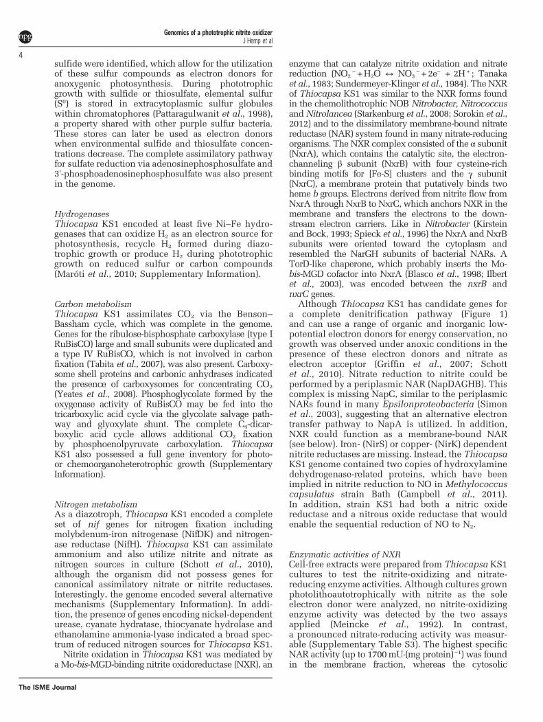
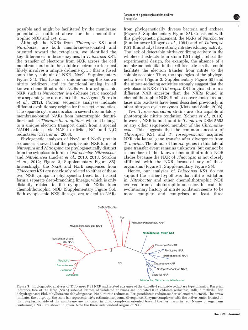
Phylogenetic analyses of NxrA and NxrB proteinsequences showed that the periplasmic NXR forms ofNitrospira andNitrospina are phylogenetically distinctfrom the cytoplasmic forms ofNitrobacter,Nitrococcusand Nitrolancea (Lücker et al., 2010, 2013; Sorokinet al., 2012; Figure 3, Supplementary Figure S5).Interestingly, the NxrA and NxrB sequences fromThiocapsa KS1 are not closely related to either of thesetwo NXR groups in phylogenetic trees, but insteadform a separate deep-branching lineage, which is onlydistantly related to the cytoplasmic NXRs fromchemolithotrophic NOB (Supplementary Figure S5).Both cytoplasmic NXR lineages are related to NARs
from phylogenetically diverse bacteria and archaea(Figure 3, Supplementary Figure S5). Consistent withthis phylogenetic placement, the NXRs of Nitrobacter(Sundermeyer-Klinger et al., 1984) and of ThiocapsaKS1 (this study) have strong nitrate-reducing activity.The lack of detectable nitrite-oxidizing activity in thewhole-cell extracts from strain KS1 might reflect theexperimental design, for example, the absence of amembrane potential in the cell-free extracts that couldfacilitate the electron transfer from nitrite to thesoluble acceptor. Thus, the topologies of the phyloge-netic trees (Figure 3, Supplementary Figure S5) andthe nitrate-reducing activities strongly suggest that thecytoplasmic NXR of Thiocapsa KS1 originated from adifferent NAR ancestor than the NXRs found inchemolithotrophic NOB. Similar conversions of reduc-tases into oxidases have been described previously inother nitrogen cycle enzymes (Klotz and Stein, 2008).
Two T. roseopersicina strains are also capable ofphototrophic nitrite oxidation (Schott et al., 2010);however, NXR is not found in T. marina DSM 5653or any other sequenced member of the Chromatia-ceae. This suggests that the common ancestor ofThiocapsa KS1 and T. roseopersicina acquiredNXR via lateral gene transfer after divergence fromT. marina. The donor of the nxr genes in this lateralgene transfer event remains unknown, but cannot bea member of the known chemolithotrophic NOBclades because the NXR of Thiocapsa is not closelyaffiliated with the NXR forms of any of theseorganisms (Figure 3; Supplementary Figure S5).
Hence, our analyses of Thiocapsa KS1 do notsupport the earlier hypothesis that nitrite oxidationin Nitrobacter and other chemolithotrophic NOBevolved from a phototrophic ancestor. Instead, theevolutionary history of nitrite oxidation seems to bemore complex and comprises at least three
Nitrospina
Scalindua
Brocadiaceae
Nitrospira
bacterial/archaealput. NAR
Halobacteraceae put. NAR
Clr/Ddh/Ebd/Ser
Pcr
Halobacteriaceae put. NAR
Thiocapsa sp. strain KS1
Thermoprotei NAR
Firmicutes NAR
proteobacterial NAR
Thermaceae NAR
Deltaproteobacteria NAR
bacterial NAR
Nitrobacter, Nitrococcus, Nitrolancea
10%
Figure 3 Phylogenetic analyses of Thiocapsa KS1 NXR and related enzymes of the dimethyl sulfoxide reductase type II family. Bayesianinference tree of the large (NxrA) subunit. Names of validated enzymes are indicated (Clr, chlorate reductase; Ddh, dimethylsulfidedehydrogenase; Ebd, ethylbenzene dehydrogenase; NAR, nitrate reductase; Pcr, perchlorate reductase; Ser, selenatereductase). The arrowindicates the outgroup; the scale bar represents 10% estimated sequence divergence. Enzyme complexes with the active center located onthe cytoplasmic side of the membrane are indicated in blue, complexes oriented toward the periplasm in red. Names of organismscontaining a NXR are shown in green. Note the three independent origins of NXR.
Genomics of a phototrophic nitrite oxidizerJ Hemp et al
7
The ISME Journal
independent origins: two for the cytoplasmic NXRsand one for the periplasmic forms (Figure 3,Supplementary Figure S5).
The limits of high-potential anoxygenic phototrophyThe high redox potential of nitrite (+430mV) posesa challenge for anoxygenic phototrophy. Currently,studied RCIIs from Proteobacteria and Chloroflexihave redox potentials in the range of +270 to+505mV (Supplementary Table S1). Expectedly,RCIIs that reduce menaquinol, such as those foundin Chloroflexi (Collins et al., 2011) and Halorhodos-pira halophila SL1 (Schoepp-Cothenet et al., 2009),have redox potentials at the lower end of this range.The RCIIs from Chloroflexi, Gemmatimonadetes andthe majority of Proteobacteria contain a tetra-hemecyt. c protein (PufC) that is bound on the periplasmicside of the RCII complex and serves as a wireconnecting the soluble electron donor with thespecial pair. The redox potentials of these hemesvary, with the direct donor to the special pair (hemec559 in Figure 2) having a potential of ~ +380mV(Nogi et al., 2005). The majority of electron donorsfor anoxygenic phototrophy have redox potentials~ 0mV or lower, providing a significant thermody-namic driving force for the overall electron transferreaction (Supplementary Figure S1). Nitrite hasa much higher redox potential—higher than PufCand very close to the redox potential of the RCIIspecial pair in Thiocapsa (+490mV in T. pfennigii;Prince, 1978). This raises a key question: how doanoxygenic phototrophs use nitrite as an electrondonor for photoautotrophic growth?
To drive nitrite oxidation, anoxygenic phototrophshave two options: either transfer electrons fromnitrite to the electron carrier pool utilized byother electron donors, possibly by expending energy,or use a high-potential cyt. c (similar to thosefound in acidophilic iron oxidizers) and a modifiedreaction center that is able to generate a higherredox potential that can produce the overpotentialneeded to drive the reaction (SupplementaryFigure S2). Thiocapsa KS1 employs the first sce-nario. This shows that a high-potential reactioncenter is not required for nitrite oxidation. However,it remains possible that other anoxygenic photo-trophs might utilize a high-potential reaction center(Supplementary Figure S2) to oxidize nitrite or otherhigh-potential substrates. In the analyses of ~ 3000RCII sequences from genomic and environmentalmetagenomic data sets, we observed no mutationsthat would confer a higher potential on any RCIIfound to date (Supplementary Table S4). Thisimplies that RCII can oxidize only substrates withredox potentials lower than ~+500mV, and thathigh-potential anoxygenic phototrophy using RCIIis either uncommon or absent in modern environ-ments. Thus, extant RCIIs offer a limited under-standing of the evolution of the high-potential
phototrophy required for the evolution of oxygenicphotosynthesis.
Conflict of Interest
The authors declare no conflict of interest.
AcknowledgementsWe are grateful to LABGeM and the National Infrastructure‘France Genomique’ for annotation support within theMicroScope platform. Support for this work was providedby the Caltech Center for Environmental Microbial Interac-tions (WWF), the David and Lucile Packard Foundation(WWF), the National Science Foundation Graduate ResearchFellowship program (JEJ), the Austrian Science Fund(FWF, grant P24101-B22), the Radboud Excellence Initia-tive and the Netherlands Organization for ScientificResearch (NWO, VENI grand 863.14.019 to SL), the DeutscheForschungsgemeinschaft, Bonn, Germany, grant Schi 180/12(BS) and the Agouron Institute (JH and WWF). JH is anAgouron Postdoctoral Scholar.
ReferencesAllen JP, Williams JC. (2014). Energetics of cofactors in
photosynthetic complexes: relationship between pro-tein–cofactor interactions and midpoint potentials.In: Golbeck JH, Van der Est A (eds), The Biophysicsof Photosynthesis, Springer: New York, NY, USA,pp 275–295.
Allen JP, Williams JC. (2011). The evolutionary pathwayfrom anoxygenic to oxygenic photosynthesis examinedby comparison of the properties of photosystem II andbacterial reaction centers. Photosyn Res 107: 59–69.
Blankenship RE. (2014). Molecular Mechanisms of Photo-synthesis, 2nd edn. Wiley-Blackwell.
Blasco F, Santos Dos JP, Magalon A, Frixon C, Guigliarelli B,Santini CL et al. (1998). NarJ is a specific chaperonerequired for molybdenum cofactor assembly in nitratereductase A of Escherichia coli.MolMicrobiol 28: 435–447.
Bock E. (1976). Growth of Nitrobacter in the presence oforganic matter. Arch Microbiol 108: 305–312.
Bryant DA, Costas AMG, Maresca JA, Chew AGM, Klatt CG,Bateson MM et al. (2007). Candidatus Chloracidobac-terium thermophilum: an aerobic phototrophic Acid-obacterium. Science 317: 523–526.
Campbell MA, Nyerges G, Kozlowski JA, Poret-Peterson AT,Stein LY, Klotz MG. (2011). Model of the molecularbasis for hydroxylamine oxidation and nitrous oxideproduction in methanotrophic bacteria. FEMS MicrobiolLett 322: 82–89.
Cava F, Zafra O, Berenguer J. (2008). A cytochrome ccontaining nitrate reductase plays a role in electrontransport for denitrification in Thermus thermophiluswithout involvement of the bc respiratory complex.Mol Microbiol 70: 507–518.
Cobley JG. (1976). Energy-conserving reactions in phos-phorylating electron-transport particles from Nitrobac-ter winogradskyi. Activation of nitrite oxidation bythe electrical component of the protonmotive force.Biochem J 156: 481–491.
Genomics of a phototrophic nitrite oxidizerJ Hemp et al
8
The ISME Journal
Collins AM, Kirmaier C, Holten D, Blankenship RE. (2011).Kinetics and energetics of electron transfer in reactioncenters of the photosynthetic bacterium Roseiflexuscastenholzii. Biochim Biophys Acta 1807: 262–269.
Edgar RC. (2004). MUSCLE: multiple sequence alignmentwith high accuracy and high throughput. NucleicAcids Res 32: 1792–1797.
Elbehti A, Brasseur G, Lemesle-Meunier D. (2000). Firstevidence for existence of an uphill electron transferthrough the bc(1) and NADH-Q oxidoreductase com-plexes of the acidophilic obligate chemolithotrophicferrous ion-oxidizing bacterium Thiobacillus ferroox-idans. J Bacteriol 182: 3602–3606.
Ferguson SJ. (1982). Is a proton-pumping cytochromeoxidase essential for energy conservation in Nitrobacter?FEBS Lett 146: 239–243.
Fischer WW, Hemp J, Johnson JE. (2016). Evolution ofoxygenic photosynthesis. Annu Rev Earth Planet Sci44: doi:10.1146/annurev-earth-060313-054810.
Gregersen LH, Bryant DA, Frigaard N-U. (2011). Mechan-isms and evolution of oxidative sulfur metabolism ingreen sulfur bacteria. Front Microbiol 2: 116.
Griffin BM, Schott J, Schink B. (2007). Nitrite, an electrondonor for anoxygenic photosynthesis. Science 316:1870.
Heering HA, Bulsink BM, Hagen WR, Meyer TE. (1995).Influence of charge and polarity on the redox poten-tials of high-potential iron-sulfur proteins: evidence forthe existence of two groups. Biochemistry 34: 14675–14686.
Ilbert M, Méjean V, Giudici-Orticoni M-T, Samama J-P,Iobbi-Nivol C. (2003). Involvement of a mate chaperone(TorD) in the maturation pathway of molybdo-enzyme TorA. J Biol Chem 278: 28787–28792.
Imhoff JF. (1984). Quinones of phototrophic purplebacteria. FEMS Microbiol Lett 25: 85–89.
Ivancich A, Kobayashi M, Drepper F, Fathir I, Saito T,Nozawa T et al. (1996). Hydrogen-bond interactions ofthe primary donor of the photosynthetic purple sulfurbacterium Chromatium tepidum†. Biochemistry 35:10529–10538.
Jenney FE, Daldal F. (1993). A novel membrane-associatedc-type cytochrome, cyt cy, canmediate the photosyntheticgrowth of Rhodobacter capsulatus and Rhodobactersphaeroides. EMBO J 12: 1283–1292.
Jenney FE, Prince RC, Daldal F. (1994). Roles of the solublecytochrome c2 and membrane-associated cytochromecy of Rhodobacter capsulatus in photosyntheticelectron transfer. Biochemistry 33: 2496–2502.
Ketchum PA, Sanders HK, Gryder JW, Nason A. (1969).Characterization of cytochrome c from Nitrobacteragilis. Biochim Biophys Acta 189: 360–365.
Kirstein K, Bock E. (1993). Close genetic relationshipbetween Nitrobacter hamburgensis nitrite oxidoreduc-tase and Escherichia coli nitrate reductases. ArchMicrobiol 160: 447–453.
Klotz MG, Stein LY. (2008). Nitrifier genomics andevolution of the nitrogen cycle. FEMS Microbiol Lett278: 146–156.
Koch H, Galushko A, Albertsen M, Schintlmeister A,Gruber-Dorninger C, Lucker S et al. (2014). Growth ofnitrite-oxidizing bacteria by aerobic hydrogen oxida-tion. Science 345: 1052–1054.
Koch H, Lücker S, Albertsen M, Kitzinger K, Herbold C,Spieck E et al. (2015). Expanded metabolic versatilityof ubiquitous nitrite-oxidizing bacteria from the genusNitrospira. Proc Natl Acad Sci USA 112: 11371–11376.
Kovács AT, Rákhely G, Kovács KL. (2003). Genes involvedin the biosynthesis of photosynthetic pigments in thepurple sulfur photosynthetic bacterium Thiocapsaroseopersicina. Appl Environ Microbiol 69: 3093–3102.
Krammer E-M, Sebban P, Ullmann GM. (2009). Profilehidden Markov models for analyzing similaritiesand dissimilarities in the bacterial reaction centerand photosystem II. Biochemistry 48: 1230–1243.
Lin X, Murchison HA, Nagarajan V, Parson WW, Allen JP,Williams JC. (1994). Specific alteration of the oxidationpotential of the electron donor in reaction centers fromRhodobacter sphaeroides. Proc Natl Acad Sci USA 91:10265–10269.
Ludwig W. (2004). ARB: a software environment forsequence data. Nucleic Acids Res 32: 1363–1371.
Lücker S, Nowka B, Rattei T, Spieck E, Daims H. (2013).The genome of Nitrospina gracilis illuminates themetabolism and evolution of the major marine nitriteoxidizer. Front Microbiol 4: 27.
Lücker S, Wagner M, Maixner F, Pelletier E, Koch H,Vacherie B et al. (2010). A Nitrospira metagenomeilluminates the physiology and evolution of globallyimportant nitrite-oxidizing bacteria. Proc Natl AcadSci USA 107: 13479–13484.
Maróti J, Farkas A, Nagy IK, Maróti G, Kondorosi E,Rákhely G et al. (2010). A second soluble Hox-typeNiFe enzyme completes the hydrogenase set inThiocapsa roseopersicina BBS. Appl Environ Micro-biol 76: 5113–5123.
Meincke M, Bock E, Kastrau D, Kroneck P. (1992). Nitriteoxidoreductase from Nitrobacter hamburgensis: redoxcenters and their catalytic role. Arch Microbiol 158:127–131.
Miller MA, Pfeiffer W, Schwartz T. (2010). Creating theCIPRES Science Gateway for Inference of Large Phylo-genetic Trees. Computing Environments Workshop(GCE); 14 November 2010; New Orleans, LA. IEEE:New Orleans, LA, USA, pp 1–8.
Muller N, Schleheck D, Schink B. (2009). Involvement ofNADH:acceptor oxidoreductase and butyryl coenzymeA dehydrogenase in reversed electron transport duringsyntrophic butyrate oxidation by Syntrophomonaswolfei. J Bacteriol 191: 6167–6177.
Nogi T, Hirano Y, Miki K. (2005). Structural and functionalstudies on the tetraheme cytochrome subunit and itselectron donor proteins: the possible docking mechan-isms during the electron transfer reaction. PhotosynRes 85: 87–99.
Olson JM. (1970). The evolution of photosynthesis. Science168: 438–446.
Overmann J, Garcia-Pichel F. (2013).The phototrophicway of life. In: Rosenberg E, Delong EF, Lory S,Stackebrandt E, Thompson F (eds), The Prokaryotes.Springer: Berlin, Heidelberg, pp 203–257.
Pattaragulwanit K, Brune DC, Trüper HG, Dahl C. (1998).Molecular genetic evidence for extracytoplasmic loca-lization of sulfur globules in Chromatium vinosum.Arch Microbiol 169: 434–444.
Pettigrew GW, Bartsch RG, Meyer TE. (1978). Redoxpotentials of the photosynthetic bacterial cytochromesc2 and the structural bases for variability. BiochimBiophys Acta 503: 509–523.
Ponomarenko NS, Li L, Marino AR, Tereshko V, Ostafin A,Popova JA et al. (2009). Structural and spectropoten-tiometric analysis of Blastochloris viridis heterodimermutant reaction center. Biochim Biophys Acta 1788:1822–1831.
Genomics of a phototrophic nitrite oxidizerJ Hemp et al
9
The ISME Journal
Prince RC. (1978). The reaction center and associatedcytochromes of Thiocapsa pfennigii: their thermo-dynamic and spectroscopic properties, and theirpossible location within the photosynthetic membrane.Biochim Biophys Acta 501: 195–207.
Przysiecki CT, Meyer TE, Cusanovich MA. (1985). Circulardichroism and redox properties of high redox potentialferredoxins. Biochemistry 24: 2542–2549.
Rappaport F, Diner BA. (2008). Primary photochemistryand energetics leading to the oxidation of the (Mn)4Cacluster and to the evolution of molecular oxygen inPhotosystem II. Coord Chem Rev 252: 259–272.
Ronquist F, Huelsenbeck JP. (2003). MrBayes 3: Bayesianphylogenetic inference under mixed models. Bioinfor-matics 19: 1572–1574.
Rutherford AW, Faller P. (2003). Photosystem II: evolu-tionary perspectives. Philos Trans R Soc Lond B BiolSci 358: 245–253.
Schoepp-Cothenet B, Lieutaud C, Baymann F, VerméglioA, Friedrich T, Kramer DM et al. (2009). Menaquinoneas pool quinone in a purple bacterium. Proc Natl AcadSci USA 106: 8549–8554.
Schott J, Griffin BM, Schink B. (2010). Anaerobic photo-trophic nitrite oxidation by Thiocapsa sp. strain KS1and Rhodopseudomonas sp. strain LQ17. Microbiology156: 2428–2437.
Shih PM, Wu D, Latifi A, Axen SD, Fewer DP, Talla E et al.(2013). Improving the coverage of the cyanobacterialphylum using diversity-driven genome sequencing.Proc Natl Acad Sci USA 110: 1053–1058.
Simon J, Sänger M, Schuster SC, Gross R. (2003). Electrontransport to periplasmic nitrate reductase (NapA) ofWolinella succinogenes is independent of a NapCprotein. Mol Microbiol 49: 69–79.
Sorokin DY, Lücker S, Vejmelkova D, Kostrikina NA,Kleerebezem R, Rijpstra WIC et al. (2012). Nitrificationexpanded: discovery, physiology and genomics of anitrite-oxidizing bacterium from the phylum Chloro-flexi. ISME J 6: 2245–2256.
Spieck E, Aamand J, Bartosch S, Bock E. (1996). Immuno-cytochemical detection and location of the membrane-bound nitrite oxidoreductase in cells of NitrobacterandNitrospira. FEMS Microbiol Lett 139: 71–76.
Stamatakis A. (2014). RAxML version 8: a tool forphylogenetic analysis and post-analysis of large phy-logenies. Bioinformatics 30: 1312–1313.
Starkenburg SR, Chain PSG, Sayavedra-Soto LA, Hauser L,LandML, Larimer FW et al. (2006). Genome sequence ofthe chemolithoautotrophic nitrite-oxidizing bacteriumNitrobacter winogradskyi Nb-255. Appl Environ Micro-biol 72: 2050–2063.
Starkenburg SR, Larimer FW, Stein LY, Klotz MG, ChainPSG, Sayavedra-Soto LA et al. (2008). Completegenome sequence of Nitrobacter hamburgensis X14and comparative genomic analysis of species withinthe genus Nitrobacter. Appl Environ Microbiol 74:2852–2863.
Sundermeyer-Klinger H, Meyer W, Warninghoff B. (1984).Membrane-bound nitrite oxidoreductase of Nitrobac-ter: evidence for a nitrate reductase system. ArchMicrobiol 140: 153–158.
Tabita FR, Hanson TE, Li H, Satagopan S, Singh J, Chan S.(2007). Function, structure, and evolution of theRubisCO-like proteins and their RubisCO homologs.Microbiol Mol Biol Rev 71: 576–599.
Tanaka Y, Fukumori Y, Yamanaka T. (1983). Purificationof cytochrome a 1 c 1 from Nitrobacter agilis andcharacterization of nitrite oxidation system of thebacterium. Arch Microbiol 135: 265–271.
Teske A, Alm E, Regan JM, Toze S, Rittmann BE,Stahl DA. (1994). Evolutionary relationships amongammonia- and nitrite-oxidizing bacteria. J Bacteriol176: 6623–6630.
Vallenet D, Belda E, Calteau A, Cruveiller S, Engelen S,Lajus A et al. (2013). MicroScope–an integratedmicrobial resource for the curation and comparativeanalysis of genomic and metabolic data. Nucleic AcidsRes 41: D636–D647.
Van Driessche G, Vandenberghe I, Devreese B, Samyn B,Meyer TE, Leigh R et al. (2003). Amino acid sequencesand distribution of high-potential iron-sulfur proteinsthat donate electrons to the photosynthetic reactioncenter in phototropic proteobacteria. J Mol Evol 57:181–199.
Yeates TO, Kerfeld CA, Heinhorst S, Cannon GC, ShivelyJM. (2008). Protein-based organelles in bacteria: car-boxysomes and related microcompartments. Nat RevMicrobiol 6: 681–691.
Zeng Y, Feng F, Medová H, Dean J, Koblížek M. (2014).Functional type 2 photosynthetic reaction centersfound in the rare bacterial phylum Gemmatimona-detes. Proc Natl Acad Sci USA 111: 7795–7800.
Supplementary Information accompanies this paper on The ISME Journal website (http://www.nature.com/ismej)
Genomics of a phototrophic nitrite oxidizerJ Hemp et al
10
The ISME Journal