subcellular localization of a neutral invertase from...
TRANSCRIPT

Faculty of Forest Sciences
Subcellular Localization of a Neutral Invertase from hybrid aspen (Populus tremula x tremuloides)
Subcellulär lokalisering av ett neutralinvertas från hybridasp (Populus tremula x tremuloides)
Marcus Andersson
Aspen fibers, vessels and ray cells (from Totte Niittylä 2014 © )
Department of Forest Genetics and Plant Physiology Master´s thesis • Examensarbete • 30 hp
Umeå 2014


Subcellular localization of a Neutral Invertase from hybrid
aspen(Populus tremula x tremuloides) Subcellulär lokalisering av ett neutralinvertas från hybridasp (Populus tremula x tremuloides)
Marcus Andersson
Supervisor: Totte Niittylä, SLU, Department of Forest Genetics and Plant Physiology
Examiner: Erling Ögren, SLU, Department of Forest Genetics and Plant Physiology
Credits: 30 hp Level: Second cycle, A2E Course title: Master's degree thesis in Biology Course code: EX0767 Programme/education: Jägmästarprogrammet
Place of publication: Umeå Year of publication: 2014 Number of part of Online publication: http://stud.epsilon.slu.se
Keywords: Developing wood, Hybrid aspen, Localization, Neutral invertase, Populus
Sveriges lantbruksuniversitet Swedish University of Agricultural Sciences
Faculty of Forest Sciences Department of Forest Genetics and Plant Physiology


Acknowledgements
I would like to thank Dr. Totte Niittylä for the enduring support I had from the start of the thesis work. A very much appreciated support in discussing and improving skills and theory, as well as providing appreciated tips.
I would also like to thank Umut Rende who helped in the start up the thesis work and reviewing the thesis. Umut also thankfully provided the construct which made the results seen in the confocal microscope possible.
I would also like to thank Valentina Floran, the people working in the Cell Wall lab, and all the nice people in the fika-room. Big thanks go out Dr. Concetta Valerio who has been very helpful with laboratory tips and supportive during my thesis work.
Special thanks to my family and friends and who have been very supportive and good friends during this thesis period!
5

Abstract
Neutral Invertases are sucrose hydrolyzing enzymes whose role in plants
remains to be understood. This thesis focuses on the subcellular localization
of a neutral invertase from hybrid aspen (Populus tremula × Populus
tremuloides), which shows increased transcript levels during secondary cell
wall formation. The study is made with a yellow fluorescent protein (YFP)
construct fused to either the C- or N-terminal end of the neutral invertase
protein. The constructs were transiently expressed in tobacco (Nicotiana
tabacum) leaf epidermis cells. The YFP-signal was mainly seen along the
border of the cells, corresponding to the cytoplasm of the epidermal cells.
The pattern from the YFP-signal appears aggregated, indicating that the
neutral invertase is associated with a compartment located in the cytoplasm.
From the interpretation of the pattern and the subcellular prediction of the
location based on the peptide sequence, the neutral invertase is plausibly
associated with the Golgi-apparatus and the synthesis of hemicellulose and
pectin. Experiments that would be able to confirm the results would be: co-
localization of the neutral invertase-YFP signal with Golgi compartment
markers, ideally in hybrid aspen cells, and Western blots to confirm the
correct size of the fusion protein. Hybrid aspen neutral invertase RNAi lines
should be characterized for their cell wall properties to investigate the role
of neutral invertase during wood biosynthesis.
6
Introduction
Sucrose metabolism in developing wood Plants use sugars as both building blocks and energy source. Sugars are
synthesized in source tissues by photosynthesis from the CO2 in the air, and
are then transported through the phloem to sink cells where the sugars are
stored and later metabolized. The primary way to transport carbon in the
phloem is in the form of sucrose. Sucrose is a non-reducing disaccharide
formed by a linkage via an ether bond between C1 on a glucose subunit, and
C2 on the fructose subunit. In addition to being an important metabolite,
sucrose also acts as a signaling molecule that can alter the gene expression,
growth development and physiology in plants (Koch 2004).
The cell wall in developing wood consists of three main components,
cellulose, hemicellulose and lignin (Albersheim et al. 2010). Cellulose is a
long polysaccharide chain and forms the major part of the cell wall structure
(Pettersen 1984). The hemicelluloses are matrix polysaccharides that
together with pectin form a ground to bind the cellulose in the cell wall
structure (Albersheim et al. 2010). Studies have concluded that the dry
weight of wood generally consists of between 40-50% of cellulose, 25-35%
of hemicelluloses, and 25-30% of lignin (Pettersen 1984). The biosynthesis
of polysaccharides such as cellulose and hemicellulose mainly use activated
sugars as building blocks (Albersheim et al. 2010). Since sucrose is the
main carbon source in the sink cell, a large portion of the cell wall structure
is derived from a sucrose source (Albersheim et al. 2010; Koch 2004).
Overview of the sucrose metabolism pathway
When the sucrose enters a sink cell, sucrose is cleaved by enzymes to be
used in the cell metabolism. According to current understanding, there are
two different forms of sucrose cleaving enzymes; sucrose synthase and
invertase (Joshi & Mansfield 2007; Koch 2004). Sucrose synthase performs
a reversible reaction (𝑠𝑠𝑠𝑠𝑠𝑠𝑠𝑠𝑠𝑠𝑠𝑠𝑠𝑠 + 𝑈𝑈𝑈𝑈𝑈𝑈) ↔ 𝑓𝑓𝑠𝑠𝑠𝑠𝑠𝑠𝑓𝑓𝑠𝑠𝑠𝑠𝑠𝑠 + 𝑈𝑈𝑈𝑈𝑈𝑈 − 𝑔𝑔𝑔𝑔𝑠𝑠𝑠𝑠𝑠𝑠𝑠𝑠𝑠𝑠.
Invertase cleaves sucrose in an irreversible reaction (𝑠𝑠𝑠𝑠𝑠𝑠𝑠𝑠𝑠𝑠𝑠𝑠𝑠𝑠 + 𝐻𝐻2𝑂𝑂) →
𝑓𝑓𝑠𝑠𝑠𝑠𝑠𝑠𝑓𝑓𝑠𝑠𝑠𝑠𝑠𝑠 + 𝑔𝑔𝑔𝑔𝑠𝑠𝑠𝑠𝑠𝑠𝑠𝑠𝑠𝑠.
7
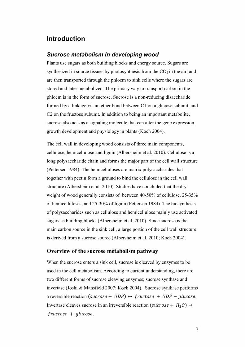
Figure 1. Sucrose metabolism pathway in sink cells leading to carbon
allocation to the cell wall.
There are several possible pathways leading towards carbon allocation for
the cell wall biosynthesis, one of them begins with sucrose cleavage by
invertase. After invertase cleaves sucrose into fructose and glucose,
hexokinase turns the glucose into glucose-6-phosphate.
Phosphoglucomutase (PGM) then turns the glucose-6-phosphate into
glucose-1-phosphate. UGP-glucose-pyrophosphorylase then catalyzes the
reactions where glucose-1-phosphate is turned into UDP-glucose
(Kleczkowski et al. 2010). The fructose cleaved by invertase is turned into
fructose-6-phosphate by fructokinase, fructose-6-phosphate, may be used to
synthesize lignin, and cell wall carbohydrates (figure 1) (Roach et al. 2012).
There is evidence that reduced fructokinase levels also affect the carbon
partitioning and the cell wall. An RNAi mediated reduction of fructokinase
in developing wood in aspen showed decreased levels of hexosephosphates
and UDP-glucose. Wood fibers also had a thinner cell walls, and there was a
reduction in cellulose (Roach et al. 2012).
Enzyme abbreviations: PMM: Phosphomannomutase PGM: Phosphoglucomutase PGI: Phosphoglucose isomerase UGPase: UGP-glucose-pyrophosphorylase SuSy: Sucrose synthase
8

The UDP-glucose is used by cellulose synthase complex as a substrate to
synthesize cellulose (Joshi and Mansfield 2007). Sucrose synthase can also
cleave sucrose directly into UDP-glucose and possibly provide UDP-
glucose directly to the cell synthase complex (Joshi and Mansfield 2007)
The origin of the UDP-glucose for the cellulose biosynthesis is disputed.
Possible enzymes responsible for supplying UDP-glucose for cell wall
biosynthesis are sucrose synthase and UGP-glucose-pyrophosphorylase
(Somerville 2006; Kleczkowski et al. 2010). Overexpression of the UDP-
glucose-pyrophosphorylase in hybrid aspen showed an increase in cellulose
content. Plants also showed reduced height growth and reduced stem
diameter (Coleman et al. 2007). Also overexpression of the UDP-glucose-
pyrophosphorylase in Jute (Corchorus capsularis), showed increased
cellulose content in stem tissues while the tree height growth was increased
(Zhang et al. 2013). Since invertase can supply substrate for UDP-glucose-
pyrophosphorylase, there might be a connection and interplay between the
two enzymes regarding the sucrose metabolism in developing wood.
Among the enzymes active in the cell wall biosynthesis are the
glycosyltransferases. The glycosyltransferases are active in the biosynthesis
and modification of the cell wall polysaccharides and glycoproteins (Rosén
et al. 2004). The glycosyltransferases can be divided into 95 families
(http://www.cazy.org/GlycosylTransferases.html), and among those are the
cellulose active subunits (CESA) and Golgi-localized type II integral
membrane proteins included (Somerville, 2006 ; Geshi et al., 2004). Golgi-
localized type II integral membrane proteins are able to synthesize the
pectin backbone (Bacic 2006), adding side chains to polysaccharides
(Persson et al. 2007) and synthesizing xylan (Faik et al. 2000; Peña et al.
2007; Cooper 2000). Hence glycosyltransferases and the Golgi-localized
type II integral membrane proteins play an important role in the biosynthesis
of parts of the cell wall.
9
Sucrose synthase Sucrose synthase (SUSY) can reversibly cleave sucrose in sink cells (Koch
2004). Some evidence points to that sucrose synthase could be directly
involved in the cellulose biosynthesis. SUSY has been found to be
associated with the plasma membrane in developing cotton fibers (Amor et
al. 1995). SUSY associated with the plasma membrane has also been shown
to be oriented in similar pattern as the cellulose microfibrils, seen in a
immunolocalization study in cotton fibers (Haigler et al. 2001). The study
concludes that SUSY associated to the plasma membrane possibly could
supply carbon directly to the cellulose synthesis (Haigler et al. 2001).
An immunoprecipitation study made in poplar suggests that SUSY co-
immunoprecipitated with the cellulose synthesis complex (Song, Shen, and
Li 2010). The author (Song, Shen, and Li 2010) also states that it is difficult
to harvest pure cellulose synthase (CESA) complex, and that some nearby
proteins could have been contaminating the samples, which could influence
the results. A similar study in Azuki bean (Vigna angularis) found that
immunolabeled SUSY with attached gold particles, was associated with a
plasma membrane structure similar to Cellulose active subunits (Fujii,
Hayashi, and Mizuno 2010). However it was not clearly established if it was
a CESA structure or not due to an uncertainty of the identification of the
SUSY protein (Fujii, Hayashi, and Mizuno 2010). Proteins may have many
variants that look similar but is slightly different in structure and function,
these variants are called isoforms. A study concluded that a SUSY isoform
SusC, that was identified in the original Amor et al. (1995) study, is mostly
located in near the cell wall (Brill et al. 2011). Considering the finding of
the SUSY isoform near the cell wall, it could indicate that SUSY possibly is
associated to the cell wall biosynthesis metabolism (Brill et al. 2011).
Interestingly there are findings that strongly indicate that SUSY is
redundant in sucrose metabolism in sink cells and cell wall biosynthesis,
and supports invertase’s role in sucrose metabolism instead. T-DNA mutant
lines in Arabidopsis with a quadruple null-mutant construct of four SUSY
isoforms SUS 1-4, showed that the null mutant SUSY did not have any
major effects on the growth and development (Barratt and Derbyshire
10

2009). T-DNA mutant lines in Arabidopsis roots with cytosolic invertase,
cinv1/cinv2 null-mutant constructs were also made, and invertase mutant
lines showed shorter root length (Barratt and Derbyshire 2009). These
results show that SUSY is redundant and invertase possibly is essential in
development and growth for sink cells in Arabidopsis. However a repeated
study in Arabidopsis made in similar conditions, claim that there are SUSY
activity in the null mutants, and that SUSY is therefore not redundant for
cellulose synthesis (Baroja-Fernández et al. 2012). A later study have found
that SUSY is not essential for cellulose biosynthesis in poplar, but that
SUSY affect the cell wall density and carbon allocation to wood (Gerber et
al. 2014). These findings raise the question which of the two sucrose
cleaving enzymes, sucrose synthase or invertase, that provides carbon for
cellulose synthesis in developing wood.
Invertases To be able to function in different compartments invertases have different
pH-optima, depending of their subcellular localization and function. The
invertases can be divided into alkaline/neutral invertases and acidic
invertases (Vargas and Salerno 2010). Invertases can also be organized after
their subcellular organization and function (Koch 2004). Vacuolar
invertases have been linked to sink initiation and generating vacuolar
hexoses leading to expansion, respiration and hexose-based sugar sensing
(Koch 2004). Cell wall invertases have been linked to functions such as;
continued sink initiation and cell expansion (Koch 2004). Further studies in
Arabidopsis have shown vacuolar invertase to possibly regulate the osmotic
potential (Sergeeva et al. 2006; Roitsch and González 2004). Another study
on the other hand speculate that invertase does not regulate the osmotic
potential, because sucrose accounts for only 1-2% of total sap osmolarity in
Arabidopsis roots (Ruan et al. 2010). The assumption is that when invertase
cleaves sucrose into two molecular compounds, invertase doubles the
amount of molecules and thus affects the osmotic pressure.
11

Alkaline/neutral invertases Neutral invertase has been shown to be important for plant growth and
development. A mutation of an alkaline/neutral invertase in rice homologue
to Arabidopsis AtCyt-inv1, showed shorter root growth, delayed flowering
and partial sterility (Jia et al. 2008). In the mutant lines there was also an
accumulation of sucrose and a reduction of hexose. Supplying glucose
exogenously could rescue the mutant (Jia et al. 2008). In Lotus japonicas a
mutant of a neutral/alkaline isoform LjINV1 showed severed growth in the
roots, shoots and impaired flowering (Welham et al. 2009). The shoot tips in
LjINV mutants showed a 9-fold increase in sucrose levels (Welham et al.
2009).
Apart from being located in the cytosol, neutral invertase has also been
found to have peptide signals that target proteins in mitochondria and
chloroplasts (Ji et al. 2005). Gene analysis and localization studies in rice
have shown invertase to be localized in mitochondria and plastids
(Murayama and Handa 2007). It has also been shown in Arabidopsis and
spinach that invertase is active inside chloroplasts (Vargas, Pontis, and
Salerno 2008). Another study in Arabidopsis examines two different
alkaline/neutral invertases, one mitochondrial and one cytosolic. The
absence of either the invertases is associated with an oxidative stress
defense gene expression. While an overexpression of the invertase genes
down-regulated the oxidative stress-responsive ascorbate peroxidase 2
(APX2) promoter (Xiang et al. 2011). This could imply that sucrose has a
regulatory role in plastids or that the sucrose pathway in plastids is more
extensive than previously thought (Murayama and Handa 2007).
12

Invertases in poplar In poplar 24 different types of invertase have been identified (Bocock et al.
2008). Of them 16 isoforms were neutral invertases predicted to be localized
with a cytosolic localization. The rest of the invertase isoforms in poplar
were acidic invertases, three of them predicted to be localized to the vacuole
and five cell wall invertases (Bocock et al. 2008). In Arabidopsis for
comparison there has only been found 9 neutral invertase isoforms (Nonis,
Ruperti, and Pierasco 2008). The fact that poplar have almost the double
number of neutral invertase isoforms compared to Arabidopsis (Bocock et
al. 2008), could indicate that neutral invertase play a more important role in
trees and
possibly wood
formation.
Figure 2. Analysis of the similarities between invertases in Populus trichocarpa and Arabidopsis, based on full-length sequences. The invertases are divided in α- and β clades based on different physical properties. (a) The protein similarity tree above is from of eight poplar and eight Arabidopsis acid invertases. (b) The protein similarity tree to the below is illustrating the difference between 12 poplar and 9 Arabidopsis alkaline/neutral invertases. Poplar has more neutral invertase isoforms, while Arabidopsis has more cell wall invertase isoforms (Bocock et al. 2008).
13

Neutral invertase expression in developing wood In poplar a neutral invertase (NIN) protein has been found to be interesting
in a screening of different genes during different stages of wood
development (Unpublished UPSC, RNA sequencing program). The NIN-
gene which we are interested in is clearly up-regulated during the secondary
cell wall synthesis during the wood formation (see figure 3). This suggests
that the NIN-gene potentially could be involved in formation of developing
wood, and be part of the cell-wall synthesis. The aim of this study is to
investigate and try to localize this NIN-protein.
1000 µm Figure 3a. Expression profile of the neutral invertase enzyme in developing wood in from a representative sample (Unpublished, UPSC RNA sequencing program). The samples are vertical slices taken from different parts of the developing wood. The Y-axis represents the strength of signal of the expression FPKM (Fragments Per Kilobase of transcript per Million mapped reads). The X-axis is in micrometers where in the developing wood profile the samples are extracted, from the left to the right, starting from the cambium to the maturation of the wood fibers (see figure 3b). The NIN peak expression profiles correspond to the secondary cell wall formation stage of wood development.
14
1000 µm
Figure 3b. Horizontal profile of the developing wood, adapted from (Mahboubi et al. 2013). The figure starts from the left with the cambium, early expansion, late expansion, secondary cell wall, and maturation. The figure can be read so that the developmental zones and scale bar corresponds with the scale bar in figure 3a.
Cam
bium
Secondary cell wall La
te e
xpan
sion
Maturation Early
exp
ansi
on
15

Methods and materials To understand more about the neutral invertase (NIN) and its role in
biosynthesis of the cell wall, a subcellular localization study was proposed.
A construct was created with the NIN-gene and an attached YFP-construct
at the N- and C-terminal side of the protein. Both N- and C-terminal
constructs were used because the targeting sequence for subcellular
localization can be found on either side of the protein, usually at the N-
terminal side. The constructs was made by using the Gateway technology™
and cloning process. With the constructs made, it was possible to transform
them into Agrobacterium and infiltrate tobacco leafs, and then visualize
YFP-signal in a confocal-microscope. The confocal-microscope works by
exciting the YFP by a laser and causing it to emit light a certain wavelength,
the emitted light then reflects back to the microscope. This makes it possible
to see fluorescence from the specific part of the cell where the YFP is
located.
Figure 4. Flow chart for Gateway cloning, process and steps from the Gateway pENTR D’TOPO manual. The end result is the binary plant transformation vector used in the transformation of Agrobacterium, and later the infiltration of tobacco plants.
16

Amplification
The NIN-gene was amplified from a cDNA library prepared from
developing wood. The amplification was made by two primers, the forward
NIN primer was CACCATGGATGGGACTAAAGAGATGGG, and the
reverse primer was GCAAGTCCAAGAAGATGATCTCCTGAG. The
amplification was done with Invitrogen® Phusion taq enzyme. The PCR
reaction was done by denaturation at 98 °C for 30 sec. Then the
amplification was done in 35 two-step cycles, with an initial denaturation
step at 98°C for 7 sec and extension step at 72°C for 1 min. Then after the
35 cycles the PCR reaction was ended with an extension step in 72°C for 10
min and after put on hold at 8°C.
The PCR products were then put onto a 100 ml 1,5% agarose gel with 5 µL
GelRed®. Then 50 µL of PCR products were put on the gel and run at 120 V
for 2 hours. Then a band was cut out from the gel corresponding to the NIN
gene size at 1674 bp. The gel fragment was then purified using a DNA gel
extraction kit from Thermo dynamics®. After the purification the
concentration of DNA was estimated by using Nanodrop®.
D’TOPO reaction The NIN gene was inserted into a pENTR™ D’TOPO® cloning kit from
Invitrogen™. Estimations from the Nanodrop® assay were used to match the
ratio between the D’TOPO® vector and the PCR product to 1:1. After
insertion the construct was transformed into chemically competent One
Shot® TOP10 E.Coli cells. The One shot® cells were incubated on ice for 30
min. The construct was added into two TOP10 cell aliquots. One aliquot
with 2 µL of D’TOPO® reagent was prepared, and in the other aliquot 6 µL
D’TOPO® reagent was added, this is ensure that enough colonies will
spawn. The cells were then heat shocked for 45 sec at 42°C. After SOC
medium addition it was incubated on ice for 2 minutes and then incubated at
37 °C for one hour. The cells were put onto plates with 25 µg/mL
kanamycin. There were two plates for each aliquot one with 50 µL of
transformants and one with 150 µL with transformants. After plating the
plates were incubated at 37°C overnight.
17

D’TOPO reaction information The Gateway D’TOPO® reactions use an enzyme Topoisomerase, which
can bind to duplex DNA at CCCTC sites. Topoisomerase originates from
Vaccinia virus (Shuman 1991). When the Topoisomerase binds to the site
on the D’TOPO® construct, it cleaves the phosphodiester backbone in one
strand. When the cleavage is done the energy is conserved by a covalent
bond formation between the 3´ phosphate of the cleaved strand, and a
tyrosyl residue (Tyr-274) of topoisomerase (Shuman 1994). After the
cleavage the 5´ hydroxyl of the original strand can attach the phosphor-
tyrosyl bond between the DNA and topoisomerase. The reaction can then be
reversed and the topoisomerase is released from the DNA strand (Shuman
1991). This process is used in the D’TOPO reaction to insert a construct into
an entry vector. The construct can then be transferred from entry vector to a
destination vector in a Gateway LR /BP ® reaction (Gateway cloning
manual).
Figure 5. Illustrating how the DNA strand looks like before and after the the D´TOPO reaction.
18

Preparing LB culture
After overnight incubation, six colonies were inoculated in 5 ml LB culture
containing 50µL/ml Spectinomycin. The cultures were then incubated
overnight with shaking at 225 rpm at 37°C. The Miniprep Qiagen kit® was
used for extracting the plasmids, and the plasmid concentrations were
measured with Nanodrop®.
LR reaction
The LR-reaction was done using the Gateway® LR Clonase™ II Enzyme
Mix kit. The incubation time for the final LR reaction was prolonged to
overnight. The LR reaction products were amplified in E.coli and the
selection of the complete construct was done using 50µL/ml Spectinomycin.
See figure 6 and 7 on the following pages for how the plasmid maps of the
destination vectors looks like.
Figure 7. Showing the plasmid map for the empty destination vector
from A Plasmid Editor®. The insert would later be placed between the
attR1 and attR2 sites between the EYFP and T35 sites. The map is
without any inserts.
20_pB7YWG211628 bp
LB 10111..10443
RB 3753..3952
attR2 1098..974
p35S 3733..2706
T35S 18..243Bar 11595..10447
attR1 2678..2554
ccdB 2553..1099
EYFP 966..247
Sm/SpR 10105..8856
19

Figure 8. Showing the plasmid map for the destination vector with the inserted NIN gene, from A plasmid editor®. The insert with the NIN-gene is showed in blue color. This is the expected result of how the destination vector with an insert would look like after the LR reaction.
LR reaction information The LR reaction is a transfer of a construct between the entry clone and the
destination vector done by LR Clonase™ II Enzyme. The LR reaction and
Gateway® system relies on mutated variants of the lambda att sites. The
variants can be made so attB1 recombines with attP1, but not with attP2, or
attP3. This means that there are little cross talk and no recombination
between non-specific sites (Katzen 2007). In the LR reaction the construct
of interest first is situated in an entry clone between two att sites, and then
transfers to the destination vector. The destination vector have two att sites
that are specific to the att sites in the entry clone (Hartley, Temple, and
Brasch 2000).
Figure 6. The red strand represent the construct of interest (Katzen 2007).
RB 7876..7677
LR_pENTRY_NIN2 Reverse Cterminal
11298 bp
attL2 10301..10310
LB 1518..1186
T35S 11281..11056
NIN2 8758..10284
EYFP 10333..11052attR2 10311..10325
source:pENTR/D-TOPO 10285..10310
p35S 7896..8757
Sm/SpR 1524..2773
Bar 34..1182
20
To ensure that the destination vector with the construct is selected and not
the entry clone, the destination vector and entry clones have different
antibiotic resistance genes. The destination vector also transfers ccdB gene
to the entry clone in the LR-reaction. The ccdB gene is a counter selectable
marker with gyrase-mediated double-stranded DNA breakage, which selects
cells of the entry clone that does not have the ccdB gene (Katzen 2007)
Restriction Digest The plasmids were tested with restriction digest using the FastDigest®
enzymes from Fermentas® and FastDigest® Green Buffer from Fermentas®.
The enzymes were Nco1, Nto1 for the D’TOPO construct, and Sal2 for the
destination vector. Then the digested plasmid products 50 µL, were put onto
gel 100 ml 1,5% agarose with 5 µL GelRed®, and was run at 120 V for 2
hours.
Sequencing Plasmids were submitted in a premixed sample, 50 µg/µL concentrations of
plasmids and 10 µMol/ml of primer. The two primers used for sequencing
were, YFP reverse primer sequence:
ACACGCTGAACTTGTGGCCGTTTA, and the sequence for the P35S
primer: CCACTATCCTTCGCAAGACCCTTC. The two samples were
submitted to Eurofins-MWG Operon in Munich.
Transformation of tobacco plants Plant material Tobacco plants (Nicotiana tabacum) were grown in 16 hours daytime and 8
hours nighttime for three weeks. The temperature was 20°C and relative
humidity was 70%.
Agrobacterium transformation The Agrobacterium infiltration started with transforming the expression
vector into Agrobacterium tumefaciens strain GV3103 (pMP90). The
Agrobacterium cells were thawed on ice in a 100 µL suspension. Then 1uL
with 2-3 ng of plasmid DNA were added by the 100 µL suspension by
mixing. The Agrobacterium suspension was transferred to an
electroporation cuvette, the cuvette was prechilled on ice.
21
The Agrobacterium suspension was then electroporated at a voltage adjusted
to the cuvette size. Then 1 ml of LB media was added to the cuvette and
transferred to a 2 ml eppendorf tube, and was grown at 28°C with shaking
for 14 hours. The solution was then plated on LB media with Gent25, Rif50
and Spectinomycin resistance. The plates were prepared and grown at 28°C
for two days. Then the culture was scaled up to 250 ml and was grown until
the OD600 reached to 1.0.
Tobacco infiltration
The Agrobacterium colonies from the expression vector transformation was
put in 5 ml falcon tubes with Rif50 and Spectinomycin resistance, and were
grown for 28°C with shaking overnight. Then 25 ml of the LB media is
grown at 28°C with shaking for 12-16 hours with 20µM Acetosyringone
added. The bacteria was precipitated at 10.000 rpm for 15 min and adjusted
to a final OD600 of 0.4. Then the culture was incubated in room temperature
for 3 hours. The infiltration was done with a syringe with the nozzle pressed
against the lower (abaxial) epidermis of the leaf. The injection was done
while exerting a counter-pressure by pressing a finger on the other side of
the leaf. The borders of the infiltrated area were marked by a permanent pen
and the plants were incubated in normal growing conditions for 2 days.
Confocal microscopy
A small square was cut from the infiltrated area was cut from the tobacco
leafs, and placed on a slide with water added. The slide were covered with a
cover-slide and put under the confocal-microscope. The confocal
microscope used was a Leica TCS SP2 AOBS spectral system with laser
excitation lines at 51/364, 405, 458, 476, 488, 496, 514, 561 and 633 nm
mounted on an inverted microscope.
22

Subcellular localization prediction The peptide sequence was tested in programs predicting the subcellular
localization. Three programs were used for general subcellular localization
(1-3). When the results from the fluorescence from the confocal-microscope
pointed to a possible Golgi localization, a Golgi specific localization
program were also used (4). Prediction programs 1-4:
1. Extensive feature detection of N-terminal protein sorting signals
iPSORT http://ipsort.hgc.jp/index.html (Bannai et al. 2002)
2. Subnuclear Compartments Prediction System
http://array.bioengr.uic.edu/subnuclear.htm (Lei and Dai 2005)
3. CELLO- subCELlular LOcalization predictor.
http://cello.life.nctu.edu.tw/cgi/main.cgi) (Su et al. 2007)
4. Prediction of Golgi Localized Transmembrane Proteins.
http://ccb.imb.uq.edu.au/golgi/golgi_predictor.shtml) (Yuan and Teasdale
2002).
23

Results The results were achieved using a construct of the neutral invertase gene
attached with YFP-tags fused at the N- and C-terminal side of the protein.
The constructs were transformed into Agrobacterium and infiltrated in
tobacco leafs, and the visualized in a confocal microscope to see the YFP-
spectrum (see figure 15-19). Prediction of the subcellular prediction was
made using the peptide sequence of the neutral invertase. The result section
is ordered by first showing the laboratory results, followed by the
microscopy results and the prediction of the subcellular localization results.
Construct laboratory results
There were several steps in the cloning procedure; from the PCR
amplification to the Gateway cloning reactions. The cloning was not
initially successful during the experimentation period, a colleague in the
group thankfully could help after with providing a cloning of the C-and N-
terminal constructs, so that I could start the tobacco infiltration with a
working construct as well as visualizing the YFP-signal with the confocal
microscope. The laboratory results below are the initial attempt of cloning
of the C-terminal construct. In the initial attempt of cloning the C-Terminal
construct, the sequencing of the products from the LR reaction did not
contain any evidence that the NIN-gene was inside the vector at the correct
location. PCR testing with NIN gene primer suggested that a construct with
the size of the NIN gene could be inside the D’TOPO vector. The
sequencing results were on the other hand convincing and it is likely that the
insert was not inside the destination vector.
The reason for the laboratory results is probably an error somewhere
between the first PCR and the D’TOPO cloning. One example of a
laboratory error could be a use of too small amounts of D’TOPO vector
compared to the recommended amount, due to a limited D’TOPO vector
supply at the time. There are numerous of other possibilities such as
handling of bacteria wrong or giving the wrong amount of an antibiotic.
24

Concluding which of the possibilities that led to the initial result is difficult,
but because of the D’TOPO vector seemed faulty and the initial PCR that
seemed correct, it is probable that the laboratory error happened before or
during the first D’TOPO cloning.
Results PCR
The picture from the electrophoresis seems to confirm that the initial
amplification and PCR was successful. The PCR result showed a band
approximately at 1700 bp (see figure 9), this is close to the equivalent of the
NIN gene size of 1674 bp.
Digestion of the products from the D’TOPO reaction
The gel pictures from the D’TOPO reaction showed ambiguous results. The
digestion and the pictures from the gel showed that there was not a
correlation between what the gels showed, and the prediction made from the
plasmid maps (see figure 10). The restriction digestion was made with
enzymes Nco1 and Nto1. The gel showed one band around 5000 bp instead
of the expected three bands (see figure 11). This could mean that the NIN-
gene insert was not there, the plasmid map was not correct or perhaps the
plasmid was cut only once and the plasmid were linear.
Restriction Digest
Restriction digestion of the plasmid product from the Gateway LR reaction
with enzyme Sal2, produced two bands (see figure 12). The bands are lower
in size than the expected (see figure 13). This means that the digestion of the
product from the LR reaction is not in correlation with the expected sizes.
PCR – LR-reaction
A PCR was also made for the different vectors. Both the bacterial E.coli
colonies from the D’TOPO-reaction and from the colonies from the LR-
reaction were tested in a PCR-reaction. Overall there seems to be bands in
the correct region of around 1700 bp for both LR destination vector
products and D’TOPO reaction products (See figure 14). One colony from
the LR reaction didn’t produce any bands at all. In some lanes there is a
25

band in the 1700 region, but it’s of bit unclear size and structure. There are
also artifacts in some lanes, which mean that the results from the PCR might
be questioned.
Results sequencing
Results from the sequencing showed that there was not any inserts in the
destination vector. The sequencing was made by using primers from the
promoter 35:S and the YFP-gene. The YFP and promoter 35:S is next to the
NIN gene according to the plasmid map (see figure 8, Methods and
materials). The sequencing results showed both correlations with the
opposite genes. In the sequencing results from the forward p35:S primer the
YFP gene sequence could be seen, and in the sequencing results from the
reverse YFP primer the p35:S gene sequence could be seen. This result
indicates that the correct destination vector is sequenced, but it is empty
without an inserted NIN gene. Two different colonies from the LR-reaction
were tested and both showed similar results.
Figure 9. Picture of the agarose gel after the PCR reaction. The green arrow indicates where the expected band from the PCR is. The ladder used are 1kb plus ladder from Thermo Scientific. The band in the left lane is situated between the ladder bands in the right lane of 1500 bp and 2000 bp.
Expected band size 1674 bp
26

¨
Figure10. The gel from a restriction digest of the D’TOPO vector after the D’TOPO reaction with restriction enzymes Nco1 and Nto1. The green arrows represent the expected band sizes after a restriction digest according to the plasmid map prediction (see figure 11). As seen the bands showing in the picture does not match with the expected band sizes shown by the green arrows.
Figure 11. Expected band distribution after the digestion of the DTOPO vector with restriction enzymes Nco1 and Not1, with a prediction made from “A plasmid editor®”.
pENTR_D-TOPO.apeSize site1 site2 Mass %
671 NotI687 NcoI957 NcoI
3968 NcoI 957 NotI 671 93270 NcoI 687 NcoI 957 616 NotI 671 NcoI 687 1
Expected bands size
27

Figure 12. The gel from restriction digest of the LR reaction products. The green arrows represent the expected band sizes after restriction digest, with a prediction made from the plasmid map (figure 8, materials and methods) (see figure 13). The five tested LR-colonies are showing in order 1-5. The green arrows points to the expected band sizes. The bands showing are not correlated to the expected bands size. The bands are lower in size compared to the expected band sizes. All five colonies show similar results.
Figure 13. Above are expected band sizes for restriction digestion for the products from the LR reaction with the enzyme Sal1, using a prediction from A plasmid editor® (figure 8, Materials and methods).
Expected bands size
28

Figure 14. Picture of the gel from with LR-reaction products. The ladder used are 1 kb plus from Thermodynamics. LR 2 and LR 3 is the destination vector plasmids extracted from the Gateway LR reaction. There are two different LR-colonies tested and three D’TOPO colonies tested. The picture shows that there are bands at the correct size corresponding to the NIN-gene at 1674 bp, from both the LR and D’TOPO reaction products. The bands on the other hand are unclear in shape and there are artifacts in many of the lanes. The PCR results are therefore a bit ambiguous with some correct bands, but with poor quality of the shape of the bands and artifacts showing.
Expected bands size 1674 bp
29

Confocal microscopy results The fluorescent pattern from the neutral invertase (NIN) C-terminal YFP-
construct showed an intervallic dotted pattern along the border of the leaf
epidermal cells (figure 15). The border part would be in the cytosol in
tobacco leaf cells (Brandizzi et al. 2002). The dotted expression indicated
that the N-terminal YFP is located in some kind of cell compartment (figure
15). If the signaling part is intact at the N-terminal side of the protein, the C-
terminal YFP-tag would be a good indicator of where the protein is located
in native conditions.
The N-terminal YFP-NIN fusion protein looks to be localized in a
fluorescent pattern in the cytoplasm in the tobacco leaf cell. The fluorescent
signal is strong and it is spread all around the cytosol (see figure 18). It is
also possible to see fluorescence in the canals in the middle of the cell
between cytosolic parts (see figure 19). In the N-terminal fluorescence
results it also looks like a pattern with yellow dots that fluorescence
brighter, this pattern would be similar to the aggregated pattern found in the
C-terminal construct results. Both the N- and C-terminal YFP-signal can be
seen in the cytoplasm, but the N-terminal signal seems more widespread,
while the C-terminal construct looks more aggregated (figure 15). The
absence of a fluorescent pattern seen in the non-infiltrated samples (see
figure 17), confirms the findings in the infiltrated samples, where it is
possible to see a fluorescent pattern. The red chlorophyll autofluorescence
signal does not correlate with the yellow YFP-signal in the C- or N-terminal
YFP-tags (see figure 16). This means that it is likely that the NIN-protein is
not localized in the chloroplasts.
30
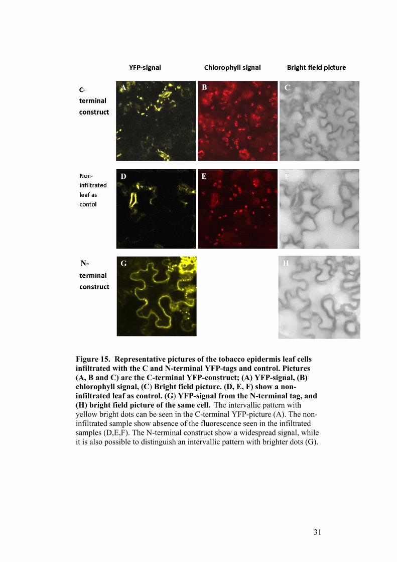
Figure 15. Representative pictures of the tobacco epidermis leaf cells infiltrated with the C and N-terminal YFP-tags and control. Pictures (A, B and C) are the C-terminal YFP-construct; (A) YFP-signal, (B) chlorophyll signal, (C) Bright field picture. (D, E, F) show a non-infiltrated leaf as control. (G) YFP-signal from the N-terminal tag, and (H) bright field picture of the same cell. The intervallic pattern with yellow bright dots can be seen in the C-terminal YFP-picture (A). The non-infiltrated sample show absence of the fluorescence seen in the infiltrated samples (D,E,F). The N-terminal construct show a widespread signal, while it is also possible to distinguish an intervallic pattern with brighter dots (G).
A B C
D
E
G
H
N-
31

Pictures NIN C-Terminal YFP-tag
Figure 16. Tobacco leaf infiltrated with C-terminal YFP-tag. YFP-signal (A), chlorophyll signal (B), bright field picture (C). In this picture the yellow fluorescence is clearly concentrated in dots distributed along the upper border of a tobacco leaf cell. The red chlorophyll signal is not at the same location at the yellow YFP-signal (B). This makes it likely that that the YFP-signal does not come from the chlorophyll containing chloroplasts.
Non-infiltrated tobacco leaf as control
Figure 17. A non-infiltrated tobacco leaf as control sample. YFP-signal (A), chlorophyll signal (B), bright field picture (C). Compared to the florescence in other infiltrated samples, an absence of the fluorescent signal can be seen in this picture. There are not any signs of a pattern other than the background florescence, the yellow fluorescence seen at the upper left of picture (A) show what looks a stomata. The non-infiltrated control pictures confirm the findings with the infiltrated samples, where there is a significant amount of yellow florescence shown.
A
A B C
A B
32

Pictures NIN N-Terminal YFP-tag
Figure 18. Tobacco leaf infiltrated with an N-terminal YFP-tag. YFP-signal (A), Bright field picture (B). In this picture the yellow fluorescence is spread all around the border of the tobacco leaf cell. There are some dots along the border that fluoresce more brightly.
Figure 19. Tobacco leaf infiltrated with an N-terminal YFP-tag. YFP-signal (A). Bright field picture (B). It is possible to distinguish canals crossing in the middle of the cell between brighter dots in the border area. These fluorescent “canals” might be cytoplasmic canals (see discussion).
A
A
B
B
33

Prediction of the subcellular localization
I analyzed the NIN-protein sequence using prediction programs for
subcellular localization. General prediction programs predicts that the
protein is not localized in the mitochondria, chloroplast and that the protein
does not contain a signal peptide (CELLO, Bannai et al 2002; IPSORT, Su
et al. 2006), one of the programs indicated that the NIN-protein is likely to
be cytoplasmic protein (CELLO, Bannai et al 2002). Another prediction
program indicates that the protein is localized in the Nuclear Lamina (SCPS,
Lei et al.2005). When the protein sequence was tested in a specific Golgi
protein localization prediction program (Yuan & Teasdale 2002
http://ccb.imb.uq.edu.au/golgi/golgi_predictor.shtml), the sequence tested
positive as a Golgi type 2 transmembrane protein. The positive prediction
result was significant with a value of 24, clearly above the threshold value
20 marking significance. The prediction as a Golgi type 2 membrane protein
stands out as being a clear significant result among the predictions from the
prediction programs.
In summary the results from the confocal microscope indicate that the NIN-
protein is located in the cytoplasm. The signal is more aggregated for the C-
terminal tag, and more widespread in the cytoplasm for the N-terminal tag.
The aggregation seen could indicate a subcellular compartment localization.
Combined with the results from the Golgi prediction program (Yuan &
Teasdale 2002 http://ccb.imb.uq.edu.au/golgi/golgi_predictor.shtml), it
gives a hint of a possible localization.
34
Discussion NIN N- and C-terminal construct results
Results from the neutral invertase (NIN) N-terminal YFP-tag show
fluorescence in the cytoplasmic region of the tobacco leaf epidermis cells.
The fluorescence is clear in the N-terminal pictures and the fluorescence can
be seen all around the cytoplasm of the tobacco leaf cell (see figure 15). The
pictures from the N-terminal YFP-NIN also show fluorescence in the
cytosolic canals in the middle of the cell connecting the cytoplasm (see
figure 19). If the signaling part of the protein has been disrupted by the N-
terminal YFP-NIN construct, the N-terminal construct would say little of the
native location of the NIN protein. There is also a pattern of more bright
dots that fluoresces in an intervallic pattern, similar to the pattern that can be
seen in fluorescence from the C-terminal NIN YFP-construct. This
similarity indicates that the N-terminal fusion protein to some extent show
aggregation in a similar way as the C-terminal YFP-NIN construct.
The results from the NIN C-terminal YFP-tag show fluorescence in the
cytoplasmic area of the tobacco leaf cell. The fluorescence from the C-
terminal YFP-tag can be seen in a dotted intervallic pattern along the border
of the leaf cells (see figure 16). The dotted pattern would indicate that the C-
terminal YFP-tag is aggregated in some way. The aggregation in an
intervallic pattern could mean that the NIN gene is targeted to a
compartment or perhaps membrane. Assuming that the C-terminal tag is less
likely to interrupt the protein targeting, it is likely that the fluorescence from
the C-terminal give a better hint of where the NIN protein natively is located
in tobacco leaf cells. The pictures show that the signal from the yellow
fluorescent protein wavelength is not correlated with the signal from the
chlorophyll autofluorescence wavelength (see figure 16-17). This finding
makes it probable that the C- or N-terminal YFP do not aggregate in or
nearby chloroplasts. The pictures from the non-infiltrated leaf show little
fluorescence in the YFP-wavelength spectrum, and indicate that the results
seen with the C-terminal YFP-tag are different from control.
35

There are a number of different locations the NIN-protein could be targeted,
including compartments such as: plastids, mitochondria, Golgi apparatus
and the ER. Since the cytoplasm in tobacco leafs cells is seen in an area near
the border, the YFP might also be in connection with the plasma membrane.
When comparing the intervallic pattern with other studies with tobacco
leaves, the results shows some resemblance to structures labeled as the
Golgi apparatus (Hanton and Brandizzi 2006; Brandizzi et al. 2002; Stefano
et al. 2006)
Combined with the conclusions from the prediction results the Golgi
apparatus is a candidate for explaining the aggregated compartment-like
pattern showing from the fluorescence results. The Golgi apparatus is where
hemicelluloses and pectin are synthesized to become part of the cell-wall
matrix during cell expansion in developing wood (Cooper 2000). If the
NIN-protein somehow is associated with the Golgi apparatus it could
support the view that neutral invertase is contributing to the cell wall
biosynthesis (Figure 3 UPSC) (Gerber et al. 2014).
It should also be noted that the experiment is done with a hybrid aspen gene
in tobacco, and the results could be different with the same experiment done
natively in hybrid aspen. A similar study made with the NIN-protein in
aspen protoplasts would be illuminating to show if the YFP-signal correlates
with those compartments. Co-localization studies are in progress in aspen
with markers for Tonoplast, ER, Golgi and plastids (Takata & Eriksson
2012). These experiments would make it possible to see if the NIN-YFP
signal match with the compartment tags or not.
Prediction results
Results from the prediction programs for subcellular localization indicate
that the peptide sequence is matching a Golgi localization. Some general
prediction programs claimed the protein to be either cytoplasmic or Nuclear
Lamina (CELLO, Bannai et al. 2002; IPSORT, Su et al. 2006; SCPS, Lei
and Dai 2005), but the clearest significant prediction came from a Golgi-
protein prediction program (Yuan and Teasdale 2002). The program
36

predicted that the protein sequence as a Golgi Type II transmembrane
protein.
The prediction result was significant with a value of 24, clearly above the
threshold value marking significance. Golgi Type II membrane proteins are
responsible for synthesizing the pectin, xylan and adding side chains to
polysaccharides (Bacic 2006; Persson et al.2007; Pena et al. 2007). Golgi
Type II membrane proteins are therefore important in the assembly of the
cell wall components hemicelluloses and polysaccharides. Since invertase is
not a typical Golgi type II membrane protein such as glycosyltransferases
(Cooper 2000), it raises the question of the function of invertase at the same
possible localization as Golgi Type II membrane proteins.
Considering the results, literature and the up-regulation of the NIN-gene in
the developing wood in hybrid aspen; the prediction results gives reason to
suspect that the NIN-protein could be involved in supplying the Golgi cell
wall biosynthesis machinery with sucrose derived hexose sugars.
Cell wall properties
The Golgi apparatus is important for sorting, modifying and packaging
proteins to different locations in the plant cell. Golgi apparatus enzymes are
able to modify proteins by both phosphorylation and glycosylation (Pavelka
and Mironov 2008). The Golgi apparatus is where the biosynthesis for the
glycosaminoglycans takes place and where the hemicelluloses in the cell
wall are modified (Cooper 2000). If the NIN is located in the Golgi
apparatus, an absence of the NIN in the NIN RNAi-null mutants would
supposedly affect synthesizing proteins in the Golgi-apparatus. It would
therefore be advisable to look for the effects of an absence of the supplier of
sucrose derived hexose sugars possibly would cause. For example if the
NIN feeds one of the glycosyltransferases in the Golgi-apparatus that affect
the synthesis of hemicelluloses, it would be worthwhile to test for changes
in the hemicellulose composition (Cooper 2000) in the NIN RNAi-mutant
lines.
37

But there are many possible glycosyltransferases and many have different
functions. A localization study made with isotope tagging found 197 Golgi
apparatus proteins, of which 41 of are glycosyltransferases described in the
CAZy database (Nikolovski et al. 2012)
(http://www.cazy.org/GlycosylTransferases.html). A way to further
investigate is by comparing the phenotype of the NIN RNAi-mutant lines,
with RNAi null mutants of previously investigated Golgi-
glycosyltransferases, affecting the cell wall biosynthesis (Scheible et
al.2004; Nikolovski et al. 2012).
After this study was finished very preliminary evidence suggests (Rende, U,
Unpublished) that the pectin levels may have changed in mutant NIN RNAi-
lines. A previous described glycosyltransferase mutant “QUASIMODO1”
have been found to be associated with pectin synthesis (Bouton et. al 2002),
and an isoform have been located at a Golgi-localized type II integral
membrane protein location (Miao et al. 2011). It is an unproven association,
but a possibility of using previous studies when looking at the cell wall
properties in the NIN RNAi-mutant lines.
According to (Scheible et al.2004) many RNAi-null mutants of
glycosyltransferases that affects the cell wall biosynthesis, show little or no
phenotypical variation compared to control. It could then be even more
difficult to find changes in the phenotype of an NIN RNAi null-mutant,
possibly feeding carbon to a glycosyltransferase affecting the cell wall
biosynthesis. If the NIN supplies carbon from sucrose to the Golgi
apparatus, it would also be surprising that only one aspect of the Golgi-
machinery is affected, and other Golgi functions that require sucrose derived
hexose sugars would remain relatively unaffected.
38

Conclusion The results from the neutral invertase investigation look promising. My
hypothesis considering the results is that it is plausible that the neutral
invertase protein is localized in the Golgi apparatus supplying the Golgi
Type II membrane proteins with carbon; thus affecting the secondary cell
wall growth. The Golgi Type II membrane proteins would need sugars to
synthesize pectin and hemicelluloses (Bacic 2006 ; Persson et al.2007 ; Pena
et al. 2007), and the neutral invertase protein could perhaps be the link to
supply the needed carbon from sucrose. The hypothesis that the neutral
invertase protein is near and is part of the supply pathway to the
glycosyltransferases is based on very preliminary results and would need
much testing and experimenting to be verified. Such tests could be
localization studies made in hybrid aspen with fluorescence tags for each of
the compartments (Takata and Eriksson 2012), Western-blot to confirm the
fusion protein size, and also testing the properties of hybrid aspen mutants
to see how the cell-wall properties change.
If the NIN-protein is shown to be inside one compartment such as Golgi, it
could also be interesting to investigate if there is any connection to the
osmotic regulation such as theorized by (Sergeeva et al. 2006; Roitsch &
González 2004; Ruan et al.2010). In previous studies and characterization of
the Neutral and Alkaline invertases in Populus trichocarpa, it is predicted
that all 16 Neutral and Alkaline invertases are localized in the cytosol
(Bocock et al.2008). If the neutral invertase protein is localized in the Golgi
apparatus, it would also mean that the way of thinking about neutral and
alkaline invertases in hybrid aspen would have to be rewritten.
Further studies also could confirm the importance of invertase versus
sucrose synthase in hybrid aspen. If the neutral invertase protein proves to
be involved in the cell wall biosynthesis pathway, it would support studies
that claim that sucrose synthase can be by-passed in the cell wall
biosynthesis pathway (Gerber et al. 2014) by invertase.
39
Reference list
Albersheim, Peter, Alan Darvill, Keith Roberts, Ron Sederoff, and Andrew Staehelin. 2010. Plant Cell Walls. 1st ed. Garland Science.
Amor, Y, C H Haigler, S Johnson, M Wainscott, and D P Delmer. 1995. “A Membrane-Associated Form of Sucrose Synthase and Its Potential Role in Synthesis of Cellulose and Callose in Plants.” Proceedings of the National Academy of Sciences of the United States of America 92 (20) (September 26): 9353–7. http://www.pubmedcentral.nih.gov/articlerender.fcgi?artid=40983&tool=pmcentrez&rendertype=abstract.
Bacic, Antony. 2006. “Breaking an Impasse in Pectin Biosynthesis.” Proceedings of the National Academy of Sciences of the United States of America 103 (15) (April): 5639–40. doi:10.1073/pnas.0601297103.
Bannai, Hideo, Yoshinori Tamada, Osamu Maruyama, Kenta Nakai, and Satoru Miyano. 2002. “Extensive Feature Detection of N-Terminal Protein Sorting Signals.” Bioinformatics (Oxford, England) 18 (2) (February): 298–305.
Baroja-Fernández, Edurne, Francisco José Muñoz, Jun Li, Abdellatif Bahaji, Goizeder Almagro, Manuel Montero, Ed Etxeberria, Maite Hidalgo, María Teresa Sesma, and Javier Pozueta-Romero. 2012. “Sucrose Synthase Activity in the sus1/sus2/sus3/sus4 Arabidopsis Mutant Is Sufficient to Support Normal Cellulose and Starch Production.” Proceedings of the National Academy of Sciences of the United States of America 109 (1) (January 3): 321–6. doi:10.1073/pnas.1117099109. http://www.pubmedcentral.nih.gov/articlerender.fcgi?artid=3252950&tool=pmcentrez&rendertype=abstract.
40
Barratt, DHP, and Paul Derbyshire. 2009. “Normal Growth of Arabidopsis Requires Cytosolic Invertase but Not Sucrose Synthase.” Proceedings of the … 106 (31). http://www.pnas.org/content/106/31/13124.short.
Bocock, Philip N, Alison M Morse, Christopher Dervinis, and John M Davis. 2008. “Evolution and Diversity of Invertase Genes in Populus Trichocarpa.” Planta 227 (3) (February): 565–76. doi:10.1007/s00425-007-0639-3. http://www.ncbi.nlm.nih.gov/pubmed/17938954.
Bouton, Sophie, Edouard Leboeuf, Gregory Mouille, Marie-thérèse Leydecker, Joël Talbotec, Fabienne Granier, Marc Lahaye, Herman Höfte, and Hoai-nam Truong. 2002. “QUASIMODO1 Encodes a Putative Membrane-Bound Glycosyltransferase Required for Normal Pectin Synthesis and Cell Adhesion in Arabidopsis” 14 (October): 2577–2590. doi:10.1105/tpc.004259.bone.
Brandizzi, Federica, Erik L Snapp, Alison G Roberts, Jennifer Lippincott-Schwartz, and Chris Hawes. 2002. “Membrane Protein Transport between the Endoplasmic Reticulum and the Golgi in Tobacco Leaves Is Energy Dependent but Cytoskeleton Independent: Evidence from Selective Photobleaching.” The Plant Cell 14 (6) (June): 1293–309. doi:10.1105/tpc.001586.1294.
Brill, Elizabeth, Michel van Thournout, Rosemary G White, Danny Llewellyn, Peter M Campbell, Steven Engelen, Yong-Ling Ruan, Tony Arioli, and Robert T Furbank. 2011. “A Novel Isoform of Sucrose Synthase Is Targeted to the Cell Wall during Secondary Cell Wall Synthesis in Cotton Fiber.” Plant Physiology 157 (1) (September): 40–54. doi:10.1104/pp.111.178574. http://www.pubmedcentral.nih.gov/articlerender.fcgi?artid=3165887&tool=pmcentrez&rendertype=abstract.
41

Coleman, Heather D, Thomas Canam, Kyu-Young Kang, David D Ellis, and Shawn D Mansfield. 2007. “Over-Expression of UDP-Glucose Pyrophosphorylase in Hybrid Poplar Affects Carbon Allocation.” Journal of Experimental Botany 58 (15-16) (January): 4257–68. doi:10.1093/jxb/erm287. http://www.ncbi.nlm.nih.gov/pubmed/18182429.
Cooper, GM. 2000. The Cell: A Molecular Approach. 2nd ed. Sinauer Associates.
Faik, a, M Bar-Peled, a E DeRocher, W Zeng, R M Perrin, C Wilkerson, N V Raikhel, and K Keegstra. 2000. “Biochemical Characterization and Molecular Cloning of an Alpha-1,2-Fucosyltransferase That Catalyzes the Last Step of Cell Wall Xyloglucan Biosynthesis in Pea.” The Journal of Biological Chemistry 275 (20) (May): 15082–9. doi:10.1074/jbc.M000677200.
Fujii, Satoshi, Takahisa Hayashi, and Koichi Mizuno. 2010. “Sucrose Synthase Is an Integral Component of the Cellulose Synthesis Machinery.” Plant & Cell Physiology 51 (2) (February): 294–301. doi:10.1093/pcp/pcp190. http://www.ncbi.nlm.nih.gov/pubmed/20056592.
Gerber, Lorenz, Bo Zhang, Melissa Roach, Umut Rende, András Gorzsás, Manoj Kumar, Ingo Burgert, Totte Niittylä, and Björn Sundberg. 2014. “Deficient Sucrose Synthase Activity in Developing Wood Does Not Specifically Affect Cellulose Biosynthesis, but Causes an Overall Decrease in Cell Wall Polymers.” The New Phytologist 203 (4) (September): 1220–30. doi:10.1111/nph.12888.
Geshi, Naomi, Bodil Jørgensen, and Peter Ulvskov. 2004. “Subcellular Localization and Topology of beta(1-->4)galactosyltransferase That Elongates beta(1-->4)galactan Side Chains in Rhamnogalacturonan I in Potato.” Planta 218 (5) (March): 862–8. doi:10.1007/s00425-003-1168-3.
42

Haigler, C H, M Ivanova-Datcheva, P S Hogan, V V Salnikov, S Hwang, K Martin, and D P Delmer. 2001. “Carbon Partitioning to Cellulose Synthesis.” Plant Molecular Biology 47 (1-2) (September): 29–51. http://www.ncbi.nlm.nih.gov/pubmed/11554477.
Hanton, Sally L, and Federica Brandizzi. 2006. “Fluorescent Proteins as Markers in the Plant Secretory Pathway.” Microscopy Research and Technique 69 (3) (March): 152–9. doi:10.1002/jemt.20276.
Hartley, James L, Gary F Temple, and Michael A Brasch. 2000. “DNA Cloning Using In Vitro Site-Specific Recombination”: 1788–1795. doi:10.1101/gr.143000.that.
Ji, Xuemei, Wim Van den Ende, Andre Van Laere, Shihua Cheng, and John Bennett. 2005. “Structure, Evolution, and Expression of the Two Invertase Gene Families of Rice.” Journal of Molecular Evolution 60 (5) (May): 615–34. doi:10.1007/s00239-004-0242-1. http://www.ncbi.nlm.nih.gov/pubmed/15983871.
Jia, Liqiang, Botao Zhang, Chuanzao Mao, Jinhui Li, Yunrong Wu, Ping Wu, and Zhongchang Wu. 2008. “OsCYT-INV1 for Alkaline/neutral Invertase Is Involved in Root Cell Development and Reproductivity in Rice (Oryza Sativa L.).” Planta 228 (1) (June): 51–9. doi:10.1007/s00425-008-0718-0. http://www.ncbi.nlm.nih.gov/pubmed/18317796.
Joshi, Chandrashekhar P, and Shawn D Mansfield. 2007. “The Cellulose Paradox--Simple Molecule, Complex Biosynthesis.” Current Opinion in Plant Biology 10 (3) (June): 220–6. doi:10.1016/j.pbi.2007.04.013. http://www.ncbi.nlm.nih.gov/pubmed/17468038.
Katzen, Federico. 2007. “Gateway(®) Recombinational Cloning: A Biological Operating System.” Expert Opinion on Drug Discovery 2 (4) (April): 571–89. doi:10.1517/17460441.2.4.571.
43

Kleczkowski, Leszek, Sabine Kunz, and Malgorzata Wilczynska. 2010. “Mechanisms of UDP-Glucose Synthesis in Plants.” Critical Reviews in Plant Sciences 29 (4) (July): 191–203. doi:10.1080/07352689.2010.483578.
Koch, Karen. 2004. “Sucrose Metabolism: Regulatory Mechanisms and Pivotal Roles in Sugar Sensing and Plant Development.” Current Opinion in Plant Biology 7 (3) (June): 235–46. doi:10.1016/j.pbi.2004.03.014. http://www.ncbi.nlm.nih.gov/pubmed/15134743.
Lei, Zhengdeng, and Yang Dai. 2005. “An SVM-Based System for Predicting Protein Subnuclear Localizations.” BMC Bioinformatics 6 (January): 291. doi:10.1186/1471-2105-6-291.
Mahboubi, Amir, Christine Ratke, András Gorzsás, Manoj Kumar, Ewa J Mellerowicz, and Totte Niittylä. 2013. “Aspen SUCROSE TRANSPORTER3 Allocates Carbon into Wood Fibers.” Plant Physiology 163 (4) (December): 1729–40. doi:10.1104/pp.113.227603. http://www.ncbi.nlm.nih.gov/pubmed/24170204.
Murayama, Seiji, and Hirokazu Handa. 2007. “Genes for Alkaline/neutral Invertase in Rice: Alkaline/neutral Invertases Are Located in Plant Mitochondria and Also in Plastids.” Planta 225 (5) (April): 1193–203. doi:10.1007/s00425-006-0430-x. http://www.ncbi.nlm.nih.gov/pubmed/17086397.
Nikolovski, Nino, Denis Rubtsov, Marcelo P Segura, Godfrey P Miles, Tim J Stevens, Tom P J Dunkley, Sean Munro, Kathryn S Lilley, and Paul Dupree. 2012. “Putative Glycosyltransferases and Other Plant Golgi Apparatus Proteins Are Revealed by LOPIT Proteomics.” Plant Physiology 160 (2) (October): 1037–51. doi:10.1104/pp.112.204263. http://www.pubmedcentral.nih.gov/articlerender.fcgi?artid=3461528&tool=pmcentrez&rendertype=abstract.
44

Nonis, Alberto, Benedetto Ruperti, and Alessandro Pierasco. 2008. “Neutral Invertases in Grapevine and Comparative Analysis with Arabidopsis, Poplar and Rice.” Planta 229 (1) (December): 129–42. doi:10.1007/s00425-008-0815-0. http://www.ncbi.nlm.nih.gov/pubmed/18800225.
Pavelka, Margit, and Alexander Mironov. 2008. The Golgi Apparatus: State of the Art 110 Years after Camillo Golgi’s Discovery. Berlin: Springer.
Peña, Maria J, Ruiqin Zhong, Gong-Ke Zhou, Elizabeth a Richardson, Malcolm a O’Neill, Alan G Darvill, William S York, and Zheng-Hua Ye. 2007. “Arabidopsis Irregular xylem8 and Irregular xylem9: Implications for the Complexity of Glucuronoxylan Biosynthesis.” The Plant Cell 19 (2) (February): 549–63. doi:10.1105/tpc.106.049320.
Persson, Staffan, Kerry Hosmer Caffall, Glenn Freshour, Matthew T Hilley, Stefan Bauer, Patricia Poindexter, Michael G Hahn, Debra Mohnen, and Chris Somerville. 2007. “The Arabidopsis Irregular xylem8 Mutant Is Deficient in Glucuronoxylan and Homogalacturonan, Which Are Essential for Secondary Cell Wall Integrity.” The Plant Cell 19 (1) (January): 237–55. doi:10.1105/tpc.106.047720.
Pettersen, Roger. 1984. The Chemistry of Solid Wood. Edited by Roger Rowell. doi:10.1021/ba-1984-0207.
Roach, Melissa, Lorenz Gerber, David Sandquist, András Gorzsás, Mattias Hedenström, Manoj Kumar, Marie Caroline Steinhauser, et al. 2012. “Fructokinase Is Required for Carbon Partitioning to Cellulose in Aspen Wood.” The Plant Journal : For Cell and Molecular Biology 70 (6) (June): 967–77. doi:10.1111/j.1365-313X.2012.04929.x.
Roitsch, Thomas, and Mari-Cruz González. 2004. “Function and Regulation of Plant Invertases: Sweet Sensations.” Trends in Plant Science 9 (12) (December): 606–13. doi:10.1016/j.tplants.2004.10.009. http://www.ncbi.nlm.nih.gov/pubmed/15564128.
45

Rosén, Maria L, Maria Edman, Michael Sjöström, and Ake Wieslander. 2004. “Recognition of Fold and Sugar Linkage for Glycosyltransferases by Multivariate Sequence Analysis.” The Journal of Biological Chemistry 279 (37) (September): 38683–92. doi:10.1074/jbc.M402925200.
Ruan, Yong-Ling, Ye Jin, Yue-Jian Yang, Guo-Jing Li, and John S Boyer. 2010. “Sugar Input, Metabolism, and Signaling Mediated by Invertase: Roles in Development, Yield Potential, and Response to Drought and Heat.” Molecular Plant 3 (6) (November): 942–55. doi:10.1093/mp/ssq044. http://www.ncbi.nlm.nih.gov/pubmed/20729475.
Scheible, Wolf-Rüdiger, and Markus Pauly. 2004. “Glycosyltransferases and Cell Wall Biosynthesis: Novel Players and Insights.” Current Opinion in Plant Biology 7 (3) (June): 285–95. doi:10.1016/j.pbi.2004.03.006. http://www.ncbi.nlm.nih.gov/pubmed/15134749.
Sergeeva, Lidiya I, Joost J B Keurentjes, Leónie Bentsink, Jenneke Vonk, Linus H W van der Plas, Maarten Koornneef, and Dick Vreugdenhil. 2006. “Vacuolar Invertase Regulates Elongation of Arabidopsis Thaliana Roots as Revealed by QTL and Mutant Analysis.” Proceedings of the National Academy of Sciences of the United States of America 103 (8) (February 21): 2994–9. doi:10.1073/pnas.0511015103. http://www.pubmedcentral.nih.gov/articlerender.fcgi?artid=1413815&tool=pmcentrez&rendertype=abstract.
Shuman, S. 1991. “Recombination Mediated by Vaccinia Virus DNA Topoisomerase I in Escherichia Coli Is Sequence Specific.” Proceedings of the National Academy of Sciences of the United States of America 88 (22) (November 15): 10104–8. http://www.pubmedcentral.nih.gov/articlerender.fcgi?artid=52876&tool=pmcentrez&rendertype=abstract.
46

Shuman, S. 1994. “Novel Approach to Molecular Cloning and Polynucleotide Synthesis Using Vaccinia DNA Topoisomerase.” The Journal of Biological Chemistry 269 (51) (December 23): 32678–84. http://www.ncbi.nlm.nih.gov/pubmed/7798275.
Somerville, Chris. 2006. “Cellulose Synthesis in Higher Plants.” Annual Review of Cell and Developmental Biology 22 (January): 53–78. doi:10.1146/annurev.cellbio.22.022206.160206. http://www.ncbi.nlm.nih.gov/pubmed/16824006.
Song, Dongliang, Junhui Shen, and Laigeng Li. 2010. “Characterization of Cellulose Synthase Complexes in Populus Xylem Differentiation.” The New Phytologist 187 (3) (August): 777–90. doi:10.1111/j.1469-8137.2010.03315.x. http://www.ncbi.nlm.nih.gov/pubmed/20546138.
Stefano, Giovanni, Luciana Renna, Laurent Chatre, Sally L Hanton, Patrick Moreau, Chris Hawes, and Federica Brandizzi. 2006. “In Tobacco Leaf Epidermal Cells, the Integrity of Protein Export from the Endoplasmic Reticulum and of ER Export Sites Depends on Active COPI Machinery.” The Plant Journal : For Cell and Molecular Biology 46 (1) (April): 95–110. doi:10.1111/j.1365-313X.2006.02675.x.
Su, Emily Chia-Yu, Hua-Sheng Chiu, Allan Lo, Jenn-Kang Hwang, Ting-Yi Sung, and Wen-Lian Hsu. 2007. “Protein Subcellular Localization Prediction Based on Compartment-Specific Features and Structure Conservation.” BMC Bioinformatics 8 (January): 330. doi:10.1186/1471-2105-8-330.
Takata, Naoki, and Maria E Eriksson. 2012. “A Simple and Efficient Transient Transformation for Hybrid Aspen (Populus Tremula × P. Tremuloides).” Plant Methods 8 (1) (January): 30. doi:10.1186/1746-4811-8-30.
47
Vargas, Walter a, Horacio G Pontis, and Graciela L Salerno. 2008. “New Insights on Sucrose Metabolism: Evidence for an Active A/N-Inv in Chloroplasts Uncovers a Novel Component of the Intracellular Carbon Trafficking.” Planta 227 (4) (March): 795–807. doi:10.1007/s00425-007-0657-1. http://www.ncbi.nlm.nih.gov/pubmed/18034262.
Vargas, Walter a., and Graciela L. Salerno. 2010. “The Cinderella Story of Sucrose Hydrolysis: Alkaline/neutral Invertases, from Cyanobacteria to Unforeseen Roles in Plant Cytosol and Organelles.” Plant Science 178 (1) (January): 1–8. doi:10.1016/j.plantsci.2009.09.015. http://linkinghub.elsevier.com/retrieve/pii/S0168945209002635.
Welham, Tracey, Jodie Pike, Irmtraud Horst, Emmanouil Flemetakis, Panagiotis Katinakis, Takakazu Kaneko, Shusei Sato, et al. 2009. “A Cytosolic Invertase Is Required for Normal Growth and Cell Development in the Model Legume, Lotus Japonicus.” Journal of Experimental Botany 60 (12) (January): 3353–65. doi:10.1093/jxb/erp169. http://www.pubmedcentral.nih.gov/articlerender.fcgi?artid=2724688&tool=pmcentrez&rendertype=abstract.
Xiang, Li, Katrien Le Roy, Mohammad-Reza Bolouri-Moghaddam, Mieke Vanhaecke, Willem Lammens, Filip Rolland, and Wim Van den Ende. 2011. “Exploring the Neutral Invertase-Oxidative Stress Defence Connection in Arabidopsis Thaliana.” Journal of Experimental Botany 62 (11) (July): 3849–62. doi:10.1093/jxb/err069. http://www.pubmedcentral.nih.gov/articlerender.fcgi?artid=3134342&tool=pmcentrez&rendertype=abstract.
Yuan, Zheng, and Rohan D Teasdale. 2002. “Prediction of Golgi Type II Membrane Proteins Based on Their Transmembrane Domains.” Bioinformatics (Oxford, England) 18 (8) (August): 1109–15.
48

Zhang, Gaoyang, Jianmin Qi, Jiantang Xu, Xiaoping Niu, Yujia Zhang, Aifen Tao, Liwu Zhang, Pingping Fang, and Lihui Lin. 2013. “Overexpression of UDP-Glucose Pyrophosphorylase Gene Could Increase Cellulose Content in Jute (Corchorus Capsularis L.).” Biochemical and Biophysical Research Communications 442 (3-4) (November 20): 153–158. doi:10.1016/j.bbrc.2013.11.053. http://www.ncbi.nlm.nih.gov/pubmed/24269810.
49