stefanie l. mcfadden icsh co - international society …reproduced by permission from elsevier...
TRANSCRIPT
Stefanie L. McFaddenICSH Morphology Panel Co‐Chair
1
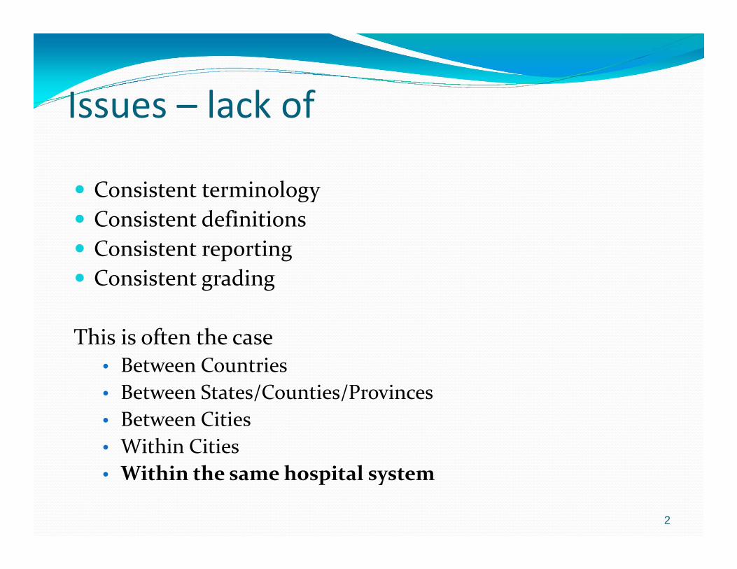
Issues – lack of
Consistent terminology Consistent definitions Consistent reporting Consistent grading
This is often the case• Between Countries• Between States/Counties/Provinces• Between Cities• Within Cities • Within the same hospital system
2

Value to Physicians
Need consistent terminology Need consistent methods of reporting and grading
Physicians need dependable/reproducible information in order to better treat patients
3

Why is this important
Changes over the years: Consolidation of facilities Hub and Spoke Laboratories Core Laboratories Health Care System with multiple hospital laboratories
Within the same town Within the same state Across different states Across different countries
4

Current Status – Wide Variation
Various literature Local regional publications from national societies:
College of American Pathologists (CAP) United Kingdom National External Quality Assessment Service (UKNEQAS)
Japanese Society for Laboratory Hematology Royal College of Pathologists of Australasia Quality Assessment Programs (RCPA QAP)
5

Global Consensus Need recognized for a global consensus guideline dealing with cell morphology.
The aim of this ICSH committee is to provide a guideline for the nomenclature and the grading of red cell, white cell and platelet abnormalities.
6

Investigation Method
An international group of morphology experts was sought from Europe, America, Australasia and Asia. Pathologists, hematologists and scientists with blood film morphology expertise
Initial Survey on blood film morphology and grading
Survey results were discussed at a full day meeting in New Orleans USA (May 2011) Outcome: Recommendations and further investigation
7

Features of paper
Standardization of Peripheral Blood Cells for Red Blood Cells, White Blood Cells and Platelets
Cell Nomenclature with morphology descriptors
Association of the cell name with the image number on the two websites for easier reference
Morphology Grading
8

Cell Nomenclature / Morphology
Consensus terminology
Alternative terminology (when applicable)
Description of cell morphology
Medical Relevance
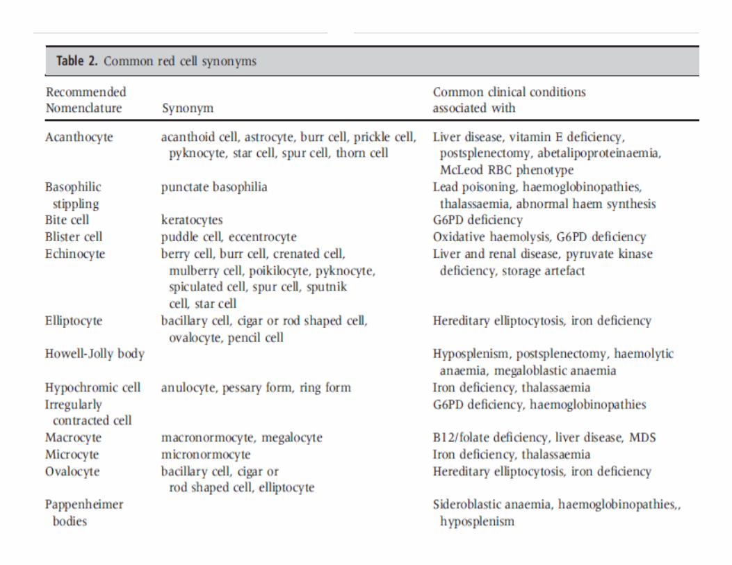
Table for common Red Cell Synonyms
9

Analyzer Grading
Encourage the use of grading some cell morphology using analyzer parameters Higher level of accuracy and precision compared with observer use of the optical light microscope
Examples for Red Cell Size Mean cell volume (MCV) for microcytosis and macrocytosis
Mean cell hemoglobin (MCH) for hypochromia and hyperchromia
10
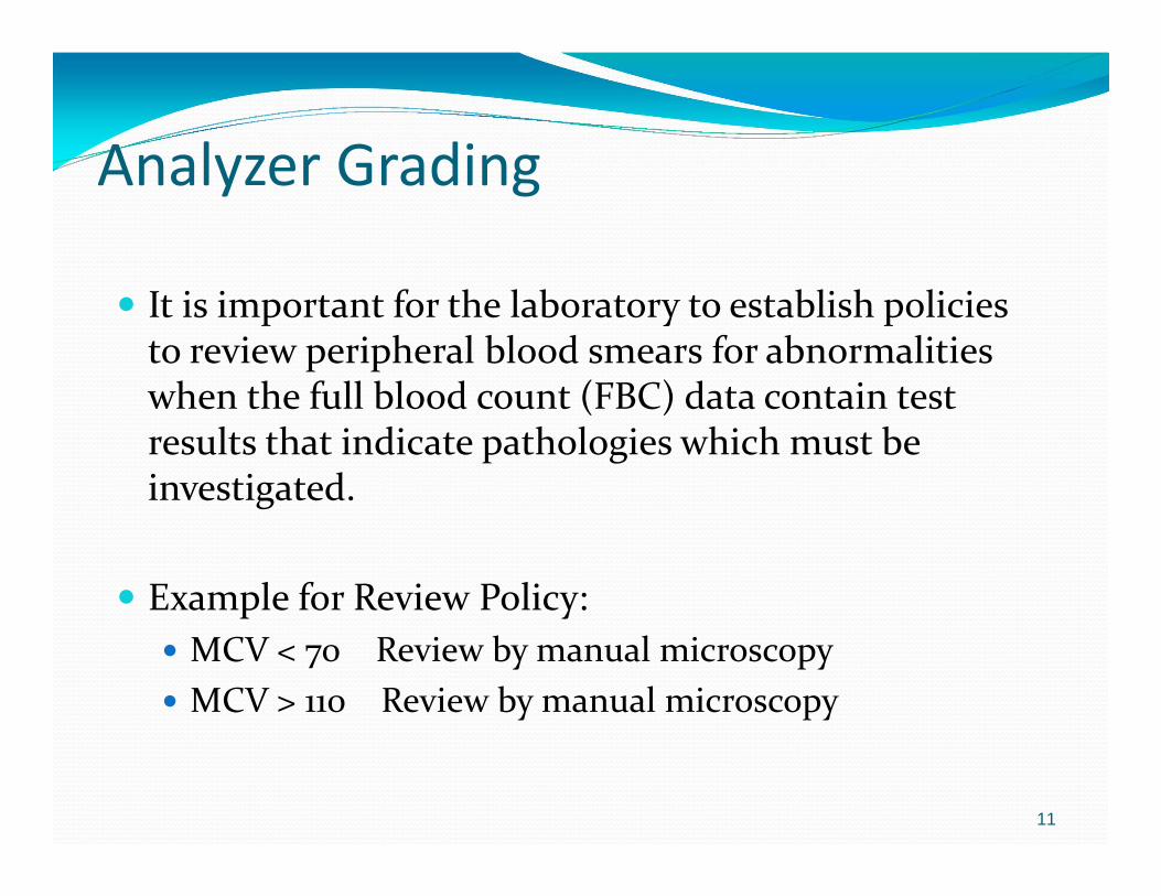
Analyzer Grading
It is important for the laboratory to establish policies to review peripheral blood smears for abnormalities when the full blood count (FBC) data contain test results that indicate pathologies which must be investigated.
Example for Review Policy: MCV < 70 Review by manual microscopy MCV > 110 Review by manual microscopy
11

Grading of Morphological Features Provide the clinician with useful information regarding the status of any abnormality in the peripheral blood.
Morphology grading table contains a two‐tiered grading system 2+ (moderate) 3+ (many)
1+ (few/rare) is reserved only for schistocytes, as the observation even in small numbers is clinically significant.
Each laboratory or laboratory system should have policies in place to ensure the consistent application of the grading criteria.
12
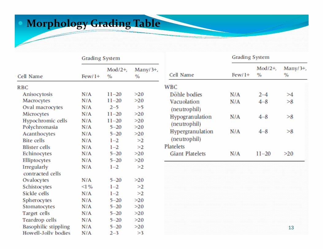
Morphology Grading Table
13
14

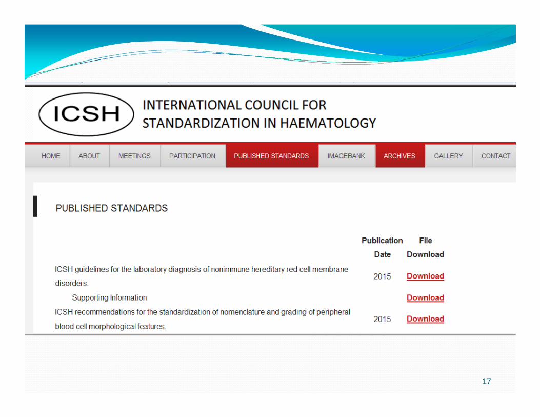
Images Repository
www.morphology.mmu.ac.ukwww.icsh.org
15

Morphology Images
ICSH.org
16
http://www.morphology.mmu.ac.uk/
17
18
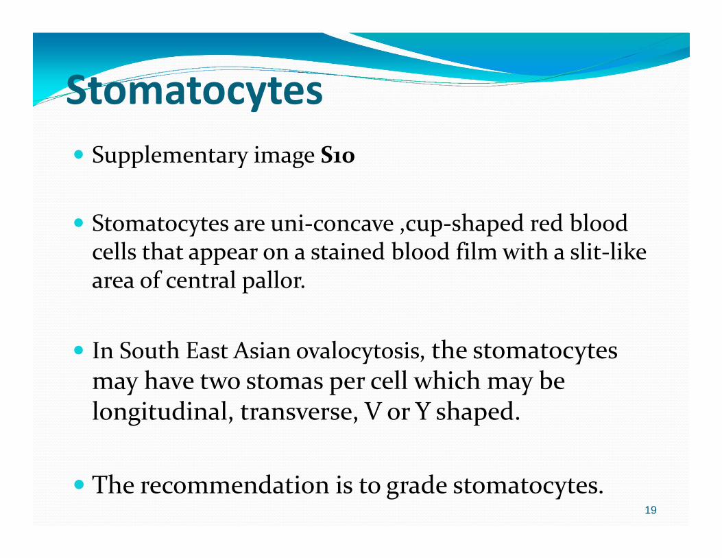
Stomatocytes Supplementary image S10
Stomatocytes are uni‐concave ,cup‐shaped red blood cells that appear on a stained blood film with a slit‐like area of central pallor.
In South East Asian ovalocytosis, the stomatocytes may have two stomas per cell which may be longitudinal, transverse, V or Y shaped.
The recommendation is to grade stomatocytes.19

20
WBC Differential WBC differential counts can be performed by automated
analyzers or manual microscopic visual examination of a blood film.
Automated analyzers count many more cells than the manual 100 or 200 cells. Therefore, more precise information can be gathered.
It is recommended that the automated analyzer WBC differential count be reported in patients with normal cell populations in the absence of analyzer flags or abnormal cell populations that cannot be reliably differentiated and classified by automated instruments.
The automated differential may also be reported after viewing a blood film due to flags or other indicators where the automated values are found to be accurate.
21
Normal cell development & morphology Metamyelocyte
The Metamyelocyte is smaller than the myelocyte with an indented or kidney‐shaped nucleus.
Nucleoli are not observed. The cytoplasm is usually clearly pink and contains granules that are clearly differentiated as neutrophilic, eosinophilic or basophilic.
Immature granulocytes (promyelocytes, myelocytes and metamyelocytes) are not usually seen in normal peripheral blood.
22

Band Neutrophil Band neutrophils are 10–14 lm in diameter and have a nucleus that is
non‐segmented or has rudimentary lobes that are connected by a thick band rather than a thread. Cytoplasm is abundant, pink and contains many small violet‐pink neutrophilic or secondary granules distributed evenly throughout the cell.
Many laboratories do not report band neutrophils on adult patients or children due to inter‐observer variation in band neutrophic classification. This is a well recognized and acceptable practice.
It is recommended that band neutrophils be counted as segmented neutrophils in the differential. Appropriate comments may be made if increased numbers are seen in the blood film.
23

Monoblast Supplementary image S25.
Monoblasts are larger than myeloblasts (20‐30 lm), with a round/oval nucleus, fine chromatin and one or two prominent nucleoli. The cytoplasm is basophilic and usually lacks granules.
The recommendation is to count these as blasts and describe them in the film report with a suitable interpretive comment.
24

25
Platelets Platelet size is of diagnostic significance particularly when considered
in relation to the platelet count.
A normal platelet measures 1.5–3 lm in diameter.
Large platelets measure 3–7 lm (roughly the diameter of a normal sized red cell)
Giant platelets are larger than normal sized red cells at 10–20 lm in diameter and are flagged by automated analyzers.
In a normal person, usually less than 5% of the platelets appear large.
It is recommended that giant platelets be graded.
A comment about the platelet count and the presence of small, large and/or giant platelets can be made with an additional interpretive film comment if appropriate.
26

27

Summary Recommendations deal with the need for a global standard in
naming, grading and reporting abnormal cells or morphological abnormalities which are observed at the time of the PB film review and manual differential count.
Primary goal is to produce clear guidelines for scientists who perform analysis of hematology samples.
The ICSH Guideline reporting system may not fit all laboratories, thus, there should be some degree of flexibility in the way laboratories report. This may to some extent be dictated by the limitations of the different Laboratory Information Systems and middleware in use.
However, one reporting system should be in use for that laboratory / networked laboratory system to ensure consistent information for the physicians and other care givers.
28
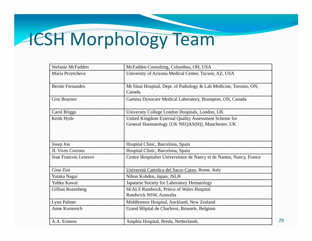
ICSH Morphology TeamStefanie McFadden McFadden Consulting, Columbus, OH, USAMaria Proytcheva University of Arizona Medical Center, Tucson, AZ, USA
Bernie Fernandes Mt Sinai Hospital, Dept. of Pathology & Lab Medicine, Toronto, ON, Canada
Gini Bourner Gamma Dynacare Medical Laboratory, Brampton, ON, Canada
Carol Briggs University College London Hospitals, London, UKKeith Hyde United Kingdom External Quality Assessment Scheme for
General Haematology [UK NEQAS(H)], Manchester, UK
Josep Jou Hospital Clinic, Barcelona, SpainJL Vives Corrons Hospital Clinic, Barcelona, SpainJean Francois Lesesve Centre Hospitalier Universitaire de Nancy et de Nantes, Nancy, France
Gina Zini Università Cattolica del Sacro Cuore, Rome, ItalyYutaka Nagai Nihon Kohden, Japan; JSLHYohko Kawai Japanese Society for Laboratory HematologyGillian Rozenberg SEALS Randwick, Prince of Wales Hospital
Randwick NSW, AustraliaLynn Palmer Middlemore Hospital, Auckland, New ZealandAnne Kornreich Grand Hôpital de Charleroi, Brussels, Belgium
A.A. Ermens Amphia Hospital, Breda, Netherlands 29

Images Compilation of the Images:
John Burthem Michelle Brereton
• Central Manchester and Manchester Children’s University Hospital, UK
• Images Provided by • John Burthem• Michelle Brereton• Gina Zini• Gillian Rozenberg*
*Reproduced by permission from Elsevier Australia from Gillian Rozenberg; Microscopic haematology: a practical guide for the laboratory 3e© 2011, Sydney, Elsevier Australia.
30

Morphology Workshop Sponsors:
Abbott Diagnostics
Beckman Coulter Diagnostics
Horiba Medical Diagnostics
Mindray
Nihon Kohden
Siemens
Sysmex Corporation
31
ICSH Morphology Grading Writing Committee
Lynn Palmer
Carol Briggs
Stefanie McFadden
Gina Zini
John Burthem
Gillian Rozenberg
Maria Proytcheva
Sam Machin
32

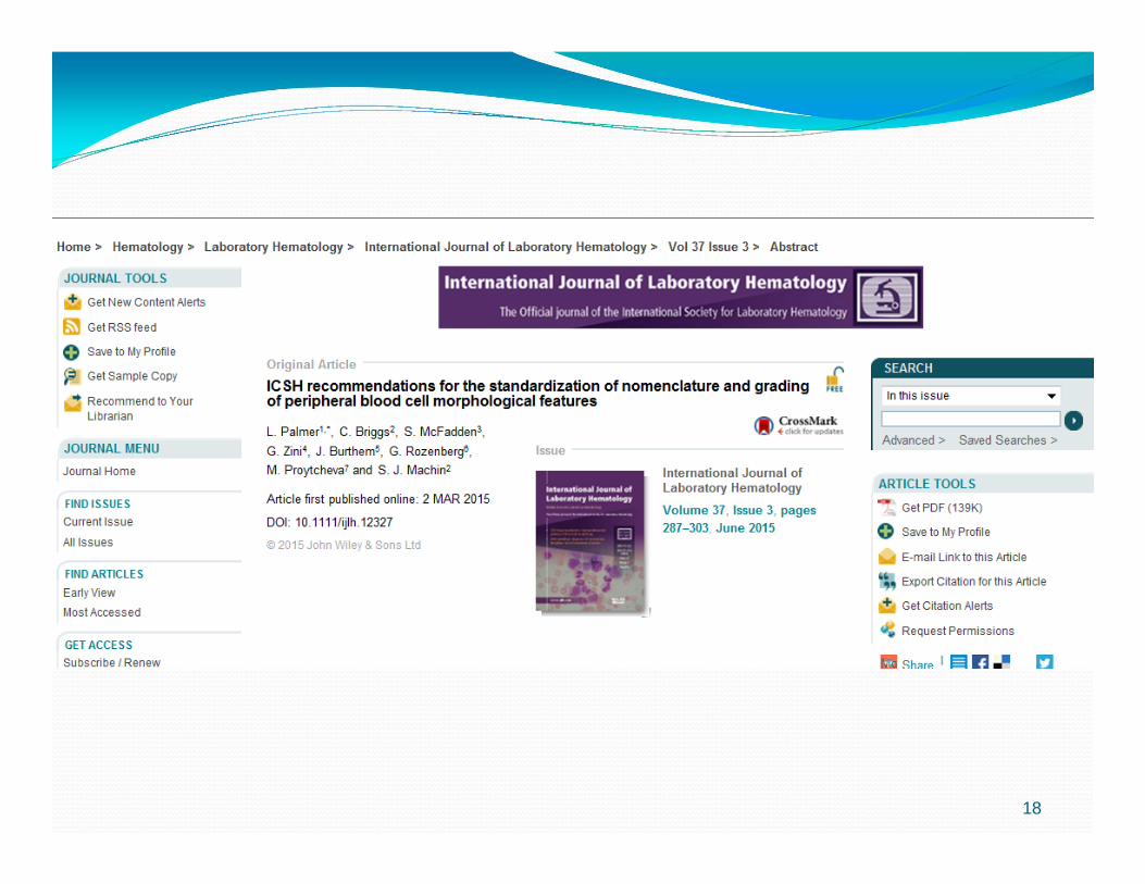
33
International Journal of Laboratory HematologyPublished June 2015

34
Thank You!