small rna-based feedforward loop with and-gate logic ... · small rna-based feedforward loop with...
TRANSCRIPT

Small RNA-based feedforward loop with AND-gatelogic regulates extrachromosomal DNA transferin SalmonellaKai Papenforta,b,c,1, Elena Espinosad, Josep Casadesúsd, and Jörg Vogelb,1
aDepartment of Molecular Biology, Princeton University, Princeton, NJ 08544; bInstitute for Molecular Infection Biology, University of Würzburg, 97080Wurzburg, Germany; cDepartment of Biology I, Ludwig-Maximilians-University Munich, 82152 Martinsried, Germany; and dDepartamento de Genética,Facultad de Biología, Universidad de Sevilla, 41080 Seville, Spain
Edited by Gisela Storz, National Institutes of Health, Bethesda, MD, and approved July 21, 2015 (received for review April 21, 2015)
Horizontal gene transfer via plasmid conjugation is a major drivingforce in microbial evolution but constitutes a complex process thatrequires synchronization with the physiological state of the hostbacteria. Although several host transcription factors are known toregulate plasmid-borne transfer genes, RNA-based regulatory circuitsfor host–plasmid communication remain unknown. We describe aposttranscriptional mechanism whereby the Hfq-dependent smallRNA, RprA, inhibits transfer of pSLT, the virulence plasmid ofSalmonella enterica. RprA employs two separate seed-pairing do-mains to activate the mRNAs of both the sigma-factor σS and theRicI protein, a previously uncharacterized membrane protein hereshown to inhibit conjugation. Transcription of ricI requires σS and,together, RprA and σS orchestrate a coherent feedforward loopwith AND-gate logic to tightly control the activation of RicI syn-thesis. RicI interacts with the conjugation apparatus protein TraVand limits plasmid transfer under membrane-damaging conditions.To our knowledge, this study reports the first small RNA-controlledfeedforward loop relying on posttranscriptional activation of twoindependent targets and an unexpected role of the conserved RprAsmall RNA in controlling extrachromosomal DNA transfer.
RprA | sRNA | feedforward control | plasmid conjugation | Hfq
Intercellular transmission of plasmid DNA is key to bacterialevolution and diversity (1). In the widespread family of F-like
conjugative plasmids, environmental cues and mating partneravailability affect conjugation frequency, and unregulated con-jugation comes with significant fitness costs for the host (2). It iswell understood how conjugation is regulated by plasmid-bornefactors. For example, conjugation of the self-transmissible F-likeplasmid pSLT [which encodes several virulence genes and isrequired for systemic disease (3)] of Salmonella species dependson TraJ, the transcriptional activator of the transfer (tra) operon(4). Synthesis of TraJ itself is precisely controlled by a cis-anti-sense RNA, FinP, which in concert with the dedicated RNAchaperone, FinO, inhibits translation of the traJ mRNA (5). As aresult, most cells reside in a conjugational OFF-state under regularphysiological conditions (6).Besides plasmid-encoded factors, core genome-encoded pro-
teins such as the leucine-responsive regulatory protein (Lrp), theArcAB two-component system, and Dam methylation affectpSLT conjugation (7, 8). These factors usually respond to spe-cific ecological cues; for example, ArcAB responds to micro-aerophilic conditions, the environment that bacteria will encounterin the intestine of infected hosts (9). In addition, host-producedcompounds such as bile salts repress pSLT conjugation, but theunderlying molecular mechanisms are unknown (10).The regulatory networks of the host that restrict plasmid
conjugation to permissive conditions must integrate variousphysiological signals. This may involve small RNAs (sRNAs) thatcan cross-connect gene expression at the posttranscriptional levelthrough their ability to repress or activate multiple target mRNAs(11, 12). Intriguingly, the RNA chaperone Hfq, which helps many
sRNAs to regulate their targets (13, 14), has been reported to affectthe transfer of F-like plasmids (15), suggesting that host–plasmidcommunication does involve regulatory activities of noncodingRNA molecules. However, Hfq-dependent sRNAs controllingplasmid conjugation were hitherto unknown.In this work, we have studied the role of RpoS regulator RNA
A (RprA) in Salmonella enterica. RprA is one of three sRNAs(the others being DsrA and ArcZ) that activate translation ofrpoS mRNA encoding the alternative sigma-factor σS (16–18).All three sRNAs act by an anti-antisense mechanism wherebytheir base pairing with the rpoS mRNA opens an inhibitory struc-ture in the 5′-untranslated region (5′-UTR) to promote translationinitiation (19). In Escherichia coli, expression of RprA is inducedduring stationary-phase growth (20) through either of two signaltransduction pathways, Rcs (21) or CpxAR (22). The fact thatboth Rcs and CpxAR respond to insults to the bacterial cell en-velope (23, 24) suggests a role for RprA in the extracytoplasmicstress responses and membrane homeostasis.Here, we describe that in Salmonella RprA controls a large set
of mRNAs in addition to rpoS, including ricI (STM4242) mRNA.Similar to its known rpoS target (21), RprA activates the ricImRNA through opening of an inhibitory structure in the 5′-UTR.However, unlike rpoS regulation, which is regulated through the5′ end of RprA, it is the conserved 3′ end of RprA that recog-
Significance
Horizontal gene transfer is a major force in bacterial evolution,and a widespread mechanism involves conjugative plasmids.Albeit potentially beneficial at the population level, plasmidtransfer is a burden for individual cells. Therefore, assembly ofthe conjugation machinery is strictly controlled, especially un-der stress. Here, we describe an RNA-based regulatory circuit inhost–plasmid communication where a regulatory RNA (RprA)inhibits plasmid transfer through posttranscriptional activationof two genes. Because one of the activated factors (σS) isnecessary for transcription of the other (RicI), RprA forms thecenterpiece of a posttranscriptional feedforward loop withAND-gate logic for gene activation. We also show that the syn-thesis of RicI, a membrane protein, inhibits plasmid transfer,presumably by interference with pilus assembly.
Author contributions: K.P., J.C., and J.V. designed research; K.P. and E.E. performed re-search; K.P., E.E., J.C., and J.V. analyzed data; and K.P., J.C., and J.V. wrote the paper.
The authors declare no conflict of interest.
This article is a PNAS Direct Submission.
Data deposition: The microarray data reported in this paper have been deposited in theGene Expression Omnibus (GEO) database, www.ncbi.nlm.nih.gov/geo (accession no.GSE67187).1To whom correspondence may be addressed. Email: [email protected] or [email protected].
This article contains supporting information online at www.pnas.org/lookup/suppl/doi:10.1073/pnas.1507825112/-/DCSupplemental.
E4772–E4781 | PNAS | Published online August 11, 2015 www.pnas.org/cgi/doi/10.1073/pnas.1507825112
nizes the ricImRNA, revealing RprA as the first (to our knowledge)regulatory RNA with two activating seed-pairing domains.From a physiological point of view, RicI is shown to inhibit
pSLT conjugation through interaction with anchor protein TraVof the type IV secretion apparatus to restrict the number of con-jugation pili. RprA activates the synthesis of RicI in the presenceof bile salts, and components of the Rcs phosphorelay as well as σSare required for this process. Thus, RprA and σS act in concert toactivate RicI synthesis via a feedforward loop (FFL) with AND-gate decision logic to control plasmid transfer. Donor cells lack-ing one component of this regulatory mechanism, i.e., RprA, σS,or RicI, display increased conjugation rates, a phenotype that isexacerbated under conditions of envelope stress.
ResultsTwo Isoforms of RprA Regulate Target mRNAs. The function ofRprA as a posttranscriptional activator of σS synthesis has beenwell established in E. coli (20, 21, 25, 26). By contrast, this “core”sRNA, which is conserved in many enterobacterial species, wasreported to have little if any role in the closely related pathogen,Salmonella Typhimurium (27), although both RprA and its tar-get region in the rpoS mRNA are fully conserved (28, 29). Toaddress this discrepancy, we monitored RprA expression inSalmonella on a Northern blot using a probe complementary tothe conserved 3′ end of the sRNA. Expression of RprA peakedduring stationary phase, and we detected two forms of RprA: afull-length transcript of ∼107 nt and a shorter processed 3′-endfragment of ∼50 nt (Fig. 1 A and B). This is in agreement withprevious studies in E. coli (30) and RNA-seq experiments inSalmonella (28, 31, 32).To identify mRNA targets of both RprA forms, we pulse-
expressed (10 min) either the full-length or the processed sRNAfrom a pBAD promoter and scored global transcriptome changeson microarrays, comparing to an empty vector control (33). In-duction of the full-length RprA altered the expression of 64genes (Fig. 1C), one of which was rpoS (+2.5-fold), rectifying thatσS activation by RprA is functionally conserved in Salmonella.As expected from the previously mapped RprA–rpoS RNA in-teraction (21, 34), processed RprA did not activate rpoS ex-pression. Twelve genes were regulated by both the full-lengthand processed RprA, 11 of which were repressed by RprA (Fig.1C). We did not observe the previously reported RprA-mediatedrepression of csgD (35, 36), perhaps because the csgD promoterwas silent under the experimental conditions used here (37). Thisnotwithstanding, our pulse expression data suggested that thetwo forms of RprA recognize several targets by different seedregions and predicted processed RprA to be a regulator in itsown right.One gene, ricI (also known as STM4242), was up-regulated by
both forms of RprA (Fig. 1C). To test the contribution of eachisoform to RicI synthesis, we added a 3×FLAG epitope to thechromosomal ricI gene and monitored RicI protein levels uponinduction of full-length or processed RprA. Indeed, both formsof RprA equally activated RicI expression (Fig. 1D), whereasonly full-length RprA induced σS production.
A Second Seed Region in RprA Activates RicI Synthesis. Next,we sought to understand how RprA activates RicI production.As our pulse expression approach suggested posttranscriptionalcontrol, we looked for evidence of activation by the aforemen-tioned “anti-antisense” mechanism (12), whereby the sRNAopens a self-inhibitory structure in the 5′-UTR of its targetmRNA (21). Indeed, in silico analysis of the secondary structureof the ricI mRNA (from the transcriptional start site to the fifthcodon; see below) using the Mfold algorithm (38) predicted adiscontinued RNA duplex formed between nucleotides 38–62 and95–119 (relative to the transcriptional start site; see below) of thericI mRNA (Fig. 2A). This structure would sequester the Shine–
Dalgarno sequence (SD) and the GUG translation start codonof ricI.To validate this predicted hairpin and its potential function in
translation control, we cloned the sequence of the ricI mRNA—
from its transcriptional start site to the 10th codon—into a gfp-based reporter plasmid designed to report posttranscriptionalregulation (39). This construct gave only modest GFP expres-sion. However, when we truncated the ricI mRNA at its 5′ end(transcription start at A48; Fig. S1A), essentially deleting thesequence predicted to sequester the translational start site,a >50-fold increase in the level of RicI::GFP was observed (Fig.S1B). Moreover, a single C→G change opposite the first nucle-otide of the start codon (42 nt downstream of the transcriptionalstart site; see below) increased the expression of the full-lengthreporter ∼13-fold (Fig. S1B). In further support of our modelthat a 5′ hairpin sequesters the ricI start codon, two other mu-tations, G44→C or C113→G (Fig. 2 A and B, compare lane 1 vs.2 and 3), also increased RicI::GFP synthesis. As expected,however, the combination of these two latter mutations restoredwild-type expression levels of RicI::GFP, most likely by restoringmRNA hairpin formation. Together, this mutational analysissuggests that RicI synthesis is intrinsically repressed by intra-molecular base pairing in the 5′ region of its mRNA.Next, we used the ricI::gfp reporter to establish that RprA
activates translation of this target by preventing self-sequestra-tion of the ricI mRNA. Indeed, coexpression of RprA from acompatible plasmid increased RicI::GFP levels by approximatelythreefold (Fig. 2C, lane 1 vs. 2), whereas it had no effect on GFPlevels expressed from the pXG-1 control plasmid (Fig. S1C) (39).As expected if RprA acted by suppressing hairpin formation, thesRNA had no effect on the truncated (Fig. S1D) or “open”(C42→G) variants of ricI::gfp (Fig. S1E).Because the processed RprA form sufficed for activation (Fig.
1 C and D), we used its sequence to search for an RprA bindingsite in the 5′-UTR of ricI. The RNA-hybrid algorithm (40) pre-dicted a consecutive stretch of nine Watson–Crick base pairsformed between the proximal end of the processed RprA and theinternal antisense element of the ricI mRNA (Fig. 2A). To testthis prediction, we constructed an RprA variant with a pointmutation in the seed region (G63→C, Fig. 2A); as expected,RprAC63 was unable to activate the ricI::gfp reporter (Fig. 2C,lane 3). Conversely, mutating the corresponding position 45 inthe ricI 5′-UTR (ricIG45::gfp reporter, Fig. 2A) abrogated re-porter activation by wild-type RprA (Fig. 2C; lane 5). Note thatthis nucleotide is not paired in the intrinsic hairpin and hencewill not alter RicI::GFP expression (Fig. 2C; lane 1 vs. 4). Bycontrast, combining both mutants (RprAC63 and ricIG45::gfp)fully restored target activation (Fig. 2C; lane 6). Thus, RprA usesa similar mechanism but different seed sequences to activate thesynthesis of σS and RicI.
Membrane Stress and the Rcs Phosphorelay Activate RicI Production.The ricI gene (also known as STM4242) is conserved in all se-quenced Salmonella species, including the ancestral Salmonellabongori and the human-specific serovar Salmonella typhi, butabsent in other enterobacterial relatives such as E. coli, Klebsiellapneumoniae, and Shigella flexneri (Fig. S2). Although its bi-ological role has not been investigated, RicI has been reported asa bile salt-induced protein (41). To address this, we monitoredproduction of 3×FLAG-tagged RicI protein in both wild-typeand ΔrprA cells upon exposure to bile. In Salmonella wild-typecells, RicI levels increased approximately fourfold within 15 minafter treatment, with a further increase to approximately eight-fold after 120 min (Fig. 3A, lanes 1–5). By contrast, the ΔrprAmutant failed to increase RicI production (Fig. 3A, lanes 6–10).These results confirm bile as a potent activator of RicI productionbut also implicate RprA as an essential factor in this process.
Papenfort et al. PNAS | Published online August 11, 2015 | E4773
MICRO
BIOLO
GY
PNASPL
US

Bile is a detergent-like substance that can disrupt bacterialmembranes (42) and thereby activate the Rcs stress response.Because the Rcs system strongly induces the rprA promoter inE. coli (21), and other cell envelope-damaging conditions triggerRprA synthesis through RcsB (21, 43–45), we hypothesized thatthe bile-induced increase in RicI synthesis is indirect, resultingfrom the Rcs-mediated activation of RprA. To test this, weconstructed single-gene deletion strains of various componentsof the Rcs signaling cascade and evaluated bile-induced changesin the levels of RprA and RicI levels in these mutants. Asexpected, wild-type cells activated both RprA and RicI expres-sion in the presence of bile (Fig. 3B, lane 1 vs. 2), whereas cellslacking rprA (lanes 3 and 4), rcsF (lanes 5 and 6), or rcsB (lanes 9and 10) failed to activate RicI. This corresponded well with a loss ofbile-induced activation of RprA in the ΔrcsB and ΔrcsF mutants,
with the ΔrcsC mutant showing intermediate RprA inductionthat seemed insufficient for RicI activation (lanes 7 and 8).Given the known relationship of RprA and σS, we also tested aΔrpoS strain. Surprisingly, bile did not increase RicI levels in theΔrpoS strain, although RprA was fully activated (Fig. 3B, lanes11 and 12). This suggested that also σS was essential for RicIactivation but it would act downstream of RprA.To better understand the role of σS in RicI activation, we
sought to override Rcs signal transduction by constitutive ex-pression of RprA (from plasmid pKP-112) in the rprA, rcsB, andrpoS mutant strains. Midexponential cultures (low endogenousRprA expression; Fig. 1B) were probed for RicI production (Fig.3C). In support of our previous results, plasmid-borne over-expression of RprA strongly induced RicI expression in wild-typecells and complemented the rprA and rcsB mutant strains (Fig.
A
B C
D
RprA
rela
tive
expr
essi
on
RprA pr
oc.
Fig. 1. Multiple target regulation by RprA in Salmonella. (A) Alignment of the rprA gene from selected enterobacterial species (ECA, Erwinia carotovora;ECO, Escherichia coli K12; KPN, Klebsiella pneumoniae; PLU, Photorhabdus luminescens; SFL, Shigella flexneri; STM, Salmonella enterica sv. Typhimurium).Transcription control regions −10 and −35 are boxed, the transcription initiation site is marked by an arrow. Scissors indicate the RprA processing site, andinverted arrows refer to the rho-independent terminator. (B) Northern blot analysis of RprA in Salmonella. Samples were collected at several stages of growth(OD600 of 0.5, 1.0, and 2.0, at 3 and 6 h after cells had reached OD600 of 2.0, and after 24 h of cultivation). 5S rRNA served as loading control. (C) Microarrayanalysis of RprA full-length and RprA processed pulse expression. Expression profiles of pulse-induced full-length and processed RprA were compared withsamples carrying control plasmids. A heat map of genes regulated by full-length RprA (more than twofold) is shown and compared with regulation byprocessed RprA. (D) Western and Northern blot analyses of σS, RicI::3×FLAG, and RprA production after pulse expression of full-length and processed RprA.Wild-type and ΔrprA carrying the indicated plasmids were grown to early stationary phase (OD600 of 1.5) and induced for pBAD expression. 5S rRNA(Northern blot) and GroEL (Western blot) served as loading controls.
E4774 | www.pnas.org/cgi/doi/10.1073/pnas.1507825112 Papenfort et al.

3C, lanes 1–6). However, RicI levels remained low in the ΔrpoSmutant, suggesting that the function of σS was independent ofRprA-mediated posttranscriptional activation of ricI mRNA.
σS Is Required for Transcription of ricI. To further address the re-quirement of σS for RicI synthesis, we tested whether RprAactivated the ricI::gfp reporter (Fig. 2B, lane 1) in the ΔrpoSmutant strain. The ricI::gfp reporter gene is transcribed from aconstitutive PLtetO promoter that is insensitive to absence of σS.There was no difference to the previously observed approxi-mately threefold activation in wild-type Salmonella (Fig. S3A),suggesting that σS influences RicI expression at an earlier step,i.e., transcription.The transcriptional start site of ricI, mapped here by 5′-RACE
(Fig. S3B) and previously by global dRNA-seq analysis of theSalmonella transcriptome (46), is a guanine that lies 114 nt up-stream of the start codon (Fig. 4A). Intriguingly, the associatedpromoter contains a highly conserved cytosine at position −13,which is a hallmark of σS-dependent promoters; this nucleotidecontacts amino acid E458 in σS and counterselects for binding ofthe housekeeping σ70 (47). To test a potential σS dependence ofthe ricI promoter, we inserted a lacZ reporter gene downstreamof it in the Salmonella chromosome (48). Promoter activity as-says in the wild-type and in a ΔrprA mutant revealed comparabletranscriptional activity of the two strains with peaking activityunder stationary-phase growth conditions (Fig. 4B). In contrast,Salmonella lacking the rpoS gene failed to activate the ricI pro-moter under all conditions tested, indicating that σS controlsricI transcription.This was further confirmed by mutating C-13, which elimi-
nated the σS dependency of ricI transcription as expected; i.e., agfp reporter gene fused to a C-13→G variant of the ricI promoterwas insensitive to the presence or absence of an intact rpoS gene(Fig. 4C). Collectively, these results suggest that both transcrip-tional activation by σS and posttranscriptional activation byRprA are essential for ricI expression.
An RNA-Controlled FFL with AND-Gate Logic Regulates RicI Production.The dual requirement of RprA and σS in the activation of RicIresembles a coherent type 1 FFL (49). However, whereas suchFFLs are typically controlled by transcription factors, the type 1FFL activating RicI depends on dual base-pairing interactions of a
regulatory RNA, which we predicted to work through an AND-gate logic: the up-regulation of RicI synthesis requires both σS andRprA (Fig. 5A).To examine the effectivity of this regulatory scheme, we mutated
the rprA gene at two positions (Fig. 5B): we changed adenosine 37to cytosine (RprAC37) abolishing activation of the rpoS mRNA(21), and guanine 63 to cytosine (RprAC63) to abrogate activationof the ricImRNA (Fig. 2 A and C). These mutant rprA alleles wereexpressed from an inducible pBAD promoter to test their ability toup-regulate a chromosomal ricI::lacZ translational reporter underthe control of the endogenous ricI promoter. Induction of wild-typeRprA resulted in a >50-fold increase in reporter activity over thecourse of 60 min (Fig. 5C). By contrast, neither the RprAC63 northe RprAC37/C63 double mutant could activate the reporter (Fig.5C). Likewise, the RprAC37 single mutant, which fully activatesthe posttranscriptional ricI::gfp reporter (Fig. S4A), failed to acti-vate the translational ricI::lacZ reporter (Fig. 5C). To further testthis scheme, we treated ΔrprA cells carrying either the rprA,rprAC37, or rprAC63 allele (on a low-copy plasmid) with 3,4-dichlorobenzyl carbamimidothioate (A22) and followed the ki-netics of RprA, σS, and RicI production. A22 inhibits the actin-like MreB protein and provides superior activation of the Rcsphosphorelay compared with bile salts (Fig. S4B) (45, 50). Asexpected from our previous results (Fig. 5C), only wild-typeRprA provided full induction of RicI and σS, whereas RprAC63failed to activate RicI and RprAC37 displayed only reduced σS
RicI
GroEL
[min]
WT ΔrprA
0’ 0’15’ 15’ 30’ 30’ 60’ 60’120’ 120’
- - - - - -+ + + + + + WT ΔrprA ΔrcsF ΔrcsC ΔrcsB ΔrpoS
RicI
GroEL
bile
5S rRNA
1
1
1
2
2
2
3
3
3
4
4
4
5
5
5
6
6
6 7
7
7
8
8
8 9 10
9 10 11 12
A
C
B
WT ΔrprA ΔrpoSΔrcsB
RicI
GroEL
full-length RprA
processed RprA
pctr.
pctr.
pctr.
pctr .
pRpr
A
pRpr
A
pRpr
A
pRpr
A
Fig. 3. Bile-induced expression of RicI. (A) Western blot analysis of RicI::3×FLAGexpression. Wild-type and ΔrprA cells were grown to late exponential phase(OD600 of 1.0) and treated with bile salts (3% final concentration). Whole-protein samples were collected at the indicated time points and probed forRicI::3×FLAG production. (B) Wild-type and the indicated Salmonella mutants(ΔrprA, ΔrcsF, ΔrcsC, ΔrcsB, and ΔrpoS) were cultivated in LB media (with orwithout 3% bile salts) to OD600 of 1.0 and probed for bile-mediated activationof RicI::3×FLAG (Western blot) and RprA (Northern blot). (C) RicI::3×FLAGproduction in the context of RprA overexpression. Wild-type, ΔrprA, ΔrcsB,and ΔrpoS cells transformed with a control plasmid (pctr.) or the RprAoverexpression plasmid (pRprA) were tested for RicI::3×FLAG expressionon Western blot.
A
C
B
GroEL
ricI::gfp ricIG45::gfp
GFP
pctr.
pctr.
pRpr
A
RprA
pRpr
A
pRpr
AC
63
pRpr
AC
63
1
1
2
2
3
3
4
4
5 6
AA AGUGUUCA C
AUAGUUCGCCUA
A
A
U
C5’ 38 62
6238
95119
113
44
32 nt
3’GGUAGA
UGUCAUAGAUCGUC
AGC
ricI
UAGUUCGCCU
C
GGFP
GroEL
wild
-type
G44
C
G44
C/C
113G
C11
3G
AAGUG
5’
95119
32 nt
3’UCAAUCG AUAGGUAGA AUAG
UUCACAGCAAUAGUUCGCCUCUGUC
C
G
5’
3’GUGUCGUUA
6068
ricIG45
RprAC63
Fig. 2. Anti-antisense activation of ricI. (A) Graphical presentation of the ricI5′-UTR alone (Top) or in complex with RprA (Bottom). Numbering of ricI andRprA is relative to their transcription start site. ricI base pairs with the 5′terminal end of the processed RprA form. Arrows denote mutations in-troduced in ricI::gfp and RprA, respectively. (B) Western blot analysis of ricI::gfp variants (as indicated in A, Top) expressed in Salmonella ΔrprA cells.(C) Western blot analysis of ΔrprA Salmonella harboring plasmid pRprA ormutant plasmid, pRprAC63, in combination with either wild-type ricI::gfp ormutant ricIG45::gfp fusion plasmids. GroEL served as loading control.
Papenfort et al. PNAS | Published online August 11, 2015 | E4775
MICRO
BIOLO
GY
PNASPL
US

induction and did not up-regulate RicI (Fig. S4C). Together,these data indicate that RprA acts in a sequential order: acti-vation of rpoS precedes activation of ricI because σS must acti-vate ricI transcription first.To test this circuit under more physiological conditions, i.e.,
without RprA overexpression, we monitored expression of a ricI::lacZ fusion at various stages of Salmonella growth. In wild-typecells, reporter activity peaked in stationary phase after in-creasing ∼12-fold from exponential phase (Fig. 5D). As expected,introduction of either a ΔrpoS or a ΔrprA allele abrogated thisincrease in RicI::LacZ levels. To uncouple transcriptional ac-tivity of σS from posttranscriptional regulation by RprA, we in-troduced the mutation G44→C (Fig. 2A) in the ricI 5′-UTR(ricI*) on the Salmonella chromosome. This “ON” mutation in-terferes with stem-loop formation of the ricI untranslated leader,resulting in high RicI::GFP production in the absence of RprA(Fig. 2B). Similarly, this mutation induced RicI::LacZ expressionby ∼16-fold at early stages of growth (Fig. 5D) and renderedreporter activity insensitive to a secondary deletion of the rprA
gene, because translation is already derepressed. Introduction ofthe ricI* allele into a rpoS mutant also increased RicI::LacZproduction at early stages of growth but failed to increase ex-pression at higher cell densities (Fig. 5D). These results supportthe combinatorial activity (AND-function) of σS and RprA inactivation of RicI.A key characteristic of type 1 FFLs is their delay function upon
signal perception (51). This is easy to understand in the contextof RprA, σS, and RicI: RprA has to activate rpoS until sufficientσS is produced to generate the ricI mRNA serving as a secondtarget for RprA. To investigate whether production of σS pre-cedes RicI expression, we treated cells with A22 and followed thekinetics of RprA, σS, and RicI production. As predicted fromthe circuit (Fig. 5A), we detected approximately fourfold ele-vated σS expression already 10 min after addition of A22 (Fig. 5E,lane 1 vs. 2), and production increased further to approximatelyeightfold after 80 min when the experiment was terminated (lane5). Induction of σS occurred synchronously to RprA, indicatingimmediate posttranscriptional activation of the rpoS mRNA. Bycontrast, a cross-comparison of σS and RicI levels showed thatRicI production was significantly delayed. RicI levels increased∼2.5-fold after 20 min of treatment and increased afterward to∼15-fold (Fig. 5E, lanes 1, 3, and 5). Using a similar approach,we also monitored expression of RprA, RicI, and σS followingdeactivation of the circuit. Specifically, we treated wild-type cellswith A22 for 30 min and collected and washed the cells followedby reinoculation into fresh media. We discovered that shutoffof σS production is almost immediate, whereas reduction of RicIexpression to “prestress” levels required ∼60 min (Fig. S4D).These differences in protein levels might depend on specificproteolytic factors targeting the σS protein (50). Interestingly, wefound that σS degradation also preceded inhibition of RprAexpression: expression levels of full-length RprA and processedRprA reached the prestress status ∼15 min after cells werereinoculated in fresh media. Together, our data provide evidencefor a previously unidentified variant of the type 1 FFL networkthat functions through the regulatory activity of two base-pairingdomains of a single sRNA. Deactivation of the circuits dependson the individual stabilities of the three components, with RicIbeing most stable and σS showing almost immediate degradationonce the stress is removed.
RicI Inhibits Salmonella Virulence Plasmid Transfer. The RprA-mediated up-regulation of RicI and the evident connection withthe σS stress response network prompted us to investigate thebiological role of these three factors more closely. Although thebiological function of RicI was unknown, BLAST-P searchessuggested similarity of RicI to a variety of proteins from differentbacterial genera (Fig. S5), most of which with candidate functionsin plasmid conjugation.To investigate a potential role of RprA-mediated RicI acti-
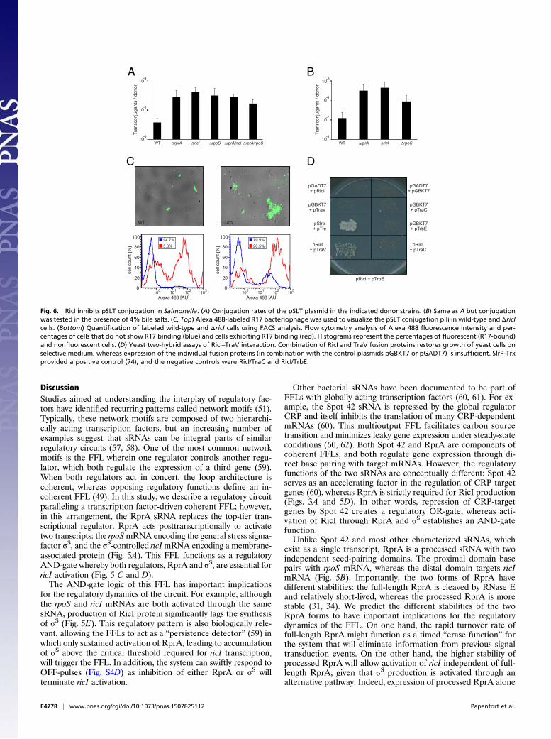
vation in conjugation of the pSLT virulence plasmid in Salmo-nella, we compared the plasmid transfer rates of ΔrprA, ΔricI,and ΔrpoS donors with the transfer rate of the wild type. De-letions of ΔrprA, ΔricI, or ΔrpoS increased plasmid conjugation∼8- to 11-fold (Fig. 6A, bars 1–4), suggesting an inhibitoryfunction for RicI in pSLT transfer. Double mutants ΔrprA ΔricIand ΔrprA ΔrpoS yielded conjugation rates similar to those of thesingle-mutant variants (Fig. 6A), suggesting that RprA, RicI, andσS act in the same biological pathway to inhibit pSLT transfer.Bile salts are an important factor for Salmonella pathogenicity
(52) and have been reported to decrease pSLT transfer (9, 10).Because RprA expression is induced by bile (Fig. 3B), we won-dered whether bile salts could affect conjugation frequencythrough RprA, σS, and RicI. To test this hypothesis, we com-pared conjugation rates of wild type, ΔrprA, ΔricI, and ΔrpoSmutants in the presence of 4% bile. As expected, bile stronglydecreased conjugal transfer from wild-type donors (compare
A
B
C
Fig. 4. σS activates ricI transcription. (A) Alignment of the ricI promotersequence from Salmonellae and Enterobacter species (ESP, Enterobacter sp.638; SAG, S. enterica sv. Agona; SAR, S. enterica sv. arizonae; SBO, Salmo-nella bongori; SEB, S. enterica sv. Bovismorbificans; SSG, S. enterica sv.Schwarzengrund; STM, S. enterica sv. Typhimurium). Transcription controlregions −10 and −35 are boxed, and the transcription initiation (+1) site ismarked by an arrow. Residue C-13 is shown in bold. (B) Wild-type, ΔrprA,and ΔrpoS cells carrying the transcriptional ricI::lacZ reporter were moni-tored for β-galactosidase production at the indicated stages of growth.(C) Wild-type and ΔrpoS cells transformed with either a wild-type (pricI::gfp)or the mutant [pricI::gfp (G13)] reporter were cultivated to early stationaryphase (OD600 of 2.0) and assayed for GFP production.
E4776 | www.pnas.org/cgi/doi/10.1073/pnas.1507825112 Papenfort et al.

Fig. 6 A and B). However, under the same conditions, ΔrprA,ΔricI, or ΔrpoS donors displayed up to ∼36-fold (ΔricI) increasedconjugation rates (Fig. 6B), similar to those of wild-type strainsgrown in rich medium (Fig. 6A). These data indicate a restrictiverole for RicI during Salmonella virulence plasmid conjugation un-der membrane stress conditions.Next, we sought to understand how RicI controls pSLT trans-
fer. We first tested whether increased pSLT transfer of ΔricISalmonella was also reflected in a higher rate of F-pili produc-tion. To this end, we treated cells with a fluorescently labeledderivative of bacteriophage R17. R17 specifically binds F-like piliand allows for accurate quantification of pili assembly (53). Weused flow cytometry to compare pSLT-encoded F-like pili pro-duction in wild-type and ΔricI cells. Approximately 5% of wild-type cells displayed F-like pili on their surface, whereas this fre-quency was increased to >20% in ΔricI mutants (Fig. 6C). Thesedata indicate that RicI inhibits production of pSLT-encoded piliand suggests that the increased conjugation rates of ΔricI mu-tants (Fig. 6 A and B) might be a consequence of increased F-likepili formation.To investigate the gene-regulatory pattern underlying these
phenotypes, we tested whether RicI affected expression of traJ.In F-family plasmids, TraJ is the major transcriptional activatorof the tra operon encoding most of the proteins necessary forconjugation (54). Therefore, elevated expression of TraJ couldwell explain the increased F-pili production ofΔricI cells (Fig. 6C).However, expression of the transcriptional reporters traJ::lacZ andtraB::lacZ [activated by TraJ (7)] remained unchanged in the ricI
mutant, whereas levels of both reporters (Fig. S6 A and B) weresignificantly increased in dam-deficient cells, which served as apositive control (8). These data indicate that RicI inhibits plasmidtransfer through a mechanism independent of TraJ.Cell fractionation assays showed that RicI localizes to the in-
ner membrane or periplasm of Salmonella (Fig. S6C), suggestingthat RicI does not act at the gene-regulatory level but ratherthrough interaction with other proteins. To identify interactionpartners of RicI, we performed protein coimmunoprecipitation(co-IP) experiments in lysates of Salmonella expressing RicI::3×FLAG. Visualization of copurified protein by silver stainingrevealed enrichment of a small protein (∼18 kDa) in cellsexpressing the RicI::3×FLAG protein, compared with co-IP inwild-type cells used for control (Fig. S6D). Mass spectrometryidentified this protein as TraV, which is a membrane-boundlipoprotein that functions as an anchor of the type IV secre-tion apparatus (55). To validate the predicted interaction ofRicI and TraV, we made use of a yeast-two-hybrid system inwhich reconstitution of the GAL4 protein through two inter-acting protein partners is required to drive the expression ofthe HIS3 and ADE2 genes, which are required for cellulargrowth (56). Indeed, we observed that plasmid-borne ex-pression of neither RicI nor TraV fusion proteins alone wouldallow growth on selective plates, whereas combination of thetwo would restore HIS3 and ADE2 expression (Fig. 6D).Together, our data indicate that the RprA-activated RicIprotein inhibits F-pili production through an interactionwith TraV.
Fig. 5. An FFL with AND-gate logic controls RicI production. (A) Schematic display of the FFL regulating RicI production. Both RprA and σS are required forRicI expression. Dashed lines indicated posttranscriptional regulation, and solid lines denote control at the transcriptional level. (B) Secondary structure ofRprA. Mutations tested in C are indicated by arrows. Scissors mark the RprA processing site. (C) Salmonella carrying the translational ricI::lacZ reporter weretransformed with the indicated plasmids and tested for β-galactosidase production upon induction of pBAD expression. (D) The indicated strains (wild type,ΔrpoS, ΔrprA, ΔricI*, ΔrpoS/ricI*, and ΔrprA/ricI*) carrying the translational ricI::lacZ reporter were assayed for β-galactosidase production at the indicatedtime points of growth. (E) Analyses of σS, RicI::3×FLAG, and RprA expression after A22-mediated induction of the Rcs pathways. Samples were collected at theindicated time points and probed for σS and RicI::3×FLAG (Western blot) as well as RprA (Northern blot) production. GroEL and 5S rRNA served as loadingcontrols.
Papenfort et al. PNAS | Published online August 11, 2015 | E4777
MICRO
BIOLO
GY
PNASPL
US
DiscussionStudies aimed at understanding the interplay of regulatory fac-tors have identified recurring patterns called network motifs (51).Typically, these network motifs are composed of two hierarchi-cally acting transcription factors, but an increasing number ofexamples suggest that sRNAs can be integral parts of similarregulatory circuits (57, 58). One of the most common networkmotifs is the FFL wherein one regulator controls another regu-lator, which both regulate the expression of a third gene (59).When both regulators act in concert, the loop architecture iscoherent, whereas opposing regulatory functions define an in-coherent FFL (49). In this study, we describe a regulatory circuitparalleling a transcription factor-driven coherent FFL; however,in this arrangement, the RprA sRNA replaces the top-tier tran-scriptional regulator. RprA acts posttranscriptionally to activatetwo transcripts: the rpoSmRNA encoding the general stress sigma-factor σS, and the σS-controlled ricImRNA encoding a membrane-associated protein (Fig. 5A). This FFL functions as a regulatoryAND-gate whereby both regulators, RprA and σS, are essential forricI activation (Fig. 5 C and D).The AND-gate logic of this FFL has important implications
for the regulatory dynamics of the circuit. For example, althoughthe rpoS and ricI mRNAs are both activated through the samesRNA, production of RicI protein significantly lags the synthesisof σS (Fig. 5E). This regulatory pattern is also biologically rele-vant, allowing the FFLs to act as a “persistence detector” (59) inwhich only sustained activation of RprA, leading to accumulationof σS above the critical threshold required for ricI transcription,will trigger the FFL. In addition, the system can swiftly respond toOFF-pulses (Fig. S4D) as inhibition of either RprA or σS willterminate ricI activation.
Other bacterial sRNAs have been documented to be part ofFFLs with globally acting transcription factors (60, 61). For ex-ample, the Spot 42 sRNA is repressed by the global regulatorCRP and itself inhibits the translation of many CRP-dependentmRNAs (60). This multioutput FFL facilitates carbon sourcetransition and minimizes leaky gene expression under steady-stateconditions (60, 62). Both Spot 42 and RprA are components ofcoherent FFLs, and both regulate gene expression through di-rect base pairing with target mRNAs. However, the regulatoryfunctions of the two sRNAs are conceptually different: Spot 42serves as an accelerating factor in the regulation of CRP targetgenes (60), whereas RprA is strictly required for RicI production(Figs. 3A and 5D). In other words, repression of CRP-targetgenes by Spot 42 creates a regulatory OR-gate, whereas acti-vation of RicI through RprA and σS establishes an AND-gatefunction.Unlike Spot 42 and most other characterized sRNAs, which
exist as a single transcript, RprA is a processed sRNA with twoindependent seed-pairing domains. The proximal domain basepairs with rpoS mRNA, whereas the distal domain targets ricImRNA (Fig. 5B). Importantly, the two forms of RprA havedifferent stabilities: the full-length RprA is cleaved by RNase Eand relatively short-lived, whereas the processed RprA is morestable (31, 34). We predict the different stabilities of the twoRprA forms to have important implications for the regulatorydynamics of the FFL. On one hand, the rapid turnover rate offull-length RprA might function as a timed “erase function” forthe system that will eliminate information from previous signaltransduction events. On the other hand, the higher stability ofprocessed RprA will allow activation of ricI independent of full-length RprA, given that σS production is activated through analternative pathway. Indeed, expression of processed RprA alone
A B
DC
Fig. 6. RicI inhibits pSLT conjugation in Salmonella. (A) Conjugation rates of the pSLT plasmid in the indicated donor strains. (B) Same as A but conjugationwas tested in the presence of 4% bile salts. (C, Top) Alexa 488-labeled R17 bacteriophage was used to visualize the pSLT conjugation pili in wild-type and ΔricIcells. (Bottom) Quantification of labeled wild-type and ΔricI cells using FACS analysis. Flow cytometry analysis of Alexa 488 fluorescence intensity and per-centages of cells that do not show R17 binding (blue) and cells exhibiting R17 binding (red). Histograms represent the percentages of fluorescent (R17-bound)and nonfluorescent cells. (D) Yeast two-hybrid assays of RicI–TraV interaction. Combination of RicI and TraV fusion proteins restores growth of yeast cells onselective medium, whereas expression of the individual fusion proteins (in combination with the control plasmids pGBKT7 or pGADT7) is insufficient. SlrP-Trxprovided a positive control (74), and the negative controls were RicI/TraC and RicI/TrbE.
E4778 | www.pnas.org/cgi/doi/10.1073/pnas.1507825112 Papenfort et al.

can be sufficient to activate RicI production in stationary-phasecells when σS is present (Fig. 1D).Why does Salmonella limit pSLT transfer when the membrane
is damaged? Despite the potential benefit of plasmid transfer atthe population level, assembly of the conjugation pilus is a bur-den for individual donor cells and requires tight control, espe-cially under stress conditions. In fact, synthesis of F-like pilicauses bile sensitivity in E. coli (63) and bile inhibits transfer ofpSLT, the Salmonella virulence plasmid (10). Synthesis of RicIinhibits conjugal transfer of pSLT (Fig. 6A), and rationalizingfrom its interaction with the pSLT-encoded periplasmic proteinTraV (Fig. 6D), RicI is likely to directly interfere with pilus as-sembly (Fig. 7). This view is supported by our observation thatadsorption of phage R17 occurs at reduced levels upon RicIproduction (Fig. 6C). Bile salts are bactericidal (64), and theRicI protein may provide a safety device that protects Salmonellafrom the danger of conjugation apparatus assembly when enve-lope integrity is in jeopardy. Since synthesis of both RprA andRpoS is activated in the presence of bile, inhibition of pilusformation might protect Salmonella against the membrane-damaging activities of bile salts and down-regulate the energy-intensive assembly of transenvelope machineries such as theconjugation apparatus. Interestingly, the CpxAR pathway hasbeen shown to fulfill an analogous function in enteropathogenicE. coli: CpxAR-mediated activation of the protease–chaperonepair, HslVU, results in degradation of TraJ, the major tran-scriptional activator of plasmid transfer genes (65). Because allof these components are also conserved in Salmonella, the CpxARand Rcs pathway might work in concert to control transfer of
pSLT. In fact, two or more redundantly acting pathway couldaccount for the residual inhibition of conjugation observed forbile-treated ΔrprA, ΔricI, or ΔrpoS donor cells (Fig. 6B).The regulatory AND-gate involving RprA illustrates how sRNAs
can function as specialization devices in global regulons. E. coli andSalmonella control the production of σS at multiple levels (16, 17),and in turn σS controls a large regulon (66, 67). Nonetheless, eventhough a variety of environmental stresses activate σS production,not every of these stress conditions requires repression of plasmidtransfer. The strict requirement for posttranscriptional activation ofricI through RprA ensures that conjugation is only inhibited whenthe membrane integrity is compromised and the Rcs or CpxARpathways are activated. Other environmental factors activating σSindependent of RprA will not affect RicI expression and plasmidtransfer. In E. coli, a similar diversification of the σS regulon ispresent with csgD and ydaM: although σS activates these twogenes for curli fiber and cellulose production, RprA repressestheir mRNAs (35, 68), suggesting that RprA promotes certainstress-related functions of the σS regulon but inhibits the functionsof CsgD and YdaM.Although our study has focused on understanding the activa-
tion of RicI synthesis by RprA, other putative RprA targetspredicted here suggest additional roles for this sRNA in thecontrol of pSLT-mediated functions. For example, our pulseexpression results predict RprA to repress the pSLT-encodedtraT mRNA (Fig. 1C). TraT belongs to the group of surfaceexclusion proteins, which block conjugative transfer of plasmidsto cells bearing identical or closely related plasmids (69). Giventhat bile salts can induce curing of pSLT plasmid (10), repression
SRcs system is inactive, RprA is not transcribed, and RicIare not produced, conjugation is activated
Membrane damage, Rcs system active, RprA activates and RicI, RicI binds TraV, conjugation inhibited
V
RcsF
V V
IgaA
?
IgaARcsC RcsC
P PP P
P P
P P
P P
RcsD RcsD
RcsB
rprArprA
ricI
RicI
5'
5'5'
5'5'
3'3'
rpoSrpoS ricI
RcsF RicI RicIV
SNH
NH2S NHNH2
S NHNH2
Fig. 7. Model of RicI-mediated conjugation inhibition in Salmonella. (Left) Under regular growth conditions (no membrane stress), the Rcs system is inactiveand RprA is not produced. Therefore, RprA cannot activate rpoS and ricI will not be expressed. Expression and assembly of the pSLT conjugation apparatus ispermitted. (Right) When the Rcs system is activated (e.g., by bile salts or A22), full-length RprA will activate the rpoS mRNA leading to σS production. σS
activates the transcription of ricI, and the ricI mRNA can be activated by the processed RprA variant. Finally, RicI interacts with TraV to inhibit assembly of thepSLT conjugation pilus.
Papenfort et al. PNAS | Published online August 11, 2015 | E4779
MICRO
BIOLO
GY
PNASPL
US

of traT by RprA could allow the uptake of new plasmids. Otherpotential targets are the Salmonella-specific SL2594 and SL2705loci from prophage regions. Similar to the repression of plasmidconjugation via RicI, RprA could inhibit the assembly of phage-derived structures under conditions of envelope stress. Thisregulatory pattern might further extend into virulence functionsof Salmonella: invasion of the host cell epithelium requires a type3 secretion system (T3SS) of virulence proteins into the host cell.The rtsA mRNA encodes an activator of this T3SS (70), and itsrepression by RprA (Fig. 1C) suggests that RprA inhibits theassembly of this T3 secretion apparatus when the bacterial en-velope is damaged. The list of RprA targets also suggests addi-tional regulatory circuits. For example, the yqaE gene is activatedby CpxR (22) and RprA, which may constitute another FFLfeaturing both transcriptional and posttranscriptional control.For certain target candidates (e.g., guaA), we observed oppositeregulation by both the full-length and processed RprA variants(Fig. 1C); the causes for such opposite regulations remain to beexplored.To our knowledge, RprA is the first processed sRNA con-
trolling distinct sets of target mRNAs through two differentisoforms (Fig. 1C), but additional work will be required to un-derstand its regulatory full scope. We note that, of 64 potentialRprA targets, 34 are predicted to be up-regulated (Fig. 1C). Thisnumber is unusually high compared with other well-character-ized sRNAs, but it shrinks to a single activated target (ricI) whenonly the processed form of RprA is induced. This high number ofactivated genes could well result from the significant amount of σSprotein that is produced even after short induction (15 min) of thefull-length RprA sRNA (Fig. 1D). Indeed, cross-comparison witha recently published list of σS-dependent Salmonella genes (66)revealed that 22 of the 34 activated targets are regulated by σS;further experiments will tell which of these genes are directlyregulated by σS, RprA, or both. This notwithstanding, our studysuggests that the widely conserved RprA sRNA has a morecomplex biological role than previously anticipated.
Materials and MethodsBacterial Strains and Growth. Bacterial strains and details on their construc-tion are listed in Table S1. Strains were grown at 37 °C in Luria–Bertani (LB)broth or on LB plates. Ampicillin (100 μg/mL), kanamycin (50 μg/mL), chlor-amphenicol (20 μg/mL), and L-arabinose (0.2%) were added where appro-priate. Salmonella wild-type (SL1344) or mutant strains were transformedby electroporation.
Plasmids and Oligonucleotides. Plasmids and DNA oligonucleotides are listedin Tables S2 and S3, respectively. Details on plasmid construction are pro-vided in SI Materials and Methods. Target fusions to gfp were constructed asdescribed previously (39).
Western Blot Analysis, Fluorescence, and β-Galactosidase Assays. Culture sam-ples were taken according to 1 [OD600], centrifuged for 4 min at 16,000 × g at4 °C, and pellets were resuspended in sample loading buffer to a finalconcentration of 0.01 OD/μL. Following denaturation for 5 min at 95 °C, 0.1
OD equivalents of sample were separated on SDS gels. Western blot analysesof σS, GFP, FLAG fusion proteins, and fluorescence assays followed publishedprotocols (71). Quantitative Western blot data were obtained using aFuji LAS-3000 imaging system (GE Healthcare), and band intensities werequantified using the AIDA software (Raytest). Probing for GroEL servedas loading control. β-Galactosidase assays were performed as describedbefore (72).
Northern Blot and Microarray Experiments. Total RNA was prepared andseparated in 5% or 6% (vol/vol) polyacrylamide–8.3 M urea gels (5–10 μg ofRNA per lane) and blotted as described (73). Membranes were hybridized at42 °C with gene-specific 32P–end-labeled DNA oligonucleotides in Rapid-hybbuffer (GE Healthcare). Microarray experiments were carried out as describedbefore (33). Plasmids pKP15 and pKP22 allowed pulse expression of full-lengthand processed RprA, respectively. Microarray data have been deposited atGEO (www.ncbi.nlm.nih.gov/geo/) (accession code GSE67187).
pSLT Conjugation Assays and R17 Phage Labeling. Conjugation rates of pSLTplasmid were determined as described previously (10). Labeling of conju-gation pili followed established protocols (53). For flow cytometry mea-surements, cells (20 μL) and R17 conjugated to Alexa 488 (5 μL) were mixedat room temperature for 60 min. Cells and bound bacteriophages wereharvested by sedimentation for 4 min at 16,100 × g. Cell pellets were sus-pended in 1 mL of PBS. Fluorescent R17 was measured by flow cytometry.Data acquisition and analysis were performed using a Cytomics FC500-MPLcytometer (Beckman Coulter). Gates were drawn to separate cell showinghigh forward side (cells exhibiting R17 binding, higher cell size), and cellsdisplaying low forward side (cells without R17 binding, smaller cell size).Alexa 488 fluorescence intensity was analyzed within the gates set as high orlow forward side. Data were obtained and analyzed with MXP and FlowJo8.7 software, respectively.
Yeast Two-Hybrid Assay. Plasmids pIZ1872 (ricI) and pIZ1878 (traV) weretransformed into strains Y2HGold and Y187, and transformants were se-lected on selective media (SD-Trp for pIZ1872 and SD-Leu for pIZ1878).Y2HGold/pIZ1872 and Y187/pIZ1878 were mixed on a YPD plate and in-cubated for 24 h at 30 °C. Mating mixtures were patched on yeast dropoutmedium (SD) lacking tryptophan and leucine (Clontech) and were incubatedat 30 °C for 2 d before replica plating on agar lacking tryptophan, leucine,histidine, and adenine and on agar plates lacking tryptophan and leucine.
ACKNOWLEDGMENTS. We thank Nassos Typas, Chase Beisel, Cynthia Sharma,Kathrin Fröhlich, and members of the Vogel Laboratory for comments onthe manuscript; Barbara Plaschke and Tim Welsink for excellent technicalassistance; Philip Silverman for the gift of Alexa 488-labeled R17 bacterio-phage; Maria Antonia Sánchez-Romero for help with FACS experiments; andJay Hinton and Sacha Lucchini for assistance with the transcriptomic analy-ses. We also thank Modesto Carballo, Laura Navarro, and Cristina Reyes ofthe Servicio de Biología of the Centro de Investigación, Tecnología e Inno-vación de la Universidad de Sevilla for help with experiments performed atthe facility. This work was funded by support from the Bavarian BioSysNetProgram and the Bundesministerium für Bildung und Forschung ProjecteBio:RNAsys (to J.V.), and Grants BIO2013-44220-R and CSD2008-00013 fromthe Ministerio de Economía y Competitividad (MINECO) of Spain and theEuropean Regional Fund, and CVI-5879 from the Consejería de Innovación,Ciencia y Empresa, Junta de Andalucía, Spain (to J.C.). K.P. was supported bya long-term fellowship from the Human Frontiers Science Program, and E.E.was supported by a fellowship from the MINECO program “Formación delPersonal Investigador.”
1. Thomas CM, Nielsen KM (2005) Mechanisms of, and barriers to, horizontal genetransfer between bacteria. Nat Rev Microbiol 3(9):711–721.
2. Haft RJ, Mittler JE, Traxler B (2009) Competition favours reduced cost of plasmids tohost bacteria. ISME J 3(7):761–769.
3. Fàbrega A, Vila J (2013) Salmonella enterica serovar Typhimurium skills to succeed inthe host: Virulence and regulation. Clin Microbiol Rev 26(2):308–341.
4. Wong JJ, Lu J, Glover JN (2012) Relaxosome function and conjugation regulation inF-like plasmids—a structural biology perspective. Mol Microbiol 85(4):602–617.
5. Arthur DC, et al. (2003) FinO is an RNA chaperone that facilitates sense-antisense RNAinteractions. EMBO J 22(23):6346–6355.
6. Koraimann G, Wagner MA (2014) Social behavior and decision making in bacterialconjugation. Front Cell Infect Microbiol 4:54.
7. Serna A, Espinosa E, Camacho EM, Casadesús J (2010) Regulation of bacterial conjugation inmicroaerobiosis by host-encoded functions ArcAB and sdhABCD. Genetics 184(4):947–958.
8. Camacho EM, Casadesús J (2002) Conjugal transfer of the virulence plasmid of Sal-monella enterica is regulated by the leucine-responsive regulatory protein and DNAadenine methylation. Mol Microbiol 44(6):1589–1598.
9. García-Quintanilla M, Ramos-Morales F, Casadesús J (2008) Conjugal transfer of the Sal-monella enterica virulence plasmid in the mouse intestine. J Bacteriol 190(6):1922–1927.
10. García-Quintanilla M, Prieto AI, Barnes L, Ramos-Morales F, Casadesús J (2006) Bile-induced curing of the virulence plasmid in Salmonella enterica serovar Typhimurium.J Bacteriol 188(22):7963–7965.
11. Gottesman S, Storz G (2010) Bacterial small RNA regulators: Versatile roles and rapidlyevolving variations. Cold Spring Harb Perspect Biol 3(12)pii: a003798.
12. Papenfort K, Vanderpool CK (2015) Target activation by regulatory RNAs in bacteria.FEMS Microbiol Rev 39(3):362–378.
13. De Lay N, Schu DJ, Gottesman S (2013) Bacterial small RNA-based negative regulation:Hfq and its accomplices. J Biol Chem 288(12):7996–8003.
14. Vogel J, Luisi BF (2011) Hfq and its constellation of RNA. Nat Rev Microbiol 9(8):578–589.
15. Will WR, Frost LS (2006) Hfq is a regulator of F-plasmid TraJ and TraM synthesis inEscherichia coli. J Bacteriol 188(1):124–131.
16. Battesti A, Majdalani N, Gottesman S (2011) The RpoS-mediated general stress re-sponse in Escherichia coli. Annu Rev Microbiol 65:189–213.
E4780 | www.pnas.org/cgi/doi/10.1073/pnas.1507825112 Papenfort et al.

17. Mika F, Hengge R (2014) Small RNAs in the control of RpoS, CsgD, and biofilm ar-chitecture of Escherichia coli. RNA Biol 11(5):494–507.
18. Papenfort K, et al. (2009) Specific and pleiotropic patterns of mRNA regulation byArcZ, a conserved, Hfq-dependent small RNA. Mol Microbiol 74(1):139–158.
19. Soper T, Mandin P, Majdalani N, Gottesman S, Woodson SA (2010) Positive regulationby small RNAs and the role of Hfq. Proc Natl Acad Sci USA 107(21):9602–9607.
20. Majdalani N, Chen S, Murrow J, St John K, Gottesman S (2001) Regulation of RpoS bya novel small RNA: The characterization of RprA. Mol Microbiol 39(5):1382–1394.
21. Majdalani N, Hernandez D, Gottesman S (2002) Regulation and mode of action of thesecond small RNA activator of RpoS translation, RprA. Mol Microbiol 46(3):813–826.
22. Vogt SL, Evans AD, Guest RL, Raivio TL (2014) The Cpx envelope stress responseregulates and is regulated by small noncoding RNAs. J Bacteriol 196(24):4229–4238.
23. Majdalani N, Gottesman S (2005) The Rcs phosphorelay: A complex signal trans-duction system. Annu Rev Microbiol 59:379–405.
24. Vogt SL, Raivio TL (2012) Just scratching the surface: An expanding view of the Cpxenvelope stress response. FEMS Microbiol Lett 326(1):2–11.
25. Madhugiri R, Basineni SR, Klug G (2010) Turn-over of the small non-coding RNA RprAin E. coli is influenced by osmolarity. Mol Genet Genomics 284(4):307–318.
26. Updegrove T, Wilf N, Sun X, Wartell RM (2008) Effect of Hfq on RprA-rpoS mRNApairing: Hfq-RNA binding and the influence of the 5′ rpoS mRNA leader region.Biochemistry 47(43):11184–11195.
27. Jones AM, Goodwill A, Elliott T (2006) Limited role for the DsrA and RprA regulatoryRNAs in rpoS regulation in Salmonella enterica. J Bacteriol 188(14):5077–5088.
28. Kröger C, et al. (2012) The transcriptional landscape and small RNAs of Salmonellaenterica serovar Typhimurium. Proc Natl Acad Sci USA 109(20):E1277–E1286.
29. Soper TJ, Woodson SA (2008) The rpoS mRNA leader recruits Hfq to facilitate an-nealing with DsrA sRNA. RNA 14(9):1907–1917.
30. Argaman L, et al. (2001) Novel small RNA-encoding genes in the intergenic regions ofEscherichia coli. Curr Biol 11(12):941–950.
31. Chao Y, Papenfort K, Reinhardt R, Sharma CM, Vogel J (2012) An atlas of Hfq-boundtranscripts reveals 3′ UTRs as a genomic reservoir of regulatory small RNAs. EMBO J31(20):4005–4019.
32. Sittka A, et al. (2008) Deep sequencing analysis of small noncoding RNA and mRNAtargets of the global post-transcriptional regulator, Hfq. PLoS Genet 4(8):e1000163.
33. Papenfort K, et al. (2006) SigmaE-dependent small RNAs of Salmonella respond tomembrane stress by accelerating global omp mRNA decay. Mol Microbiol 62(6):1674–1688.
34. McCullen CA, Benhammou JN, Majdalani N, Gottesman S (2010) Mechanism of pos-itive regulation by DsrA and RprA small noncoding RNAs: Pairing increases translationand protects rpoS mRNA from degradation. J Bacteriol 192(21):5559–5571.
35. Mika F, et al. (2012) Targeting of csgD by the small regulatory RNA RprA links sta-tionary phase, biofilm formation and cell envelope stress in Escherichia coli. MolMicrobiol 84(1):51–65.
36. Latasa C, et al. (2012) Salmonella biofilm development depends on the phosphory-lation status of RcsB. J Bacteriol 194(14):3708–3722.
37. Gerstel U, Römling U (2003) The csgD promoter, a control unit for biofilm formationin Salmonella typhimurium. Res Microbiol 154(10):659–667.
38. Zuker M (2003) Mfold web server for nucleic acid folding and hybridization pre-diction. Nucleic Acids Res 31(13):3406–3415.
39. Corcoran CP, et al. (2012) Superfolder GFP reporters validate diverse new mRNAtargets of the classic porin regulator, MicF RNA. Mol Microbiol 84(3):428–445.
40. Rehmsmeier M, Steffen P, Hochsmann M, Giegerich R (2004) Fast and effective pre-diction of microRNA/target duplexes. RNA 10(10):1507–1517.
41. Prouty AM, et al. (2004) Transcriptional regulation of Salmonella enterica serovarTyphimurium genes by bile. FEMS Immunol Med Microbiol 41(2):177–185.
42. de Aguiar Vallim TQ, Tarling EJ, Edwards PA (2013) Pleiotropic roles of bile acids inmetabolism. Cell Metab 17(5):657–669.
43. Callewaert L, Vanoirbeek KG, Lurquin I, Michiels CW, Aertsen A (2009) The Rcs two-component system regulates expression of lysozyme inhibitors and is induced byexposure to lysozyme. J Bacteriol 191(6):1979–1981.
44. Laubacher ME, Ades SE (2008) The Rcs phosphorelay is a cell envelope stress responseactivated by peptidoglycan stress and contributes to intrinsic antibiotic resistance.J Bacteriol 190(6):2065–2074.
45. Cho SH, et al. (2014) Detecting envelope stress by monitoring β-barrel assembly. Cell159(7):1652–1664.
46. Ramachandran VK, Shearer N, Jacob JJ, Sharma CM, Thompson A (2012) The archi-tecture and ppGpp-dependent expression of the primary transcriptome of SalmonellaTyphimurium during invasion gene expression. BMC Genomics 13:25.
47. Typas A, Becker G, Hengge R (2007) The molecular basis of selective promoter acti-vation by the sigmaS subunit of RNA polymerase. Mol Microbiol 63(5):1296–1306.
48. Ellermeier CD, Janakiraman A, Slauch JM (2002) Construction of targeted single copylac fusions using lambda Red and FLP-mediated site-specific recombination in bac-teria. Gene 290(1-2):153–161.
49. Mangan S, Itzkovitz S, Zaslaver A, Alon U (2006) The incoherent feed-forward loopaccelerates the response-time of the gal system of Escherichia coli. J Mol Biol 356(5):1073–1081.
50. Battesti A, Majdalani N, Gottesman S (2015) Stress sigma factor RpoS degradation andtranslation are sensitive to the state of central metabolism. Proc Natl Acad Sci USA112(16):5159–5164.
51. Alon U (2007) Network motifs: Theory and experimental approaches. Nat Rev Genet8(6):450–461.
52. Gonzalez-Escobedo G, Marshall JM, Gunn JS (2011) Chronic and acute infection of thegall bladder by Salmonella Typhi: Understanding the carrier state. Nat Rev Microbiol9(1):9–14.
53. Clarke M, Maddera L, Harris RL, Silverman PM (2008) F-pili dynamics by live-cell im-aging. Proc Natl Acad Sci USA 105(46):17978–17981.
54. Will WR, Frost LS (2006) Characterization of the opposing roles of H-NS and TraJ intranscriptional regulation of the F-plasmid tra operon. J Bacteriol 188(2):507–514.
55. Harris RL, Hombs V, Silverman PM (2001) Evidence that F-plasmid proteins TraV, TraKand TraB assemble into an envelope-spanning structure in Escherichia coli. MolMicrobiol 42(3):757–766.
56. Luban J, Goff SP (1995) The yeast two-hybrid system for studying protein-proteininteractions. Curr Opin Biotechnol 6(1):59–64.
57. Beisel CL, Storz G (2010) Base pairing small RNAs and their roles in global regulatorynetworks. FEMS Microbiol Rev 34(5):866–882.
58. Gurtan AM, Sharp PA (2013) The role of miRNAs in regulating gene expression net-works. J Mol Biol 425(19):3582–3600.
59. Mangan S, Alon U (2003) Structure and function of the feed-forward loop networkmotif. Proc Natl Acad Sci USA 100(21):11980–11985.
60. Beisel CL, Storz G (2011) The base-pairing RNA spot 42 participates in a multioutputfeedforward loop to help enact catabolite repression in Escherichia coli. Mol Cell41(3):286–297.
61. Plumbridge J, Bossi L, Oberto J, Wade JT, Figueroa-Bossi N (2014) Interplay of tran-scriptional and small RNA-dependent control mechanisms regulates chitosugar up-take in Escherichia coli and Salmonella. Mol Microbiol 92(4):648–658.
62. Beisel CL, Storz G (2011) Discriminating tastes: Physiological contributions of the Hfq-binding small RNA Spot 42 to catabolite repression. RNA Biol 8(5):766–770.
63. Bidlack JE, Silverman PM (2004) An active type IV secretion system encoded by the Fplasmid sensitizes Escherichia coli to bile salts. J Bacteriol 186(16):5202–5209.
64. Hernández SB, Cota I, Ducret A, Aussel L, Casadesús J (2012) Adaptation and pre-adaptation of Salmonella enterica to bile. PLoS Genet 8(1):e1002459.
65. Lau-Wong IC, Locke T, Ellison MJ, Raivio TL, Frost LS (2008) Activation of the Cpxregulon destabilizes the F plasmid transfer activator, TraJ, via the HslVU protease inEscherichia coli. Mol Microbiol 67(3):516–527.
66. Lévi-Meyrueis C, et al. (2014) Expanding the RpoS/σS-network by RNA sequencing andidentification of σS-controlled small RNAs in Salmonella. PLoS One 9(5):e96918.
67. Weber H, Polen T, Heuveling J, Wendisch VF, Hengge R (2005) Genome-wide analysisof the general stress response network in Escherichia coli: sigmaS-dependent genes,promoters, and sigma factor selectivity. J Bacteriol 187(5):1591–1603.
68. Thomason MK, Fontaine F, De Lay N, Storz G (2012) A small RNA that regulatesmotility and biofilm formation in response to changes in nutrient availability in Es-cherichia coli. Mol Microbiol 84(1):17–35.
69. Harrison JL, Taylor IM, Platt K, O’Connor CD (1992) Surface exclusion specificity of theTraT lipoprotein is determined by single alterations in a five-amino-acid region of theprotein. Mol Microbiol 6(19):2825–2832.
70. Ellermeier CD, Ellermeier JR, Slauch JM (2005) HilD, HilC and RtsA constitute a feedforward loop that controls expression of the SPI1 type three secretion system regu-lator hilA in Salmonella enterica serovar Typhimurium. Mol Microbiol 57(3):691–705.
71. Papenfort K, Sun Y, Miyakoshi M, Vanderpool CK, Vogel J (2013) Small RNA-mediatedactivation of sugar phosphatase mRNA regulates glucose homeostasis. Cell 153(2):426–437.
72. Fröhlich KS, Papenfort K, Berger AA, Vogel J (2012) A conserved RpoS-dependentsmall RNA controls the synthesis of major porin OmpD. Nucleic Acids Res 40(8):3623–3640.
73. Papenfort K, Förstner KU, Cong JP, Sharma CM, Bassler BL (2015) Differential RNA-seqof Vibrio cholerae identifies the VqmR small RNA as a regulator of biofilm formation.Proc Natl Acad Sci USA 112(7):E766–E775.
74. Bernal-Bayard J, Ramos-Morales F (2009) Salmonella type III secretion effector SlrP isan E3 ubiquitin ligase for mammalian thioredoxin. J Biol Chem 284(40):27587–27595.
75. Guzman LM, Belin D, Carson MJ, Beckwith J (1995) Tight regulation, modulation, andhigh-level expression by vectors containing the arabinose PBAD promoter. J Bacteriol177(14):4121–4130.
76. Lutz R, Bujard H (1997) Independent and tight regulation of transcriptional units inEscherichia coli via the LacR/O, the TetR/O and AraC/I1-I2 regulatory elements. NucleicAcids Res 25(6):1203–1210.
77. Hautefort I, Proença MJ, Hinton JC (2003) Single-copy green fluorescent protein genefusions allow accurate measurement of Salmonella gene expression in vitro andduring infection of mammalian cells. Appl Environ Microbiol 69(12):7480–7491.
78. Datsenko KA, Wanner BL (2000) One-step inactivation of chromosomal genes in Es-cherichia coli K-12 using PCR products. Proc Natl Acad Sci USA 97(12):6640–6645.
79. Papenfort K, et al. (2008) Systematic deletion of Salmonella small RNA genes iden-tifies CyaR, a conserved CRP-dependent riboregulator of OmpX synthesis. MolMicrobiol 68(4):890–906.
80. Uzzau S, Figueroa-Bossi N, Rubino S, Bossi L (2001) Epitope tagging of chromosomalgenes in Salmonella. Proc Natl Acad Sci USA 98(26):15264–15269.
81. Vogel J, et al. (2003) RNomics in Escherichia coli detects new sRNA species and in-dicates parallel transcriptional output in bacteria. Nucleic Acids Res 31(22):6435–6443.
82. Pucciarelli MG, Prieto AI, Casadesús J, García-del Portillo F (2002) Envelope instabilityin DNA adenine methylase mutants of Salmonella enterica. Microbiology 148(Pt 4):1171–1182.
83. Hoiseth SK, Stocker BA (1981) Aromatic-dependent Salmonella typhimurium are non-virulent and effective as live vaccines. Nature 291(5812):238–239.
84. García-Quintanilla M, Casadesús J (2011) Virulence plasmid interchange betweenstrains ATCC 14028, LT2, and SL1344 of Salmonella enterica serovar Typhimurium.Plasmid 65(2):169–175.
85. Sittka A, Pfeiffer V, Tedin K, Vogel J (2007) The RNA chaperone Hfq is essential for thevirulence of Salmonella typhimurium. Mol Microbiol 63(1):193–217.
Papenfort et al. PNAS | Published online August 11, 2015 | E4781
MICRO
BIOLO
GY
PNASPL
US