sleep disordered breathing and nasal obstruction by ahmad younes professor of thoracic medicine...
TRANSCRIPT

Sleep Disordered Breathing And Nasal Obstruction
BYAHMAD YOUNES
PROFESSOR OF THORACIC MEDICINEMansoura Faculty Of Medicine
Functional valvular anatomy• Nsal airflow follows a parabolic curve directed superiorly through
the nostril, upwards through the nasal cavity past the turbinates, and posteriorly to the nasopharynx during inspiration.
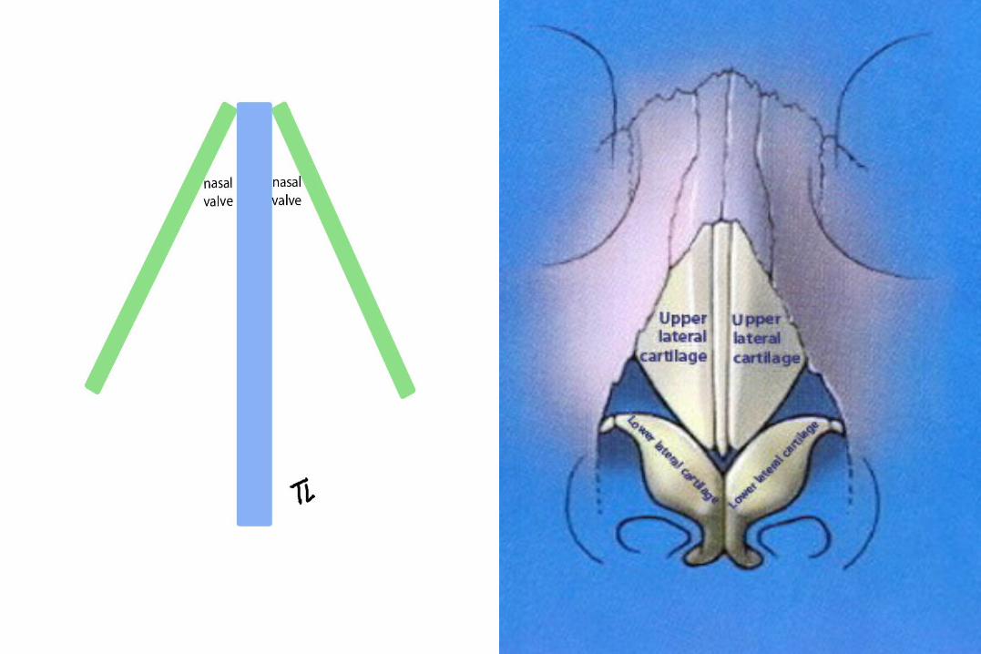
• The internal nasal valve area is the narrowest part of the nasal passage and is the major source of nasal resistance in normal patients.
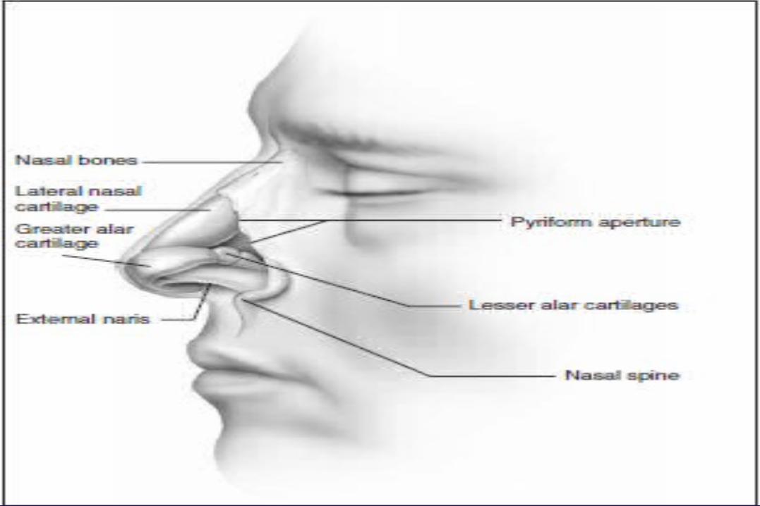
• Four structures compose the internal nasal valve: the upper lateral cartilage superiorly, nasal septum medially, pyriform aperture inferiorly, and the head of the inferior turbinate posteriorly.
• The narrowest portion of the internal nasal valve area is the region between the septum and the caudal border of the upper lateral cartilage. This structure is approximately 10–15 ° in Caucasian subjects and can be wider in subjects of African and Asian descent.
• Patients with internal nasal valve angles of <10° are more prone to internal nasal valve collapse on inspiration.
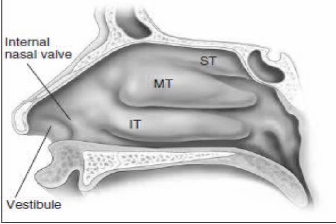
Functional valvular anatomy• The external nasal valve is composed of the nares
and the nasal vestibule.• The nasal vestibule lies just inside the external naris
and is located caudal to the internal nasal valve area.
• The septum is located medially along with the columella, and the alar sidewalls are lateral to the vestibule.
• Vibrissae are located within the vestibule under the lateral crus and function as a filter for the inspired air. They also serve to direct the air posteriorly into the nasal cavity and to slow the inspired air.
Functional valvular anatomy• The internal and external nasal valves function together to deliver a
smooth air current to the nasal cavities for humidification. • The nasal valves can have a greater influence during deep inspiration
when the nostrils flare and the diameter of the external nasal valve is increased.
• The Bernoulli principle is responsible for this effect in that the intra-luminal pressure in the internal valve area decreases when the airflow is increased through it.
• The cartilaginous structure of the nose serves as a counterbalance to this tendency toward internal nasal valve collapse.
• The investing nasal musculature consists of elevator muscles, including the procerus, levator labii superioris alaeque nasi and anomalous nasi. The depressor muscles include the alar nasalis and the depressor septi nasi while the compressor muscles include the transverse nasalis and the compressor narium minor. There are also minor dilator muscles. The alar muscles contract and dilate the internal nasal valve area in order to keep the lumen open.
• Throughout normal nasal function, the internal nasal valve area should remain unchanged.

Functional valvular anatomy• Individuals with a narrow upper cartilaginous vault
have a narrower internal nasal valve and may exhibit collapse in the resting state.
• Those subjects who possess weak upper lateral cartilages and/or lateral nasal walls may be more predisposed to collapse of the internal nasal valve during inspiration.
• Those patients who have pre-existing or traumatic septal deviation can suffer nasal obstruction.
• Insufficient support of the alar rim and alar lobule may lead to external valve collapse on inspiration.

Causes of nasal obstruction 1- Structural causes: include septal deviation, hypertrophy of the
inferior or middle turbinates and fixed and inspiratory nasal valve collapse, which may or may not be secondary to prior nasal surgery or trauma.
• Nasal valve dysfunction may contribute to symptoms in as many as 13% of adults that report chronic nasal obstruction.
• The increased respiratory efforts by OSAHS patients during episodes of apnea caused by palatal or tongue base obstruction make these patients more prone to nasal valve collapse.
• In children, in addition to developmental abnormalities like choanal atresia and craniofacial syndromes (e.g. Pierre Robin sequence), adenoidal and to a lesser degree – tonsillar hypertrophy also constitute an important cause for nasal obstruction, both of which have an important correlation with OSAHS in this patient population.
• Chronic mouth breathing in these patients actually leads to acquired craniofacial abnormalities (e.g. the ‘ adenoid face ’ ), which further compromise the stability of the upper airway.
Causes of nasal obstruction2- Alterations in the nasal mucosa lead to nasal congestion,
which involves the cavernous tissues of the turbinates. • Common causes of nasal congestion include allergic rhinitis,
vasomotor rhinitis, chronic sinusitis, and upper respiratory tract viral infections.
• Chronic inflammation, as well as conditions like asthma, aspirin sensitivity or cystic fibrosis, lead to the development of nasal or nasopharyngeal polyps, which cause obstruction.
• Intranasal corticosteroid therapy for rhinitis showed improvement of OSAHS, but not snoring,
3- Facial muscular weakness and impaired nasal reflexes (especially the ones involved in dilating the nose prior to inspiration), which occur secondary to neuropathy and facial palsy, are also important neuromuscular causes of nasal obstruction.
Diagnostic evaluation of nasal obstruction
1- the history : include whether the obstruction is uni- or bilateral, intermittent or persistent, seasonal or perennial, and whether it is worse while in the supine position, particularly at night.
• A detailed medication history is essential, in order to document the effects of medications, particularly decongestants and topical steroids.
• History of previous surgery is also an important aspect that guides the therapeutic decision making.
• There is no simple way to assess the patient’s nasal airway during sleep.
• A therapeutic trial of topical decongestants and systemic steroids may be useful in assessing the effect on snoring and overall quality of sleep in OSAHS patients.

Diagnostic evaluation of nasal obstruction
2- The physical examination : include an examination of the internal and external nasal valves and the septum by means of anterior rhinoscopy.
• The nasal valves are common sites of obstruction in sleep apneic patients even more than deviated septa.
• Fiberoptic endoscopy enables the surgeon to rule out the presence of any obstructing masses such as nasal polyps or nasopharyngeal tumors.
3- Radiologic evaluation; with CT scans may also have a confirmatory for the preoperative evaluation in select cases, but is not essential.
Diagnostic evaluation of nasal obstruction
4- Therapeutic trials : help confirm the causes and sites of nasal obstruction, and also help in determining the potential success of both medical and surgical interventions.
• Topical nasal steroids, sympathomimetic agents, and antihistamines, as well as allergic management in the form or desensitization.
• Patients that show significant improvement may choose not to undergo surgery.
• The impact of decongestants such as oxymetazoline during nighttime sleep is valuable in assessing the impact of nasal obstruction in symptoms like snoring and overall in sleep-disordered breathing.
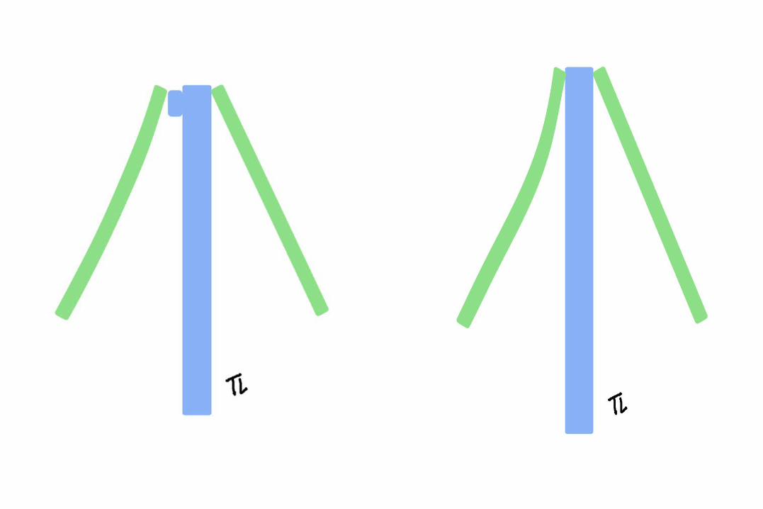
• The role of nasal valve collapse in nasal obstruction can also be confirmed with a trial of Breathe Right ™ nasal strips which help maintain the valves open through external dilatation, and prevent collapse during deep inspiration. Patients that have an adequate response in the form of reduced snoring and improved breathing are likely to benefit from nasal valve suspension procedures.


Diagnostic evaluation of nasal obstruction5- Nasal airflow evaluation: • Even a thorough clinical evaluation can be unreliable and even
contradictory.• Patients are often not completely aware of the magnitude of nasal
obstruction they might be experiencing, particularly during sleep.• In the awake state, patients with atrophic rhinitis secondary to extensive
turbinate resection have a persistent feeling of obstruction in spite of objective evidence of patency, whereas patients with deviated septa or nasal polyposis report no obstruction symptoms, despite evidence against a patent nasal airway.
• The evaluation of nasal airflow is a key element in the evaluation of improvement after surgical intervention.
• Measurements are performed at baseline and after procedures, in order to compare values and confirm improvement.
• Nasal airflow tests show a wide variety of normal values, which limits their use as stand-alone diagnostic tests.
Rhinomanometry• Rhinomanometry (RM) measures nasal airway resistance
and airflow. It has two phases, passive and active, and it can be divided into anterior and posterior.
• Active RM requires the patient to generate airflow through inspiration.
• Passive RM utilizes external generation of airflow through the nose at a constant pressure.
• Anterior RM reflect the status of the nares, nasal valves, and nasal cycling, and utilizes a device inserted into the nares.
• Posterior RM, utilizes a sensor inserted into the mouth which measures the nasopharynx, hence requiring substantial patient co-operation.
• These tests are also usually performed before and after the administration of nasal decongestants.
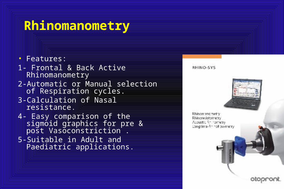
Rhinomanometry
• Features:1- Frontal & Back Active
Rhinomanometry2-Automatic or Manual selection of
Respiration cycles.3-Calculation of Nasal resistance.4- Easy comparison of the sigmoid
graphics for pre & post Vasoconstriction .
5-Suitable in Adult and Paediatric applications.

RHINOSPIR-PRO rhinomanometer
• The RHINOSPIR-PRO rhinomanometer is a PC based unit, which permits the objective assessment of nasal airway resistance, allowing the following tests to be carried out:
1-Active anterior rhinomanometry2-Active posterior rhinomanometry.3-Nasal Provocation Test (N.P.T).
Acoustic rhinometry• Acoustic rhinometry evaluates nasal obstruction by analyzing
reflected sound waves introduced through the nares.• It is not invasive, is easily reproducible and it does not require
patient co-operation.• The results are expressed as cross-sectional dimensions of
the nasal cavity.• The cross-sectional area (CSA) is measured at different points
from the nares to the closest area of narrowing, which corresponds to the nasal valve area.
• Since a ‘ normal ’CSA varies so much from patient to patient, an absolute value is not very useful for diagnosis or for confirmation of nasal valve area obstruction.
• The ratio of the CSA during inspiration to the CSA at rest is highly diagnostic of nasal valve collapse.
• Normally, deep inspiration should not decrease the CSA. If it does, it is a sign of nasal valve collapse.
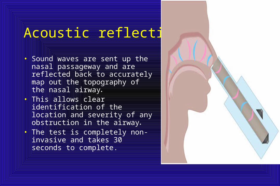
Acoustic reflection
• Sound waves are sent up the nasal passageway and are reflected back to accurately map out the topography of the nasal airway.
• This allows clear identification of the location and severity of any obstruction in the airway.
• The test is completely non-invasive and takes 30 seconds to complete.
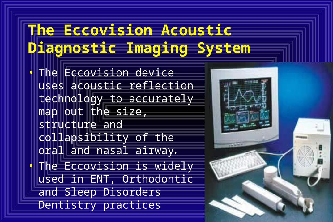
The Eccovision Acoustic Diagnostic Imaging System
• The Eccovision device uses acoustic reflection technology to accurately map out the size, structure and collapsibility of the oral and nasal airway.
• The Eccovision is widely used in ENT, Orthodontic and Sleep Disorders Dentistry practices
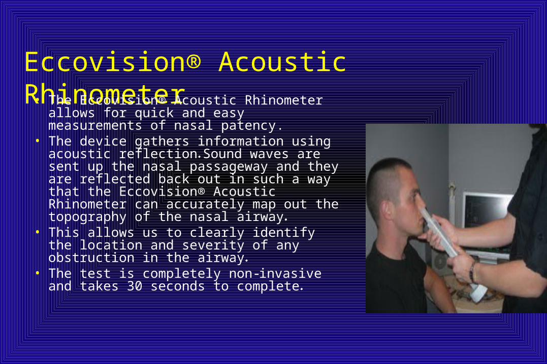
Eccovision® Acoustic Rhinometer• The Eccovision® Acoustic Rhinometer
allows for quick and easy measurements of nasal patency.
• The device gathers information using acoustic reflection. Sound waves are sent up the nasal passageway and they are reflected back out in such a way that the Eccovision® Acoustic Rhinometer can accurately map out the topography of the nasal airway.
• This allows us to clearly identify the location and severity of any obstruction in the airway.
• The test is completely non-invasive and takes 30 seconds to complete.
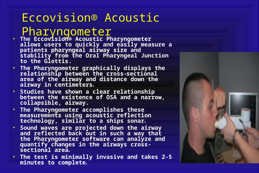
Eccovision® Acoustic Pharyngometer• The Eccovision® Acoustic Pharyngometer allows
users to quickly and easily measure a patients pharyngeal airway size and stability from the Oral Pharyngeal Junction to the Glottis.
• The Pharyngometer graphically displays the relationship between the cross-sectional area of the airway and distance down the airway in centimeters.
• Studies have shown a clear relationship between the existence of OSA and a narrow, collapsible, airway.
• The Pharyngometer accomplishes these measurements using acoustic reflection technology, similar to a ships sonar.
• Sound waves are projected down the airway and reflected back out in such a way that the Pharyngometer software can analyze and quantify changes in the airways cross-sectional area.
• The test is minimally invasive and takes 2-5 minutes to complete.
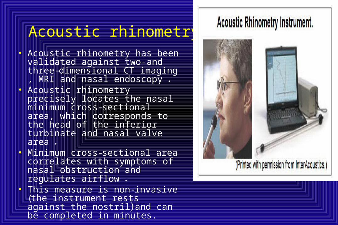
Acoustic rhinometry• Acoustic rhinometry has been
validated against two- and three-dimensional CT imaging , MRI and nasal endoscopy .
• Acoustic rhinometry precisely locates the nasal minimum cross-sectional area, which corresponds to the head of the inferior turbinate and nasal valve area .
• Minimum cross-sectional area correlates with symptoms of nasal obstruction and regulates airflow .
• This measure is non-invasive (the instrument rests against the nostril) and can be completed in minutes.

Nasal obstruction and sleep disordered breathing
• Nasal obstruction and SDB often co-exist.• Approximately 15% of patients with SDB also have nasal obstruction, but
there is no correlation between the severity of obstructive sleep apnea and an objective measure of nasal resistance – acoustic rhinometry.
• Cephalometrics, body mass index and posterior rhinometry showed that daytime nasal obstruction is an independent risk factor for obstructive sleep apnea.
• Normal patients have been shown to experience sleep disturbances – including sleep stage disruption as well as new-onset snoring and even mild obstructive sleep apnea – after acute, complete bilateral nasal obstruction such as abrupt occlusion of their nose.
• SDB was related to nasal cross-sectional area objectively assessed using acoustic rhinometry and both titrated nasal CPAP and the RDI ; however, these associations were only present in patients with normal BMI.
Nasal obstruction and sleep disordered breathing
• Besides the simple effect of nasal obstruction on breathing patterns during sleep, the nose is a major conduit for treatment of obstructive sleep apnea with positive airway pressure therapy.
• Nasal obstruction can therefore interfere with treatment.
• Nasal obstruction can increase the required CPAP in obstructive sleep apnea patients.
• Nasal obstruction can affect positive airway pressure therapy tolerance and adherence .

Three different theories have been proposed to explain the relationship between nasal obstruction and SDB.
1-Increased airway resistence: If the extra-luminal pressure was greater than the pressure of the downstream segment, flow was related not to pressure differential across the whole tube, but rather to inflow pressure minus the extra-luminal pressure.
• Applying the Starling model to the pharynx maximal airflow is based on three factors: upstream pressure, resistance in the upstream segment, and the extra-luminal pressure.

1-Increased airway resistence• Nasal resistance matches the impedance of upper
and lower airways to prolong inspiration and expiration.
• On expiration ,this prolongation has the beneficial effect of improving pulmonary compliance and increasing gas exchange.
• On inspiration, nasal resistance can augment pharyngeal resistance to promote upper airway collapse.
• The decline in tidal volume and minute ventilation has been shown in several studies.
2-Unstable oral breathing:• With nasal obstruction, patients open their mouth. This
reflexive compensation contributes to SDB by narrowing the pharyngeal lumen in two ways.
• First, the chin and rest of the mandible move posteriorly and inferiorly with mouth opening to displace the tongue in that direction. This directly narrows the pharyngeal airway.
• Second, this maneuver decreases the length and tension of the muscles surrounding the airway, and the compliance of the pharynx increases.
• It has been shown that opening of the mouth to provide a 1.5 cm separation between incisors moves the angle of the mandible posteriorly 1 cm, a substantial change.
3- Impaired nasal reflexes:
• Nasal breathing stimulates ventilation,• Not only is minute ventilation higher with nasal compared to
oral breathing, but also that augmented nasal airflow increases minute ventilation.
• Furthermore, the evidence that this response is due to neural regulation in humans comes from studies which show that application of topical anesthesia in the nose and nasopharynx specifically increases nasal and pharyngeal resistance, and that topical nasal anesthesia actually leads to increased SDB ,whether this is measured as the number of apneas, apnea duration, or a decrease in genioglossus muscle activity.

Effect of treatment:
• Treatment of nasal obstruction has three potential goals.
1-It can reduce nasal obstruction,
2- reduce the severity of , possibly even eliminating – SDB, or
3-it can facilitate SDB treatment by allowing the nose to be used more easily as a conduit for positive airway pressure therapy.
Effect of treatment: • A wide variety of treatments have demonstrated benefits
in improving nasal obstruction,• In contrast, isolated treatment of nasal obstruction does
not successfully treat obstructive sleep apnea for most patients.
• Medical treatment produced resolution of obstructive sleep apnea in 9% of patients and surgical treatment in 18%.
• Nasal corticosteroids can produce small changes in snoring and the Apnea/Hypopnea Index, but the degree of improvement varies widely.
• Less than 20% of patients achieve a 50% reduction in AHI with nasal surgery alone.
• A recent report failed to show improvement in snoring intensity or daytime sleepiness symptoms in a group of 40 patients after nasal surgery for an obstructed nasal airway, in spite of decreased nasal resistance as measured by rhinomanometry.
• Patients undergoing nasal surgery alone for the treatment of OSAHS showed that, despite subjective improvement in patients undergoing submucous resection (SMR) of the septum with or without SMR of the inferior turbinates, an overall improvement in OSAHS on the basis of polysomnographic data could not be demonstrated in most patients.
• In fact, when pre- and postnasal surgery AHI have been compared in mild, moderate, and severe OSAHS patients, a tendency towards worsening of this parameter has been observed, although statistical significance for the postoperative increase in AHI was only observed in mild OSAHS cases. This paradoxical effect has been previously described,
• Numerous hypotheses have been proposed for this paradoxical effect are based on
1- Better subjective sleep quality due to a patent nasal airway is achieved, which leads to deeper and more comfortable sleep, resulting in increased apneic episodes due to increased collapsibility of the upper airway.
2- Another possible explanation for worsening of the AHI from the first to the second night of study might occur because the patients experience what is also known as the ‘ first night effect ’ , where they become adapted to the test environment after the second or third night of study, and thus are able to sleep better.
3- The increased airflow in a patient’s nasal airway creates more turbulence by the Bernoulli effect, thus promoting the collapse of the airway at the site of greatest pressure, which corresponds to the narrowest point.
• Correction of nasal airway obstruction has been shown to have a significant impact on OSAHS when performed as a part of a multilevel approach to treat patients with OSAHS .
Patients with nasal obstruction and OSAHS achieved significantly better objective and subjective success rates when soft palate implant insertion for the treatment of snoring and mild to moderate OSAHS was performed together with nasal surgery .The lowest oxygen saturation levels (LSaO 2 ) are directly affected by the patency of the nasal airway, with or without persistent OSAHS. While the increase in LSaO 2 might be discrete, nasal interventions
might at least maintain adequate blood oxygenation levels. • Nasal treatments can facilitate the treatment of SDB by decreasing the
magnitude of the positive airway pressure necessary to treat SDB.\A reasonable improvement (9.3–6.7 cm water) for septoplasty with or without inferior turbinate reduction, and a smaller effect (8.6–8.0 cm water) was seen with an external nasal valve dilator device. Two additional studies have shown that nasal surgery can increase the adherence to continuous positive airway pressure devices.
• CPAP in itself might promote the development of nasal inflammation, and in some patients even vasomotor rhinitis, particularly when using CPAP machines that do not control the temperature of the delivered air nor humidify it.
Effects of nasal surgery on snoring and sleep apnea
• The location of obstruction in the OSAHS is variable and can often be localized to several levels in the upper airway.
• In fact, the level of obstruction can vary in the same patient among consecutive episodes of apnea.
• In normal circumstances, the normal nose contributes to 50% of upper airway resistance, adding to the resistance provided by both oropharyngeal tissues and the tongue.
• In fact, resistance at the level of the nose is more constant in both sleep and awake states, due to the more rigid frame provided by the septum and the lower and upper lateral cartilages.
• If the upper or lower lateral cartilages are weak, damaged, resected, this stability is lost. Patients in such cases are subject to an increased tendency towards collapse during sleep, even with a normal nasal airway in the awake state.
Effects of nasal surgery on snoring and sleep apnea
• Nasal obstruction interferes with pressure titration in nasal continuous positive airway pressure (nCPAP) for the management of OSAHS, and that treatment of such obstruction improves patient compliance with Ncpap.
• The cause–effect relationship of nasal obstruction and OSAHS, however, remains unclear.
• The primary sites of nasal obstruction are the nasal vestibule, the nasal valves, and the turbinates.
• The nasal septum, when deviated, also has a significant impact on these areas of obstruction.
• Of these three sites, the nasal valve is the site of major resistance. in fact, nasal valve incompetence may equal, or even exceed, septal deviation or turbinate hypertrophy as the prime cause of nasal airway obstruction.
Effects of nasal surgery on snoring and sleep apnea
• During inspiration, particularly during the forced inspiration that occurs during an apneic episode, the negative nasopharyngeal and intranasal pressures increase to generate more flow, creating a transmural pressure gradient, which, when a critical value is reached, causes collapse of the upper lateral cartilages.
• Nasal obstruction leads to mouth opening and transition to oral breathing, which contributes to airway flow limitation and collapse, mainly due to inferior movement of the mandible, a backward fall of the base of the tongue, resulting in a reduction of the posterior pharyngeal space and diameter and an increased respiratory effort that causes collapse of the pharyngeal tissues due to greater negative pressures.
Conclusions
• The patient population in whom SDB presents will commonly also show concurrent nasal obstruction.
• Selecting the potential medical and surgical treatment options requires an understanding of nasal anatomy and physiology and the combination of history, physical examination, and accurate diagnosis.
• There are three potential mechanisms for the association between nasal obstruction and SDB: an increase in airway resistance; the transition to unstable oral breathing; and the impairment of nasal reflexes.
• Treatment can effectively reduce nasal obstruction, although the effects are more variable in the reduction of SDB severity (when treating nasal obstruction alone) and the facilitation of SDB treatment.
Conclusions5- Selected patients who respond to a diagnostic trial of artificial nasal
airway improvement with topical and systemic decongestants and steroids, along with external nasal dilators (e.g. Breathe Right ™ strips), may also benefit from definitive correction of the nasal airway, with elimination of snoring and improvement in OSAHS.
6-The majority of patients with nasal obstruction and OSAHS will likely benefit from a multilevel surgical approach.
7-If nasal surgery is undertaken alone, it is important to inform patients that it may not only fail to cure, but may even aggravate snoring and OSAHS in certain cases.
8- Nasal surgery also has a significant impact in improving patients ’subjective symptoms, like daytime sleepiness and quality of sleep. It also constitutes a valuable intervention in patients that need nCPAP management for their OSAHS.
Friedman tongue position and the staging of OSAHS• Nasal, palatal and hypopharyngeal obstruction, acting alone or in concert, are
frequently identified as the cause of snoring and OSAHS.• Even in cases where a single site is primarily involved, the increase in negative
pressure may induce further obstruction in other areas. • When surgical management of OSAHS is considered, a clear understanding of
the complex relationship between the sites of obstruction is essential to surgical success.
• Numerous methods that attempt to predict the location of the upper airway obstruction include snoring sound analysis, physical examination, computed tomography, magnetic resonance imaging, cephalometric studies and fluoroscopy,
• The number of methods described is evidence of the lack of agreement that any single method is perfect.
• The most commonly used method is the Mueller maneuver (MM). This methode is used for the preoperative assessment of sleep-disordered breathing (SDB). The MM consists of having the patient perform a forced inspiratory effort against an obstructed airway with fiberoptic endoscopic visualization of the upper airway.
• The test is widely used and simple to perform. Despite this, its use is controversial and certainly no studies have been able to associate the maneuver as a tool for patient selection.
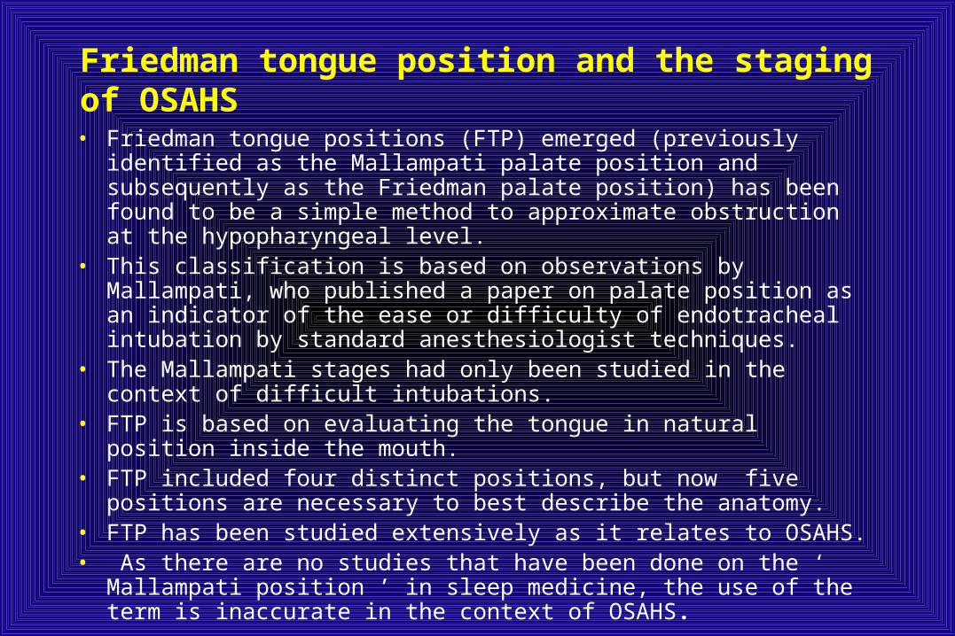
Friedman tongue position and the staging of OSAHS
• Friedman tongue positions (FTP) emerged (previously identified as the Mallampati palate position and subsequently as the Friedman palate position) has been found to be a simple method to approximate obstruction at the hypopharyngeal level.
• This classification is based on observations by Mallampati, who published a paper on palate position as an indicator of the ease or difficulty of endotracheal intubation by standard anesthesiologist techniques.
• The Mallampati stages had only been studied in the context of difficult intubations.
• FTP is based on evaluating the tongue in natural position inside the mouth.
• FTP included four distinct positions, but now five positions are necessary to best describe the anatomy.
• FTP has been studied extensively as it relates to OSAHS.• As there are no studies that have been done on the ‘ Mallampati
position ’ in sleep medicine, the use of the term is inaccurate in the context of OSAHS.
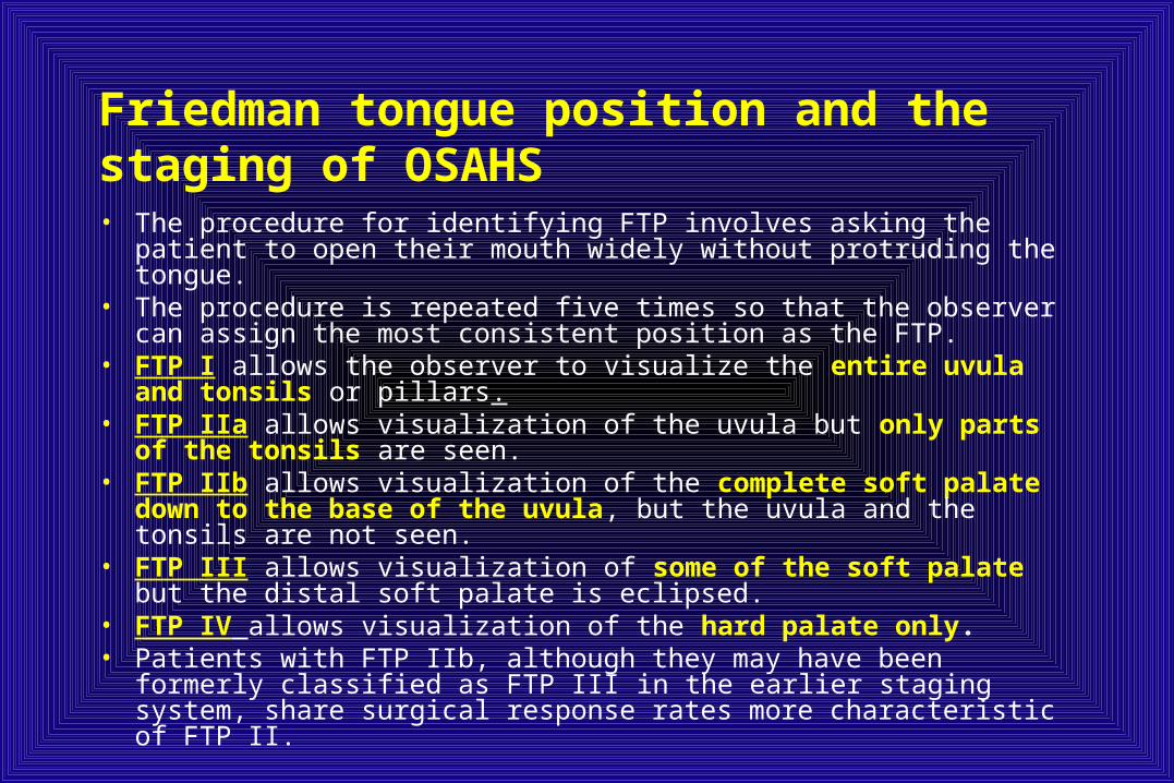
Friedman tongue position and the staging of OSAHS• The procedure for identifying FTP involves asking the patient to open
their mouth widely without protruding the tongue.• The procedure is repeated five times so that the observer can assign the
most consistent position as the FTP. • FTP I allows the observer to visualize the entire uvula and tonsils or
pillars.• FTP IIa allows visualization of the uvula but only parts of the tonsils
are seen. • FTP IIb allows visualization of the complete soft palate down to the
base of the uvula, but the uvula and the tonsils are not seen.• FTP III allows visualization of some of the soft palate but the distal soft
palate is eclipsed.• FTP IV allows visualization of the hard palate only.• Patients with FTP IIb, although they may have been formerly classified
as FTP III in the earlier staging system, share surgical response rates more characteristic of FTP II.

Friedman tongue position and the staging of OSAHS
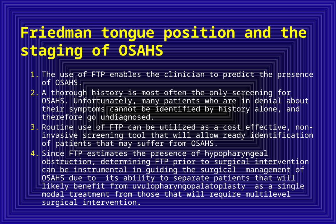
1. The use of FTP enables the clinician to predict the presence of OSAHS.2. A thorough history is most often the only screening for OSAHS.
Unfortunately, many patients who are in denial about their symptoms cannot be identified by history alone, and therefore go undiagnosed.
3. Routine use of FTP can be utilized as a cost effective, non-invasive screening tool that will allow ready identification of patients that may suffer from OSAHS.
4. Since FTP estimates the presence of hypopharyngeal obstruction, determining FTP prior to surgical intervention can be instrumental in guiding the surgical management of OSAHS due to its ability to separate patients that will likely benefit from uvulopharyngopalatoplasty as a single modal treatment from those that will require multilevel surgical intervention.
Friedman tongue position and the staging of OSAHS
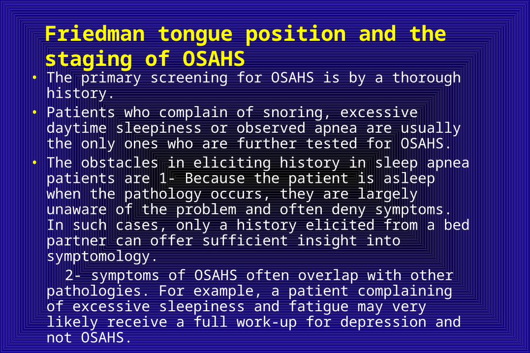
• The primary screening for OSAHS is by a thorough history. • Patients who complain of snoring, excessive daytime
sleepiness or observed apnea are usually the only ones who are further tested for OSAHS.
• The obstacles in eliciting history in sleep apnea patients are 1- Because the patient is asleep when the pathology occurs, they are largely unaware of the problem and often deny symptoms. In such cases, only a history elicited from a bed partner can offer sufficient insight into symptomology.
2- symptoms of OSAHS often overlap with other pathologies. For example, a patient complaining of excessive sleepiness and fatigue may very likely receive a full work-up for depression and not OSAHS.
Friedman tongue position and the staging of OSAHS
– The known physical findings that are associated with OSAHS include BMI, neck circumference, and tonsil size, are routinely assessed and are well defined.
– Descriptions of hypopharyngeal obstruction have not been standardized. Often times the similar physical findings are reported with many arbitrary terms such as ‘ crowded oropharynx , macroglossia ’ , ‘ retrognathia ’ ,etc. This causes much confusion in both patient care and in reporting data.
– The routine use of FTP in the context of OSAHS can help standardize the description of hypopharyngeal obstruction.
– FTP can be employed in an algorithm that can help identify patients with OSAHS. This system is based on three readily identifiable and reproducible physical exam findings and can provide a simple means for screening patients.
– The system relies on calculating the patient’s Body Mass Index (BMI), along with the assessment of the patient’s tonsil size and FTP.

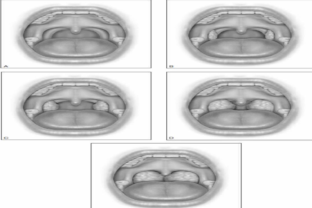
Friedman tongue position and the staging of OSAHS– Tonsil size and BMI are assessed as follows. Tonsil
size 1 implies tonsils hidden within the pillars. Tonsil size 2 implies the tonsil extending to the pillars. Size 3 tonsils are beyond the pillars but not to the midline. Tonsil size 4 implies tonsils that extend to the midline.
– The BMI is graded as grade 0 (< 20 kg/m 2 ), grade 1 (20–25 kg/m 2 ) grade 2 (25–30 kg/m 2 ), grade 3 (30–40 kg/m 2 ), and grade 4 ( > 40 kg/m 2 ).
– OSAHS score = FTP(0- IV) + tonsil size (0-4) +BMI (0-4)

Friedman tongue position and the staging of OSAHS
• Any value above 8 is considered as a positive OSAHS score, whereas any value below 4 is considered a negative OSAHS score.
• A positive score has a positive predictive value of moderate OSAHS (Apnea/Hypopnea Index (AHI) > 20) of 90%, and was 74% effective in predicting severe OSAHS (AHI > 45).
• A negative score was 67% effective in predicting an AHI of < 20. • With the use of FTP and the OSAHS score, the number of
undiagnosed cases may also fall.• In cases where history is unclear, this algorithm may help
identify patients that may have otherwise gone unnoticed. In other cases, this algorithm may provide the impetus for eliciting a more detailed history and performing further tests to confirm suspicion of OSAHS.
PETCO2•
Surgical staging of OSAHS• Uvulopalatopharyngoplasty (UPPP) is the most common
surgical procedure performed by otolaryngologists for the treatment of OSAHS.
• Unfortunately, only 40-79% of patients had a ‘ successful ’ surgery defined by an AHI reduction of 50% and a postoperative AHI < 20 or an Apnea Index (AI) reduced by 50% and a postoperative AI <10.
• The ideal system would identify those patients with a high likelihood of successful UPPP and separate them from those with a high likelihood of failure, thus guiding patient selection and improving outcomes.
• The most common method used to identify patients for surgery is based on the misconception that patients with mild/moderate disease are better candidates for UPPP than those patients with severe disease. Therefore, the procedure is often recommended for patients with mild/moderate OSAHS.
Surgical staging of OSAHS
• Severity of disease as determined by clinical symptoms, polysomnography results, or tools such as the Epworth Sleepiness Scale has been shown not to correlate with surgical success.
• Studies have shown that patients with mild SDB based on clinical and polysomnographic data have no better chance of successful treatment with UPPP than patients with severe disease.
• In fact, one study demonstrated that UPPP performed on unselected patients with mild OSAHS (AHI<15) not only does not cure disease in 60% of cases but often makes it worse .
Surgical staging of OSAHS• It is well known that OSAHS involves obstruction of
the airway at multiple levels.• Although palatal obstruction accounts for a large
portion of the obstruction, hypopharyngeal obstruction can also play a significant role.
• UPPP alleviates obstruction at the level of the soft palate and tonsils, but does not address obstruction at the level of the hypopharynx. This is clearly a significant cause of the failure of UPPP.
• Therefore when devising a system that is intended to predict UPPP outcomes, the anatomical considerations must be incorporated.
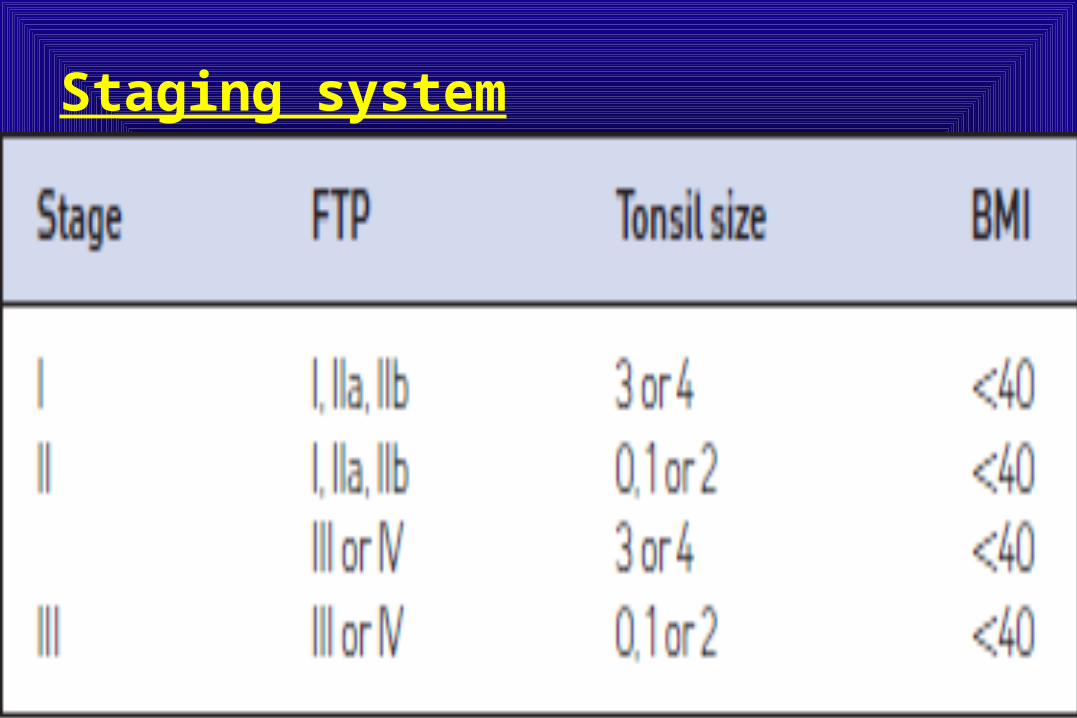
Staging system

Surgical staging of OSAHS• Most surgeons have found that patients with BMI <40 have a poor
prognosis for corrective UPPP and therefore these patients are automatically assigned to stage IV.
• All patients with skeletal deformities such as micrognathia or mid-face hypoplasia are considered stage IV.
• The rationale for such a staging system is that the success of UPPP is highly dependent on the anatomical relationship between palatal and hypopharyngeal obstruction. Stage I patients are those with favorable tongue position (I, IIa, IIb) indicating minimal hypoglossal obstruction and large tonsils. They are most likely to benefit from UPPP with tonsillectomy as hypopharyngeal obstruction does not represent a significant component of their disease. Stage III patients are on the opposite extreme of the spectrum with small or no tonsils and unfavorable tongue position (III or IV) indicating significant hypopharyngeal obstruction. They are least likely to benefit from UPPP as UPPP will not address their hypopharyngeal obstruction. Stage II patients are those with either large tonsils and unfavorable tongue position or small tonsils and a favorable tongue position. UPPP was most likely achieved in stage I patients (80.6%) and least likely in stage III patients (8.1%).
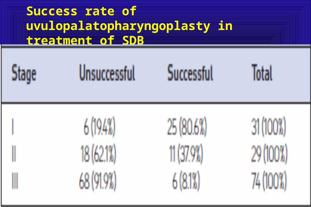
Success rate of uvulopalatopharyngoplasty in treatment of SDB

Success rate of uvulopalatopharyngoplasty in treatment of SDB
• The use of the anatomic staging system in OSAHS offers a cost-effective, non-invasive, reproducible method to stratify patients based on anatomic variations.
• The use of this system in addition to detailed clinical examination, cephalometrics and Mueller maneuver can help improve surgical outcomes in OSAHS.
• Patients with stage I disease have better than an 80% chance of success with UPPP and should therefore undergo the procedure when non-surgical treatment has failed. Even patients with severe SDB had an 80% success rate if they had stage I disease based on this staging system.
• Patients with stage III disease should never undergo UPPP alone as a surgical cure for SDB. With an 8.1% success rate, the surgery is destined to fail.
• They should be treated with a combination of procedures that address both the palate and the hypopharynx. Patients with stage II disease do not fall into either extreme but probably can be treated similar to stage III patients.
• Allergic rhinitis affects up to 40% of the population, and its prevalence appears to be increasing.
• Nasal congestion is a prominent and troublesome symptom of both allergic and non-allergic rhinitis.
• Additional symptoms of allergic rhinitis include rhinorrhea, sneezing, and pruritus of the eyes, nose, and throat, particularly in patients with perennial allergic rhinitis (PAR).
• Typical sleep-related problems seen include sleep-disordered breathing, sleep apnea, and snoring, all of which are associated with nasal congestion / obstruction.
Rhinitis and sleep
Rhinitis• Allergic rhinitis can affect the mucous membranes of the nose, eyes,
eustachian tubes, middle ear, sinuses, and pharynx and is usually triggered by immunoglobulin E in response to an allergen.
• Mediators involved in clinical symptoms include histamine, tryptase, chymase, heparin as well as leukotrienes.
• Non allergic rhinitis is a term used to describe a variety of diseases including, infectious, vasomotor, occupational, hormonal, gustatory, and drug induced rhinitis.
• In non-allergic etiologies, autonomic parasympathetic stimuli lead to localized nasal swelling and secretions.
• In a recent survey of individuals with allergic rhinitis, 68% of respondents with perennial allergic rhinitis (PAR) and 48% with seasonal allergic rhinitis (SAR) reported that their condition interfered with sleep.
• Sleep disturbance can be caused by the mechanical obstruction of nasal congestion. However, additional symptoms of rhinitis and the release of inflammatory mediators may influence sleep and resultant daytime fatigue and somnolence.
• Treatments that improve the symptoms of allergic rhinitis, particularly those that relieve nasal congestion, have been shown to improve patients’ sleep and quality of life.
Rhinitis• Sleep disturbances associated with allergic rhinitis include
sleep disordered breathing (ranging from snoring to obstructive sleep apnea and/or hypopnea) and micro-arousals, although one study has suggested that allergic rhinitis is not a major risk factor for obstructive sleep apnea syndrome (OSAS).
• Individuals with frequent night-time symptoms of rhinitis have been shown to be more likely to have chronic excessive daytime sleepiness or chronic non-restorative sleep than those who rarely or never have such symptoms.
• Studies using actigraphy have demonstrated objective evidence that adults with PAR had sleep disturbance when compared to normal controls.
Mechanisms of sleep impairment :• The daytime fatigue experienced by patients with allergic rhinitis
could be attributed to sleep impairment resulting from nasal congestion or other rhinitis symptoms or to the effects of inflammatory cytokines on sleep or producing fatigue directly.
• The current evidence suggests that symptoms and the underlying pathophysiologic changes of the disease contribute to reduced sleep quality, insomnia, hypersomnolence,and daytime somnolence.
• Nasal congestion is a common and bothersome symptom and occurs when the cavernous tissues of the nasal turbinates swell as a result of dilation of the capacitance vessels.
• Nasal congestion reduces the internal nasal diameter, increases airway resistance to nasal airflow, and can also cause nasal obstruction.
• The end result of airway resistance is the same irrespective of its etiology including both allergic and non-allergic etiologies.
• The degree of congestion (nasal patency) can be evaluated objectively by measures of nasal airflow (such as peak nasal inspiratory flow), assessment of airways resistance /conductance (rhinomanometry), and acoustic rhinometry, which assesses the volume and area of the nasal cavity by analyzing reflected sound waves .
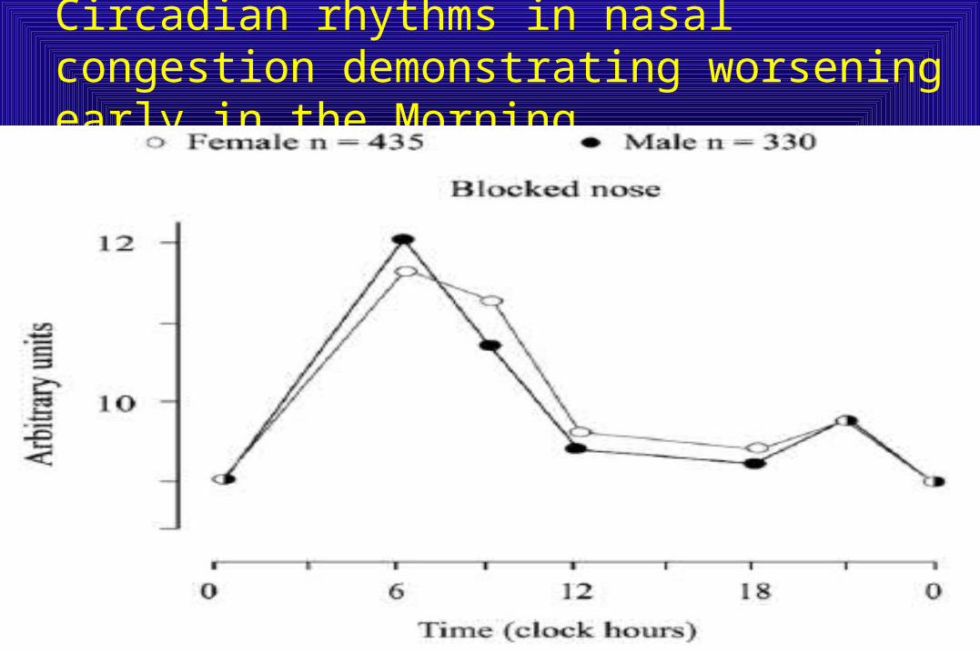
• Nasal congestion is often worse at night and first thing in the morning.
• Congestion tends to increase when an individual lies down.• The normal overnight decline in serum cortisol is likely to
contribute to nighttime worsening.• Similarly in patients with asthma, lower cortisol levels are
associated with greater airway obstruction, particularly at night.
• Nasal congestion worsened overnight and peaked at approximately 6 AM. This shows a clear, large-amplitude, circadian variation, similar, but slightly delayed from what we see in asthma .
Circadian rhythms in nasal congestion demonstrating worsening early in the Morning.
• Approximately half patients stated that congestion was the most bothersome symptom, that nasal congestion woke them up during the night, and that it made it difficult to fall asleep.
• These effects were more pronounced for those experiencing more severe congestion.
• Nasal congestion had a negative effect on emotions and the ability to perform daily activities. These effects may, in part, be a consequence of the effects of nasal congestion on sleep.
• Using objective assessments of sleep indicated that nasal congestion in patients with allergic rhinitis is associated with an increase in the number of micro-arousals and episodes of Apnea.
• Simple nasal occlusion by a nose clip in healthy subjects resulted in an increase in sleep apnea and transient arousals.
• These data support that congestion alone is a major factor interfering with quality of sleep and congestion alone may cause disturbance of sleep and daytime somnolence and other poor outcomes.
• In patients with allergic rhinitis, greater nasal congestion was associated with obstructive sleep apnea. Thus, suggesting a direct correlation.
• Subjects with nasal congestion due to allergic rhinitis were 1.8 times more likely to have moderate to severe sleep-disordered breathing than subjects with allergic rhinitis without nasal congestion.
• The switch to oral breathing that occurs as a result of nasal congestion may be the key factor behind this association, compromising the airway and leading to sleep-disordered breathing.
• Other symptoms, such as sneezing, rhinorrhea, and nasal pruritus, may contribute to reduced sleep quality and sleep disturbance in allergic rhinitis.
• Rhinorrhea was troublesome to patients with allergic rhinitis and interfered with sleep.
• Ocular itching has also been demonstrated as a cause of sleep disturbance .
• The inflammatory mediators, such as histamine and cytokines released during allergic reactions, may directly affect the central nervous system, which contributes to disturbed sleep and fatigue or sleepiness during the day.
• Histamine is involved in the regulation of the sleepewake cycle and arousal.
• Higher levels of the cytokines interleukin (IL)-1b, IL-4, and IL-10 in allergic patients than in healthy subjects have been shown to correlate with increased latency to rapid eye movement (REM) sleep, decreased time in REM sleep, and decreased latency to sleep onset.
• Because the restorative function of REM sleep is important, its disruption may contribute to daytime fatigue, difficulty concentrating, and worse performance in individuals with allergic rhinitis.
• Inflammatory cells and mediators show circadian variation, with levels peaking during the early morning hours.
• These changes of cytokines could explain the higher level of allergic rhinitis symptoms upon waking and why overnight sleep is particularly affected.
• Similar changes in cytokines have also been seen associated with obstructive sleep apnea with increases noted in Interleukin-1, tumor necrosis factor (TNF), and IL-6.
• The changes noted may promote a T-helper cell type 2 phenotype further causing allergic type inflammation leading to increased congestion.
• Elevation of some of the cytokines seen in both disorders, such as TNF, IL-6 and IL-1, can cause fatigue and other non-specific constitutional symptoms during the day.
• The extent that excessive cholinergic activity and the decrease in adrenergic tone has on the congestion and associated sleep disturbance is unknown.
• We do know that automonic disturbance is associated with mild obstructive sleep apnea (OSA).
• The abnormalities noted may be secondary, but may also precede the OSA and possibly be part of the pathologic mechanism.
Sleep impairment and quality of life• Individuals with allergic rhinitis suffer impaired cognitive
function and reduced work productivity and performance.• Allergic rhinitis can affect children’s learning ability and
performance at school and cause somnolence and inability to concentrate in children. These effects may be a direct result of allergic symptoms but are likely to be exacerbated by sleep impairment.
• Sleep disordered breathing and sleep disturbance are known to be directly associated with decreased quality of life in the general population. This is made evident by experimentally induced sleep fragmentation in healthy subjects associated with impaired mental flexibility and attention, increased daytime sleepiness, and impaired mood.
• Daytime fatigue, difficulty concentrating, and impaired psychomotor performance are commonly reported by individuals with allergic rhinitis and may reduce their ability to perform the physical and social tasks of daily living.
Disease-specific quality-of-life measures1-Rhinoconjunctivitis quality of life questionnaire (RQLQ)
include a domain that measures the effects of disease and/or treatment on sleep. These questionnaires focus on the problems for which patients seek help, and so are more sensitive to changes in patients’ quality of life than generic health-status questionnaires or those specific for obstructive sleep apnea.
2-Nocturnal rhinoconjunctivitis quality of life questionnaire(NRQLQ) assesses the functional impairments most problematic for patients with nocturnal symptoms, including sleep problems, symptoms during sleep time and on waking in the morning, and problems during waking hours.
General questionnaires that assess daytime sleepiness and sleep quality
1-Epworth sleepiness scale2- Pittsburgh sleep quality index, Poor sensitivity and specificity may limit their use in assessing the
mild-to-moderate sleep disturbance often noted in rhinitis.
Polysomnography
• Polysomnography in seasonal allergic rhinitis and healthy volunteers showed that there were statistically significant differences between the 2 groups in sleep parameters, including increases in the apnea index, hypopnea index , apneaehypopnea index, snoring time,amount of REM sleep, and sleep latency.
• The changes were not clinically relevant, as values remained within normal limits.
• The study found a statistically significant increase in daytime sleepiness (assessed subjectively using the Epworth sleepiness scale) , suggesting the known poor correlation between subjective and objective measures of sleep disturbance.
Effects of therapy
• Sedating antihistamines are contraindicated in those experiencing daytime sedation, fatigue, and functional impairment.
• Nonsedating oral antihistamines are widely used to treat allergic rhinitis and relieve nasal symptoms such as rhinorrhea, sneezing, and pruritus, but have less effect on nasal congestion .
• While non-sedating antihistamines are tolerated well and often improve sleep and quality of life, data have suggested that the increased sedation secondary to first generation antihistamines and topical intranasal administration of sedating antihistamines may exacerbate daytime somnolence.
Effects of therapy• Oral decongestants reduce nasal congestion, but may have
adverse effects on sleep because of their stimulatory effects and their association with systemic side effects, such as tachycardia and urinary retention. Topical decongestants appear to improve sleep in patients with nasal obstruction, but because of the risk of rhinitis medicamentosa (“rebound” congestion), they should not be used for long periods .
• The anticholinergic agent ipratropium bromide is not considered effective in relieving nasal congestion; however, limited data suggest that sleep and quality of life may be improved following treatment.
• Leukotriene receptor antagonists or a combination of an antihistamine and a leukotriene receptor antagonist have shown some efficacy in improving sleep and quality of life in patients with allergic rhinitis or sleep-disordered breathing.
• The cause of the improved sleep and daytime somnolence may be a reduction of congestion or a reduction of inflammatory mediators or a combination of both.

Effects of therapy• Intranasal corticosteroids are considered first-line therapy when nasal
congestion is a major symptom. • Intranasal corticosteroids relieve the nasal symptoms, including nasal
congestion, and decrease inflammatory mediators secreted from activated lymphocytes, epithelial cells, mast cells and other inflammatory cells.
• The improvement in nasal congestion is both subjective and objective with the objective evidence assessed by an increase in peak nasal inspiratory flow in allergic rhinitis patients treated with intranasal corticosteroids.
• Intranasal budesonide reduces nasal congestion, subjective daytime somnolence, and fatigue and improves sleep quality and patients’ quality of life.
• Another intranasal corticosteroid, flunisolide, improved subjectively assessed nasal congestion and sleep problems in patients with PAR compared with placebo, although daytime fatigue and sleepiness did not show significant improvement.
• Flunisolide was superior to azelastine nasal spray in improving nasal congestion, sleep, and daytime sleepiness in patients with PAR.
• Intranasal fluticasone significantly improved subjective assessments of sleep compared with placebo and decreased daytime sleepiness and fatigue by more than 10% (although this was not statistically significant).
Effects of therapy
• Fluticasone propionate nasal spray improved nasal symptoms, quality of life, and verbal memory in children with SAR.
• Intranasal fluticasone improved allergic rhinitis associated with OSAS; treatment resulted in a significantly lower frequency of apnea/hypopnea and subjective improvements in nasal congestion and daytime alertness, although snoring noise and sleep quality were unchanged compared with placebo.
• All intranasal corticosteroids improve quality of life in patients with allergic rhinitis.
• Intranasal triamcinolone acetonide relieved nasal congestion and improved health-related quality of life (including sleep), as assessed using the RQLQ, the NRQLQ, and the Pittsburgh sleep quality index.
Conclusions• Sleep impairment associated with rhinitis has a significant impact on
patients’ quality of life.• Nasal congestion, one of the most common and bothersome symptoms
of these conditions, is thought to be a major cause of sleep impairment and sleep-disordered breathing.
• The poor sleep associated with nasal congestion is an important therapeutic target.
• Other rhinitis symptoms, inflammatory mediators released because of rhinitis can also disturb sleep and lead to an increase in daytime somnolence and limitations.
• Treatment with intranasal corticosteroids has been shown to significantly reduce nasal congestion in allergic rhinitis and in those with congestion should be considered the drug of choice.
• Montelukast may be an alternative therapy in those intolerant to intranasal corticosteroids.
• Sedating antihistamines should be avoided since they may amplify the sedation and decrease productivity in those already compromised.
• Immunotherapy has to improve sleep quality and daytime somnolence.
Practice points
1) Sleep impairment associated with rhinitis has a significant impact on patients’ quality of life with nasal congestion thought to be a major cause of sleep impairment and sleep-disordered breathing.
2) Treatment with intranasal corticosteroids has been shown to significantly reduce nasal congestion in allergic rhinitis and in those with congestion should be considered the drug of choice.
3) Sedating antihistamines should be avoided since they may amplify the sedation and decrease productivity in those already compromised