central sleep apnea /hypoventillation syndrome by ahmad younes professor of thoracic medicine...
TRANSCRIPT
CENTRAL SLEEP APNEA /Hypoventillation Syndrome
BY
AHMAD YOUNES
PROFESSOR OF THORACIC MEDICINEMansoura faculty of medicine
CENTRAL SLEEP APNEA /Hypoventillation Syndrome
• The central sleep apnea (CSA) syndromes include a diverse group of disorders associated with the presence of central apnea during sleep.
• In some of the disorders the patients have primarily nocturnal hypoventilation (increased PaCO2) due to decreased tidal volume and/or respiratory rate with relatively few discrete central apneas.
• Discussion of patients with central apnea and hypoventilation syndromes together is useful clinically because they have many similar aspects of pathophysiology and treatment.
Central sleep apnea
• Central sleep apnea (CSA) is characterized by repetitive apneic episodes lasting at least 10 seconds each occurring during sleep without associated ventilatory efforts.
• Heterogeneous group of sleep-related breathing disorders in which respiratory effort is absent in an intermittent or cyclical fashion.
• Many patients with CSA also have some obstructive respiratory events.
CSA is considered to be the primary diagnosis when 50% of apneas are scored as central in origin
• CSA, like OSA, is associated with important complications, 1- frequent nighttime awakenings,
2- excessive daytime sleepiness 3- increased risk of adverse cardiovascular outcomes. Manifestations of CSA. 1-Hgh altitude-induced periodic breathing2-Cheyne-Stokes breathing (CSB). 3- Idiopathic CSA (ICSA),4-Narcotic-induced central apnea5- Obesity hypoventilation syndrome (OHS)
Prevalence of CSA•CSA is estimated to represent about 5% to 10% of patients with sleep-related breathing disorders. •Most healthy individuals will have periodic breathing on high-altitude ascent, provided the magnitude of the ascent is sufficient to cause substantial alveolar hypoxia. •Given the global increase in obesity, the prevalence of OHS is likely on the rise.•ICSA is relatively uncommon and may constitute < 5% of patients referred to a sleep•patients with heart failure and left ventricular ejection fraction < 45% revealed that 37% of patients had CSA. Interestingly, OSA is not uncommon in this population at 12%

Risk Factors• High CO2 ventilatory drive• Sleep disturbance: Increased frequency of sleep-wake
transitions• Gender: Men are more likely to have central apneas due to a
higher apneic threshold during NREM sleep. The lower apneic threshold in women than in men could be mediated by both female and male hormones
• Age: Central apneas are more common in older adults due to the increased prevalence of underlying medical disorders (eg, CHF,neurologic disorders), or greater sleep disturbance and awakenings
• Altitude: Central apneas can develop acutely following ascent to high altitudes
• Nasal obstruction
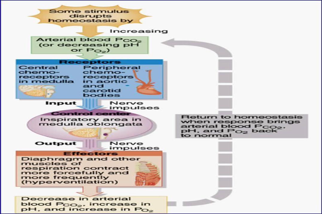
During the waking state, respiration is controlled by three processes, namely the metabolic (automatic), the wake-related drive to breathe and
behavioral (voluntary) systems
• During NREM sleep, the wake-related drive to breathe and behavioral control systems are abolished, and respiration is controlled entirely by the metabolic control system, primarily by the hypercapnic ventilatory drive and to a lesser degree by the hypoxic ventilatory drive
• A PaCO2 above the apneic threshold stimulates ventilation, whereas a PaCO2 below this threshold leads to a central apnea that continues until PaCO2 increases and once again exceeds the apneic threshold.
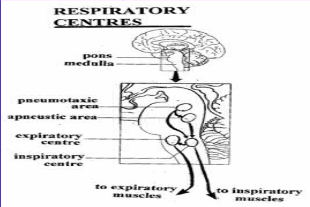
Mechanism of CSA• Chemoreceptor inputs (medullary neurons and peripherally at the carotid
body) play a key role in modulating ventilation. The ventilatory output to a given change in PaO2 or PaCO2 ("chemo-sensitivity") can vary greatly between individuals and with disease status.
• Highly sensitive chemo-responses can place an individual at risk for unstable breathing patterns because these individuals "over-respond" to small changes in chemical stimuli.
• The inherent delays in the negative feedback loop controlling ventilation also contribute to the risk for developing instability.
• individuals with high chemo-responsiveness will hyperventilate markedly to a perturbation potentially, lowering PaCO2 below the eupnic level and leading to hypoventilation and potential apnea.
• Similarly, an individual with a long delay in the loop, such as individuals with reduced cardiac output, may have more prolonged hyperventilation, also leading to greater hyperventilation and a subsequent unstable breathing
12
Mechanism of CSA• Several respiratory control mechanisms are down
regulated at sleep onset.• Upper airway (UA) dilator and respiratory pump
muscle tone is reduced, and there is an accompanying increase in UA resistance leading to a reduction in ventilation for a given level of drive.
• Chemosensitivity is also likely reduced at sleep onset. Although of variable magnitude and rate, these normal physiologic responses occur in all individuals.
Mechanism of CSA
• During sleep all individuals are susceptible to breathing cessation should the PaCO2 fall below a critical threshold known as the apnea threshold. The apnea threshold is usually 2 to 6 mm Hg below the eucapnic sleeping PaCO2 level. This typically equates to the wakefulness eucapnic PaCO2 level or marginally lower
• Regardless of the underlying cause of arousal from sleep (ie, spontaneous arousal, periodic leg movements, respiratory load induced arousal), an individual with a low arousal threshold (ie, susceptible to waking up easily) will be vulnerable to sleep state instability. That is, the combination of a predisposition to sleep transition apnea and a low arousal threshold may be sufficient to facilitate a repetitive CSA cycle as the individual oscillates between wakefulness and sleep.
The arousal threshold increase with progressively deeper sleep stage, as does breathing stability provided slow wave sleep can be achieved.
• The rapid switch from sleep to wakefulness that occurs with arousal causes a sudden shift in the underlying homeostatic control of the cardio-respiratory system. The eucapnic set point rapidly shifts from the sleep set point (approximately 45 mm Hg) to the wakefulness level (approximately 40 mm Hg) creating a state of relative hypocapnia. In addition, sleep-induced UA resistance is removed and the wakefulness drive is reintroduced. The brisk ventilatory response causes a rapid reduction in PaCO2, such that central apnea may ensue during subsequent sleep if the hypocapnia is sufficient to cross the apnea threshold
Central hypopneas• The American Academy of Sleep Medicine (AASM) scoring
manual discourages identification of central hypopnea unless an accurate method to quantify respiratory effort is being used (esophageal pressure or respiratory inductance plethysmography).
• In general, central hypopneas are characterized by a proportionate decrease in both airflow and respiratory effort .
• Usually, there is no snoring or chest-abdominal paradox and the nasal pressure or PAP device flow signal is fairly rounded (minimal or no signs of airflow limitation).
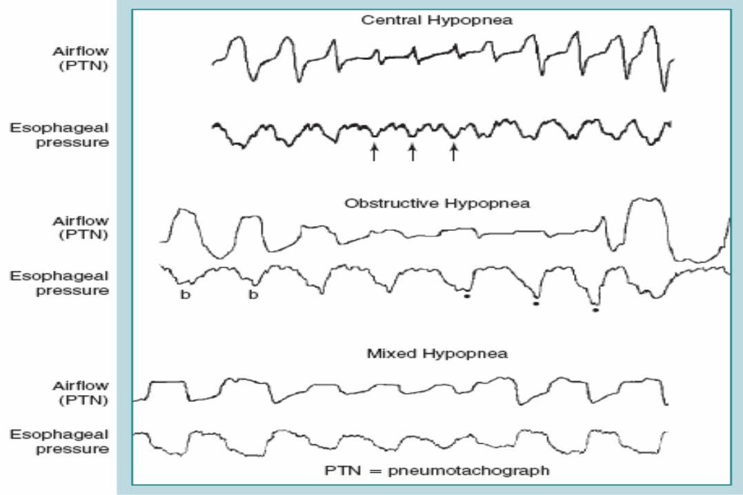
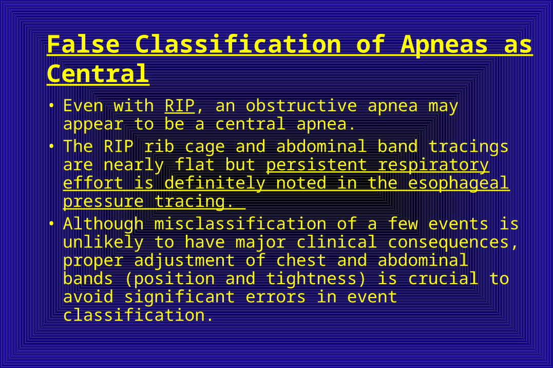
False Classification of Apneas as Central
• Even with RIP, an obstructive apnea may appear to be a central apnea.
• The RIP rib cage and abdominal band tracings are nearly flat but persistent respiratory effort is definitely noted in the esophageal pressure tracing.
• Although misclassification of a few events is unlikely to have major clinical consequences, proper adjustment of chest and abdominal bands (position and tightness) is crucial to avoid significant errors in event classification.
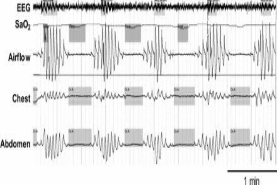
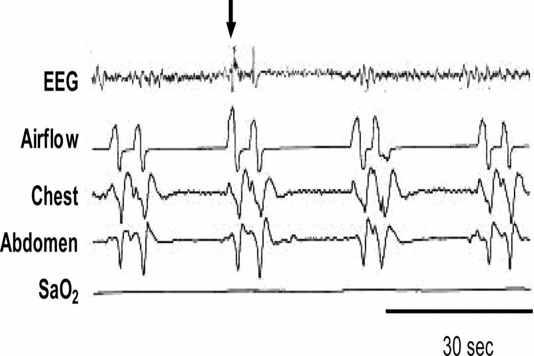
PSG features of central apneas• Pauses in respiration and absent ventilatory effort lasting 10 seconds or
longer• Loss of chest and abdominal movement (strain gauge or respiratory
inductance plethysmography)• No EMG activity of the respiratory muscles including diaphragm• No change in intrathoracic pressures (esophageal balloon)• Central hypopneas have a rounded profile on nasal pressure monitoring• Associated with oxygen desaturation (generally mild) and, occasionally,
arousals• In patients with obstructive, central and mixed apneas, at least 50% of the
respiratory events are central in nature• At least five central apneas per hour of sleep• Snoring may occur (less prominent than in obstructive sleep apnea)• Increased NREM stages 1 and 2 sleep(N1,N2)• Decreased NREM stages 3 and 4 sleep(N3).
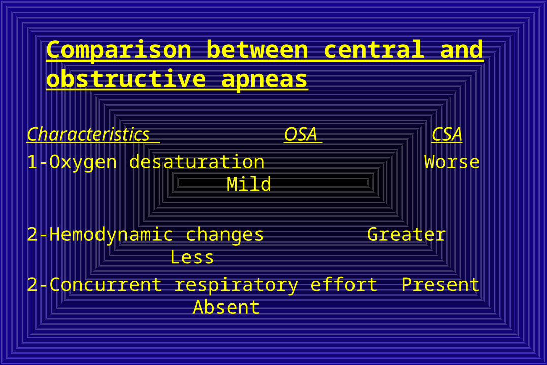
Comparison between central and obstructive apneas
Characteristics OSA CSA
1-Oxygen desaturation Worse Mild
2-Hemodynamic changes Greater Less
2-Concurrent respiratory effort Present Absent
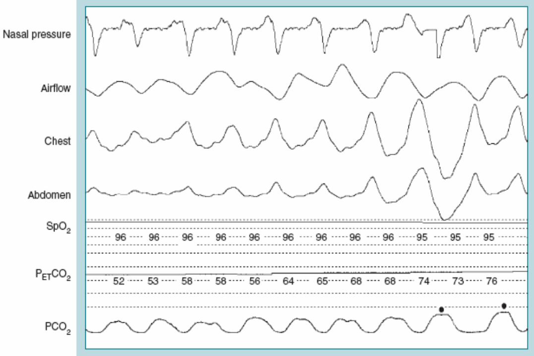
Hypoventilation Rule (AASM Scoring Manual• Score hypoventilation during sleep if there is a ≥10 mm Hg
increase in PaCO2 during sleep in comparison with an awake supine value
• Both end-tidal carbon dioxide pressure (PETCO2) and transcutaneous carbon dioxide pressure (TcPCO2) may be used as surrogate measures of PaCO2 if demonstrated to be reliable and valid.
• Continuous arterial blood gas monitoring during PSG is not practical (or well tolerated).
• It is observed that finding an increase in the PaCO2 obtained immediately upon awakening is suggestive of sleep hypoventilation.
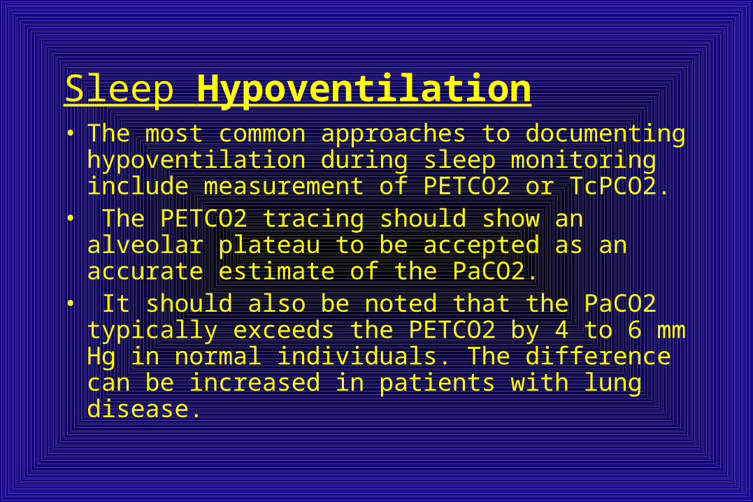
Sleep Hypoventilation• The most common approaches to documenting
hypoventilation during sleep monitoring include measurement of PETCO2 or TcPCO2.
• The PETCO2 tracing should show an alveolar plateau to be accepted as an accurate estimate of the PaCO2.
• It should also be noted that the PaCO2 typically exceeds the PETCO2 by 4 to 6 mm Hg in normal individuals. The difference can be increased in patients with lung disease.

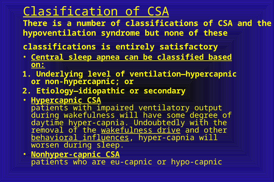
Clasification of CSAThere is a number of classifications of CSA and the hypoventilation syndrome but none of these classifications is
entirely satisfactory • Central sleep apnea can be classified based on:1. Underlying level of ventilation—hypercapnic or non-
hypercapnic; or2. Etiology—idiopathic or secondary• Hypercapnic CSA
patients with impaired ventilatory output during wakefulness will have some degree of daytime hyper-capnia. Undoubtedly with the removal of the wakefulness drive and other behavioral influences, hyper-capnia will worsen during sleep.
• Nonhyper-capnic CSApatients who are eu-capnic or hypo-capnic
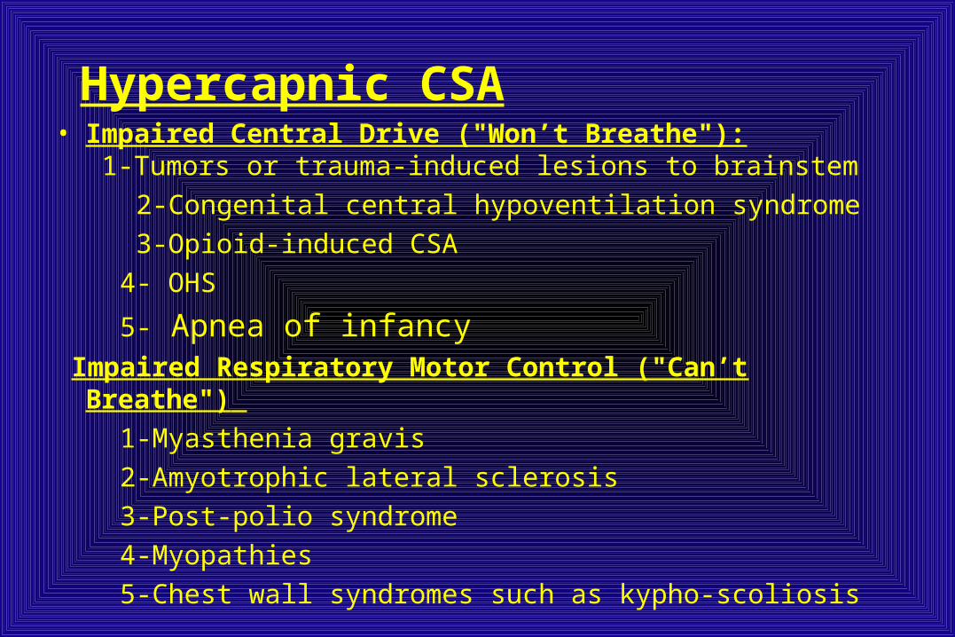
Hypercapnic CSA• Impaired Central Drive ("Won’t Breathe"):
1-Tumors or trauma-induced lesions to brainstem
2-Congenital central hypoventilation syndrome
3-Opioid-induced CSA
4- OHS
5- Apnea of infancy Impaired Respiratory Motor Control ("Can’t Breathe")
1-Myasthenia gravis
2-Amyotrophic lateral sclerosis
3-Post-polio syndrome
4-Myopathies
5-Chest wall syndromes such as kypho-scoliosis
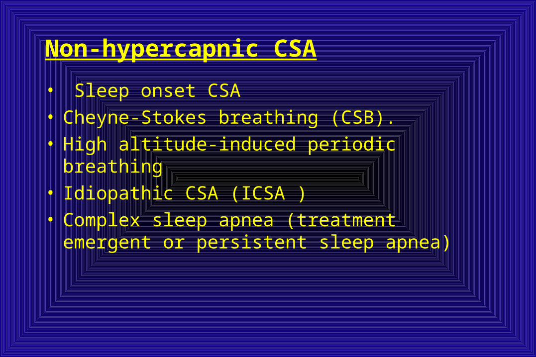
Non-hypercapnic CSA
• Sleep onset CSA• Cheyne-Stokes breathing (CSB). • High altitude-induced periodic breathing• Idiopathic CSA (ICSA )• Complex sleep apnea (treatment emergent or
persistent sleep apnea)
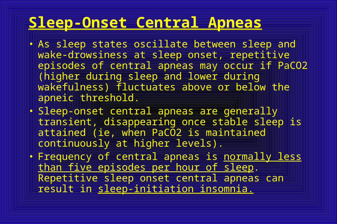
Sleep-Onset Central Apneas• As sleep states oscillate between sleep and wake-
drowsiness at sleep onset, repetitive episodes of central apneas may occur if PaCO2 (higher during sleep and lower during wakefulness) fluctuates above or below the apneic threshold.
• Sleep-onset central apneas are generally transient, disappearing once stable sleep is attained (ie, when PaCO2 is maintained continuously at higher levels).
• Frequency of central apneas is normally less than five episodes per hour of sleep. Repetitive sleep onset central apneas can result in sleep-initiation insomnia.
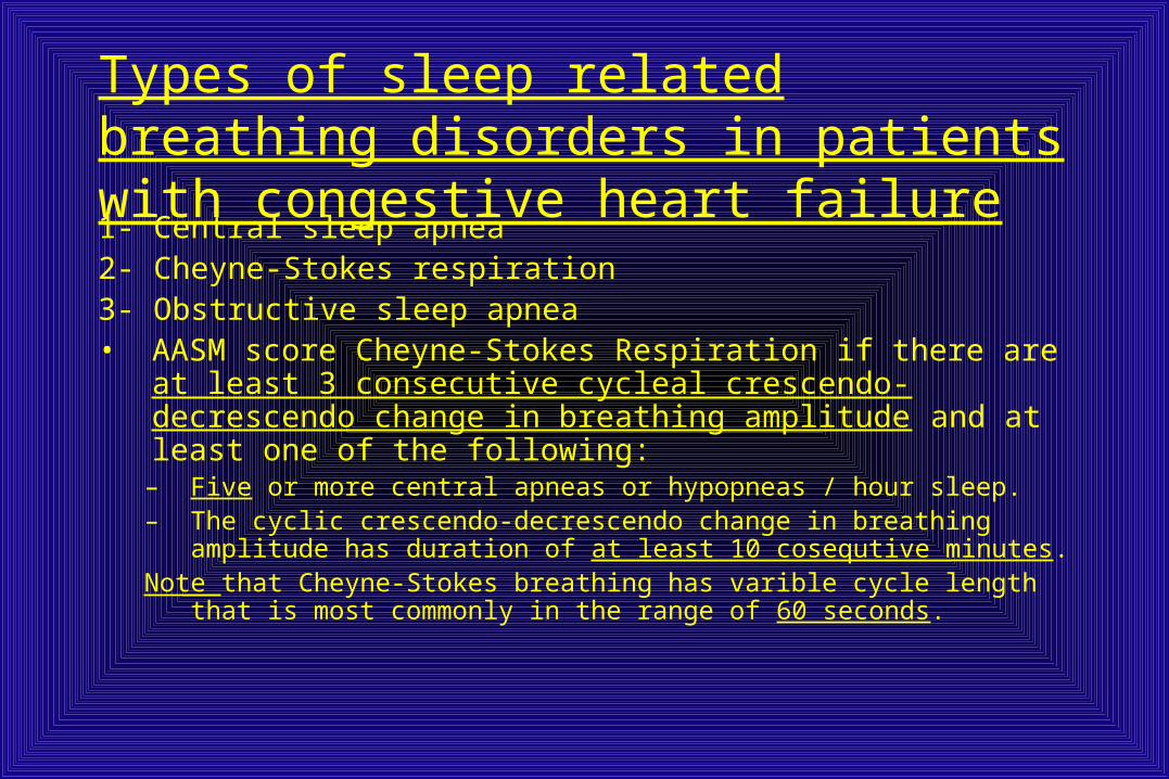
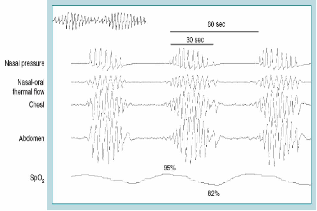
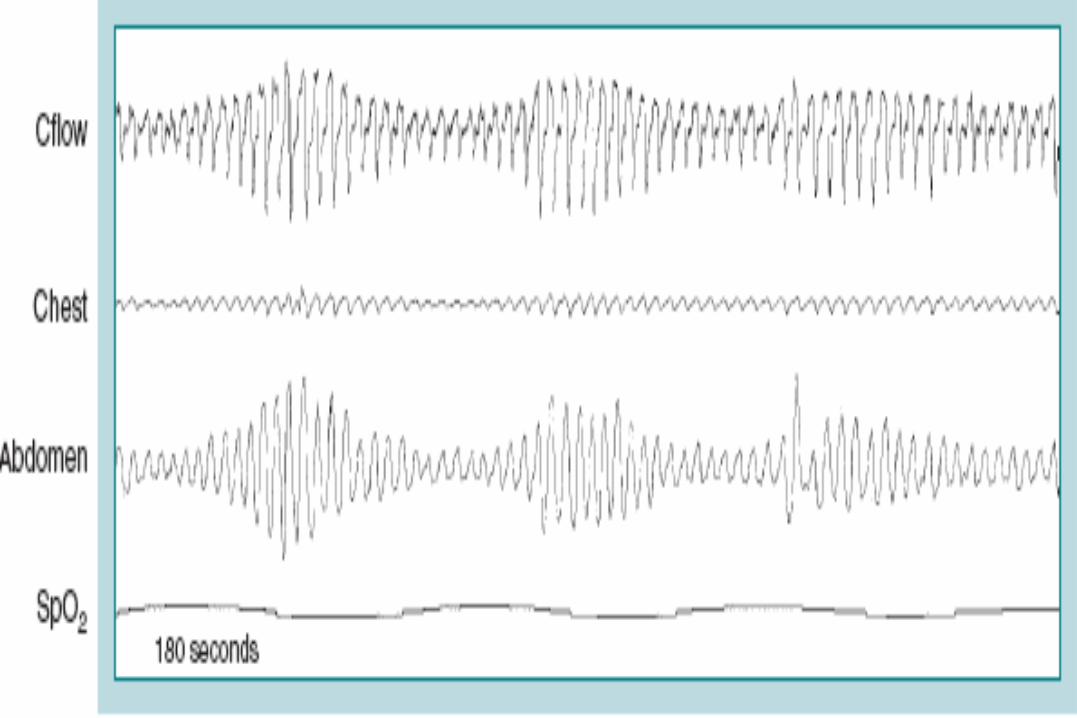
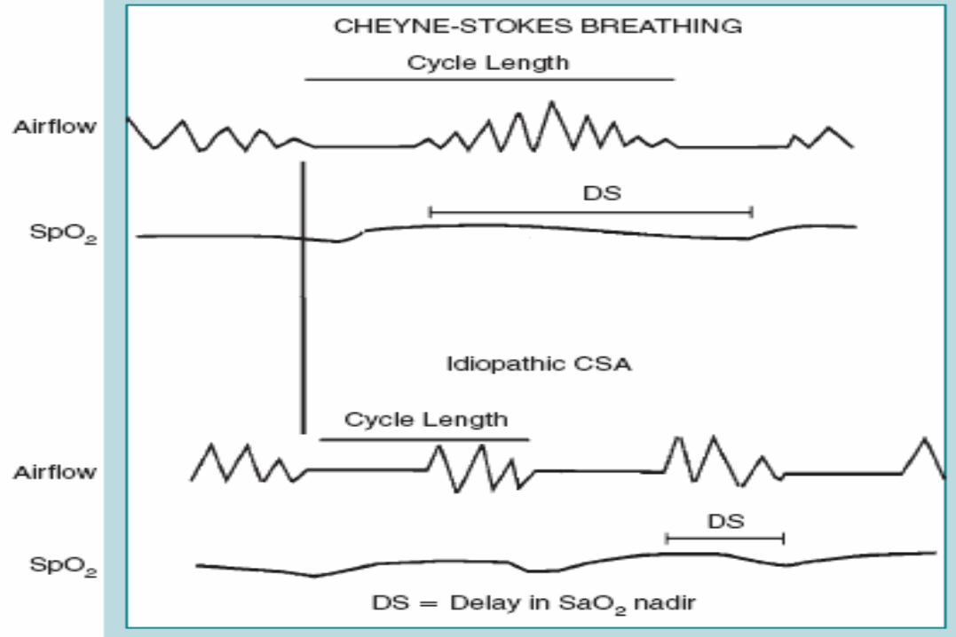
Types of sleep related breathing disorders in patients with congestive heart failure1- Central sleep apnea2- Cheyne-Stokes respiration3- Obstructive sleep apnea• AASM score Cheyne-Stokes Respiration if there are at
least 3 consecutive cycleal crescendo-decrescendo change in breathing amplitude and at least one of the following:
– Five or more central apneas or hypopneas / hour sleep. – The cyclic crescendo-decrescendo change in breathing amplitude
has duration of at least 10 cosequtive minutes.Note that Cheyne-Stokes breathing has varible cycle length that is most
commonly in the range of 60 seconds.

Evaluation: • Patients with CSA-CSB may complain of the typical
symptoms of sleep apnea including disturbed sleep or daytime sleepiness.
• However, in most studies, the majority of patients do not complain of subjective excessive daytime sleepiness. Thus, a high index of suspicion is needed to suspect the presence of CSB.
• Nocturnal sleep complaints are often assumed to be secondary to CHF rather than to co-morbid sleep apnea.
Therapy: • Optimize medical management • Supplemental oxygen• CPAP (effective in ~40–50%)• BPAP with backup rate (BPAP-ST)• ASV• Transplant
Neurologic Disorders• Several neurologic and neuromuscular disorders can decrease central
ventilatory drive and give rise to CSA. Patients are usually hypercapnic.Periodic Breathing Secondary to High Altitude• Periodic breathing, or cycles of central apneas and hyperpneas, can occur
on ascent to high altitude (usually > 4000 to 7600 meters). • Severity of symptoms is influenced by elevation, speed of ascent, and
individual predisposition. Persons withse incread hypoxic ventilatory chemoresponsiveness appear to have a greater risk for developing high-altitude–related periodic breathing
• Hyperventilation due to hypoxia on ascent to altitude gives rise to hypocapnic (low PaCO2) alkalosis that, in turn, results in central apneas during sleep, particularly NREM sleep, on the first few nights at altitude. Ventilation resumes as PaCO2 rises and PaO2 falls during the apneic episode.
• The pattern of periodic breathing at high altitude resembles Cheyne–Stokes breathing in heart failure, with one exception. The cycle of periodic breathing is short.
Therapy of Periodic Breathing Secondary to High Altitude • Therapy consists of either oxygen therapy or
administration of acetazolamide (250 mg /6h) reduce central apnea over 1-2 weeks
• Acetazolamide causes metabolic acidosis and decreases PaCO2. In spite of a lower (than normal) PaCO2, central apneas decrease. This is because the apneic threshold PCO2 decreases more than that of PaCO2. This results in a widening of the two PCO2 set points. which will decrease the likelihood of developing central apnea.
Central Sleep Apnea Related to Medication Use
• Central apneas can develop during administration of opiate drugs.
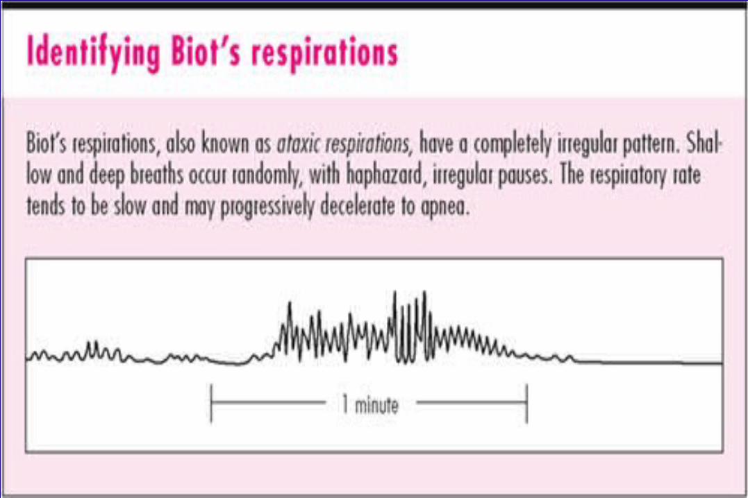
• Other respiratory pattern abnormalities, such as periodic breathing, Biot respiration, and obstructive hypoventilation, can develop due to receptor-related depression of the hypercapnic ventilatory drive and increase in hypoxic ventilatory drive.
Central Apnea During Positive Airway Pressure Titration
• Central sleep apnea may emerge during CPAP titration in patients with OSA.
• This may be due to unmasking of a previously concurrent central apnea that becomes apparent when the more predominant obstructive events are controlled by CPAP therapy.
• Alternatively, it may be due to recurrent arousals related to the use of CPAP with central apnea occurring in the post-arousal period.
Complex sleep apnea • CompSA is defined as a form of CSA identified by the
persistence or emergence sleep of central apneas or hypopneas upon exposure to CPAP or BPAP without a backup rate when obstructive events have disappeared.
• These patients have predominantly obstructive or mixed apneas during the diagnostic portion of the study occurring 5/hr or more.
• With use of CPAP or BPAP without a backup rate, they show a pattern of apneas and hypopneas that meets the definition of CSA.

Treatment of Complex sleep apnea • In the absence of CSB-CSA or opioids, it appears that a significant
portion will improve with chronic CPAP alone. The only problem with watchful waiting is that if the AHI remains high, sleep quality will be impaired and adherence may decrease. Therefore, it is prudent to assess patients soon after starting CPAP.
• Many CPAP devices provide the ability to estimate residual events. • It is also important to remember that the patient is being treated,
not the AHI. Therefore, if a patient reports good sleep and objective adherence is good, intervention may not be necessary if the residual AHI remains mildly elevated (5–10/hr). If patients do not improve or if the residual AHI is high, most clinicians would order a titration with ASV.
• Another option would be treatment with BPAP-ST (backup rate) if ASV is not effective.
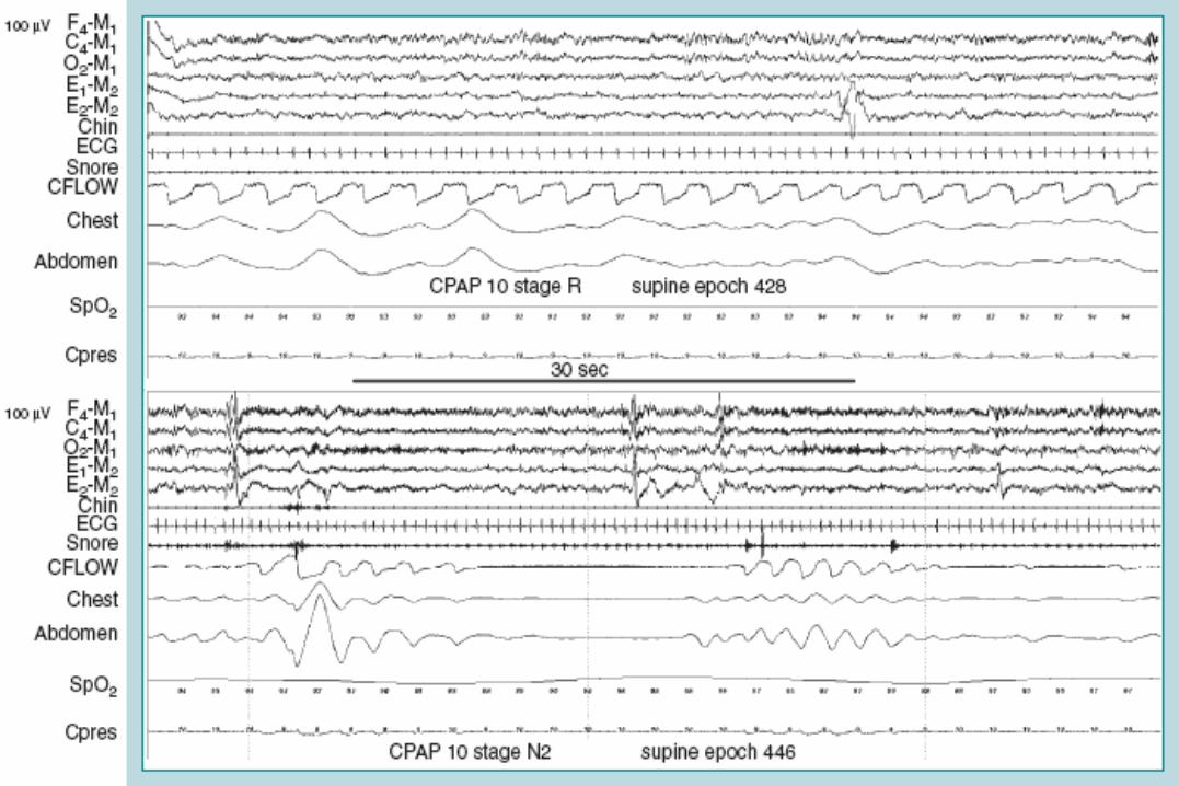
Obesity Hypoventilation Syndrome• The two essential features of obesity hypoventilation
syndrome (OHS) are the presence of a body mass index >30 kg/m2 and PaCO2 > 45 mmHg during wakefulness. Hypoventilation is not due to another respiratory or neuromuscular disorder.
• Many patients with OHS also have OSA (80-90 %)• The American Academy of Sleep Medicine has
incorporated the definition of OHS into a broader term of sleep hypoventilation syndrome
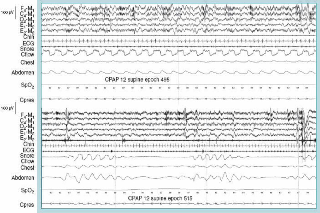
Pathophysiology:• Patients with the OHS are a heterogeneous group.• Up to 20% of OHS patients have an AHI of 5/hr or less but exhibit both
daytime hypercapnia and severe sleep sleep-related hypoventilation and arterial oxygen desaturation.
Etiology of hypercapnia in obesity hypoventilation syndrome1-Increased production of CO2 : Greater work of breathing Decreased chest wall compliance2- Decreased ventilation: Decreased expiratory reserve volume Decreased tidal volume Increased resistive load Increased dead space3-Decreased ventilatory response to hypercapnia and hypoxemia4- coexistence of obstructive sleep apnea (OSA)
Clinical Features
• Clinical manifestations include excessive sleepiness, sleep fragmentation with frequent arousals (insomnia), decreased attention or concentration, peripheral edema and cyanosis. Chronic hypoxemia can result in polycythemia, pulmonary hypertension, and cor pulmonale.
Prevalence • The prevalence of OHS is from 5% to 31% of obese
adults, and it is uncommon but possible in obese children
Treatment of OHS:• Some OHS patients could be adequately treated with CPAP
alone.• Others still had hypoventilation despite the absence of
apnea or hypopnea. • Some with persistent airflow limitation responded to higher
levels of CPAP• Some patients required the addition of supplemental oxygen
along with CPAP.• Another group of patients required either nasal bilevel
positive airway pressure (BPAP) or mechanical ventilation with or without oxygen.
• Tracheostomy can be life-saving in patients noncompliant with PAP treatment who have repeated bouts of hypercapnic respiratory failure.
Overlap Syndrome• Patients with the OSA and COPD may have daytime
hypoventilation and severe nocturnal oxygen desaturation.
• Patients with mild COPD have no higher incidence of OSA than the general population.
• However, because both COPD and OSA are common, the combination is common even if by chance.
Idiopathic Central Sleep Apnea• The etiology of idiopathic CSA is unknown. • In patients with idiopathic CSA, carbon dioxide
(CO2) ventilatory drive is increased. Thus, PaCO2 is low during wakefulness and sleep.
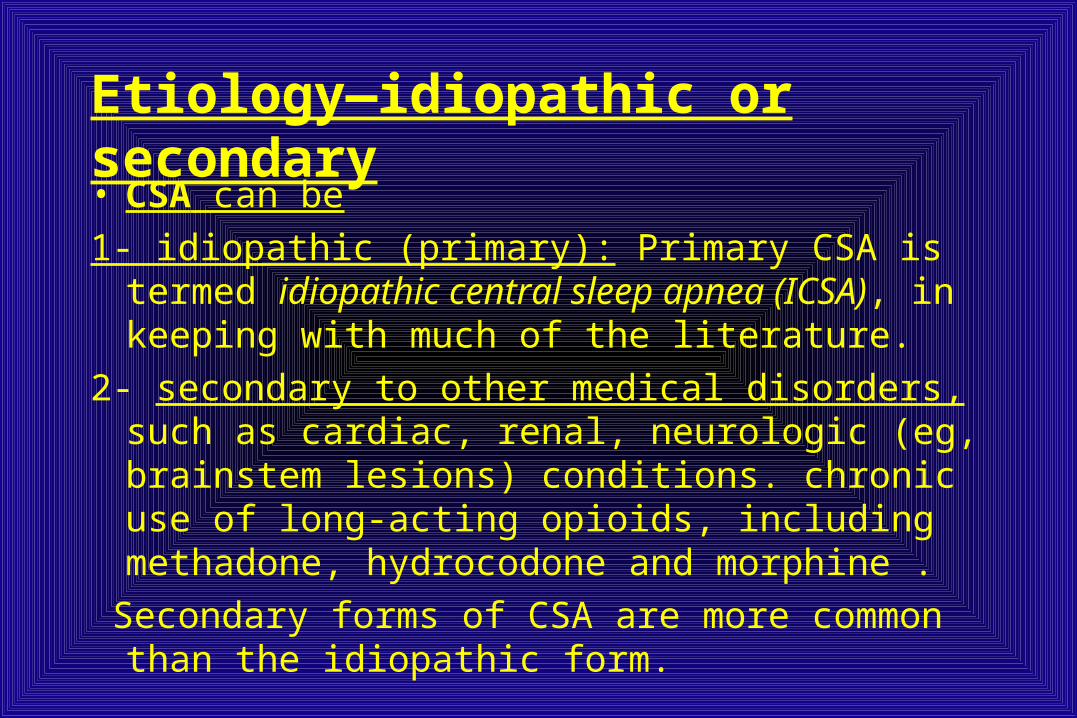
Etiology—idiopathic or secondary• CSA can be
1- idiopathic (primary): Primary CSA is termed idiopathic central sleep apnea (ICSA), in keeping with much of the literature.
2- secondary to other medical disorders, such as cardiac, renal, neurologic (eg, brainstem lesions) conditions. chronic use of long-acting opioids, including methadone, hydrocodone and morphine .
Secondary forms of CSA are more common than the idiopathic form.
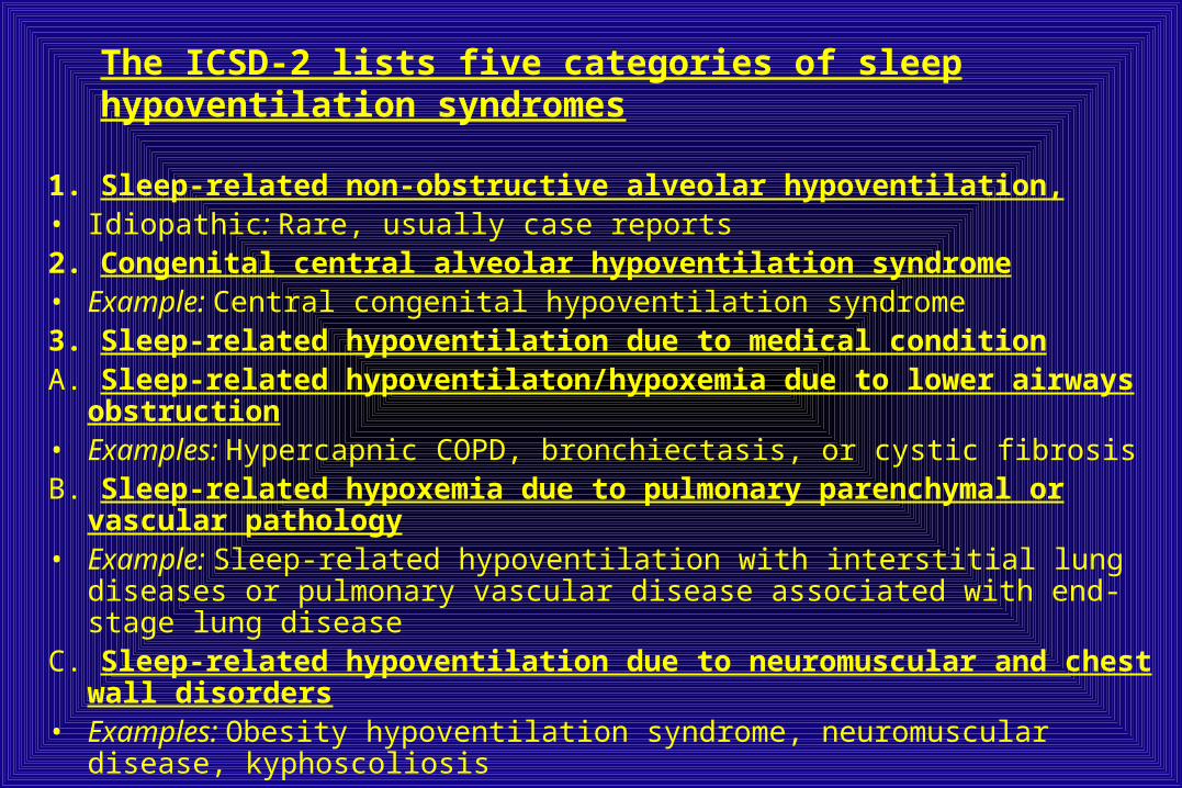
The ICSD-2 lists five categories of sleep hypoventilation syndromes
1. Sleep-related non-obstructive alveolar hypoventilation,• Idiopathic: Rare, usually case reports2. Congenital central alveolar hypoventilation syndrome• Example: Central congenital hypoventilation syndrome3. Sleep-related hypoventilation due to medical conditionA. Sleep-related hypoventilaton/hypoxemia due to lower airways obstruction• Examples: Hypercapnic COPD, bronchiectasis, or cystic fibrosisB. Sleep-related hypoxemia due to pulmonary parenchymal or vascular
pathology• Example: Sleep-related hypoventilation with interstitial lung diseases or pulmonary
vascular disease associated with end-stage lung diseaseC. Sleep-related hypoventilation due to neuromuscular and chest wall disorders• Examples: Obesity hypoventilation syndrome, neuromuscular disease,
kyphoscoliosis
Clinical Features
• Sleep disturbance with repeated nocturnal awakenings• Nocturnal sensation of dyspnea• Insomnia• Morning headaches• Excessive sleepiness
Note: Patients may be asymptomatic.
Associated features• Cardiovascular disorders• Systemic hypertension• Pulmonary hypertension,Cor pulmonale• Cardiac arrhythmias • Respiratory insufficiency• Polycythemia• Depression• Cognitive impairment• ImpotencePhysical features• Peripheral edema• Cyanosis• Shallow breathing

Therapeutic Interventions • improve cardiac status for patients with an underlying heart
condition may attenuate SDB • Gradual dose reduction of opioid medication • Weight loss is likely to lead to improvement of SDB in
patients with, morbidly obese patients with OHS.• Acetazolamide and theophylline have been shown to
improve CSA in patients with heart failure. Acetazolamide has also been shown to improve SDB in ICSA.
• Nasal CPAP has been shown to be effective in some patients with ICSA.