skeletal system part ii - fisiokinesiterapia
TRANSCRIPT

Anatomy and Physiology
Skeletal System part II

Skeletal Organization
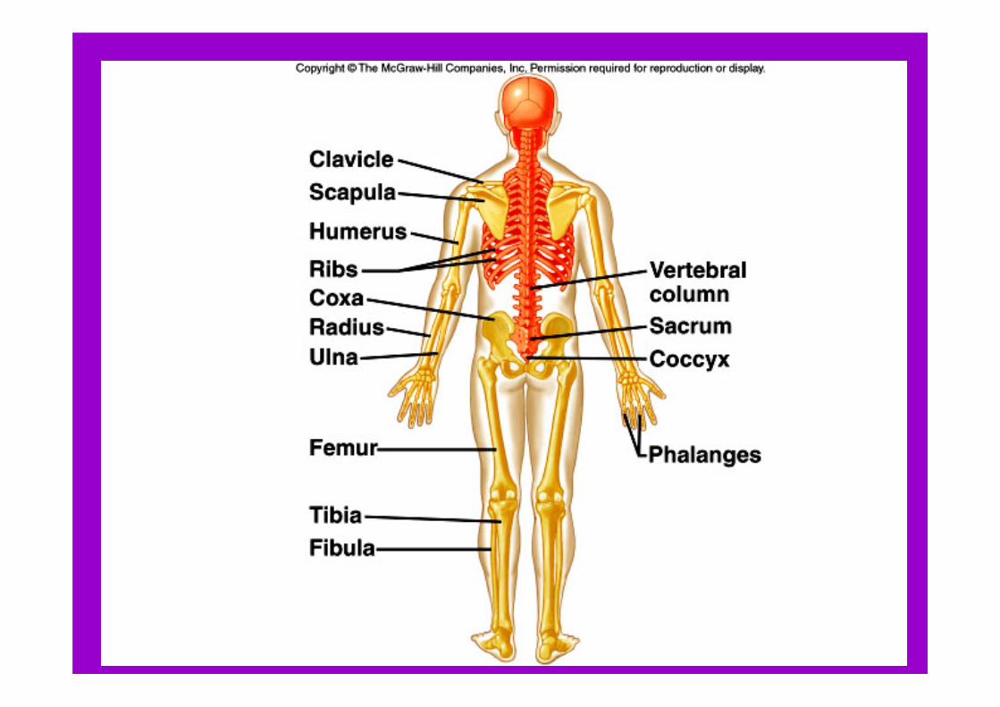
• The skeleton can be divided into – Axial portion (head, neck, and trunk)– Appendicular portion (arms and legs).

• The axial skeleton consists of:– The skull– Hyoid bone– Vertebral column– Thoracic cage.

• The appendicular skeleton consists of:– Pectoral girdle– Upper limbs– Pelvic girdle– Lower limbs

• 206 bones in an adult skeleton
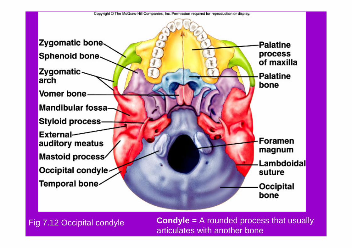
Fig 7.12 Occipital condyle Condyle = A rounded process that usually articulates with another bone

Fig 7.27 Iliac crest Crest = A narrow, ridgelike projection

Fig 7.16 and 7.17 Rib facet of thoracic vertebrae
Facet = A small, nearly flat surface

Fig 7.15 Anterior fontanelFontanel = A soft spot in the skull where membranes cover the space between bones

Fig 7.12 Foramen magnumForamen = A opening through a bone that usually is a passageway for blood vessels, nerves, or ligaments

Fossa = a relatively deep pit or depressionFig 7.23 Olecranon fossa of humerus

Fig 7.29 Fovea capitus of femur Fovea= a tiny pit or depression

Fig 7.23 Head of humerus Head = An enlargement on the end of a bone
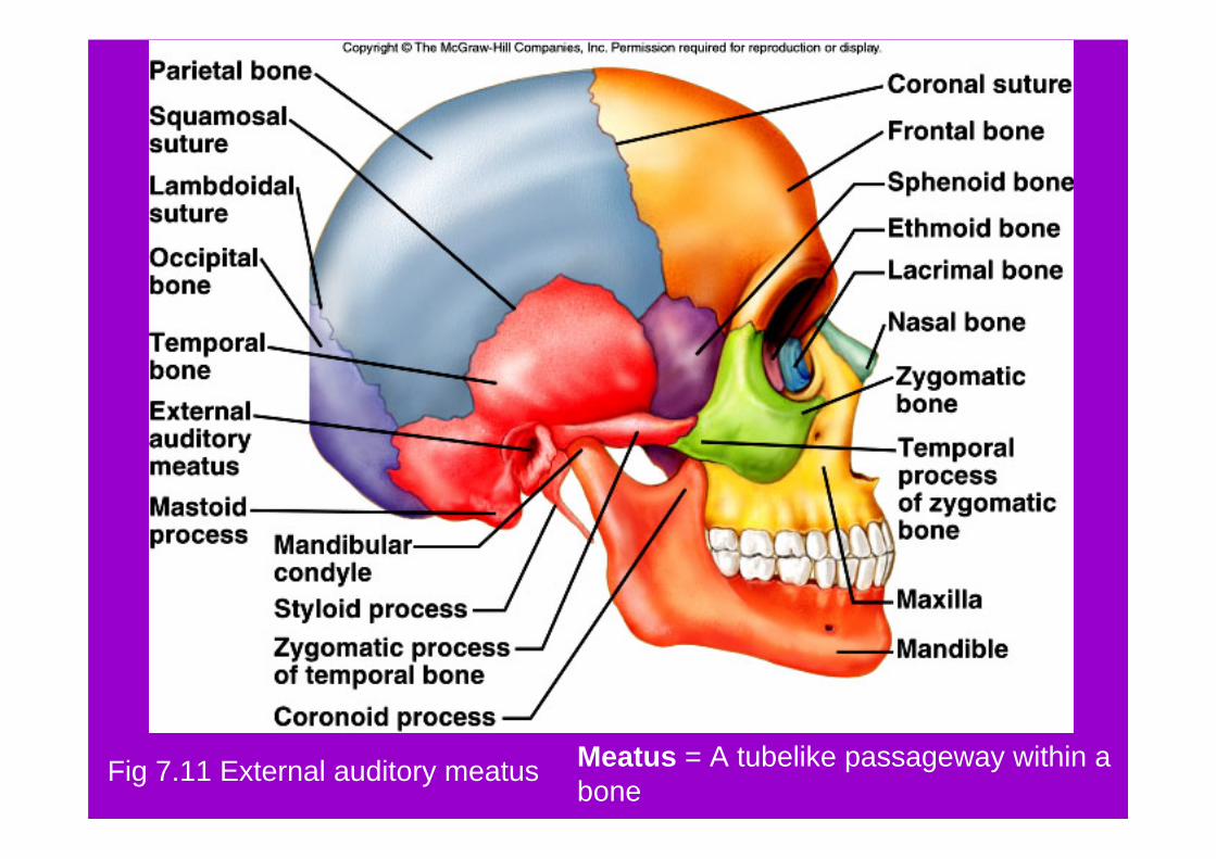
Fig 7.11 External auditory meatus Meatus = A tubelike passageway within a bone

Fig 7.11 Mastoid process Process = A prominent projection on a bone

Fig 7.14 Frontal sinus Sinus = A cavity within a bone

Fig 7.22 Spine of scapula Spine = A thornlike projection

Fig 7.11 Lambdoidal suture Suture = An interlocking line of union between bones
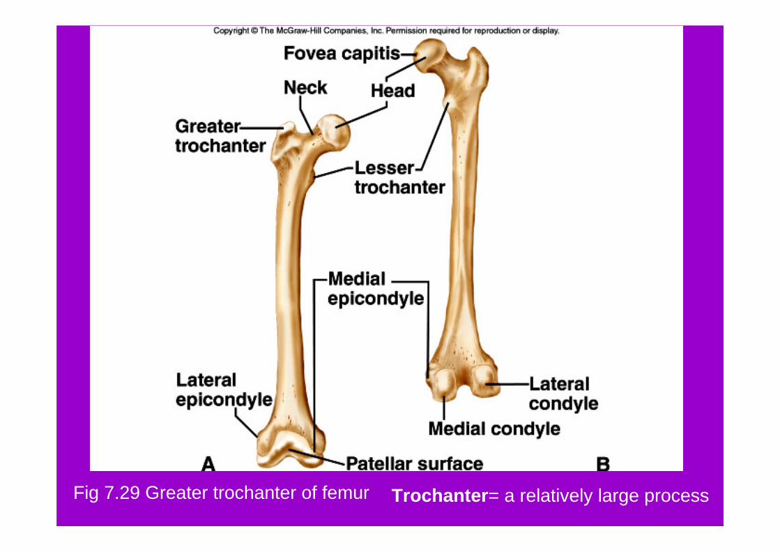
Fig 7.29 Greater trochanter of femur Trochanter= a relatively large process

Fig 7.23 Greater tubercle of humerus Tubercle= a small, knoblike process

Fig 7.24 Radial tuberosity of radius
Tuberosity = a knoblike process usually larger than a tubercle


Skull
• The skull consists of twenty-two bones.– 8 cranial bones– 14 facial bones (13 + 1 mandible)

Skull
• Cranium (braincase)– The cranium encloses and protects the
brain– Surface of the cranium provides
attachments for muscles used in chewing and head movements

– Some cranial bones contain air-filled paranasal sinuses.• Sinuses are lined with mucous membranes and
are connected to the nasal cavity
• Sinuses reduce the skull’s weight and increase voice intensity by resonance


Cranial bones include: • Frontal bone• Parietal bones• Occipital bone• Temporal bone• Sphenoid bone• Ethmoid bone


– Sutures: immovable joint along which flat bones of the cranium are joined


Facial Skeleton
• Facial bones – Form the basic shape of the face – Provide attachments for muscles that move
the jaw– Control facial expressions

Facial SkeletonFacial bones include: – Maxillae
– Palatine bones– Zygomatic bones– Lacrimal bones– Nasal bones– Vomer bone– Inferior nasal
conchae– Mandible


Mandible
• The lower jawbone• The only movable bone of the skull • Held to the cranium by ligaments

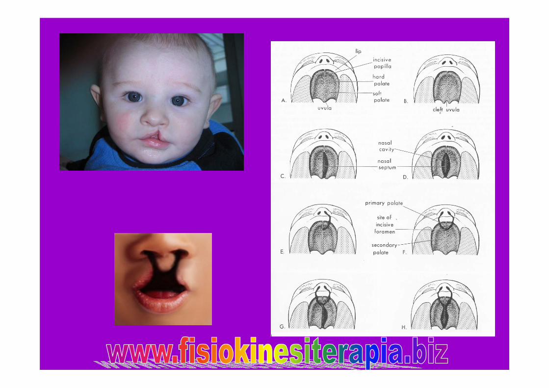
Cleft Palate
• Incomplete fusion of the palatine processes of the maxillae
• Causes problems with sucking, eating, and speaking
• Corrective surgery is needed to close the opening between the oral and nasal cavities


Infantile Skull
• Proportions of the infantile skull are different from those of an adult skull.– Small face with prominent forehead and
large orbits– Smaller jaw and nasal cavity– Frontal bone in two parts


Infantile Skull
• Fontanels (fibrous membranes) connect incompletely developed bones– Called “soft spots”– Permit movement between bones so that
developing skull can change shape as it moves through the birth canal
– Eventually close as cranial bones grow together


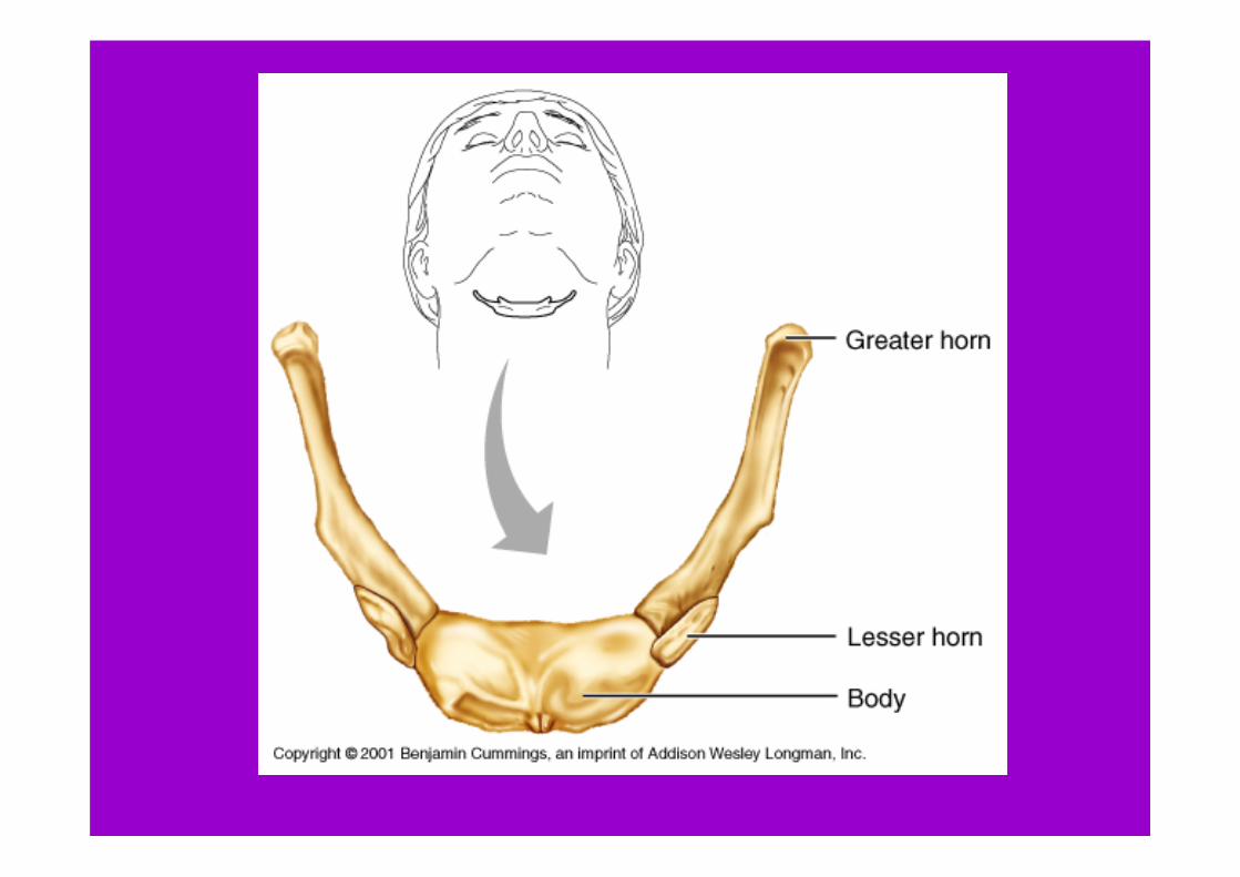
Hyoid Bone
• Located in the neck between the lower jaw and the larynx
• Supports tongue and attachment site for muscles used during swallowing
Vertebral Column
• Backbone or Spine• The vertebral column extends from the
skull to the pelvis

Vertebral Column
• Protects the spinal cord which passes through the vertebral canal
• Supports the head and trunk
Vertebral Column (backbone)
• It is composed of vertebrae, separated by intervertebral disks and are connected to one another by ligaments


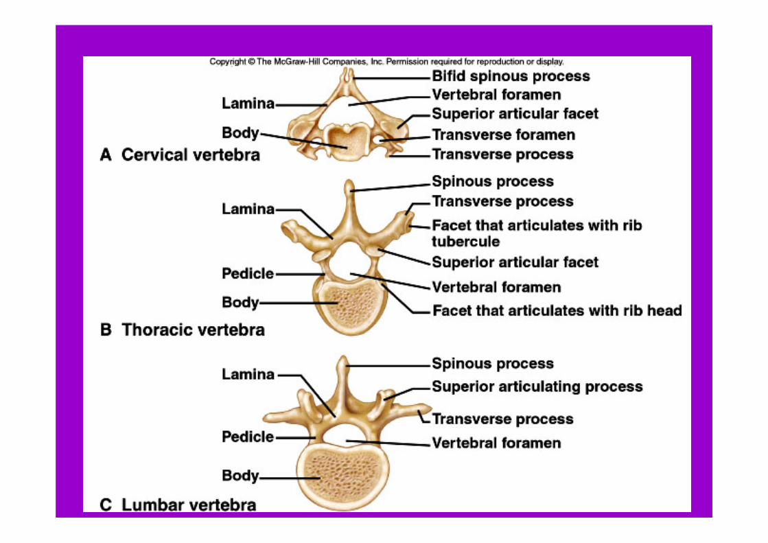
A Typical Vertebra
• A typical vertebra consists of:– Body: forms the thick anterior portion – Bony vertebral arch: surrounds the spinal
cord.
• Notches on the upper and lower surfaces provide intervertebralforamina (openings) through which spinal nerves pass.

Spina Bifida
• Occurs if the laminae of the vertebral arch fail to unite during development.
• Contents of the vertebral canal protrude outward, most commonly in the lumbrosacral region.

Vertebral Column
• Cervical Vertebrae (7)• Thoracic Vertebrae (12)• Lumbar Vertebrae (5)• Sacrum (5 fused)• Coccyx (4 fused)


Cervical Vertebrae (7)
• Transverse processes (projections) bear transverse foramina, which are passageways for arteries leading to the brain
• Forked bifid spinous process provides attachments site for muscles

Cervical Vertebrae (7)
• The Atlas:– First vertebra – Supports and balances the head. – Articulates with occipital condyles of the
cranium
• The Axis– Second vertebra – Provides a pivot for the atlas when the
head is turned side to side.


Thoracic Vertebrae (12)• Thoracic vertebrae are larger than
cervical vertebrae.• Facets on the side articulate with the
ribs.• Long spinous process• Increase in size inferiorly (as you go
downward). Adapted to bear increasing loads of body weight.


Lumbar Vertebrae (5)
• Vertebral bodies are large and strong.• They support more body weight than
other vertebrae.
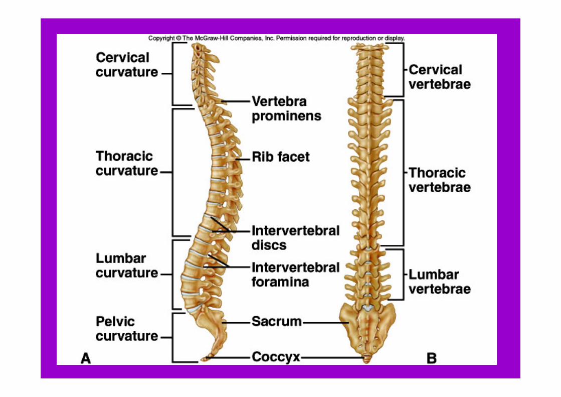


Sacrum (5 fused)
• The sacrum is a triangular structure formed of five fused vertebrae.
• Vertebral foramina form the sacral canal.
• Part of the pelvis


Coccyx (4 fused)
• Tailbone• Composed of four fused vertebrae• Forms the lowest part of the vertebral
column.• Acts as a shock absorber when a person
sits.

Intervertebral Disks
• Composed of tough outer layer of fibrocartilage with an elastic central mass
• Degenerates with age, loses firmness, outer layer thins, weakens, cracks

Ruptured/ Herniated Disk
• Pressure from lifting may break outer layer and allow it to squeeze out or rupture.
• Pressure on the spinal cord or nerve causes pain, numbness, loss of muscular function


Thoracic Cage
• The thoracic cage includes – The ribs– Thoracic vertebrae– Sternum– Costal cartilages (attach ribs to sternum).
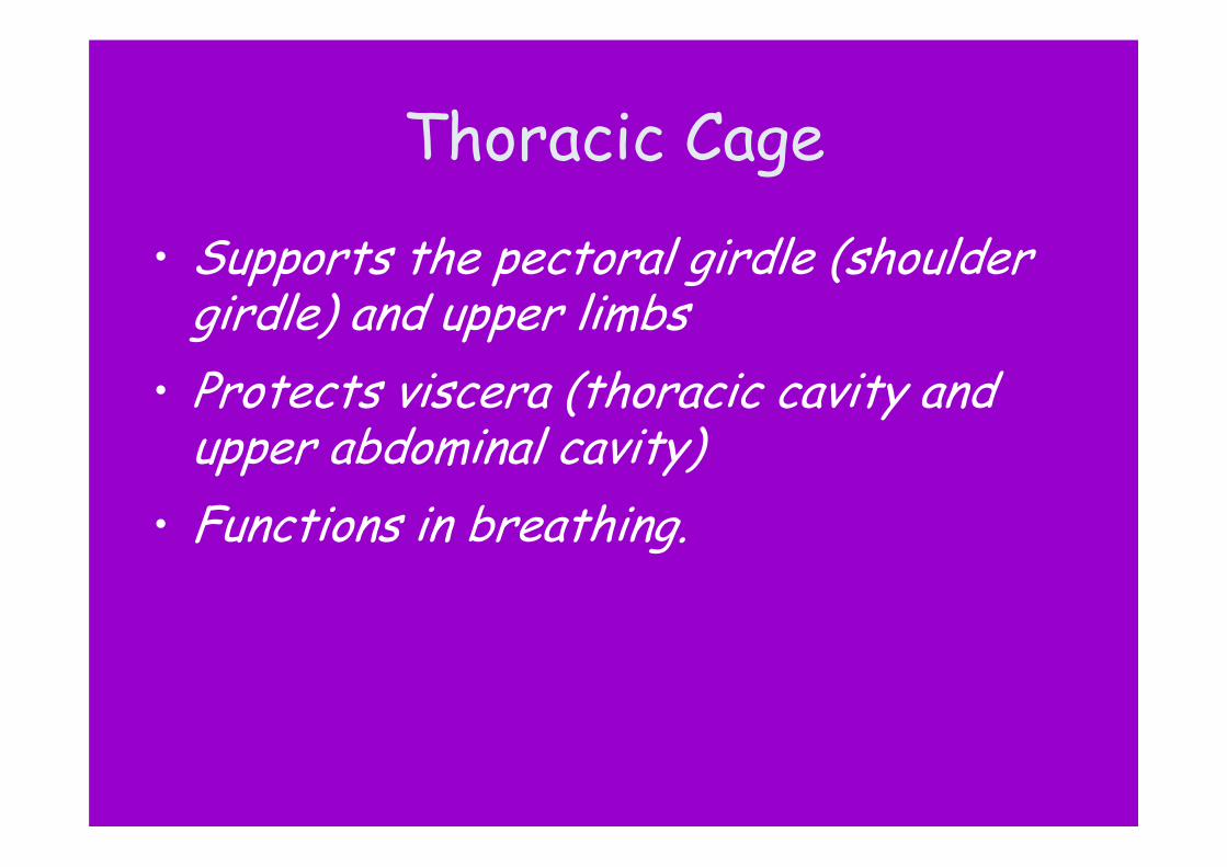
Thoracic Cage
• Supports the pectoral girdle (shoulder girdle) and upper limbs
• Protects viscera (thoracic cavity and upper abdominal cavity)
• Functions in breathing.

Ribs
• Twelve pairs of ribs attach to the twelve thoracic vertebrae and articulate posteriorly
• A typical rib has a shaft, a head, and tubercles that articulate with the vertebrae.

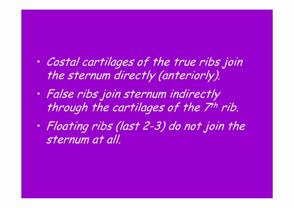
• Costal cartilages of the true ribs join the sternum directly (anteriorly).
• False ribs join sternum indirectly through the cartilages of the 7th rib.
• Floating ribs (last 2-3) do not join the sternum at all.

Sternum
• Breastbone• Located on the midline in the anterior
portion (front) of the thoracic cage.• The sternum consists of a manubrium
(upper part), body and xiphoid process.• The manubrium articulates with the
clavicles.

Sternum (breastbone)
• Red marrow in the sternum produces blood cells into adulthood. – Easily reached for marrow samples in
disease diagnosis. – Called “sternal puncture”. – Cells also sampled from iliac crest of coxal
bone

Pectoral Girdle
• Shoulder girdle• Composed of two clavicles and two
scapulae• Forms an incomplete ring that supports
the upper limbs and provides attachments for muscles.
• Connects bones of the upper limbs to the axial skeleton and aids in upper limb movement.


Clavicles
• Collarbones• Rod like bones located between the
manubrium and scapulae.• Hold the shoulders in place and provide
attachments for muscles of upper limbs, chest, and back.

Scapulae
• Shoulder blades• Broad, triangular bones• Articulate with the humerus of each
upper limb and provide attachment for muscles.


Upper Limbs
• Bones of the upper limb provide the frameworks and attachments of muscles
• Function in levers that move the limb and its parts.

Humerus
• Arm bone• The humerus extends from the scapula
to the elbow.• It articulates with the radius and the
ulna at the elbow and with the scapula at the shoulder.


Radius
• Forearm bone• Located on the thumb side of the
forearm between the elbow and wrist.• Articulates with the humerus, ulna, and
wrist.

Ulna
• Forearm bone• Longer than the radius • Overlaps the humerus posteriorly.• Articulates with the radius laterally and
with a disk of fibrocartilage inferiorly which joins a wrist bone.


Hand
• Composed of a wrist, a palm, and five fingers.
• Includes:– 8 carpal bones (wrist bones) that form a
carpus– 5 metacarpal bones (palm) – 14 phalanges (finger bones: 3/finger,
2/thumb).


Pelvic Girdle
• The pelvic girdle consists of two coxalbones (hip bones) that articulate with each other anteriorly and with the sacrum posteriorly.
• The sacrum, coccyx, and pelvic girdle form the bowl-shaped pelvis.


Pelvic Girdle
• Supports the trunk of the body • Connects the bones of the lower limbs
to the axial skeleton.• Protects the urinary bladder, distal
ends of the large intestine, and internal reproductive organs.

Coxal bone
• Consists of an ilium, ischium, and pubis, which are fused in the region of the acetabulum (depression on the side). Figure 7.27 p159


Ilium
• Hip, iliac crest• Largest portion of the coxal bone.• Joins the sacrum at the sacroiliac joint

Ischium
• Lowest portion of the coxal bone.• Supports body weight when sitting.

Pubis
• Anterior portion of the coxal bone.• Pubic bones are fused anteriorly at the
symphysis pubis.

• The female pelvis is usually wider in all diameters and roomier than that of the male. Figure 7.26 p158


Lower Limb
• Bones of the lower limb provide frameworks of the thigh, leg, and foot.

Femur
• Thigh bone• Longest bone in the body.• Extends from the hip to the knee.• Articulates proximally with the coxal
bone and distally with the tibia and the patella.


Patella
• Knee cap• Articulates with the femur’s anterior
surface. • Located within a tendon that passes
over the knee.


Tibia
• Shin bone• Located on the medial side of the leg• Larger of the two lower leg bones.• The femur and the tibia articulate with
each other at the knee joint where the patella covers the anterior surface.
• Articulates with the talus of the ankle and with the fibula on the lateral side.

Fibula
• Lower leg bone• Located on the lateral side of the tibia • More slender than the tibia.• Articulates with the ankle but does not
bear body weight.• Does not enter the knee joint.• Protrudes on the lateral side of the
ankle.

Foot
• Consists of an ankle, an instep, and five toes.
• Includes:– 7 tarsal bones (ankle bones) that form the
tarsus – 5 metatarsal bones (form the instep and
the ball of the foot) – 14 phalanges (toes).

Foot• Talus (one of the tarsals) moves freely where
it joins the tibia and fibula.• Calcaneus: largest ankle bone, projects
backward to form the heel.• Arches:
– Form longitudinally and transversely by the tarsalsand metatarsals which are bound by ligaments.
– Arches provide a stable, springy base for the body.

