skeletal system - · pdf file–composed of compact bone –location of yellow marrow...
TRANSCRIPT

Skeletal System
Chapter 6.1
Human Anatomy & Physiology
Overview of Skeletal System
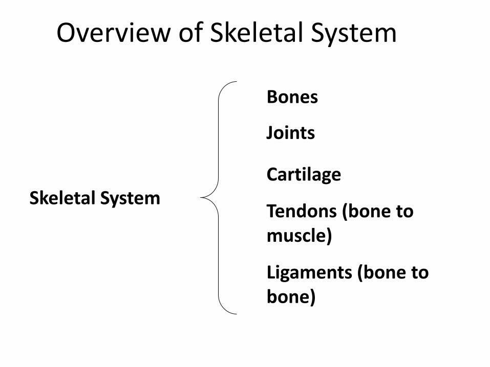
Skeletal System
Bones
Joints
Cartilage
Ligaments (bone to bone)
Tendons (bone to muscle)

Function of the Skeletal System
• Support of the body
• Protection of soft organs
• Movement due to attached skeletal muscles
• Storage of minerals and fats
• Blood cell formation

Types of Bone Tissue
• Compact Bone: Hard outer layer of bone
• Spongy bone: Less dense, small needle-like pieces of bone (trabeculae) with many open spaces
• Bone marrow: Soft tissue inside bone that produces blood cells
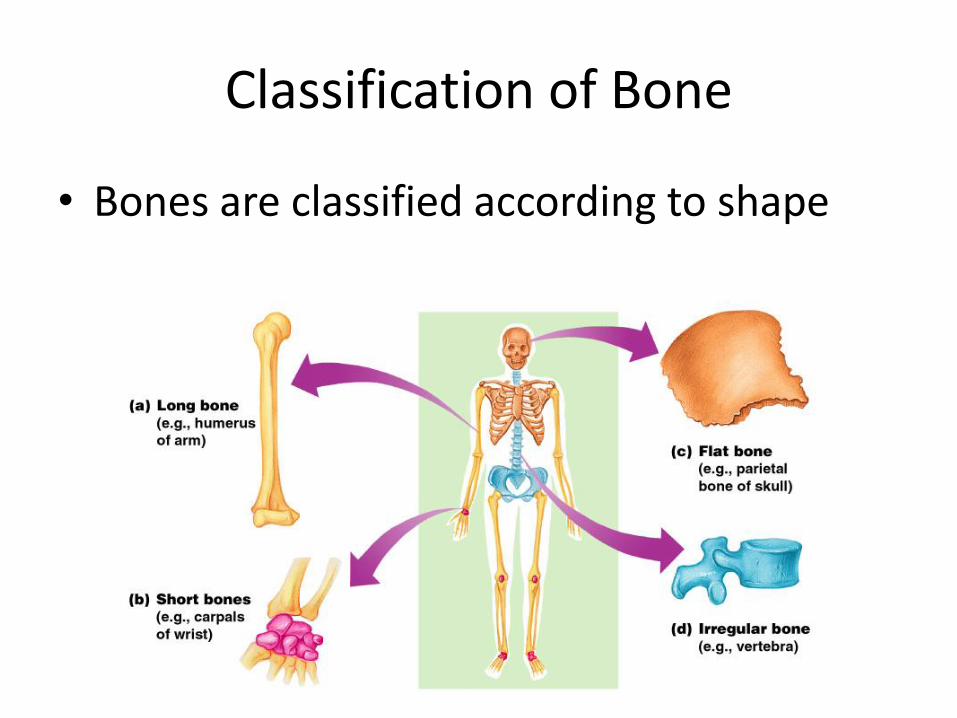
Classification of Bone
• Bones are classified according to shape
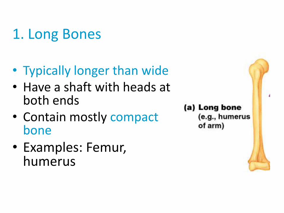
1. Long Bones • Typically longer than wide • Have a shaft with heads at
both ends • Contain mostly compact
bone
• Examples: Femur, humerus

2. Short bones
• Generally cube-shape
• Contain mostly spongy bone
• Examples: Carpals, tarsals

3. Flat bones
• Thin and flattened
• Usually curved
• Thin layers of compact bone around a layer of spongy bone
• Examples: Skull, ribs, sternum

4. Irregular bones
• Irregular shape
• Do not fit into other bone classification categories
• Example: Vertebrae and hip
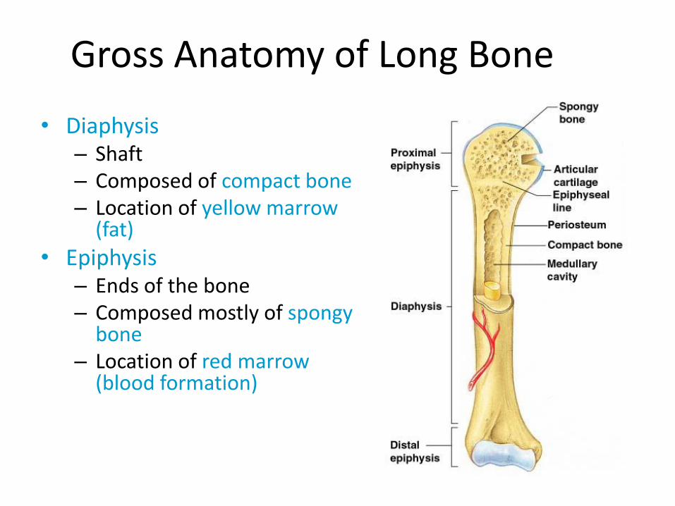
Gross Anatomy of Long Bone
• Diaphysis – Shaft – Composed of compact bone – Location of yellow marrow
(fat)
• Epiphysis – Ends of the bone – Composed mostly of spongy
bone – Location of red marrow
(blood formation)

• Periosteum
– Outside covering of the diaphysis
– Fibrous connective tissue membrane
• Arteries
– Supply bone cells with nutrients

• Articular cartilage – Covers the external surface
of the epiphyses
– Made of hyaline cartilage
– Decreases friction at joint surfaces
• Medullary cavity – Cavity of the shaft
– Contains yellow marrow (mostly fat) in adults
– Contains red marrow (for blood cell formation) in infants
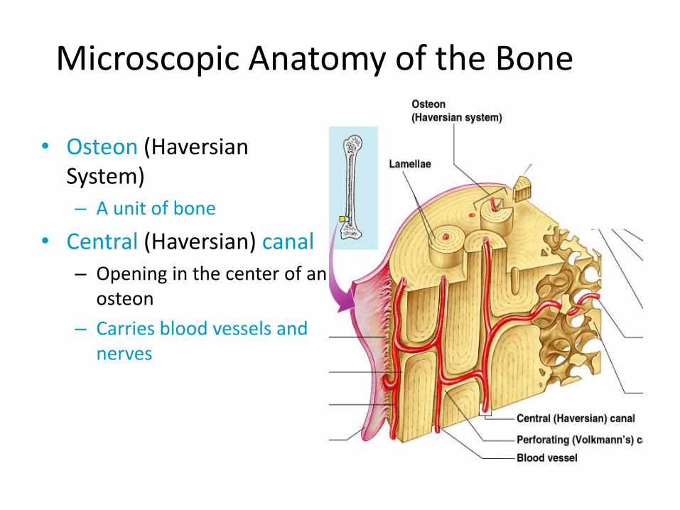
Microscopic Anatomy of the Bone
• Osteon (Haversian System)
– A unit of bone
• Central (Haversian) canal
– Opening in the center of an osteon
– Carries blood vessels and nerves

• Lacunae – Cavities containing bone
cells (osteocytes) – Arranged in concentric
rings
• Lamellae – Rings around the central
canal – Sites of lacunae
• Canaliculi – Tiny canals – Radiate from the central
canal to lacunae – Form a transport system

Ossification: Bone Growth
• Epiphyseal plates allow for growth of long bone during childhood
– New cartilage is continuously formed
– Older cartilage becomes ossified (changed to bone)
• Cartilage is broken down
• Bone replaces cartilage
• Bones are remodeled and lengthened until growth stops
– Bones change shape somewhat
– Bones grow in width


Types of Bone Cells
• Osteocytes
– Mature bone cells
• Osteoblasts
– Bone-forming cells for bone growth
• Osteoclasts
– Bone-destroying cells
– Break down bone matrix for remodeling and release of calcium
• Bone remodeling is a process done by both osteoblasts and osteoclasts

Ticket out the Door
• Identify and Describe the 5 functions of the skeletal system.

Vocabulary Quiz
• Write a sentence for each. Include a word that correctly uses the roots below.
ROOT
Adipo- Chondro- Lacuna- Osteo- -gli(o/a) Erythro- Leuko- Stria- Neuro- Oligo-

Bone Fractures
• A break in a bone
• Types of bone fractures
– Closed (simple) fracture – break that does not penetrate the skin
– Open (compound) fracture – broken bone penetrates through the skin
• Bone fractures are treated by reduction and immobilization
– Realignment of the bone

Common Types of Fractures

Repair of Bone Fractures • Hematoma (blood-filled swelling) is formed
• Break is splinted (immobilized) by fibrocartilage to form a callus
• Fibrocartilage callus is replaced by a bony callus
• Bony callus is remodeled to form a permanent patch
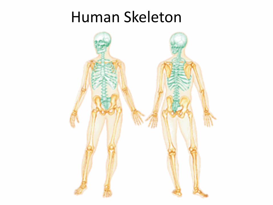
Human Skeleton
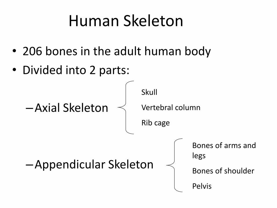
Human Skeleton
• 206 bones in the adult human body
• Divided into 2 parts:
–Axial Skeleton
–Appendicular Skeleton
Skull
Vertebral column
Rib cage
Bones of arms and legs
Bones of shoulder
Pelvis

• Axial Skeleton
• Appendicular Skeleton

• Divided into three parts
– Skull
– Vertebral column
– Rib Cage (bony thorax)
The Axial Skeleton

The Skull
• Two sets of bones
– Cranium
– Facial bones
• Skull bones are joined by sutures
• Only the mandible is attached by a freely movable joint

Bones of the Cranium (Skull) • Protects the brain
• Bones are attached by immovable joints called sutures
• Made up of 8 flat bones – 1 frontal bone
– 2 parietal bones
– 1 occipital bone
– 2 temporal bones
• Foramen Magnum – opening in the occipital bone – Area where spinal cord joins the brain

• Lateral View of Skull

• Superior View of Skull
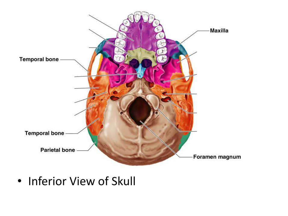
• Inferior View of Skull