skeletal system, chapter 6 - dr. scott croes' website system, chapter 6 ... after studying this...
TRANSCRIPT

1
Skeletal System, Chapter 6 Outline of class notes Objectives: After studying this chapter you should be able to:
1. Explain the main components of the skeletal system 2. Describe the six main functions of the skeletal system. 3. Explain how bones are classified by shape. 4. Discuss the basic structure of a long bone 5. Describe the components of bone tissue including different types of cells and the bone matrix. 6. Discuss the characteristics of compact and spongy bone. 7. Describe the difference between intramembranous and endochondral ossification. 8. Explain how bone grows in length and thickness. 9. Describe the processes involved in bone remodeling. 10. Describe the following types of bone fractures: Compound (open), simple (closed), complete,
partial, greenstick, impacted, stress, and compression. 11. Discuss the following conditions: Rickets and osteoporosis. 12. Describe the structures that make up a synovial joint 13. Explain the components of the carpal tunnel and describe Carpal Tunnel Syndrome 14. Describe the characteristics of the following forms of arthritis: rheumatoid, osteoarthritis, gout.
Main Components of the Skeletal System • The skeletal system is composed of:
– Bones – Associated
Axial and Appendicular Skeleton
• There are 206 named bones in the human body. • Each belongs to one of two large groups:
– Axial skeleton (80 bones) • Forms long axis of the body. • Includes bones of the
– Appendicular skeleton (126 bones) • Bones of upper & lower limbs and the girdles (shoulder bones and hip bones)
that attach them to the axial skeleton. Functions of the Skeletal System
1. Support: Provides the framework for the body. • Attachment points for
2. Protection: Internal organs are protected from injuries. 3. Movement
• Skeletal muscles use the bones as levers to move the body. 4. Mineral storage and release
• Large amounts of 5. Energy Storage
• Adipose tissue is found in the yellow marrow. • Provides
6. Blood cell production:
Red marrow produces: Red blood cells,
Process called Hematopoiesis (hemopoiesis).

2
Bones
1. Parietal
2. Occipital
3. Clavicle
4. Scapula
5. Humerus
6. Vertebra
7. Coxal (Hip) bone
8. Femur
9. Patella
10. Talus
11. Metatarsals
12. Frontal
13. Maxilla
14. Mandible
15. Sternum
16. Rib
17. Radius
18. Sacrum
19. Ulna
20. Carpals
21. Metacarpals
22. Phalanges
23. Tibia
24. Fibula
25. Tarsals
26. Phalanges
Parietal
3
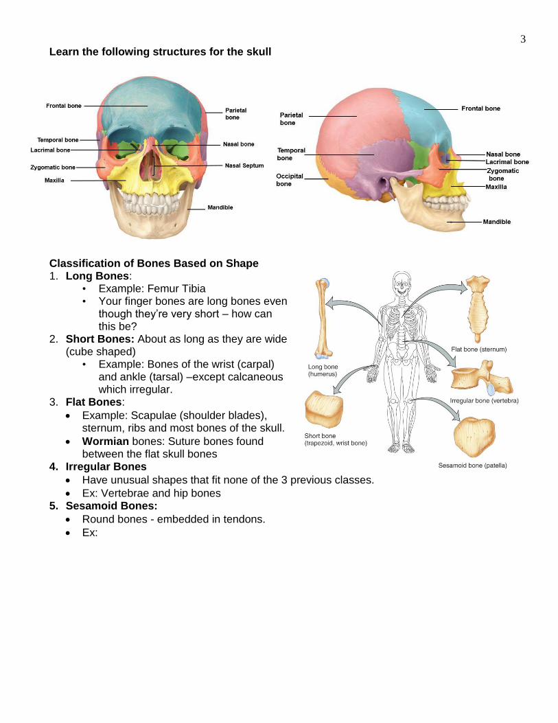
Learn the following structures for the skull
Classification of Bones Based on Shape 1. Long Bones:
• Example: Femur Tibia • Your finger bones are long bones even
though they’re very short – how can this be?
2. Short Bones: About as long as they are wide (cube shaped)
• Example: Bones of the wrist (carpal) and ankle (tarsal) –except calcaneous which irregular.
3. Flat Bones:
Example: Scapulae (shoulder blades), sternum, ribs and most bones of the skull.
Wormian bones: Suture bones found between the flat skull bones
4. Irregular Bones
Have unusual shapes that fit none of the 3 previous classes.
Ex: Vertebrae and hip bones 5. Sesamoid Bones:
Round bones - embedded in tendons.
Ex:

4
Long Bone Structure • Consists of a shaft (diaphysis) and two expanded
extremities (epiphysis) – Diaphysis: Consists of a thick collar of
• Medullary cavity contains: – Yellow bone marrow – fat (energy
source) – Epiphysis: Consists of a thin layer of compact
bone covering an interior of spongy bone. – Metaphysis: Portion of a long bone between the
diaphysis and epiphysis – contains the epiphyseal plate.
• Epiphyseal plate (growth plate): Composed of hyaline cartilage and found in long bones that are still growing in length.
• Becomes the epiphyseal line (disk) when bone growth stops
– Cartilage of the • Articular Cartilage:
– A thin layer of hyaline cartilage that covers the ends of the epiphyses
• Located in areas where one bone articulates with another bone.
– Functions: Reduces friction and
• Periosteum: – Tough sheath that covers the external surface
except for the articular cartilage. – Function:
• Essential for bone growth in diameter and repair • Point of attachment for
• Endosteum: Thin membrane that lines internal surfaces of bone. –
Bone Marrow • Bone marrow: General term for the soft tissue
occupying the medullary cavity of a long bone and the spaces amid the trabeculae of spongy bone.
• There are two types: red & yellow. – Red bone marrow: Blood cell forming tissue =
hematopoietic tissue – Yellow bone marrow:

5
]
Bone Cell Types 1. Osteogenic Cells
– Stem cells that – Located within the periosteum, endosteum, and the
central canal 2. Osteoblasts
– Bone-building cells. – Synthesize and secrete collagen fibers and other organic components of bone matrix. – Initiate the
3. Osteocytes
– Mature bone cells. – Osteoblasts that have become trapped by the secretion of matrix become osteocytes. – No longer secrete matrix. – Responsible for
4. Osteoclasts – Large cells derived from the fusion of – Cells that digest bone matrix – this process is called bone resorption and is part of
normal bone growth, development, maintenance, and repair. – Concentrated in the periosteum and endosteum. – On the side of the cell that faces the bone surface, the plasma membrane is deeply
folded into a ruffled border. Here, the osteoclast secretes digestive enzymes (how might this occur?) to resorb the bone matrix. It also pumps out hydrogen ions to create an acid environment that eats away at the matrix. What advantage might a ruffled border confer?
– Why do we want a cell that eats away at bone? (Hint: bone is a very dynamic tissue.) Bone Matrix • Bone Matrix is the nonliving structural part and Consists of:
– Water (~25%) – – Crystalized mineral salts (50%)
• Collagen fibers – Provide the bone with flexibility – gives it resilience and the ability to resist stretching
and twisting. – Frame work for calcification and

6
• Crystalized Mineral Salts – Mainly hydroxyapatite (interaction of calcium phosphate and calcium carbonate. – Trace amounts of – Minerals are deposited among the collagen fibers and crystallize giving bone its
characteristic hardness. • Mineral salts are like the concrete and collagen fibers are like the metal rods in concrete.
– Without the collagen fibers, bone would be very brittle. Compact and Spongy Bone • Two categories of bone:
1. Compact bone: 2. Spongy (cancellous)
bone: Contains lots of spaces in a honeycomb of needle-like projections called trabeculae
• Surrounded by compact bone and contains red marrow
3. All 206 bones are a combination of compact and spongy bone
Organization of Compact Bone • Osteons or Harversian
Systems are the fundamental units of compact bone which are multiple cylindrical structural units.
– Imagine these osteons as weight-bearing pillars that are arranged parallel to one another along the long axis of a compact bone.
Osteon Components • Each osteon consists of a single longitudinal central (Haversian) canal, surrounded by
concentric layers of calcified bone matrix called lamellae. – Central canals allow the passage of blood vessels,
Volkmann’s Canals • Perforating (Volkmann’s) canals.
– Connect the blood and nerve supply between the periosteum, central (Haversian) canals and the medullary cavity.
Osteocytes and Canaliculi • Osteocytes: Occupy cavities known as lacunae at the junctions of the lamellae. • Canaliculi: Canals that connect the lacunae to each other and to the central canal.
– Allow osteocytes to exchange nutrients, wastes, and chemical signals to each other
7
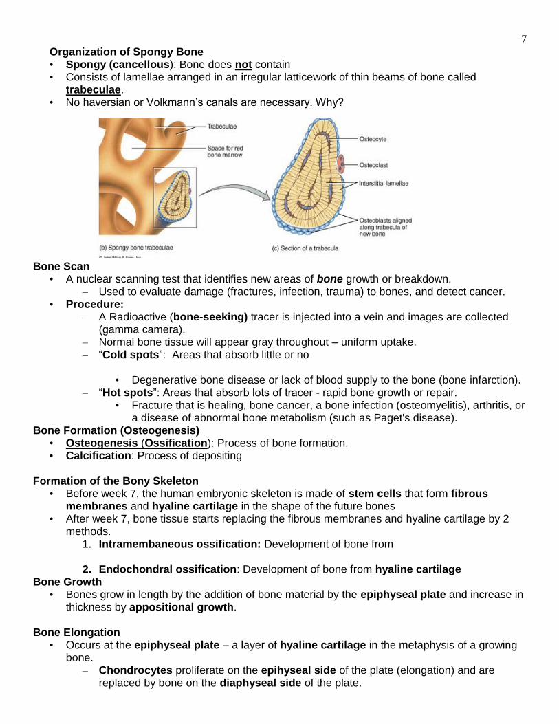
Organization of Spongy Bone • Spongy (cancellous): Bone does not contain • Consists of lamellae arranged in an irregular latticework of thin beams of bone called
trabeculae. • No haversian or Volkmann’s canals are necessary. Why?
Bone Scan • A nuclear scanning test that identifies new areas of bone growth or breakdown.
– Used to evaluate damage (fractures, infection, trauma) to bones, and detect cancer. • Procedure:
– A Radioactive (bone-seeking) tracer is injected into a vein and images are collected (gamma camera).
– Normal bone tissue will appear gray throughout – uniform uptake. – “Cold spots”: Areas that absorb little or no
• Degenerative bone disease or lack of blood supply to the bone (bone infarction). – “Hot spots”: Areas that absorb lots of tracer - rapid bone growth or repair.
• Fracture that is healing, bone cancer, a bone infection (osteomyelitis), arthritis, or a disease of abnormal bone metabolism (such as Paget's disease).
Bone Formation (Osteogenesis) • Osteogenesis (Ossification): Process of bone formation. • Calcification: Process of depositing
Formation of the Bony Skeleton
• Before week 7, the human embryonic skeleton is made of stem cells that form fibrous membranes and hyaline cartilage in the shape of the future bones
• After week 7, bone tissue starts replacing the fibrous membranes and hyaline cartilage by 2 methods.
1. Intramembaneous ossification: Development of bone from 2. Endochondral ossification: Development of bone from hyaline cartilage
Bone Growth • Bones grow in length by the addition of bone material by the epiphyseal plate and increase in
thickness by appositional growth. Bone Elongation
• Occurs at the epiphyseal plate – a layer of hyaline cartilage in the metaphysis of a growing bone.
– Chondrocytes proliferate on the epihyseal side of the plate (elongation) and are replaced by bone on the diaphyseal side of the plate.

8
Halt to Elongation: Formation of the Epiphyseal Line • Epiphyseal line: Bony structure that results from the closure of the epiphyseal plate.
– Closure happens at ~ age – Signifies the bone has stopped growing in length. – Fractures that damage the epiphyseal plate can result in premature closure and a
shorter bone. Appositional Bone Growth (Diameter) Steps in the Process
1. Osteoblasts beneath the periosteum secrete bone matrix on the external surface of the bone.
– Osteoblasts become surrounded by – This makes the bone thicker
2. At the same time, osteoclasts on the endosteum of the medullary cavity break down bone and thus widen the medullary cavity.
• This results in an increase in shaft diameter even though the actual amount of bone in the shaft is relatively unchanged.
Bone Remodeling
• Bone remodeling: The replacement of old • Bone will grow or remodel in response to the forces
or demands placed on it – it’s a dynamic tissue. – Constantly reappropriates its matrix along
lines of mechanical stress. • Involves bone resorption by osetoclasts and bone depostion of minerals and
collagen by osteoblasts – Renewal rate for compact bone is about 4%/year and for spongy bone is about
20%/year Test Your Knowledge
• Why might you suspect someone who has been a powerlifter for 15 years to have heavy, massive bones, especially at the point of muscle insertion?
• Astronauts tend to experience bone atrophy after they’re in space for an extended period of time. Why?

9
Fractures • Fracture: • Fractures are often classified according to the position of the bone
ends after the break: – Open (compound): Bone ends protrude through the skin.
– Closed (simple): Bone ends don’t penetrate the skin. – Complete: A complete break across the bone – broken in two
or more pieces – Partial: Break across bone is incomplete – Greenstick: Bone breaks incompletely. One
• Common in children whose bone contains more collagen and are less mineralized.
– Spiral: Ragged break caused by
• Common in sports injury/Injury of abuse. – Impacted: One end of the fractured bone is forcefully driven into the
medullary space (interior) of the other. – Compression: The bone is crushed, causing the broken bone to be
wider or flatter in appearance. – Stress: Bone fractures (microscopic fissures) without
• Difficult to detect with x-rays – can see them in a bone scan • Result from repeated strenuous activities (running, jumping,
etc) on hard surfaces. Nutritional Effects on Bone
• Normal bone growth/maintenance cannot occur with out sufficient dietary intake of calcium and phosphate salts.
• Calcium and phosphate are not absorbed in the intestine unless the hormone calcitriol is present. Calcitriol is made in the kidneys and its synthesis is dependent on the availability of the steroid cholecalciferol (Vitamin D) which may be synthesized in the skin with exposure to UV rays from the sun or obtained from the diet.
• Vitamins Vitamin Deficiencies: Clinical Considerations
• Rickets: Vitamin D or calcium deficiency in children results in
– Soft matrix laid down by the osteoblasts fails to calcify. Weight of body causes the bones of the legs to bow.
• Osteomalacia: Vitamin D or calcium deficiency in adults.

10
Osteoporosis • Osteoporosis is characterized by a decrease in bone mass and susceptibility to fractures • Results from decreased • Sex hormones (estrogens and testosterone) stimulate osteoblast
activity and synthesis of bone matrix – decreased amounts weaken bone.
• Treatment of postmenopausal women include: • Hormone replacement therapy (HRT)
• Combination of estrogen and progesterone • Calcium supplements and weight bearing exercise. • Evista (Raloxifene): Drug that mimics the effects of estrogen on bone without increased
risk of breast cancer. • Fosamax (Alendronate):
Synovial Joints
• Synovial Joints – Have a synovial cavity between articulating bones. – Freely moveable joints
• Synovial joints have 5 structural features: – Joint cavity – Articular cartilage – Articular capsule – Synovial fluid – Accessory ligaments and menisci
Structures of Synovial Joints
1. Joint (synovial) Cavity: The potential space between the bones. 2. Articular Cartilage: (hyaline cartilage) covers articulating ends of bones
– Functions to cushion and reduce friction 3. Articular Capsule: Encloses the synovial joint and is composed of two layers 4. Synovial Fluid: A viscous fluid that lubricates the joint.
• Formed by secretion of fibroblast cells and the interstitial fluid filtered from blood plasma of the synovial membrane
• Warming up before exercise stimulates the production of synovial fluid
5. Accessory Ligaments and Menisci
Ligaments consists of dense regular connective tissue and stabilizes the joint. • Double-jointed-ness results from
extra-stretchy ligaments and joint capsules. Is this necessarily a good thing?
• Menisci: Pads of fibrocartilage that lie between bones and act as shock absorbers as well as improve the fit between bone ends, thus stabilizing the joint.
• Found in the knee joint

11
Carpal Tunnel • The "carpal tunnel" is formed by the bones,
tendons and ligaments that surround the median nerve. – The median nerve supplies the thumb and 1st 3 fingers.
• Carpal Tunnels Syndrome: Occurs when the median nerve is squeezed at the wrist. – Swelling of the tendon sheaths causes a too tight carpal tunnel and puts pressure on
the median nerve. How Does Carpal Tunnel Syndrome Develop?
• The tendons of the hand are encased in sheaths (bursa), through which the tendons slide. The inner wall of the sheaths contains cells that produce a slippery fluid to lubricate the tendons. Lubrication is essential for the normal and smooth functioning of the tendons.
• With repetitive or excessive movement of the hand, the lubrication system may malfunction. It may not produce enough fluid or it may produce a fluid with poor lubricating qualities. Failure of the lubricating system creates friction between the tendon and its sheath causing inflammation and swelling of the tendon area. In turn, the swelling squeezes the median nerve in the wrist or carpal tunnel. Repeated episodes of inflammation cause fibrous tissue to form. The fibrous tissue thickens the tendon sheath, and hinders tendon movement.
Clinical Conditions
• Rheumatism: Any painful disorder of the supporting structures of the body – bones, ligaments, tendons or muscles
• Arthritis: Joint inflammation causing swelling, stiffness and pain.
– A form of rheumatism. – Common types: Rheumatoid, gout, and osteoarthritis
Osteoarthritis
• Osteoarthritus (degenerative joint disease) – Most common: Results from the gradual “wear and
tear” of joint cartilage – Normal joint use prompts the release of cartilage-
damaging enzymes. If cartilage destruction exceeds cartilage replacement, we’re left with roughened, cracked, eroded cartilages.
• Spurs of osseous tissue are deposited and restrict movement – Treatment: Stop doing the movement that stresses the joint, however mild exercise has
been shown to slow joint degeneration and enhance mobility. Damaged joint can be surgically removed and replaced

12
Rheumatoid Arthritis • Autoimmune disease
• Body creates antibodies which attack the joint surfaces
• The synovial membrane can inflame and eventually thicken into a pannus – an abnormal tissue that clings to the articular cartilage.
• The pannus erodes the cartilage and eventually scar tissue forms and connects the two bone ends. This scar tissue can later ossify, fusing the bones together. This is known as ankylosis.
Gouty Arthritis (Gout)
• Caused by the buildup of uric acid in the blood. • When nucleic acids are metabolized uric acid is produced.
Normally uric acid is excreted in the urine. • If blood uric acid concentration rises due to decreased excretion
or increased production, it may begin to form needle-shaped crystals in the soft tissues of joints.
• Inflammation ensues causing painful arthritis. • Eventually, the articular cartilage is destroyed and the bones
fuse. • Treatment: Anti-inflammatory drugs and surgical removal of the
pannus before fusion of the joint. • Damaged joint can be surgically replaced.