the skeletal system chapter 5. the skeletal system parts of the skeletal system bones joints ...
TRANSCRIPT
The Skeletal SystemChapter 5

The Skeletal System Parts of the skeletal system
Bones
Joints
Cartilage
Ligaments
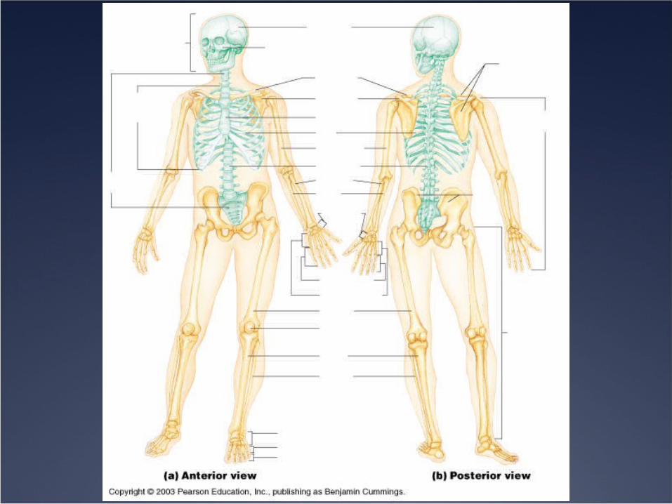
Divided into two divisions
Axial Skeleton – torso and head
Appendicular Skeleton - limbs

Functions of the Bones
Support of the body
Protection of the soft organs
Movement due to attached skeletal muscles
Storage of Minerals and fats
Blood cell formation

Bones of the Human Body The skeleton has 206 bones
Two basic types of bone tissue Compact bone
Homogeneous Very dense and strong
Spongy bone Small needle-like
pieces of bone Many open spaces Purpose?
Figure 5.2b

Classification of Bones• Long Bones
• Description? Examples?
• Short Bones• Description? Examples?
• Flat bones• Description? Examples?
• Irregular Bones• Irregular shapes• Do not fit into any other bone classification
category• Examples?

Classification of Bones on the Basis of Shape
Figure 5.1
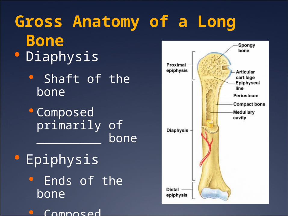
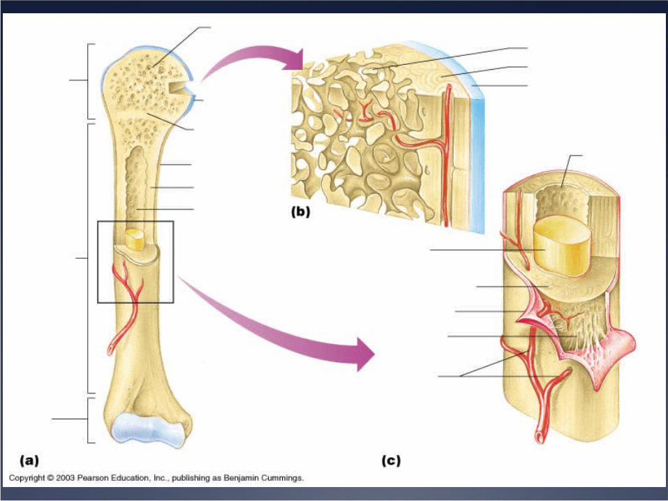
Gross Anatomy of a Long Bone
Diaphysis
Shaft of the bone
Composed primarily of _________ bone
Epiphysis
Ends of the bone
Composed primarily of _________ bone
Figure 5.2a
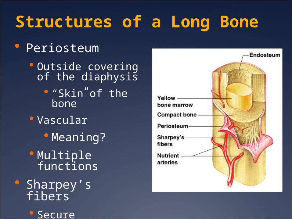
Structures of a Long Bone
Periosteum Outside covering of
the diaphysis
“Skin of the bone”
Vascular
Meaning?
Multiple functions
Sharpey’s fibers Secure periosteum to
underlying boneFigure 5.2c

Structures of a Long Bone
Articular cartilage
Covers external surfaces of epiphysis
Hyaline cartilage
Function?
Figure 5.2a

Structures of a Long Bone
Medullary cavity
Cavity of the shaft
Contains yellow marrow (mostly fat) in adults
Contains red marrow (for blood cell formation) in infants Figure 5.2a

Structures of Long Bone
Process – Projection from the bone
Purposes
Site of attachments for muscles
Create joints
Pathway for nerves, blood vessels

Microscopic Anatomy of Bone
Osteon (Haversian System) Bone is built around a “canal” Tube-like openings in the bone
DQ - What would these canals be used for?
Two types of Canals Central (Haversian) canal
Run longitudinally in the bode Perforating (Volkman’s) canal
Perpendicular to the Haversian Canal

Microscopic Anatomy of Bone
Figure 5.3

Microscopic Anatomy of Bone
Lacunae Tiny cavities
containing osteocytes (bone cells)
Lamellae Rings around the
central canal
Sites of lacunae
Figure 5.3

Microscopic Anatomy of Bone
Canaliculi
Tiny canals
Radiate from the central canal to lacunae
Not the same as Volkmann’s canal
Purpose = diffusionFigure 5.3

Bone Cells
3 Types of Bone cells
Osteocytes - Mature bone cell
Osteoblast – Bone forming cell
Osteoclast – Bone Destroying cell
Break down bone matrix for remodeling and release of calcium

Skeletal Functions
• Support and Protection:• Bones create the shape of our body
• Examples?• Bones provide a hard protective barrier
around vital organs• Examples?
• Movement:• Muscles attach to the bones across
joints• Work like levers
Skeletal Functions
• Blood cell production:• Hematopoiesis – the process of forming
blood cells• Not always in the bone marrow
• As embryo develops, production is in the liver and spleen
• Then switches to the marrow• 2 types of Marrow
• Red• Yellow
Hematopoiesis
• Red marrow produces erythrocytes, leukocytes, and thrombocytes• Which is RBC? WBC? Platelets?
• Red color is due to hemoglobin• Infants have mostly red marrow
• Why would this be?
• As aging occurs, most red marrow is replaced with yellow marrow• Yellow stores fat

Storage of Minerals
• Minerals account for about 70% of bone matrix• Calcium #1
• When blood calcium levels are low hormones stimulate osteoclasts to break down bone tissue• Why would they do this?
• High blood calcuim levels stimulate osteoblasts to form bone

Changes in the Human Skeleton
In a fetus, the skeleton is primarily hyaline cartilage
What happens to the cartilage as we develop?
Replaced by bone
Cartilage remains in isolated areas
Where?

Bone Growth and Development (Endochondral)
Replacement of cartilage begins in the primary ossification center Occurs in the diaphysis What type of cell is active?
Continues in secondary ossification sites in epiphysis Epiphyseal plate is created between primary
and secondary ossification sites. You might know this by a different name

Bone Growth and Development (Endochondral)
• During this process the Medullary Cavity must be formed.
• How is this done?• Growth Hormone (GH) and sex hormones
control bone growth• DQ - When does bone growth stop?• When the primary and secondary ossification
sites grow together, closing the epiphyseal plate.
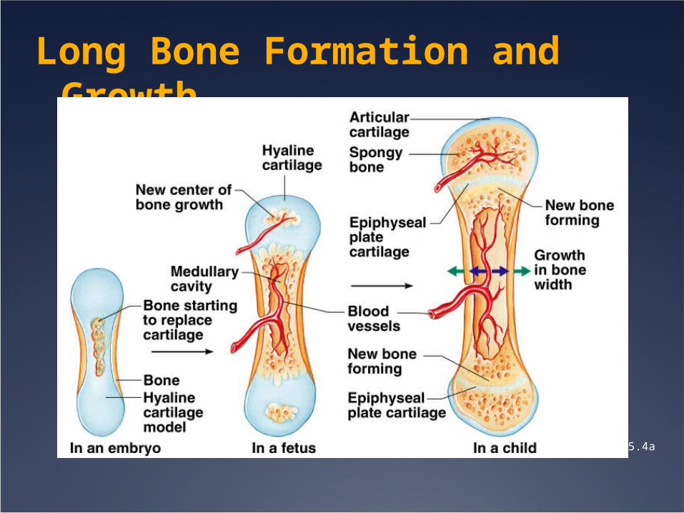
Long Bone Formation and Growth
Figure 5.4a

Long Bone Formation and Growth
Figure 5.4b

Bone Homeostasis
• To stay healthy, bone is continually resorbed and deposited
DQ - Why would this be? So old bone is broken down and new bone can
be formed. Controlled by two factors
Calcium levels in blood Stress on bones
Example: Running If this process becomes unbalanced bones
lose their mass and become weaker
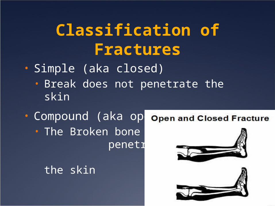
Classification of Fractures
• Simple (aka closed)• Break does not penetrate the skin
• Compound (aka open)• The Broken bone
penetrates through the skin


Types of fractures• Greenstick –
• incomplete, only one side of bone
• Transverse –• Complete break to right angle to lengthwise of
long bone. Usually traumatic
• Oblique -• Rare. Break at an angle
• Comminuted –• More than two fragments broken off. traumatic

Types of fractures

Types of fractures
• Impacted –• Occurs after a fall, vertebral column
compressed, and cracks
• Pathologic -• Disorder that weakens the bone, leading to
a fracture
• Stress -• A bone becomes stressed from over use. • Can cause slight breaks

Repair Bone fractures
• Break causes blood vessels to rupture• What does this cause?
• Osteoclasts will remove bone fragments
• New vessels and Fibrocartilage form around break• Cartilage will be replaced by a bony
callus• Cell types - osteoblasts
Repair of fracture (Bone remodeling)
• There is typically more bone produced at site of healing• Why would this be?
• How does the bone get back to normal?
• Osteoclasts will reshape to like original bone

Skeletal Differences
• Adult vs. Infant skull
• Infant - face is small in comparison to cranial bones• How much of an adults length is made of
the head? Infants?
• Adults = about 1/8 Infants = about 1/4
• Fontanels - soft spots• What is the purpose of fontanels?
• Provides room for the brain to grow
Skeletal Disorders• Use your book to come up with a one
sentences summary of each of the following disorders.• Osteoarthritis• Rheumatoid arthritis• Gouty arthritis (Gout)• Osteoporosis• Scoliosis• Kyphosis• Lordosis• Osteomyelitis – bacterial infection of the
bone, causing pain and discomfort.• Paget disease – Bone remodeling is not
balanced leading to abnormal and enlarged but brittle bones.

Skeletal Disorders
Due to poor posture, Helga has felt like she is constantly leaning forward. An X-ray reveals excessive flexion in the thoracic curvature.
Kyphosis
Jimbo was a four sport athlete in high school and has continued with high impact excercises. He has complained of stiffness in his knees. He has also started to develop bone spurs, which hinder movement.
Osteoarthritis
Skeletal Disorders
Phoebe’s phalanges have fused together, so she is unable to flex his fingers. Her family has a history of this disorder and it is discovered that she has a high quantity of uric acid in her blood.
Gouty Arthritis
Whitney’s muscles in her lumbar region are excessively tight. This has caused the lumbar vertebrae to curve laterally towards the tightened muscles.
Scoliosis

Skeletal disorders
Gertrude has experienced a dull pain in her lower back. An x-ray revealed a fracture of her L2, yet Gertrude does not recall any impact that may have caused the break.
Osteoporosis
Will-i-am experienced a compound fracture a week ago and the bones were reduced while on a hunting trip. He has since developed a fever and severe pain in the area of injury. It is suspected that he has a bacterial infection.
Osteomyelitis
Skeletal Disorders
Mac has experienced pain in his bones. Through an x-ray it has been determined that his femur is misshapen. He has also been told by his doctor that he has a high alkaline phosphatase level in his blood.
Paget’s Disease
Marge’s joints have become swollen, reddened and tender. It has been very painful to move. This seems to to go away, but it keeps coming back.
Rheumatoid Arthritis
Joints
• Every bone in the body articulates with another bone• Except the hyoid
• Not all joints are movable.• Where would immovable joints be found?
• 3 Types of Joints• Fibrous• Cartilaginous• Synovial

Types of Joints• Fibrous
• Immovable• Examples?
• Cartilaginous• Both ends connected by cartilage• Immovable to limited movement• Examples?
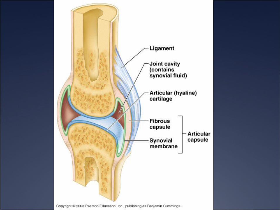
• Synovial• surrounded by joint cavity• Contain hyaline cartilage, ligaments, and
synovial fluid• Examples?

Knee injuries Torn ACL, MCL,
PCL, LCL
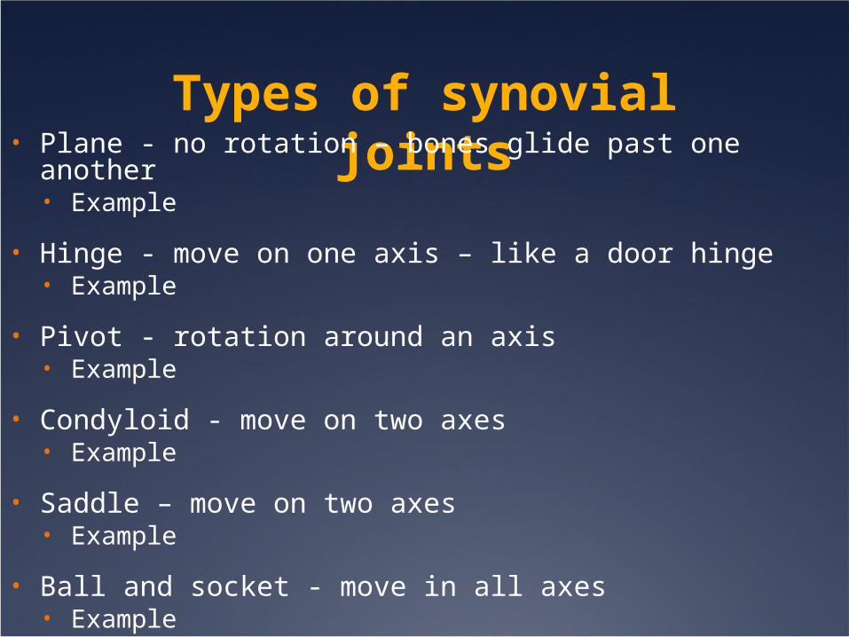
Types of synovial joints• Plane - no rotation – bones glide past one another
• Example
• Hinge - move on one axis – like a door hinge• Example
• Pivot - rotation around an axis• Example
• Condyloid - move on two axes• Example
• Saddle – move on two axes• Example
• Ball and socket - move in all axes• Example

Types of Synovial Joints Based on Shape

Types of Synovial Joints Based on Shape
Figure 5.29d–f


Motions• Flexion-Angle
decreases
• Extension -
• Rotation – Move around an axis
• Abduction -
• Adduction -
• Circumduction -
• Dorsiflexion – raising foot to the shin
• Plantar flexion -
• Elevation – raise a body part
• Depression – lower a body part
• Supination -
• Pronation -