similarities of the golgi apparatus membrane and the plasma...
TRANSCRIPT

J. Cell Sci. ao, 167-182 (1976)
Printed in Great Britain
SIMILARITIES OF THE GOLGI APPARATUS
MEMBRANE AND THE PLASMA MEMBRANE
IN RAT LIVER CELLS
S. HODSONUnit of Electron Microscopy
AND G. BRENCHLEYDepartment of Medical Biochemistry, The Welsh National School of Medicine,Heath Park, Cardiff CF4. 4XN, U.K.
SUMMARYA Golgi apparatus-rich fraction and a plasma membrane-rich fraction were isolated from
a common homogenate of rat liver. Their respective buoyant densities, appearances in theelectron microscope and 5'-nucleotidase and UDP-galactose ovalbumin galactosyltransferaseactivities were in accord with published data on separately isolated Golgi apparatus-rich andplasma membrane-rich fractions. Contamination by endoplasmic reticulum and mitochondriawas low.
Gel electrophoresis of the membrane proteins of the Golgi apparatus-rich and plasmamembrane-rich fractions (separately and mixed) showed a close similarity. After Neville'sdemonstration that electrophoretic patterns of membrane protein subunits from differentsubcellular fractions are easily distinguishable, the present work demonstrates an unusuallyclose relationship between the Golgi apparatus membrane and the cell membrane. It ispossible that membrane similarity may be mediated by the transfer of membrane-boundvesicles from the Golgi apparatus to the cell membrane.
INTRODUCTION
The Golgi apparatus plays a common role in many cell types. It is the site wheresugar chains are attached to a variety of macromolecules (Northcote & Pickett-Heaps, 1966; Ronzio, 1973; Chabaud, Bouchilloux, Ronin & Ferrand, 1974). Vesicles,rilled with the product of the particular cell type, bud off from the Golgi apparatusand migrate across the cell until they contact the cell membrane (Northcote &Pickett-Heaps, 1966; Neutra & Leblond, 1966; Cuminge & Dubois, 1972; Coulomb& Coulomb, 1973; Moussel & Moussel, 1973). The Golgi vesicular membrane usuallyfuses with the cell membrane (but see Neutra & Leblond, 1966) and the contents ofthe Golgi vesicle are deposited into the extracellular space. Examination of thiswidespread process, which seems an important route for the export of macro-molecules out of the cell, suggests that as the Golgi vesicle membrane originates in theGolgi apparatus and terminates in the plasma membrane, there should be featurescommon to the membranes which delineate the Golgi apparatus and the cell itself.
Neville & Glossmann (1971) have shown that the protein subunit compositionof any membrane is unique. Although erythrocyte ghosts, kidney brush border

168 S. Hodson and G. Brenchley
membranes and liver plasma membranes of rat may have a few common proteinsubunits - ' the differences are so striking that a single glance at the gel pattern issufficient to identify the source of the membrane'. Although the protein subunitcompositions of kidney and liver mitochondrial membranes are similar, they showfew or no bands in common with the plasma membranes. The protein subunitcomposition of endoplasmic reticulum is also different from either plasma membraneor mitochondrial membrane.
This paper compares the protein subunit compositions of a Golgi apparatus-richfraction and a plasma membrane-rich fraction prepared from a common homogenateof rat liver.
METHODS
Fractionation
Male Wistar rats, weighing about 200 g and fed ad libidum were used. All sucrose solutionswere prepared in 89-5 mM K,HPO4, 105 mM citric acid, pH 70 . The rat was killed byneck-breaking and its liver was dissected. Seven grammes of liver were quickly minced witha razor blade and transferred to 15 ml ice-cold buffered, 1 4 6 % (w/w), sucrose (d = 1-067 gcm"3). After 10 min of gentle agitation, the sucrose solution was decanted and the dicedliver was transferred to 10-5 ml ice-cold buffered, 14-6% (w/w), sucrose in a stout tube ofdiameter 25 cm.
Tissue was homogenized with a Polytron PT20 (Camlab, Cambridge) modified by turningdown the removable rotating head to give a radial clearance of 1 mm. The liver was homo-genized for 90 s to 2 min at tap 0 6 . Homogenization was so gentle that the tube had to bemanipulated around the Polytron head. Homogenization was completed when the lastdistinguishable particle of liver had disappeared. Further homogenization, up to a total timeof 3 min, did not cause any detectable differences in the collected fractions. The homogenatewas made up to 24 ml with 146 % (w/w) buffered sucrose, and then centrifuged for 10 minat 600 g. The supernatant designated fraction iS was recovered in 17 ± 0 5 ml. The precipitate,mainly red coloured with a thin top layer of white material was designated fraction 1 PPT.The supernatant (iS) was applied to step sucrose gradients in 3 centrifuge tubes. The gradientof buffered sucrose was made of: 4 ml 23-7 % w/w (d = 1-108 g cm"'); 4 ml 308 % w/w(d = 1-137 gem-"); 4 ml 33-8% w/w (d = 1-154 g cm"1); and 4 ml 38-1 % w/w (d = 1-175 Scm"*). The tubes were spun at 27000 rev/min (105000 g max.) in a 3 x 25 ml swing out headof an MSE Superspeed 50 for 2 h. Visible bands appeared at the d = 1-108/1-137 g c m J
interface (designated the Golgi apparatus-rich fraction, G), and the d = 1154/1-175 g cm"1
interface (designated a plasma membrane-rich fraction, PM). A much fainter band appearedat the intermediate d = 1-137/1-154 g cm"3 interface (designated the intermediate fraction I).
Additionally in each tube there was a precipitate designated fraction 2PPT.
Enzymic activitiesThe homogenate and all fractions were assayed for enzymic activity.Prior to assay, the fractions were washed in 7 mM Tris pH 72 , although except for deter-
mination of glucose-6-phosphate phosphohydrolase activity, where background phosphateof the buffer masked enzyme activity, the washing was not essential. The fractions weremade up to 1 mg protein/ml. 5'-Nucleotidase was assayed by the method of Newby, Luzio &Hales (1975). Samples (5 fi\) of homogenate or subcellular fractions were incubated for 1 h at37 °C with 500 fi\ assay mixture containing 50 mM Tris (adjusted to pH 8 0 with HC1), 2 mMmagnesium chloride, 0-2 mg adenosine, 200 /tM AMP and tracer 'H-AMP (20000—30000 cpm).The incubation was terminated by the addition of 100/tl 0-15 M zinc sulphate. UnhydrolysedAMP was precipitated by addition of 100 fil 0-15 M barium hydroxide; 500 /tl of supernatantwere added to 5 ml of scintillant prepared by adding 8 g PPO, 200 mg POPOP and 1 1. Triton

Golgi and plasma membranes 169
X-100 to 2 1. toluene. Glucose-6-phosphate phosphohydrolase was measured by the methodof de Lamirande, Morais & Blackstein (1967). Cytochrome-c oxidase was measured by themethod of Cooperstein & Lazarow (1951). UDP-galactose ovalbumin galactosyltransferasewas assayed by a modification of the method of Freilich, Richmond, Reppucci & Silbert(1975). To 100/tl of reagent (25 mM Tris pH 6-4, 2-0 mg ovalbumin, 10 mM MnClj, o-i %Triton X-100, UDP-[I4C]galactose, 30000 dpm sp.act. 297 mCi/mmol) was added 10 fi[enzyme. After incubation at 37 °C for 1 h, the reaction was terminated with 3 ml ice-cold10 % trichloroacetic acid (TCA). The precipitate was washed in 10 % TCA (x 5) at thecentrifuge and then resuspended in 1 ml of 2 % sodium dodecyl sulphate (SDS) in 8 M ureaat 50 CC for 3 h. Radioactivity was counted in 10 ml of scintillant consisting of 5 g PPO + 0 2 gPOPOP in 1 1. toluene plus 500 ml Triton X-100. Protein was determined by the methodof Lowry, Rosebrough, Farr & Randall (1951) against bovine serum albumin as standard.
Gel electropJioresis
For gel electrophoresis, the fractions were centrifuged in 150 mM NaCl at 105000 g for1 h (x 2), and then resuspended in 50 mM NajCOj to a final protein concentration of2 mg/ml. The fractions were then treated in a similar fashion to that described by Neville(1971). Fractions were exposed to 8 mg SDS per mg of protein and 10% by volume of/?-mercaptoethanol, and heated on a boiling water bath for 1 min, when the suspensionclarified. They were then dialysed overnight in the cold against 0-04 M boric acid, 0041 MTris, o-i % SDS, 0-05% dithiothreitol, 2 % sucrose. Three buffer systems were used fordiscontinuous electrophoresis.
(a) Upper reservoir buffer: 004 M boric acid, 0041 M Tris, plus 0 1 % SDS.(b) Spacer gel buffer: 50 ml 02164 M Tris titrated against o-i N HaSOj to pH 6-i and
then made up to 200 ml. 3-2 g Acrylamide, plus 0-2 g iV.iV'-methylenebisacrylamide (Bis)dissolved in 100 ml spacer gel buffer make the spacer gel solution. To polymerize the spacergel 20 fil TEMED, plus 100 fi\ 5 % ammonium persulphate were added to 10 ml spacer gelsolution.
(c) Lower reservoir buffer: 00308 N HC1, plus 04244 M Tris. I I - I g Acrylamide, plusc i g Bis in 100 ml of lower reservoir buffer make the separation gel solution. To polymerizethe separation gel 30 fil TEMED, plus 200 /tl 5 % ammonium persulphate were added to20 ml of separation gel solution.
Gels were run in glass tubes 8 cm long, i.d. 5 mm scrupulously cleaned with chromic acidfollowed by immersion in detergent in an ultrasonic bath. After applying and gelling theseparation and spacer gels and filling the upper and lower reservoirs, current was applied at0 5 mA/tube for 10 min, then 1-5 mA/tube to complete the run in about 2 h. During therun, the Kohlrauach fronts between the separation gel buffer/spacer gel buffer and spacergel buffer/upper reservoir buffer, were clearly visible. At the end of the run, the gels weretaken out of the tubes and stained with Coomassie blue (Weber & Osborn, 1969). Gels werestored in 7-5 % acetic acid. Every run was regulated against 20 fil of a mixture of markerproteins each in an amount of about 2 fig. The mixture had been treated identically to thecell fractions and consisted of: lysozyme (14300), carbonic anhydrase (29000), lactatedehydrogenase (36000), ovalbumin (43000), pyruvate kinase (57000), phosphorylase a (94000),/?-galactosidase (130000) (polypeptide chain molecular weight quoted by Weber & Osborn,1969).
Electron microscopy
Most fractions in most trials (70) were examined in the electron microscope. The fractionswere fixed in 20 vol. of 2-5 % glutaraldehyde in 67 mM potassium phosphate (pH 74),containing o-i mM CaClj and were then pelleted at 1200 g max. for 10 min. They werepostfixed in 1 % OsO4 in o-i M sodium cacodylate adjusted to pH 7-0 and dehydrated throughan ethanol series which was more gradual than is commonly used (10 %, then in 10 % incrementsto absolute alcohol). The purpose of this gradual dehydration was to prevent breaking up ofthe delicate pellet. The pellets were taken, gradually, into 100 % epoxypropane at which stagethey were quite firm, and then transferred into Araldite. Sections were cut on a Cambridge

170 S. Hodson and G. Brenchley
Huxley Mk. II ultramicrotome. It was particularly noticeable that the designated Golgiapparatus-rich fraction was very difficult to section. The other fractions were not. Sectionswere stained in lead citrate (Reynolds, 1963) at 30 °C for 25 min in the staining pot (Hodson,1974). They were examined in a Philips EM 300.
Chemicals
Lactate dehydrogenase and pyruvate kinase were purchased from the Boerhinger Corporation.Carbonic anhydrase, lysozyme, ovalbumin, phosphorylase a, /?-galactosidase, bovine serumalbumin and sodium dodecyl sulphate (SDS) were purchased from Sigma. UDP-[14C]galactoseand 'H-AMP were purchased from the Radiochemical Centre, Amersham. PPO, POPOP,toluene and Triton X-100 were purchased from Koch-Light. Acrylamide, iVjiV'-methylene-bisacrylamide (Bis) were purchased from B.D.H. All reagents used for electron microscopywere purchased from Taab. All other chemicals used in this work were purchased fromHopkin and Williams.
RESULTS
Electron microscopy
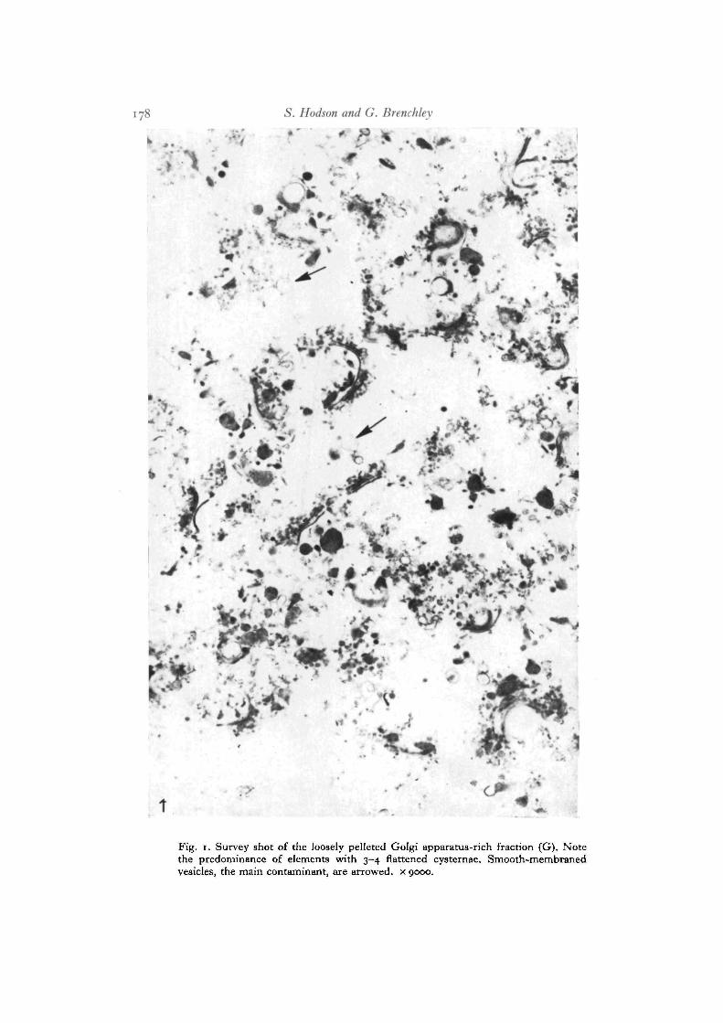
The Golgi apparatus-rich fraction (G). Examination of representative sectionsthrough 32 preparations revealed only smooth membrane structures. Mitochondriaand rough endoplasmic reticulum were not recognized. The pellet was always looselypacked (Fig. 1) but most elements of the pellet showed the 3 or 4 stacked cysternaewhich are characteristic of the liver Golgi apparatus. The 2 characteristic Golgivesicles of the hepatocyte (the 0-22-0-30 /tm diameter vesicle filled with very lowdensity lipoproteins, arrowed Fig. 2, and the 50-nm diameter vesicle, arrowedFig. 4) were present in the isolated Golgi apparatus-rich fractions (arrowed, Figs. 3,5). The flattened cysternae of isolated Golgi bodies were less regular (double arrows,Fig. 5) than when they were in vivo. Smooth-membraned vesicles in the Golgiapparatus-rich fraction (arrow, Fig. 1) contributed less than 5 % of total membrane.
The plasma membrane-rich fraction (PM). The fraction consisted mainly of smooth-membraned vesicles (Figs. 6, 7). An occasional mitochondrion or fragment of roughendoplasmic reticulum contaminated the fraction. The smooth-membraned vesicleranged in diameter from 0-14 to 5-0 /tm, but the medial vesicle was about 0-7/imin diameter. Most of the vesicles contained no obvious inclusions, other than small'pegs' of material which have been widely interpreted as microvilli, and theirmembranes seemed flaccid. About 10% of the vesicles were filled with what mightbe cytoplasm (arrow, Fig. 7). After washing with 0-9% (w/v) NaCl solution onlyempty vesicles were seen in the fraction.
The intermediate fraction (I). This fraction showed no unique features. About10% of the profiles were of Golgi bodies and the rest were empty vesicles (medialdiameter about i-o/tm). The intermediate fraction looked like an overlap ofthe heavy tail of fraction G and the light tail of fraction PM. Nuclei, glycogen,mitochondria and rough endoplasmic reticulum were seen in the first precipitate.Rough endoplasmic reticulum and mitochondria with some glycogen were seen inthe second precipitate.

Golgi and plasma membranes 171
Enzymic activities
The results are presented in Table 1 as the specific activity of each fractiontogether with the total protein. The cytochrome oxidase and glucose-6-phosphatasewere markers for mitochondria and endoplasmic reticulum. In the Golgi, intermediateand plasma membrane fractions the marker enzymes showed considerably lowerspecific activities than either the tissue homogenate or the supernatant from which theywere prepared.
UDP-galactose ovalbumin galactosyltransferase activity was almost completelyrecovered in the Golgi apparatus-rich fraction. Of the total activity in the homo-genate, about 70% was recovered in the supernatant of the first spin. The remainingactivity was located in the precipitate. Nearly all the galactosyltransferase activitycould be eluted from the precipitate by a second wash and this second wash activitybanded at the same density as the Golgi apparatus-rich fraction (G). The galactosyl-transferase activity in the first precipitate seems to represent simple entrapment ofa small part of the Golgi apparatus-rich fraction. Nearly all the galactosyltransferaseactivity in the supernatant (— 110%) banded in the Golgi apparatus-rich fraction(G). The specific activity of this fraction showed greater than 50-fold enrichmentover the homogenate. In contrast, the plasma membrane-rich fraction showed a3-fold depression of specific activity over homogenate. Some activity (3 %) waspresent in the intermediate density band (I) of specific activity 8-fold greater thanthe homogenate.
Specific activity of 5'-nucleotidase was highest in the plasma membrane-richfraction (PM). The specific activity was 10-fold enriched over the homogenate and22-fold enriched over the supernatant from which it was prepared. Golgi apparatus-rich fraction (G) also showed a persistent enrichment of its 5'-nucleotidase specificactivity over the homogenate, but it was much less than that of fraction PM. In spiteof the enrichment only 7 % of total 5'-nucleotidase activity was recovered in fractionPM. Most of the activity was found in the first (68 %) and second (37 %) precipitates.
Gel electrophoresis
Protein patterns of the saline-washed membranes of the Golgi apparatus-rich andplasma membrane-rich fractions were examined. Aliquots were run singly and inequal weight mixtures (total weight in the range 5-100 fig protein) from fractionsisolated from 12 separate rats. The Golgi apparatus-rich fraction patterns isolatedfrom the 12 separate rats were not distinguishable. There was some slight variationin the plasma membrane-rich fraction. In all cases, the patterns of the Golgi apparatus-rich fraction and the plasma membrane-rich fractions were similar. Figs. 8-10show patterns from the 2 fractions isolated from the rat livers which showed thegreatest difference. Each figure shows the patterns of 3 gels after application ofa fixed quantity of membrane protein, coded G (membrane proteins of the Golgiapparatus-rich fraction), PM (membrane proteins of the plasma membrane-richfraction), or G + PM (mixed half weights of G and PM). Fig. 8 shows gels after12 fig protein application. The 7 major bands run and stain identically. Fig. 9 shows


Golgi and plasma membranes 173
gels after 20 fig protein application. The extra band in gel PM is arrowed. Thisband is only faintly visible in gel G and is visible in the mixture (G + PM). Fig. 10shows gels after 70 fig protein application. The major band difference is againarrowed. All 12 isolations were treated by this method of increasing protein appli-cation. In the fractions illustrated in Figs. 8-10 the major difference appears inband 8 (protein bands numbered in terms of decreasing visibility). In two isolationsthe major difference appeared in band 12 (again at the same Rf position in the gel).In one gel there was no distinguishable difference in the first 26 bands. Up to36 bands could be distinguished in each fraction. The major 7 bands of all gelshad the following molecular weights (in decreasing order of their staining capacitywith Coomassie brilliant blue): 1, 53000; 2 and 3 (nearly equal), 63000 and 85000;4 and 5 (nearly equal), 59000 and 103000; 6, 70000; and 7, 98000.
DISCUSSION
The purity of the Golgi apparatus-rich fraction and plasma membrane-rich fractions
The Golgi apparatus-rich fraction and the plasma membrane-rich fraction of ratliver float at different buoyant densities. Golgi apparatus-rich fractions are normallyprepared from rat livers homogenized in hypertonic sucrose (0-5 M). Plasma membrane-rich fractions are prepared from rat livers homogenized in either hypotonic solutions(1 mM NaHCO3) or isotonic sucrose (0-25 M). The buoyant density of the plasmamembrane fraction seems to be independent of the osmotic pressure of the homo-genization medium but the same is not true for the Golgi apparatus-rich fraction.The method described here, which isolates the 2 fractions from a common homo-genate, is based on these considerations. The tissue was homogenized in 0-45 Msucrose.
The Golgi apparatus-rich fraction after homogenization in hypertonic sucrose isreported to collect in the density range 1-064—1-162 8 cm"3 (Mahley, Hamilton &LeQuire, 1969; Leelavathi, Estes, Feingold & Lombardi, 1970; Morre", Cheetham &Nyquist, 1972; Gang, Lieber & Rubin, 1973; Ovtracht, Morre, Cheetham & Mollen-hauer, 1973). More exactly, van Golde, Fleischer & Fleischer (1971) report theGolgi apparatus-rich fraction to have a density of 1-12-1-14 g cm"3. In this work,the Golgi apparatus-rich fraction was collected in the density range 1 • 108-1 • 137 g cm~3.The plasma membrane-rich fraction whether prepared from livers homogenized inhypotonic solution (Emmelot & Bos, 1969; Dorling & le Page, 1973) or isotonicsolution (Berman, Gram & Spirtes, 1969; Nigam, Morais & Karasaki, 1971) collectsin the density range 1-16-1-18 g cm~3. In this work, the plasma membrane-richfractions were collected in the density range 1-154-1-175 g cm"3.
The Golgi apparatus-rich fraction had the characteristics of high yield and highpurity. Galactosyltransferase activity (Table 1) was mainly confined to the Golgiapparatus-rich fraction. The 20% of total activity found in the first precipitaterepresents simple entrapment of Golgi particles as it could be readily eluted fromthe pellet by a second wash which subsequently banded in the Golgi apparatus-richfraction. Therefore, more than 95 % of the galactosyltransferase activity was found in

174 S. Hodson and G. Brenchley
the Golgi apparatus-rich fraction and most of the remaining activity was found inthe intermediate fraction. The intermediate fraction was designed into the preparationto provide a clear barrier between the Golgi apparatus-rich and plasma membrane-rich fractions. The Golgi apparatus of rat liver has previously been shown to bethe sole locus of galactosyltransferase activity (Fleischer, Fleischer & Ozawa, 1969;Fleischer & Fleischer, 1970). The yield of membrane protein in the Golgi apparatus-rich fraction (correcting for galactosyltransferase activity) is 2-26 mg protein/gliver. The figure is in agreement with other published results. The variation in theyield from different male Wistar rats recorded in this series of experiments was fargreater than the measured error. This strongly suggests that in rats fed ad Ubidumthere is significant variation (up to 50 %) in the concentration (or possibly size, butthis was not detected in trials) of Golgi apparatus in liver. Electron-microscopicexamination of many sections confirmed the purity of the Golgi apparatus-richfraction.
The plasma membrane-rich fraction showed no visual or enzyme activity para-meters similar to the Golgi apparatus-rich fraction. Galactosyltransferase activitywas nearly undetectable and less than 1 % of the specific activity of the Golgiapparatus-rich fraction. The profiles seen in the electron microscope were similarto those reported for plasma membrane-rich fractions isolated from tissue homo-genized in isotonic sucrose (Touster, Aronson, Dulaney & Hendrickson, 1970;Nigam et al. 1971) although cell-cell junctions reported in fractions isolated fromtissue homogenized in hypotonic solutions (Emmelot, Bos, Benedetti & Riimke,1964; Dorling & le Page, 1973) were not observed. 5'-Nucleotidase specific activity(x 10 over the homogenate; x 22 over the supernatant from which it was prepared)was consistent with the values given in the literature. Published enrichments include:homogenate activity x 12 (Nigam et al. 1971), homogenate activity x 17 (Coleman,Michell, Finean & Hawthorne, 1967), homogenate activity x 12 (Berman et al. 1969),homogenate activity x 15 (Stein, Widnell & Stein, 1968), post nuclear supernatantactivity x 25 (Dorling & le Page, 1973). The yield of protein in the plasma membrane-rich fraction seemed relatively low. Protein yield and 5'-nucleotidase total activitycould both be increased by increasing the higher density range of the band fromd = 1-175 to d = I - I85g cm"3. Contamination from endoplasmic reticulum (markedby glucose-6-phosphatase) rose steeply. The plasma membrane-rich fraction desig-nated in this paper contains about 7 % of the total 5'-nucleotidase activity. By otherprocedures, up to 17% of total 5'-nucleotidase activity has been recovered in aplasma membrane-rich fraction of rat liver (Dorling & le Page, 1973). In rat fat cellsmore than 80% of total 5'-nucleotidase activity is located on the outside of theplasma membrane (Newby et al. 1975). From such observations, it is clear thatthe designated plasma membrane-rich fraction of this paper does not include allthe plasma membrane of rat cells.
Similarities of tlie Golgi apparatus-rich and plasma membrane-rich fractions
Gel electrophoresis of the 2 fractions showed a close similarity and in one case(out of 12) complete identity of the protein subunit patterns. The identity consisted

Golgi and plasma membranes 175
not only of the Rf values but also of each band's staining capacity with Coomassiebrilliant blue. This dye is not a quantitative marker for total protein. For instance,ovalbumin stains considerably less than an equal weight of pyruvate kinase. Never-theless, the relative staining capacity of each band in the electrophoretic patternmay be taken as correlative evidence of similarity.
The close similarity of the 2 common patterns should be set in the followingcontext of reports of rat liver fractions. The patterns have no bands in commonwith rough endoplasmic reticulum membrane stripped of its ribosomes and isolatedfrom the same homogenate (Hodson & Miller, unpublished observation). TheGolgi pattern has no bands in common with microsomal membrane proteins (Fleischer& Fleischer, 1970). The plasma membrane pattern has no major bands in commonwith mitochondrial membrane or endoplasmic reticulum including the ribosomes(Neville & Glossman, 1971). Smooth microsomal membranes, rough microsomalmembranes, inner mitochondrial membranes and outer mitochondrial membranesall have readily distinguishable electrophoretic protein patterns (Schnaitman, 1969).
The similarity of the Golgi apparatus-rich fraction membrane subunits and theplasma membrane-rich fraction subunits suggests an intimate relationship betweenthe 2 membranes. An identity could never be established by the present method fortwo reasons: I. Gel electrophoresis can provide only correlations in band comparisonand the many minor bands of the fraction cannot easily be visualized. Presumablythis is why the galactosyltransferase, certainly present in G and certainly absent inPM was never seen as there were no recognizable bands present in G and absentin PM. And II, the plasma membrane fraction reported here does not representthe whole plasma membrane. It is often claimed that the plasma membrane may bea mosaic of separate organelles (see Wallach, 1967) and that citrate (used in thebuffer solution in the present preparation) promotes vesiculation of the plasmamembrane (Emmelot & Bos, 1969). It may well be that the present preparationselects only an element of the total plasma membrane mosaic. The yield althoughslightly greater than that reported for isotonic homogenization is less than half thatreported for hypotonic homogenization. It has been stated without published evidencethat the protein subunit patterns of differently isolated plasma membrane fractionsare distinct and different (Berman et al. 1969).
The Golgi apparatus membrane and at least a fraction of plasma membrane ofrat liver have a common protein subunit composition. This relationship betweendifferent cellular organelles seems to be unique (although lysosomal membranesappear not to have been studied) in rat liver cells and is possibly mediated bymembrane-bound vesicles which originate in the Golgi apparatus and terminateat the plasma membrane. In hepatocytes, the vesicles are filled with very low densitylipoprotein (Mahley et al. 1969; Chapman, Mills & Taylaur, 1972, 1973). In hamsterseminal vesicle there is a suggestion that the opposite process may occur, wherepinocytotic vesicles bud off the plasma membrane and fuse into the Golgi apparatus(Mata & David-Ferreira, 1973). In non-growing, non-dividing cells where Golgivesicles fuse into the plasma membrane, there has to be some mechanism for strippingthe excess membrane from the plasma membrane. The dynamics of the process

176 S. Hodson and G. Brenchley
with special reference to the origin and continuity of the Golgi apparatus and theplasma membrane, promise to be full of interest.
We thank Dr. Paul Luzio for his advice and help, and Tenovus for financial support to G.B.
REFERENCES
BERMAN, H. M., GRAM, W. & SPIRTES, M. A. (1969). An improved reproducible method ofpreparing rat liver plasma cell membranes in buffered isotonic sucrose. Biochim. biophys.Ada 183, 10-18.
CHABAUD, O., BOUCHILLOUX, S., RONIN, C. & FERRAND, M. (1974). Localisation in a Golgi-rich thyroid fraction of sialyl-, galactosyl-, and iV-acetylglucosaminyltransferases. Biochimie56, 119-130.
CHAPMAN, M. J., MILLS, G. L. & TAYLAUR, C. E. (1972). Lipoprotein particles from the Golgiapparatus of guinea-pig liver. Biochem. J. 128, 779-787.
CHAPMAN, M. J., MILLS, G. L. & TAYLAUR, C. E. (1973). The effect of a lipid-rich diet onthe properties and composition of lipoprotein particles from the Golgi apparatus of guinea-pig liver. Biochem. J. 131, 177-185.
COLEMAN, R., MICHELL, R. H., FINEAN, J. B. & HAWTHORNE, J. N. (1967). A purified plasmamembrane fraction isolated from rat liver under isotonic conditions. Biochim. biophys. Ada135- 573-579-
COOPERSTEIN, S. J. & LAZAROW, A. (1951). A microspectrophotometric method for thedetermination of cytochrome oxidase. J. biol. Cliem. 189, 665-670.
COULOMB, P. & COULOMB, C. (1973). Notion de GERL et d'appareil reticulaire interne deGolgi, dans le m6risteme de la courge. C. r. hebd. Seanc. Acad. Sci., Paris 277, 1577-1580.
CUMINGE, D. & DUBOIS, R. (1972). fitude autoradiographique, au microscope electronique,des 6bouches gonadiques du poulet apres une incorporation de galactose triti6, en cultureorganotypique: r61e des appareils de Golgi. J. Microscopic 14, 299-326.
DE LAMIRANDE, G., MORAIS, R. & BLACKSTEIN, M. (1967). Intracellular distribution of 5'-ribonuclease and s'-phosphodiesterase in rat liver. Arclis Biocliem. Biophys. 118, 347-351.
DORLING, P. R. & LE PAGE, R. N. (1973). A rapid high yield method for the preparation ofrat liver cell plasma membranes. Biochim. biophys. Ada 318, 33-44.
EMMELOT, P. & Bos, C. J. (1969). Studies on plasma membranes. IX. Cancer 4, 705-722.EMMELOT, P., BOS, C. J., BENEDETTI, E. L. & RUMKE, P. (1964). Studies on plasma membranes.
I. Biochim. biophys. Ada 90, 126—145.FLEISCHER, B. & FLEISCHER, S. (1970). Preparation and characterisation of Golgi membranes
from rat liver. Biochim. biophys. Ada 219, 301-319.FLEISCHER, B., FLEISCHER, S. & OZAVVA, H. (1969). Isolation and characterization of Golgi
membranes from bovine liver. J. Cell Biol. 43, 59-79.FRELLICH, L. S., RICHMOND, M. E., REPPUCCI, A. C. & SILBERT, J. (1975). A micromethod
for simultaneous determination of galactosyltransferase and s'-nucleotidase activities incell fractions. Biochem. J. 146, 741-743.
GANG, H., LIEBER, C. S. & RUBIN, E. (1973). Ethanol increases glycosyl transferase activityin the hepatic Golgi apparatus. Nature, New Biol. 243, 123-125.
HODSON, S. (1974). A batch technique for electron microscope radioautography. Micron 5,175-189.
LEELAVATHI, D. E., ESTES, L. W., FEINGOLD, D. S. & LOMBARDI, B. (1970). Isolation ofa Golgi-rich fraction from rat liver. Biochim. biophys. Ada 211, 124-138.
LOWRY, O. H., ROSEBROUGH, N. J., FARR, A. L. & RANDALL, R. J. (1951). Protein measure-ment with the Folin phenol reagent. J. biol. Cliem. 193, 265-275.
MAHLEY, R. W., HAMILTON, R. L. & LEQUIRE, V. S. (1969). Characterisation of lipoproteinparticles isolated from the Golgi apparatus of rat liver. J. Lipid Res. 10, 433-439.
MATA, L. R. & DAVID-FERREIRA, J. F. (1973). Transport of exogenous peroxidase to Golgicisternae in the hamster seminal vesicle. J. Microscopie 17, 103-106.
MORRE, D. J., CHEETHAM, R. D. & NYQUIST, S. E. (1972). A simplified procedure for isolationof Golgi apparatus from rat liver. Prep. Biochem. 2, 61-69.

Golgi and plasma membranes 177
MOUSSEL, B. & MOUSSEL, C. (1973). Evolution rythmique de l'appareil de Golgi et du plasma-lemme en liaison avec la croissance de la paroi enveloppant le prothalle cenocytique deI'Ephedra distachya L. Caryologia 25, 97-108.
NEUTRA, M. & LEBLOND, C. P. (1966). Synthesis of the carbohydrate of mucus in the Golgicomplex as shown by electron microscope radioautography of goblet cells from rats injectedwith glucose-H3. J. Cell Biol. 30, 119-136.
NEVILLE, D. M. (1971). Molecular weight determination of protein-dodecyl sulfate complexesby gel electrophoresis in a discontinuous buffer system. J. biol. Chem. 246, 6328-6334.
NEVILLE, D. M. & GLOSSMANN, H. (1971). Plasma membrane protein sub-unit composition.jf. biol. Client. 246, 6335-6338.
NEWBY, A. C , LUZIO, J. P. & HALES, C. N. (1975). The properties and extracellular locationof s'-nucleotidase of the rat fat-cell plasma membrane. Biochem. J. 146, 625-633.
NlGAM, V. N., MORAIS, R. & KARASAKI, S. (1971). A simple method for the isolation of ratliver cell plasma membranes in isotonic sucrose. Biochim. biophys. Ada 349, 34—40.
NORTHCOTE, D. H. & PICKETT-HEAPS, J. D. (1966). A function of the Golgi apparatus inpolysaccharide synthesis and transport in the root cap cells of wheat. Biochem. J. 98, 159-167.
OVTRACHT, L., MORRE, D. J., CHEETHAM, R. D. & MOLLENHAUER, H. H. (1973). Subfractionof Golgi apparatus from rat liver: method and morphology. J. Microscopie 18, 87-102.
REYNOLDS, S. (1963). The use of lead citrate at high pH as an electron-opaque stain in electronmicroscopy. J. Cell Biol. 17, 208-211.
RONZIO, R. A. (1973). Glycoprotein synthesis in the adult rat pancreas. Archs Biochem.Biophys. 159, 777-784.
SCHNAITMAN, C. A. (1969). Comparison of rat liver mitochondrial and microsomal membraneproteins. Proc. natn. Acad. Set. U.S.A. 63, 412-419.
STEIN, Y., WIDNELL, C. & STEIN, O. (1968). Acylation of lysophosphatides by plasma mem-brane fractions of rat liver. J. Cell Biol. 39, 185-192.
TOUSTER, O., ARONSON, N. N., DULANEY, J. T. & HENDRICKSON, H. (1970). Isolation of ratliver plasma membranes. J. Cell Biol. 47, 604-618.
VAN GOLDE, L. M. G., FLEISCHER, B. & FLEISCHER, S. (1971). Some studies on the metabolismof phospholipids in Golgi complex from bovine and rat liver in comparison to other sub-cellular fractions. Biochim. biophys. Ada 249, 318-330.
WALLACH, D. F. H. (1967). Isolation of plasma membranes of animal cells. In The Specificityof Cell Surfaces (ed. B. D. Davis & L. Warren), pp. 129-163. New York: Prentice Hall.
WEBER, K. & OSBORN, M. (1969). The reliability of molecular weight determinations bydodecyl sulfate-polyacrylamide gel electrophoresis. J. biol. Chem. 244, 4406-4412.
(Received 11 June 1975)
S. Hodson and G. Brenchley
Fig. 1. Survey shot of the loosely pelleted Golgi apparatus-rich fraction (G). Notethe predominance of elements with 3-4 flattened cysternae. Smooth-membranedvesicles, the main contaminant, are arrowed, x 9000.

Golgi and plasma membranes
~ • * •
Fig. 2. Golgi apparatus in a hepatocyte. Small vesicles associated with the complexare arrowed, x 39000.Fig. 3. An isolated Golgi apparatus with associated small vesicles (arrowed), x 49000.
12-2

i8o S. Hodson and G. Brenchley
Fig. 4. Golgi apparatus in hepatocyte. Vesicles filled with lipoprotein of very lowdensity are arrowed, x 39000.Fig. 5. Isolated Golgi apparatus. Vesicles filled with lipoprotein of very low densityare indicated by a single arrow. Golgi apparatus which show some disorganizationof their cystemae are indicated by paired arrows, x 45 000.

Golgi and plasma membranes 181
7*.#*
Fig. 6. The designated plasma membrane-rich fraction, x 21000.Fig. 7. The designated plasma membrane-rich fraction. Occasional vesicles showelectron-dense contents (arrows) which are absent after washing in 0-9 % (w/v) NaClsolution. X 21000.

182 S. Hodson and G. Brenchley
PM G + PM PM G + PM
r
8
G + PM
10Figs. 8-io. Patterns of protein subunits after electrophoresis of solubilized membraneproteins of the Golgi apparatus-rich fraction (G), the plasma membrane-richfraction (PM) and equal half weights of each fraction mixed (G + PM). All resultsare from the rat liver homogenate which showed the greatest difference in the2 patterns. Fig. 8, 12/tg protein in each tube; Fig. 9, 20 fig protein in each tube;Fig. 10, 70 fig protein in each tube. The major difference is a band which appears inthe PM tube, arrowed in Figs. 9 and 10.