separation and structure of components of nuclear polyhedrosis virus of the silkworm
TRANSCRIPT
JOURNAL OF VIROLOGY, Feb. 1971, p. 267-273Copyright ( 1971 American Society for Microbiology
Vol. 7, No. 2Prinited in U.S.A.
Separation and Structure of Components of NuclearPolyhedrosis Virus of the Silkworm
TOSHIYUKI KHOSAKA, MICHIO HIMENO, AND KONOSHIN ONODERA
Laboratory of Biochemistry, Department of Agricultural Chemistry, Kyoto Untiversity, Kyoto, Japan
Received for publication 21 September 1970
Morphology of structural components of nuclear polyhedrosis virus (NPV)particles of the silkworm (Bombyx mori Linne) was studied by electron microscopeusing negative staining. NPV particles isolated from polyhedra could be separatedinto five structural components by centrifugation in sucrose density gradients. Thelowest band (band I) was found to consist of thick rod-shaped particles (330 by80 nm) with knobby surfaces and with occasional protrusion at one end. Thesecond band from the bottom (band II) was shown to consist mainly of slender rod-shaped particles (360 by 60 nm), in which internal structures were visible as a densemass. Regular striations were also seen on the surface of these particles. By treat-ment with mercaptoethanol, these particles were drastically damaged, and in some
cases the internal substances were partially released, producing empty inner mem-
branes of various degrees of disintegration. In bands III and IV, both empty spheri-cal and empty rod-shaped membranes were present. Band III was rich in emptyspherical membranes which were shown to be the outer membranes of thick rod-shaped particles. The empty rod-shaped membranes, the inner membranes, were
mainly located in band IV and have cross striations on the surface. It is remarkablethat the uppermost band (band V) consisted purely of small spherical particles,somewhat heterogeneous in size and shape (around 20 to 25 nm in diameter), in-dicating the particles to be the degradation product of the virus particles. Similarparticles could also be observed within the empty inner membranes.
Although the structure of nuclear polyhedrosisvirus (NPV) of insects has been investigated by anumber of workers, and was found to consist ofcomplex rod-shaped particles, the fine structureof the particles still remains obscure. Earlier ob-servations on the shadowed preparations of virusparticles isolated from polyhedra by treatmentwith weak alkali (1, 2, 3) have led to the workingmodel that several spherical subunits with a diam-eter of about 40 nm were enveloped with a set ofmembranes (outer and inner membranes) toform rod-shaped particles (4). On the contrary,Krieg (8) observed that the rod-shaped particleshave a helical structure similar to that of tobaccomosaic virus and concluded that the subunits donot represent viral developmental stages butartifacts produced by alkaline degradation of rod-shaped particles. Such structural componentswere also found in negatively stained virus prep-arations but were interpreted as discs or groupsof discs of the inner membranes (5).Examination of ultrathin sections of polyhedra
(3, 9, 10), however, gave no evidence for support-ing the existence of a subunit structure in rod-shaped particles. More recently, Kozlov and
Alexeenko (7) observed the central helical corein virus rods treated with chloroform or urea andsuggested that the structure of the inner core issimilar to threadlike bacteriophage fd.
In a previous report (6), it was shown by thinsectioning of tissue infected with NPV of the silk-worm that, at the earlier stages of the maturationprocess, the flexuous rod particles loosely packedwith inner membrane first appeared in the cellnucleus and have laterally striated internal struc-tures. These particles were, then, enveloped withthe outer membrane to form a compact rigidstructure before embedding in polyhedral protein.
In the present study, the fine structure of virusparticles isolated from polyhedra was investigatedby separating viral structural components bymeans of sucrose density gradient centrifugationand by examination of each component by elec-tron microscopy.
MATERIALS AND METHODS
Preparation of polyhedra. The sources for prepa-ration of polyhedra were the hemolymph, collectedshortly before death, of experimentally diseased fifthinstar larvae of the silkworm. The polyhedra were
267
Dow
nloa
ded
from
http
s://j
ourn
als.
asm
.org
/jour
nal/j
vi o
n 20
Feb
ruar
y 20
22 b
y 17
7.38
.5.1
56.

KHOSAKA, HIMENO, AND ONODERA
collected from the hemolymph by settling out for afew days in the cold. The sediment containing poly-hedra was placed on and centrifuged through a 50%c;sucrose layer at 8,000 X g for 15 min at 2 C. As a re-sult, the polyhedra were recovered as a pellet at thebottom of the centrifuge tube, whereas the majorityof the impurities remained over the sucrose layer.After washing several times by cycles of suspendingin distilled water and centrifugation (3,000 X g,15 min, 2 C), the polyhedra were further treatedwith massive doses of lipase and trypsin for 2 hr atroom temperature to remove any residual impurities.Finally, the polyhedra were again washed extensivelywith distilled water and stored as dry powder in thecold.
Isolation of virus. Virus particles were isolatedfrom polyhedra by treatment with Na2CO3 solutionof various concentrations (from 0.005 M to 0.05 M)containing 0.05 M NaCl for 1 to 3 hr at 5 C. Afterremoval of impurities by low-speed centrifugation,centrifuge. The pelleted virus particles were suspendedthe suspension was centrifuged for I hr at 105,000 X gat 5 C in the RP 30 rotor of a Hitachi 55P-2 ultra-in -0.067 M phosphate buffer (PB), pH 7.8, andpurified by another cycle of low- and high-speedcentrifugation.
Sucrose density gradient centrifugation. The puri-fied virus was suspended in a small amount of -0.067M PB (pH 7.8), and Tween 80 was added to give afinal concentration of 0.05% The suspension waslayered onto 30 to 50%X, gradient of sucrose in thesame buffer, and centrifuged for 2 hr at 25,000 rev/min, 5 C, in a swinging bucket rotor. After the run,fractions were collected through a needle by punc-turing the bottom of the tube, and measured asabsorbance at 260 nm.
Electron microscopy. The structural componentsthus separated were removed from sucrose either byhigh-speed centrifugation or by filtration through ashort column (I by 20 cm) of Seplhadex G-25, equi-librated with -0.067 M PB, pH 7.2. The pellets ob-tained in the former case were resuspended in asmall volume of the same buffer. Each drop of thesamples was mixed on dental wax with one drop of2%' phosphotungstic acid adjusted to pH 6.8, andthe specimens were examined immediately in aJEM 7A electron microscope.
RESULTS
As described by Bergold (4), NPV of insects iseasily purified, practically free of host materials,because polyhedra can readily be obtained in avery pure form for the following reasons. (i) Theyare highly characteristic macromolecules severalmicrons in diameter with high density (2), and (ii)although proteinaceous in nature, they are resist-ant to the usual proteolytic enzymes. In additionpolyhedra consist only of virus particles besidespolyhedral protein matrix (3, 10).Examination of isolated virus particles in the
electron microscope revealed the presence ofseveral structural components. To separate these
E0cs 0.1 5 Band II Band V
uX 0.10 m \ BandIVIc / \ BandI>/
1i 0.050.0
10 20 30 40Tube Number
FIG. 1. SUcrose delnsity gradienit profile of} NPVstru(ctlurlal componzenits of B. mori. Viruts particles wereisolated from polyhedra by treatment with 0.01 f
Na2C&O soluitionl conztaininiig 0.05 m NaCl for I h1ratS C. A 0.3-ml anmounlit of virues suspenisionl in 0.067,iiPB (pH 7.8) vith 0.05(%, Tween 80 was layered o0ito4.4 ml oJ 30 to 50%(i, sucrose gradient anid centriJifiged at25,000 rev,lmiii for 2 hr- at 5 C in a Hitachi SW 40Rotor. Fractionis were collected as described in the text.Sedimnentationi is from right to leJt.
components, sucrose density gradient centrifuga-tion was found to be useful. Under the conditionsused, virus particles, isolated by dissolving poly-hedra with 0.01 M Na2CO3 solution containing0.05 M NaCI for 1 hr, could be separated into fivebands in 30 to 50% sucrose density gradients bycentrifugation in the SW 40 rotor at 25,000 rev,min for 2 hr at 5 C (Fig. 1).
It is shown in Fig. 2 that the lowest band (bandI) is found to consist of thick rod particles, meas-uring 330 by 80 nm in average size, which have anirregular knobby surface structure and occasion-ally a terminal protrusion at one end. When en-larged, however, further details could not be ob-tained, since the staining merely outlined theparticles.Band II (Fig. 3a) consists mainly of slender rod
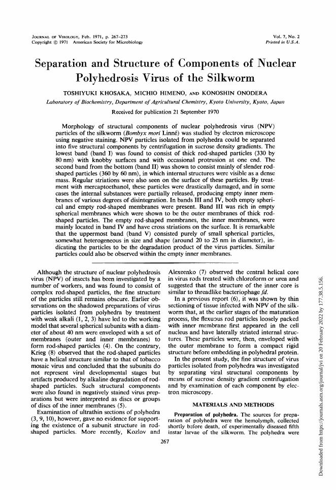
particles, 360 nm long and 60 nm wide. As seen inFig. 3b, this particle appears to have transverseregular banding on the surface, in which a densemass of internal structure can also be seen. Ofthese particles, some were observed to be distortedin shape and some were partially degraded. Whentreated with 1 (j' mercaptoethanol, the slenderrods were drastically damaged, resulting in partialrelease of the internal substance in some particlesand producing empty rod membranes with vari-ous degrees of disintegration (Fig. 4).Bands III and IV were fairly close together so
that the two bands could not be easily distin-guished. For further separation, these two bandswere combined and recentrifuged in 20 to 40%csucrose gradient at 25,000 rev/min for 2 hr at 5 C.
268 J. VIROL.
Dow
nloa
ded
from
http
s://j
ourn
als.
asm
.org
/jour
nal/j
vi o
n 20
Feb
ruar
y 20
22 b
y 17
7.38
.5.1
56.

MORPHOLOGY OF NUCLEAR POLYHEDROSIS VIRUS
FIG. 2. Virus particles, negatively stained with 2%c phosphotunigstic acid, from the lowest band (ban1d 1) in the'gradient. Kniobby surfaced rod-shaped particles, 330 by 80 tim in average size, are seen. A termital protrusion isobserved at one end of some particles. X 50,000.
a
FIG. 3. (a) Virus particles found in band 11. These slender rod-shaped particles, about 360 nim long and 60 tinmwide, are partially penetrated by the stain and are somewhat elongated longitudinally and distorted in shape.X160,000. (b) Slender rod-shaped particles. On the surface, regular banding is visible. Note that an internfalsubstatice can also be seen inside the particle. X 160,000.
VOL. 7, 1971 269
Dow
nloa
ded
from
http
s://j
ourn
als.
asm
.org
/jour
nal/j
vi o
n 20
Feb
ruar
y 20
22 b
y 17
7.38
.5.1
56.
KHOSAKA, HIMENO, AND ONODERA
After the run, however, only single a diffuse bandappeared in the gradient. When fractionated andmeasured by optical density at 280 nm, two widelydiffused, overlapping bands could be resolved,indicating a considerable size variation of thesecomponents. Examination of these bands in theelectron microscope revealed that both emptyspherical and empty rod-shaped membranes werepresent.
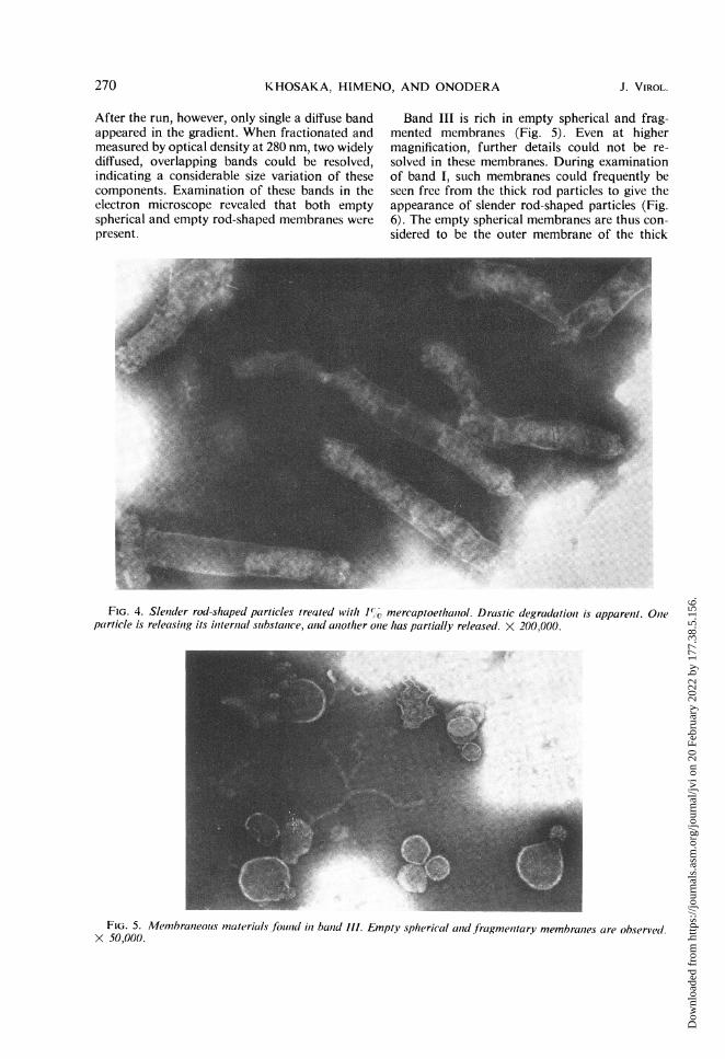
Band III is rich in empty spherical and frag-mented membranes (Fig. 5). Even at highermagnification, further details could not be re-solved in these membranes. During examinationof band I, such membranes could frequently beseen free from the thick rod particles to give theappearance of slender rod-shaped particles (Fig.6). The empty spherical membranes are thus con-sidered to be the outer membrane of the thick
FIG. 4. Slenider rod-shaped particles treated With 1%O mercaptoethaniol. Drastic degradationz is apparenit. Olneparticle is releasin7g its intternial suibstance, anid aiiother onte has partially released. X 200,000.
FIG. 5. Menmbranleolis materials found in ban7d Ill. Empty spherical ald fragme,itary membranies are observel.X 50,000.
270 J. VIROL.
Dow
nloa
ded
from
http
s://j
ourn
als.
asm
.org
/jour
nal/j
vi o
n 20
Feb
ruar
y 20
22 b
y 17
7.38
.5.1
56.

MORPHOLOGY OF NUCLEAR POLYHEDROSIS VIRUS
rods. Similar findings have been reported in theisolated virus preparations from some species ofNPV polyhedra (2, 7). The empty rod-shapedmembranes were mainly located in band IV. Onthe surface, regular striations are clearly visible(Fig. 7) as can be seen with the slender rod-shapedparticles (Fig. 3b). It is noted that the same ob-servation has been made by Kozlov andAlexeenko (7) with NPV of the silkworm. It seemsobvious that these membranes are the inner mem-brane of the thick rod-shaped particles.The uppermost band (band V) appeared at the
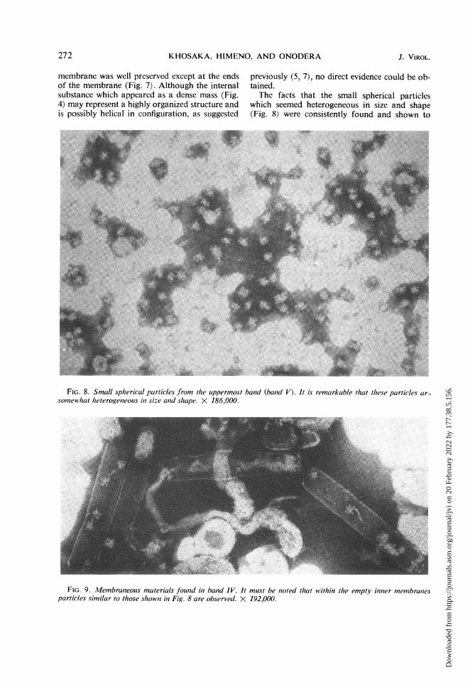
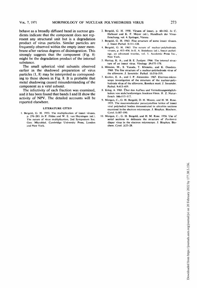
sucrose-suspension interface as a sharp band andwas found to consist purely of small sphericalparticles with a diameter of about 20 to 25 nm(Fig. 8). It must be noted that the particles aresomewhat heterogeneous in size and shape. Whenanalyzed in a 10 to 40c% sucrose density gradientby centrifugation for 3 hr at 25,000 rev min, theseparticles behaved as a broad diffuse band, indi-cating size heterogeneity. It is also noteworthythat within the empty inner membranes found inband IV, similar particles are frequently observed(Fig. 9).
DISCUSSION
NPV of the silkworm was shown to be de-graded in a progressive fashion from thick rodparticles to subviral structural components by theaction of alkali.The thick rod particles observed in the lowest
band (Fig. 2) are apparently identical to thosefound in ultrathin section of polyhedra (3, 9, 10)and were elucidated to consist of an internal sub-stance enveloped with two layers of membrane,outer and inner membranes (Fig. 3a, 3b, 5, and7). The outer membrane may serve to retain thecompact rigid structure of virus rods, since theslender rods, which had lost the outer membrane,were elongated longitudinally and frequently weredistorted in shape during the process of electronmicroscopy (Fig. 3a). This is supported by theresults obtained by thin sectioning of the tissueinfected with NPV of the silkworm (6).
It is evident that when slender rods degrade,further breakages may occur preferentially at theend of the particles to release internal substances(Fig. 4) because the structure of the empty inner
~~~~~~~~~~~,.
lip/A
FIG. 6. Outer membranies are freeing from thick rod-shaped particles as
menited membranles. This sample was takeni from balnd I. X 50,000.empty spherical membrcanies or fraig-
FIG. 7. Empty rod, iinnier memnbranes from bauid IV. Reglalar striations are clearly seeui oni the suirfcace. X 150,000.
VOL. 7, 1971 271
Dow
nloa
ded
from
http
s://j
ourn
als.
asm
.org
/jour
nal/j
vi o
n 20
Feb
ruar
y 20
22 b
y 17
7.38
.5.1
56.
KHOSAKA, HIMENO, AND ONODERA
membrane was well preserved except at the endsof the membrane (Fig. 7). Although the internalsubstance which appeared as a dense mass (Fig.4) may represent a highly organized structure andis possibly helical in configuration, as suggested
previously (5, 7), no direct evidence could be ob-tained.The facts that the small spherical particles
which seemed heterogeneous in size and shape(Fig. 8) were consistently found and shown to
::k.: IF'"4
a,
P..
i.
FIG. 8. Small spherical particles from the uppermost band (band V). It is remarkable that these particles ar.somewhat heterogeneouis in size and shape. X 186,000.
FIG. 9. Membraneouis materials found in band IV. It must be noted that within the empty inner membranesparticles similar to those shownI in Fig. 8 are observed. X 192,000.
272 J. VIROL.
n.4,,.i '.v ...t,f :.'
11
AM
Av.
Dow
nloa
ded
from
http
s://j
ourn
als.
asm
.org
/jour
nal/j
vi o
n 20
Feb
ruar
y 20
22 b
y 17
7.38
.5.1
56.
MORPHOLOGY OF NUCLEAR POLYHEDROSIS VIRUS
behave as a broadly diffused band in sucrose gra-dients indicate that the component does not rep-resent any structural unit but is a degradationproduct of virus particles. Similar particles arefrequently observed within the empty inner mem-brane after various degrees of disintegration. Thisstrongly suggests that the component (Fig. 8)might be the degradation product of the internalsubstance.The small spherical viral subunits observed
earlier in the shadowed preparation of virusparticles (1, 8) may be interpreted as correspond-ing to those shown in Fig. 8. It is probable thatmetal shadowing caused misunderstanding of thecomponent as a viral subunit.The infectivity of each fraction was examined,
and it has been found that bands I and II show theactivity of NPV. The detailed accounts will bereported elsewhere.
LITERATURE CITED
1. Bergold, G. H. 1953. The multiplication of insect viruses,p. 276-283. In P. Fildes and W. E. van Heyningen (ed.),The nature of virus multiplication, 2nd Symposium Soc.Gen. Microbiol. Cambridge University Press, Londonand New York.
2. Bergold, G. H. 1958. Viruses of insect, p. 60-142. In C.Hallauer and K. F. Meyer (ed.), Handbuch der Virus-forschung, vol. 4. Springer, Vienna.
3. Bergold, G. H. 1963. Fine structure of some insect viruse3.J. Insect Pathol. 5:111-128.
4. Bergold, G. H. 1963. The nature of nuclear-polyhedrosis.viruses, p. 413-456. In E. A. Steinhaus (ed.), Insect pathol-ogy, an advanced treatise, vol. 1. Academic Press Inic.,New York.
5. Harrap, K. A., and B. E. Juniper. 1966. The internal struc-ture of an insect virus. Virology 29:175-178.
6. Himeno, M., S. Yasuda, T. Khosaka, and K. Onodera.1968. The fine structure of a nuclear-polyhedrosis virus ofthe silkworm. J. Invertebr. Pathol. 11:516-519.
7. Kozlov, E. A., and I. P. Alexeenko. 1967. Electron-micro-scope investigation of the structure of the nuclear-poly-hedrosis virus of the silkworm, Bombyx mori. J. Invertebr.Pathol. 9:413-419.
8. Krieg, A. 1961. Uber den Aufbau und Vermehrungsmoglich-keiten von stabchenformigen Insekten-Viren. II. Z. Natur-forsch. 16b:1 15-117.
9. Morgan, C., G. H. Bergold, D. H. Moore, and H. M. Rose.1955. The macromolecular paracrystalline lattice of insectviral polyhedral bodies demonstrated in ultrathin sectionsexamined in the electron microscope. J. Biophys. Biochem.Cytol. 1:187-190.
10. Morgan, C., G. H. Bergold, and H. M. Rose. 1956. Use ofserial sections to delineate the structure of Porthetrisdispar virus in the electron microscope. J. Biophys. Bio-chem. Cytol. 2:23-28.
VOL. 7, 1971 273
Dow
nloa
ded
from
http
s://j
ourn
als.
asm
.org
/jour
nal/j
vi o
n 20
Feb
ruar
y 20
22 b
y 17
7.38
.5.1
56.