segmented cmr acquisition with iterative sense reconstruction using l1-regularization in the...
TRANSCRIPT
WORKSHOP PRESENTATION Open Access
Segmented CMR acquisition with iterative SENSEreconstruction using L1-regularization in theevaluation of right ventricular systolic functionAbraham Bogachkov1*, Maria Carr1, Bradley D Allen2, Marie Wasielewski2, Karissa Campione2,Bruce S Spottiswoode3, Michaela Schmidt4, Michael O Zenge4, Mariappan S Nadar5, James C Carr2,Jeremy D Collins2
From 17th Annual SCMR Scientific SessionsNew Orleans, LA, USA. 16-19 January 2014
BackgroundCardiac MR (CMR) has emerged as the gold standard inassessing biventricular size and systolic function withsegmented balanced steady-state free-precession (bSSFP)cine acquisitions. The application of a novel prototypeiterative reconstruction technique to sparsely under-sampled segmented bSSFP cine acquisitions may enablehigher acceleration factors while shortening imageacquisitions, thus maintaining adequate image qualityfor quantitative analysis. The purpose of this study is toevaluate the clinical utility of a segmented sparselysampled 2D CINE imaging technique, with a prototypeiterative SENSE reconstruction using L1-regularization,in the quantitative assessment of right ventricular (RV)systolic function.
Methods9 healthy volunteers (44.3 ± 13.5 yrs) and 29 patients(54.3 ± 13.8 yrs) with suspected cardiac pathology werescanned on a 1.5T scanner (MAGNETOM Aera, SiemensAG, Healthcare Sector, Erlangen, Germany). All subjectswere imaged using a conventional segmented bSSFP cinesequence with GRAPPA factor 2 acceleration (“CINESeg”, temp res = 40 msec, 25 phases, slice = 6 mm,in-plane res = 1.5 × 1.5 mm2, acq time 8.4 sec) and asegmented in-plane sparsely sampled acquisition withT-PAT factor 4 acceleration and prototype iterativeSENSE reconstruction using L1-regularization (“CINESeg-IR”, eff temp res = 39 msec, 20 phases, slice = 6 mm,in-plane res = 1.9 × 1.8 mm2, acq time 4 sec). Quantitative
RV systolic function analysis was performed by a singlereviewer on a dedicated workstation (QMass 5.2, Medis,Leiden, Netherlands). Continuous variables were analyzedusing linear regression. A blinded reviewer scored imagesfor overall image quality, noise, and artifacts using a 5-point Likert scale. 14 patients were re-analyzed to evaluateintraobserver agreement using the intraclass correlationcoefficient (ICC).
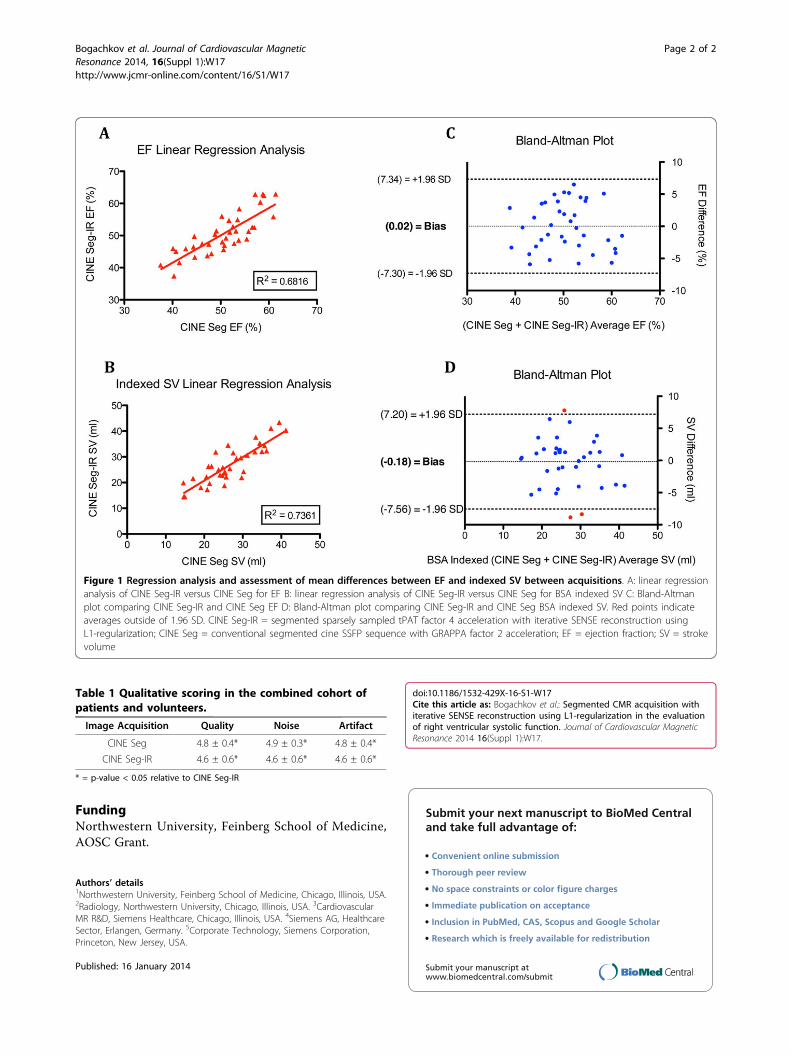
ResultsAcceptable CINE Seg-IR acquisitions were successfullyacquired in all subjects. The mean difference in ejectionfraction (EF) and indexed stroke volume (SV) betweenCINE Seg and CINE Seg-IR were -0.02 ± 3.74% and0.18 ± 3.76 mL/m2, respectively. The R2 value for linearregression of CINE Seg-IR versus CINE Seg is shown inFigure 1. Qualitative review yielded comparable valuesfor CINE SEG versus CINE Seg-IR, as seen in Table 1.ICC values for CINE Seg and CINE Seg-IR, were 0.878and 0.941 for EF, and 0.955 and 0.922 for SV,respectively.
ConclusionsSegmented sparsely sampled cine bSSFP imaging withT-PAT factor 4 and iterative SENSE reconstructionusing L1-regularization at 1.5T can both accurately andreliably quantitate RV systolic function parameters ascompared to conventional segmented cine sequenceswith GRAPPA factor 2 acceleration. This technique hasthe potential to shorten examination times by 50% whileimproving imaging options in patients with arrhythmiasor difficulty with breath-holding.1Northwestern University, Feinberg School of Medicine, Chicago, Illinois, USA
Full list of author information is available at the end of the article
Bogachkov et al. Journal of Cardiovascular MagneticResonance 2014, 16(Suppl 1):W17http://www.jcmr-online.com/content/16/S1/W17
© 2014 Bogachkov et al.; licensee BioMed Central Ltd. This is an Open Access article distributed under the terms of the CreativeCommons Attribution License (http://creativecommons.org/licenses/by/2.0), which permits unrestricted use, distribution, andreproduction in any medium, provided the original work is properly cited. The Creative Commons Public Domain Dedication waiver(http://creativecommons.org/publicdomain/zero/1.0/) applies to the data made available in this article, unless otherwise stated.
FundingNorthwestern University, Feinberg School of Medicine,AOSC Grant.
Authors’ details1Northwestern University, Feinberg School of Medicine, Chicago, Illinois, USA.2Radiology, Northwestern University, Chicago, Illinois, USA. 3CardiovascularMR R&D, Siemens Healthcare, Chicago, Illinois, USA. 4Siemens AG, HealthcareSector, Erlangen, Germany. 5Corporate Technology, Siemens Corporation,Princeton, New Jersey, USA.
Published: 16 January 2014
doi:10.1186/1532-429X-16-S1-W17Cite this article as: Bogachkov et al.: Segmented CMR acquisition withiterative SENSE reconstruction using L1-regularization in the evaluationof right ventricular systolic function. Journal of Cardiovascular MagneticResonance 2014 16(Suppl 1):W17.
Submit your next manuscript to BioMed Centraland take full advantage of:
• Convenient online submission
• Thorough peer review
• No space constraints or color figure charges
• Immediate publication on acceptance
• Inclusion in PubMed, CAS, Scopus and Google Scholar
• Research which is freely available for redistribution
Submit your manuscript at www.biomedcentral.com/submit
Figure 1 Regression analysis and assessment of mean differences between EF and indexed SV between acquisitions. A: linear regressionanalysis of CINE Seg-IR versus CINE Seg for EF B: linear regression analysis of CINE Seg-IR versus CINE Seg for BSA indexed SV C: Bland-Altmanplot comparing CINE Seg-IR and CINE Seg EF D: Bland-Altman plot comparing CINE Seg-IR and CINE Seg BSA indexed SV. Red points indicateaverages outside of 1.96 SD. CINE Seg-IR = segmented sparsely sampled tPAT factor 4 acceleration with iterative SENSE reconstruction usingL1-regularization; CINE Seg = conventional segmented cine SSFP sequence with GRAPPA factor 2 acceleration; EF = ejection fraction; SV = strokevolume
Table 1 Qualitative scoring in the combined cohort ofpatients and volunteers.
Image Acquisition Quality Noise Artifact
CINE Seg 4.8 ± 0.4* 4.9 ± 0.3* 4.8 ± 0.4*
CINE Seg-IR 4.6 ± 0.6* 4.6 ± 0.6* 4.6 ± 0.6*
* = p-value < 0.05 relative to CINE Seg-IR
Bogachkov et al. Journal of Cardiovascular MagneticResonance 2014, 16(Suppl 1):W17http://www.jcmr-online.com/content/16/S1/W17
Page 2 of 2