role of caspase-3 level in perinatal asphyxia
TRANSCRIPT
* Corresponding author: Johnny Rompis, Department of Pediatrics, Faculty of Medicine, Sam Ratulangi, Manado, North Sulawesi, Indonesia. Tel: +6285240560766; Email: [email protected]
Please cite this paper as: Rompis J, Murdono D, Daud D, Wilar R, Hatta M, Umboh A. Role of Caspase-3 Level in Perinatal Asphyxia. Iranian
Journal of Neonatology. 2021 Oct: 12(4). DOI: 10.22038/IJN.2021.52192.1932
Original Article
Open Access Role of Caspase-3 Level in Perinatal Asphyxia Johnny Rompis1*, Darmawan Murdono1, Dasril Daud2, Rocky Wilar1, Mochammad Hatta3, Adrian Umboh1
1. Department of Pediatrics, Faculty of Medicine, Sam Ratulangi University, Manado, North Sulawesi, Indonesia 2. Department of Pediatrics, Faculty of Medicine, Hasanuddin University, Makassar, South Sulawesi, Indonesia 3. Department of Molecular Biology and Immunology, Faculty of Medicine, Hasanuddin University, Makassar, South Sulawesi, Indonesia
ABSTRACT
Background: Perinatal asphyxia is the main cause of neurodevelopmental sequelae and perinatal death. Caspase-3 is a major enzyme associated with apoptosis and increases in hypoxic-ischemic events. There is still no reliable biomarker to predict the severity and outcome of an asphyxial event. In this regard, this study aimed to determine the caspase-3 level and its role as an outcome predictor in perinatal asphyxia. Methods: This paired-group observational analytical cross-sectional study lasted from September 2016 to February 2017. In total, 50 neonates were included in the research and Caspase-3 levels were examined at two different times. Student’s t-test and logistic regression analysis were used for statistical analysis. Results: There were 23 neonates (46%) with hypoxic-ischemic encephalopathy (HIE) and an increase in Caspase-3 level by 0.3135 points from the first to the second examination (t=6.555; P<0.0001). Results of this study showed a significant correlation between the caspase-3 level and mortality in neonates with HIE during both the initial (RR=2.33; P=0.014) and subsequent examinations (RR=2.25; P=0.015). Conclusion: There is a significant increase in Caspase-3 levels in infants who suffer from perinatal asphyxia which can predict mortality in neonates with HIE. Keywords: CASP3 protein, Hypoxic-ischemic encephalopathy, Perinatal asphyxia
Introduction
Asphyxia is one of the most common causes of brain injury in the perinatal period with an incidence rate of 2-4 per 1000 births in developed countries. (1) This rate is up to 10 times higher in developing countries owing to limited access to maternal and neonatal care. It is noteworthy that 15-20% of the affected infants die during the neonatal period, and up to 25% of those who survive are left with permanent neurological deficits (2). In Indonesia, the neonatal mortality rate is 14 per 1000 live births, 21.6% of which is asphyxia-related (3). Perinatal asphyxia is characterized by impaired blood gas exchange that, if persistent, leads to progressive hypoxemia and hypercapnia. The American Academy of Paediatrics and American College of Obstetrics and Gynaecology guidelines consider all of the following criteria for the diagnosis of asphyxia: (i)
profound metabolic or mixed acidemia (pH<7.00) in the umbilical artery blood sample, if obtained, (ii) persistence of an Apgar score of 0–3 for longer than five min, (iii) neonatal neurological sequelae (e.g., seizures, coma, and hypotonia), and (iv) multiple organ involvement (e.g., kidney, lungs, liver, heart, and intestines) (4). Neonatal hypoxic-ischemic encephalopathy (HIE) is the resultant of a global hypoxic-ischemic brain injury in neonates, usually developed following an asphyxia event (5). Apoptosis, together with necrosis and autophagy, is known to be the pathomechanism of neuronal cell death during neonatal hypoxic-ischemic event (6).
Caspase is a specific cysteine-aspartic acid intercellular protein that regulates apoptosis with caspase-3 known as the main enzyme during the proteolytic event. Several in vivo studies have been

Rompis J et al Caspase-3 Level and Perinatal Asphyxia
2 Iranian Journal of Neonatology 2021; 12(4)
conducted on animals regarding the gene expression of caspase-3 mRNA whose results indicate increased caspase-3 expression in rats with amyloid beta-induced Alzheimer’s disease. The caspase-3 level was found to increase by 30-40% during asphyxial apoptosis and induced neuronal cell death sequences that cause brain damage (7, 8).
Based on the findings of another study, there is an association between the caspase-3 elevation of more than 0.2 ng/mL and 30-day mortality in patients with traumatic brain injury (9). There are currently no other studies about caspase-3 levels in perinatal asphyxia. The high morbidity and mortality rate of perinatal asphyxia indicates the need for a biomarker as an outcome predictor. This study aimed to look for caspase-3 level in asphyxia event and its correlation with the neonatal outcome, as a predictor of perinatal asphyxia.
Methods Study participants
This analytical cross-sectional study lasted from September 2016 to February 2017 in the Neonatal Intensive Care Unit (NICU) of Prof. Dr. R.D. Kandou Hospital, Manado, Indonesia. Cases of birth asphyxia were recruited in a consecutive manner. The inclusion criteria were Apgar score of < 3 on minute five and gestational age of > 34 weeks. On the other hand, the exclusion criteria were the presence of any congenital anomaly and traumatic delivery, such as cephalhematoma and subgaleal or subaponeurotic hemorrhage.
The HIE is clinically defined as a disturbance in neurological function, demonstrated by difficulty in maintaining respirations, hypotonia, altered level of consciousness, depressed or absent primitive reflexes, seizures, and poor feeding. It is graded based on Sarnat and Sarnat classification, and subjects diagnosed with HIE are treated with passive cooling.
It should be mentioned that all the methods used in this study were in accordance with all the relevant guidelines and regulations in Indonesia as well as the Declaration of Helsinki for Medical Research Involving Human Subjects. This study was approved by the Medical Ethics Committee of Prof. Dr. R.D. Kandou Hospital (reference code: 049/EC-KEPK/IV/2016) and written informed consent was obtained from all parents or guardians prior to participation in this study.
The caspase-3 examination was performed twice on all subjects. The first test was performed in the first eight h of life followed by the second
test which was performed 48 h after birth, or if the subject presented with HIE symptoms. Approximately three ml of venous blood were collected and subsequently, centrifuged at 1000 g (3000 rpm) for 15 min to isolate serum for further assays, using Human CASP/Caspase-3 ELISA Kit (LSBio Seattle, United States). In this study, the neonatal outcome is defined as the patients’ survival at the time of discharge from NICU.
Statistical analysis
Descriptive analysis was performed to describe the characteristics of the study population. Bivariate analysis regarding the association between Caspase-3 level and the outcome in perinatal asphyxia was performed using the independent samples t-test. Moreover, the correlation between the caspase-3 level and neonatal outcome was analyzed using logistic regression analysis. The overall analyses were conducted in SPSS software (version 22) while a p-value of less than 0.05 was considered statistically significant.
Results Characteristics of neonates with perinatal asphyxia
In total, 23 (46%) out of 50 subjects, consisting of 26 male and 24 female neonates, developed HIE symptoms. The majority of the neonates were delivered spontaneously with cephalic presentation (62%) and cesarean section (32%). The most common indications for cesarean section included premature rupture of membranes (42.9%) and fetal distress (28.7%). The mean birth weight of the neonates was 2837±651.4 g and the mean maternal age was 26.8±6.6 years old. It should also be mentioned that 40 (80%) neonates were alive, while 10 (20%) were declared dead. Moreover, 9 out of the 10 deceased subjects presented with HIE symptoms (90%). For better interpretation, demographic characteristics are summarized in Table 1 below.
Caspase-3 measurement
The caspase-3 level was generally elevated on both examinations. Mean caspase-3 levels on the first and second examination were 7.84±2.5 (median: 7.75; range: 3.455-12.642) ng/mL and 7.53±2.46 (median: 7.36; range: 3.308-11.732) ng/mL, respectively. There was a significant difference between the first and second examinations (0.3135 points, t=6.555; P<0.0001).
In terms of neonatal outcome, there was a

Caspase-3 Level and Perinatal Asphyxia Rompis J et al
3 Iranian Journal of Neonatology 2021; 12(4)
Table 1. Demographic characteristics of children with perinatal asphyxia
Variable HIE
N=23 (%) Non-HIE
N=27 (%) Gender Male 13 (56.6) 13 (48.1) Female 10 (43.4) 14 (51.9) Type of labor Spontaneous 15 (51.6) 15 (48.4) Sectio caesaria 7 (43.7) 9 (56.3) Bracht maneuver - 2 (100) Vacuum extraction - 1 (100) Education level of the mother College 5 (41.7) 7 (58.3) ≤High school 18 (47.4) 20 (52.6) Outcome Alive 14 (35) 26 (65) Dead 9 (90) 1 (10)
HIE: hypoxic-ischemic encephalopathy
Table 2. Caspase-3 level in perinatal asphyxia
Caspase-3 Level Variable Mean Median SD Range General First examination 7.84 7.75 2.5 3.455-12.642 Second examination 7.53 7.36 2.46 3.308-11.732 HIE First examination 9.225 11.737 2.39 3.848-12.642 Second examination 8.89 11.732 1.96 3.618-11.732 Non-HIE First examination 6.675 5.371 1.97 3.455-11.030 Second examination 6.379 4.995 1.96 3.308-11.036 Alive First examination 7.11 6.495 2.14 3.455-12.276 Second examination 6.816 6.04 2.13 3.308-11.415 Dead First examination 10.8 11.448 1.49 7.673-12.642 Second examination 10.411 11.726 1.36 7.656-11.732
HIE: hypoxic-ischemic encephalopathy
significant difference between the caspase-3 levels of the survived (0.294 points, t=6.363; P<0.0001) and deceased subjects (0.391 points, t=2.503; P=0.034). Caspase-3 level of the deceased subjects was consistently higher than that of the survivors (10.8 vs. 7.11 ng/mL on the first examination and 10.411 vs. 6.816 ng/mL on the second examination). Table 2 summarizes the difference between the caspase-3 levels of subjects based on their group and outcome.
Correlation between caspase-3 level and neonatal outcome
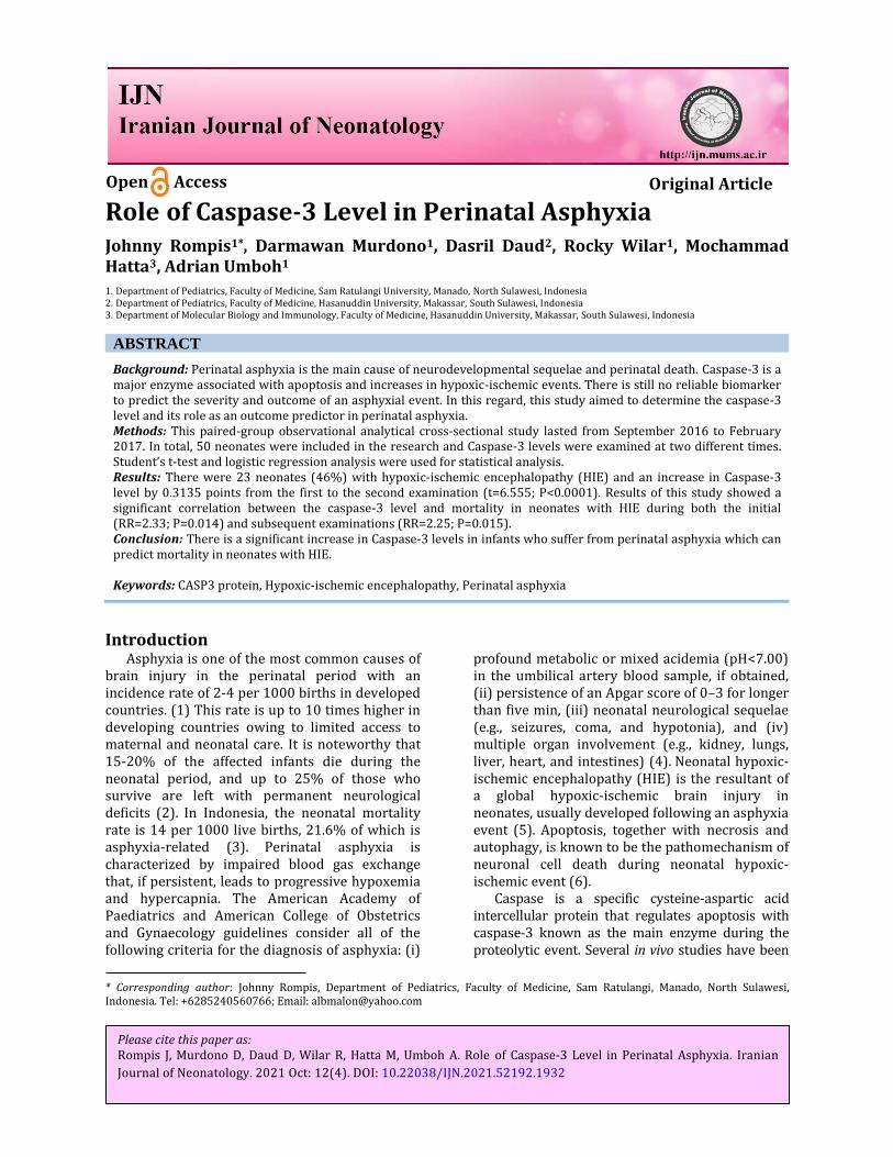
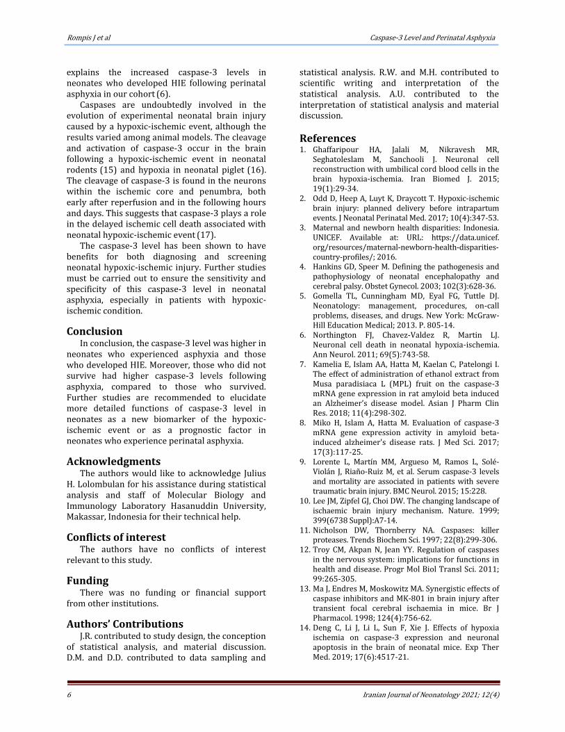
Using logistic regression analysis, a significant correlation was noted between caspase-3 level and neonatal outcome in subjects with HIE symptoms, both in the first (Figure 1) (RR=2.33; P=0.014) and second examinations (Figure 2) (RR=2.25; P=0.015). However, there was no significant correlation between the caspase-3 level and neonatal outcome in subjects without HIE symptoms in both the first (Figure 3) (RR=2.23; P=0.205) and second examinations (Figure 4)
(RR=2.47; P=0.178).
Discussion Perinatal hypoxic-ischemic brain injury is an
important cause of neurodevelopmental impairment and disability as the neonates grow older (10). In the case of hypoxic-ischemic conditions of the brain, the brain itself will eliminate the nonfunctional cells during normal development with minimal inflammation. Regarding this process, many molecules have been identified and known to participate in mediating the highly ordered disassembly of cells through apoptosis. The main agents in the central pathways of this event are a group of cysteine proteases termed caspases (11).
Caspases play an important role in apoptosis and inflammation, and can be divided into three major groups, namely initiator caspases (caspase-2, -8, -9, -10), effector caspases (caspase-3, -6, -7), and inflammatory caspases (caspase -1, -4, -5, -11, -12). Effector caspases are activated by the initiator caspases, while initiator caspases are

Rompis J et al Caspase-3 Level and Perinatal Asphyxia
4 Iranian Journal of Neonatology 2021; 12(4)
Figure 1. Scatterplot diagram between Caspase-3 level and Death outcome in neonatal with HIE symptoms (first examination)
Figure 2. Scatterplot diagram between Caspase-3 level and death outcome in neonatal with HIE symptoms (second examination)
activated through a different and more complex mechanism (12).
Caspase-3 comprises the highest proportion of the caspase molecules in the brain and appears to play a crucial role both during normal development and in certain situations, such as brain injury. According to the results of a study performed on genetically modified mice, those without caspase-3 died at 1-3 weeks of age and displayed a hyperplastic and disorganized brain, while the rest of their bodies, appeared to be
relatively normal. This indicates the particularly important role of caspase-3 in the brain (13).
In this study, the caspase level of neonates with perinatal asphyxia was evaluated twice; first, within the first eight h and then at 48 h after their births or if they developed HIE. Based on the results, the mean level of caspase-3 was significantly higher in neonates who developed HIE, compared to the non-HIE neonates in both the first (9.225 vs 6.675 ng/mL) and second examinations (8.89 vs. 6.379 ng/mL). Moreover,

Caspase-3 Level and Perinatal Asphyxia Rompis J et al
5 Iranian Journal of Neonatology 2021; 12(4)
Figure 3. Scatterplot diagram between Caspase-3 level and death outcome in neonatal without HIE symptoms (first examination)
Figure 4. Scatterplot diagram between Caspase-3 level and death outcome in neonatal without HIE symptoms (second examination)
the caspase-3 level was significantly lower in the survived group, compared to the deceased group in both the first (7.11 vs 10.8 ng/mL) and second examinations (6.81 vs 10.41 ng/mL).
A study was carried out on neonatal mice which were modeled to have the hypoxic-ischemic event. The results of the aforementioned study revealed that the protein expression levels of caspase-3 and Fas ligand detected through reverse transcription-polymerase chain reaction were significantly higher in mice with the hypoxic-ischemic event than the mice without hypoxic-
ischemic condition. This condition could lead to a significant increase in both caspase-3 expression and neuronal apoptosis in the brain of neonatal mice (14).
The expression of caspase-3 in hypoxic-ischemic events takes place due to the classic adenosine triphosphate-driven and caspase-dependant programmed cell death. In the setting of hypoxic-ischemic events, axonal damage and target deprivation in the mature nervous system seem to induce neuronal apoptosis, which is related to caspase expression. This notion
Rompis J et al Caspase-3 Level and Perinatal Asphyxia
6 Iranian Journal of Neonatology 2021; 12(4)
explains the increased caspase-3 levels in neonates who developed HIE following perinatal asphyxia in our cohort (6).
Caspases are undoubtedly involved in the evolution of experimental neonatal brain injury caused by a hypoxic-ischemic event, although the results varied among animal models. The cleavage and activation of caspase-3 occur in the brain following a hypoxic-ischemic event in neonatal rodents (15) and hypoxia in neonatal piglet (16). The cleavage of caspase-3 is found in the neurons within the ischemic core and penumbra, both early after reperfusion and in the following hours and days. This suggests that caspase-3 plays a role in the delayed ischemic cell death associated with neonatal hypoxic-ischemic event (17).
The caspase-3 level has been shown to have benefits for both diagnosing and screening neonatal hypoxic-ischemic injury. Further studies must be carried out to ensure the sensitivity and specificity of this caspase-3 level in neonatal asphyxia, especially in patients with hypoxic-ischemic condition.
Conclusion In conclusion, the caspase-3 level was higher in
neonates who experienced asphyxia and those who developed HIE. Moreover, those who did not survive had higher caspase-3 levels following asphyxia, compared to those who survived. Further studies are recommended to elucidate more detailed functions of caspase-3 level in neonates as a new biomarker of the hypoxic-ischemic event or as a prognostic factor in neonates who experience perinatal asphyxia.
Acknowledgments The authors would like to acknowledge Julius
H. Lolombulan for his assistance during statistical analysis and staff of Molecular Biology and Immunology Laboratory Hasanuddin University, Makassar, Indonesia for their technical help.
Conflicts of interest The authors have no conflicts of interest
relevant to this study.
Funding There was no funding or financial support
from other institutions.
Authors’ Contributions J.R. contributed to study design, the conception
of statistical analysis, and material discussion. D.M. and D.D. contributed to data sampling and
statistical analysis. R.W. and M.H. contributed to scientific writing and interpretation of the statistical analysis. A.U. contributed to the interpretation of statistical analysis and material discussion.
References 1. Ghaffaripour HA, Jalali M, Nikravesh MR,
Seghatoleslam M, Sanchooli J. Neuronal cell reconstruction with umbilical cord blood cells in the brain hypoxia-ischemia. Iran Biomed J. 2015; 19(1):29-34.
2. Odd D, Heep A, Luyt K, Draycott T. Hypoxic-ischemic brain injury: planned delivery before intrapartum events. J Neonatal Perinatal Med. 2017; 10(4):347-53.
3. Maternal and newborn health disparities: Indonesia. UNICEF. Available at: URL: https://data.unicef. org/resources/maternal-newborn-health-disparities-country-profiles/; 2016.
4. Hankins GD, Speer M. Defining the pathogenesis and pathophysiology of neonatal encephalopathy and cerebral palsy. Obstet Gynecol. 2003; 102(3):628-36.
5. Gomella TL, Cunningham MD, Eyal FG, Tuttle DJ. Neonatology: management, procedures, on-call problems, diseases, and drugs. New York: McGraw-Hill Education Medical; 2013. P. 805-14.
6. Northington FJ, Chavez-Valdez R, Martin LJ. Neuronal cell death in neonatal hypoxia-ischemia. Ann Neurol. 2011; 69(5):743-58.
7. Kamelia E, Islam AA, Hatta M, Kaelan C, Patelongi I. The effect of administration of ethanol extract from Musa paradisiaca L (MPL) fruit on the caspase-3 mRNA gene expression in rat amyloid beta induced an Alzheimer’s disease model. Asian J Pharm Clin Res. 2018; 11(4):298-302.
8. Miko H, Islam A, Hatta M. Evaluation of caspase-3 mRNA gene expression activity in amyloid beta-induced alzheimer's disease rats. J Med Sci. 2017; 17(3):117-25.
9. Lorente L, Martín MM, Argueso M, Ramos L, Solé-Violán J, Riaño-Ruiz M, et al. Serum caspase-3 levels and mortality are associated in patients with severe traumatic brain injury. BMC Neurol. 2015; 15:228.
10. Lee JM, Zipfel GJ, Choi DW. The changing landscape of ischaemic brain injury mechanism. Nature. 1999; 399(6738 Suppl):A7-14.
11. Nicholson DW, Thornberry NA. Caspases: killer proteases. Trends Biochem Sci. 1997; 22(8):299-306.
12. Troy CM, Akpan N, Jean YY. Regulation of caspases in the nervous system: implications for functions in health and disease. Progr Mol Biol Transl Sci. 2011; 99:265-305.
13. Ma J, Endres M, Moskowitz MA. Synergistic effects of caspase inhibitors and MK-801 in brain injury after transient focal cerebral ischaemia in mice. Br J Pharmacol. 1998; 124(4):756-62.
14. Deng C, Li J, Li L, Sun F, Xie J. Effects of hypoxia ischemia on caspase-3 expression and neuronal apoptosis in the brain of neonatal mice. Exp Ther Med. 2019; 17(6):4517-21.

Caspase-3 Level and Perinatal Asphyxia Rompis J et al
7 Iranian Journal of Neonatology 2021; 12(4)
15. Blomgren K, Zhu C, Wang X, Karlsson JO, Leverin AL, Bahr BA, et al. Synergistic activation of caspase-3 by m-calpain after neonatal hypoxia-ischemia: a mechanism of "pathological apoptosis"? J Biol Chem. 2001; 276(13):10191-8.
16. Delivoria-Papadopoulos M, Ashraf QM, Ara J, Mishra OP. Nuclear mechanism of hypoxic cerebral injury in
the newborn: the role of caspases. Semin Perinatol. 2008; 32(5):334-43.
17. Namura S, Zhu J, Fink K, Endres M, Srinivasan A, Tomaselli KJ, et al. Activation and cleavage of caspase-3 in apoptosis induced by experimental cerebral ischemia. J Neurosci. 1998; 18(10):3659-68.